Ultrastructural characteristics and histological impacts ...
7
DISEASES OF AQUATIC ORGANISMS Dis Aquat Org Vol. 90: 207–213, 2010 doi: 10.3354/dao02241 Published July 1 INTRODUCTION Myxosporea comprises > 2180 species assigned to 62 genera commonly found in fish (Lom & Dyková 2006). Myxobolus is the most common and largest myxo- sporean genus parasitizing fish. Eiras et al. (2005) pro- vided a synopsis of the relevant morphometric and morphological data of 744 nominal species of Myxobo- lus including nearly all the species described so far in fish and amphibians. Only 14 Myxobolus spp. have been described from fishes caught in Egyptian waters to date (Fahmy et al. 1971, 1975, Abdel-Ghaffar et al. 1995, 1998, 2008, Negm-Eldim et al. 1999, Ali et al. 2002, 2003, 2007). The majority of these species were described using light microscopy and diagrammatic drawings of spores to define species. Only 2 Egyptian Myxobolus spp. have been described using electron microscopy (Ali et al. 2003, 2007). Therefore, the pre- sent study was undertaken to study the ultrastructural details of M. naffari that infects the Nile labeo Labeo niloticus. The histological impact of the parasite on the host was also described. MATERIALS AND METHODS During a survey of myxosporean infections of fish species from the River Nile in Egypt, a total of 200 freshly caught Labeo niloticus were examined from March 2008 to March 2009. The examined fish ranged from 23 to 30 cm in total length. Descriptions and mea- surements of spores followed the guidelines of Lom & Arthur (1989). Measurements were based on 30 spores, and data were presented as mean ± SD (range). For histology, the infected parts were fixed in 10% phosphate-buffered formalin, embedded in paraffin, sectioned, and stained with hematoxylin and eosin (H&E). For electron microscopy, small intact cysts with © Inter-Research 2010 · www.int-res.com *Email: [email protected] Ultrastructural characteristics and histological impacts of Myxobolus naffari (Myxozoa: Myxosporea) infecting Nile labeo Labeo niloticus (Osteichthyes: Cichlidae) A. S. Abdel-Baki 1, 2, *, T. Sakran 2 , H. Fayed 3 , E. Zayed 2 1 Zoology Department, College of Science, King Saud University, PO Box 12455, Riyadh 11451, Saudi Arabia 2 Zoology Department, Faculty of Science, Beni-Suef University, Beni-Suef, Egypt 3 Zoology Department, Faculty of Science, Cairo University, Giza, Egypt ABSTRACT: We describe the ultrastructural characteristics and histological impacts of Myxobolus naffari Abdel-Ghaffar et al., 1998, which infects the Nile fish Labeo niloticus. The prevalence of infec- tion was 65%, with the maximum rate occurring during winter and a lower rate during summer. The histological impacts were manifested as a fusion of the gill epithelia, hyperplasia at the ends of the plasmodia, and atrophy of the external surface of the plasmodia. The ultrastructural study revealed that the plasmodial wall was composed of a single unit membrane and bound externally by a thick layer of collagen fibers. The earliest recognizable stage was the disporous pansporoblast. The devel- opment of the parasite was asynchronous, with mature and immature spores randomly distributed throughout the plasmodium. KEY WORDS: Myxobolus naffari · Labeo niloticus · Ultrastructure · River Nile · Histology Resale or republication not permitted without written consent of the publisher
Transcript of Ultrastructural characteristics and histological impacts ...

DISEASES OF AQUATIC ORGANISMSDis Aquat Org
Vol. 90: 207–213, 2010doi: 10.3354/dao02241
Published July 1
INTRODUCTION
Myxosporea comprises >2180 species assigned to 62genera commonly found in fish (Lom & Dyková 2006).Myxobolus is the most common and largest myxo-sporean genus parasitizing fish. Eiras et al. (2005) pro-vided a synopsis of the relevant morphometric andmorphological data of 744 nominal species of Myxobo-lus including nearly all the species described so far infish and amphibians. Only 14 Myxobolus spp. havebeen described from fishes caught in Egyptian watersto date (Fahmy et al. 1971, 1975, Abdel-Ghaffar et al.1995, 1998, 2008, Negm-Eldim et al. 1999, Ali et al.2002, 2003, 2007). The majority of these species weredescribed using light microscopy and diagrammaticdrawings of spores to define species. Only 2 EgyptianMyxobolus spp. have been described using electronmicroscopy (Ali et al. 2003, 2007). Therefore, the pre-sent study was undertaken to study the ultrastructural
details of M. naffari that infects the Nile labeo Labeoniloticus. The histological impact of the parasite on thehost was also described.
MATERIALS AND METHODS
During a survey of myxosporean infections of fishspecies from the River Nile in Egypt, a total of 200freshly caught Labeo niloticus were examined fromMarch 2008 to March 2009. The examined fish rangedfrom 23 to 30 cm in total length. Descriptions and mea-surements of spores followed the guidelines of Lom &Arthur (1989). Measurements were based on 30spores, and data were presented as mean ± SD (range).For histology, the infected parts were fixed in 10%phosphate-buffered formalin, embedded in paraffin,sectioned, and stained with hematoxylin and eosin(H&E). For electron microscopy, small intact cysts with
© Inter-Research 2010 · www.int-res.com*Email: [email protected]
Ultrastructural characteristics and histologicalimpacts of Myxobolus naffari (Myxozoa:Myxosporea) infecting Nile labeo Labeo
niloticus (Osteichthyes: Cichlidae)
A. S. Abdel-Baki1, 2,*, T. Sakran2, H. Fayed3, E. Zayed2
1Zoology Department, College of Science, King Saud University, PO Box 12455, Riyadh 11451, Saudi Arabia2Zoology Department, Faculty of Science, Beni-Suef University, Beni-Suef, Egypt
3Zoology Department, Faculty of Science, Cairo University, Giza, Egypt
ABSTRACT: We describe the ultrastructural characteristics and histological impacts of Myxobolusnaffari Abdel-Ghaffar et al., 1998, which infects the Nile fish Labeo niloticus. The prevalence of infec-tion was 65%, with the maximum rate occurring during winter and a lower rate during summer. Thehistological impacts were manifested as a fusion of the gill epithelia, hyperplasia at the ends of theplasmodia, and atrophy of the external surface of the plasmodia. The ultrastructural study revealedthat the plasmodial wall was composed of a single unit membrane and bound externally by a thicklayer of collagen fibers. The earliest recognizable stage was the disporous pansporoblast. The devel-opment of the parasite was asynchronous, with mature and immature spores randomly distributedthroughout the plasmodium.
KEY WORDS: Myxobolus naffari · Labeo niloticus · Ultrastructure · River Nile · Histology
Resale or republication not permitted without written consent of the publisher
Dis Aquat Org 90: 207–213, 2010
minimal surrounding tissue were isolated and fixedin 3% glutaraldehyde in 0.1% sodium cacodylate(pH 7.4), washed in the same buffer, and post-fixedwith 2% OsO4 in the same buffer. The tissue pieceswere dehydrated in graded ethanol and embedded inaraldite. Ultrathin sections were stained with uranyleacetate and lead citrate and examined with a Philips(208) electron microscope at 80 to 100 kV.
RESULTS
The prevalence of infection was 65% (130/200), withthe maximum rate occurring during winter and theminimum rate in summer.
Light microscopy
Plasmodia. Plasmodia appeared as white aggregatesof tiny rods on the gill filament. Fish were infected with2 to 16 cysts, the average dimensions of which were0.6 ± 0.4 (range: 0.3 to 1.26) mm in length and 0.2 ±0.07 (range: 0.1 to 0.2) mm in width.
Spores. Spores were subspherical to ellipsoidal inthe frontal view with rounded anterior and posteriorends (Fig. 1). They measured 10.9 ± 0.4 (range: 10.5 to12.0) µm in length and 8.6 ± 0.5 (range: 7 to 9.5) µm inwidth (Fig. 1). A small intercapsular process was pre-sent. Spore valves were thin, smooth, and symmetrical.Polar capsules were oval, equal in size, and occupiednearly half of the spore length. They measured 5.7 ±0.2 (range: 5.2 to 6.3) µm in length and 3.3 ± 0.4 (range:2.6 to 4.0) µm in width. Polar filaments showed 6 to 8coils slightly oblique along the axis of the polar cap-sule. The sporoplasm was binucleated, with a largeand rounded to oval iodinophilous vacuole, measuring3.6 ± 0.4 (range: 2.3 to 4.0) µm in diameter.
Histological impact. Plasmodia were located insidethe lining epithelium covering the gill filaments. Theplasmodial mass occupied a large part of the gill fila-ment (Fig. 2). The host reaction was detected as theformation of a thin fibrous connective tissue capsulearound the plasmodia (Fig. 2). The histological impactswere manifested as a fusion of the gill epithelia, hyper-plasia at the ends of the plasmodia, and atrophy of theexternal surface of the plasmodia (Fig. 2).
Electron microscopy
Plasmodia. The plasmodia were surrounded by sin-gle unit membranes (Fig. 3). External to the mem-brane, there was a thick layer composed of collagenfibers in contact with the host cell (Fig. 3). The plas-
modium was composed of 2 distinct layers, the outerectoplasm and an inner endoplasm. The ectoplasmwas an amorphous layer with many pinocytotic vesi-cles, whereas the endoplasm contained the differentdevelopmental stages (Fig. 3).
Sporogenesis. The early sporogenic stages were notobserved. The earliest recognizable stages were themonosporous and the disporous pansporoblast (Figs. 4& 5). As the spore matured, there was a structural pro-gression in the capsulogenesis, sporoplasm matura-tion, and valvogenesis (Figs. 4 to 9).
Capsulogenesis. Capsulogenic cells occurred at theapical pole of the developing spore. Together with thesporoplasm they formed the central core of spore thatwas ensheathed by valvogenic cells (Figs. 4, 7 & 9).Thedifferentiation of the capsulogenic cell started with theappearance of a club-shaped structure, which was theinitial stage of the capsular primordium (Fig. 5). Grad-ually, the initial stage differentiated into the bulbousprimordium and the associated external tube (Figs. 5 &6). The external tube was later internalized into theprimordium to make the polar filament coils with 6 to8 turns (Figs. 6, 7 & 9).
The developing polar capsule had a homogeneouscore of medium electron density, surrounded by anelectron-lucent layer and an outer layer of mediumdensity (Fig. 6). A stopper with a triangular cap-likecover plugged the apex of each mature polar capsule(Fig. 6). Maturation of the 2 polar capsules was usuallysynchronous (Figs. 7 & 9)
Sporoplasm. As the polar capsule primordiumappeared, the sporoplasm could be easily recognized.In the mature spore, it filled nearly all the spacebeneath the polar capsules (Figs. 4, 7 & 9). The sporo-plasm contained 2 nuclei (Fig. 4) and exhibited densematrices of sporoplasmosomes (Figs. 4, 7 & 9). A smallarea of sporoplasm in mature spores was occupied byglycogen granules that were often vacuolated (Figs. 7& 9).
Valvogenesis. Valvogenesis was initiated by thegradual envelopment of the capsulogenic cells andsporoplasm by valvogenic cells. Their nuclei were flat-tened and located laterally (Fig. 8). In the course ofdevelopment, these cells gave rise to the 2 shell valvessurrounding each spore and the sutural ridge joiningthe valves (Figs. 7 & 8). At the spore apex, the wall ofthe valve formed a triangular thickening (Fig. 7).
DISCUSSION
Light microscopy
Many species of Myxobolus fall within the morpho-logical and dimensional ranges of the present species.
208

Abdel-Baki et al.: Ultrastructural and histological impacts of Myxobolus naffari 209
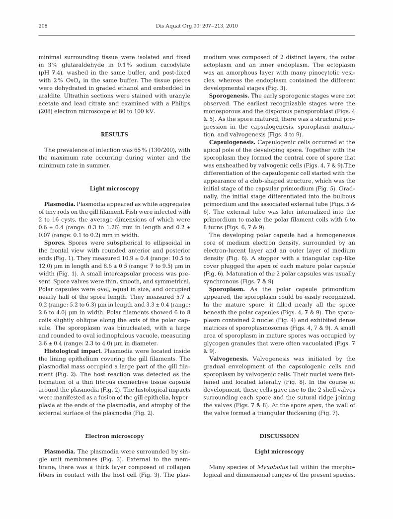
Figs. 1 to 6. Fig. 1. Fresh spores of Myxobolus naffari Abdel-Ghaffar et al., 1998. Scale bar = 5 µm. Fig. 2. Longitudinal section ininfected gill filaments showing many plasmodia (P) with hyperplasia in the gill filament epithelia (Hp) and degeneration (D)along the external surface of the plasmodium. H&E. Scale bar = 0.2 mm. Fig. 3. Electronmicrograph through a part of M. naffariplasmodium showing the plasmodial wall with a single unit membrane (arrows) from which vesicles (v) originated. The plas-modial wall is composed of ectoplasm (Ecto) and endoplasm (Endo). Externally, the plasmodium was surrounded by a layer of col-lagen fiber (Cf) in contact with the host cell (HC) and host nucleus (HN). Scale bar = 2 µm. Fig. 4. Trans-section through maturespore containing 1 mature polar capsule (PC), sporoplasm (Sp) with 2 nuclei (N1, N2), and sporoplasmosomes (white arrowheads)and surrounded by valvogenic cells (VC). Scale bar = 1 µm. Fig. 5. Trans-section through disporic pansporoblast consisting of 2asynchronous spore-producing units, each containing capsular primordium (Cp). Sporoplasm (Sp) with sporoplasmosomes(arrows). Scale bar = 1 µm. Fig. 6. Trans-section through mature polar capsule (PC). The polar capsule is composed of an electron-dense outer layer (small arrow), an inner translucent (Lu), and a central dense core. The polar capsules contain polar filament (PF)
and have a triangular plug (P) at the apex. Scale bar = 500 nm

Dis Aquat Org 90: 207–213, 2010
These include M. muelleri Bütschli, 1882 (cited in Lom& Dyková 1992); M. aldrichetti Su & White, 1994 fromthe gills of Aldrichetta forsteri in Australia; M. cognatiCone et al., 1996 from the operculum of Cottus cogna-tus in Canada; M. mesopotamiae Molnár et al., 1996from the connective tissue of fins of Barbus grypus inIran; M. bulbocordis Masoumian et al., 1996 from theheart region of B. sharpeyi in Iran; and M. naffariAbdel-Ghaffar et al., 1998 and M. imami Ali et al.,2002, both from the gills of Labeo niloticus in Egypt.
Comparatively, Myxobolus muelleri and M. bulbo-cordis are larger than M. naffari in all dimensions,while M. aldrichetti and M. cognati have pyriform po-lar capsules and lack an intercapsular process, and M.
mesopotamiae is smaller in all dimensions and lacks anintercapsular process. M. imami differs from the pre-sent species in the number of polar filament turns (9 vs.7) and the absence of an intercapsular process. Thepresent species matches M. naffari in all morphometricaspects, host, and site of infection. Therefore, the pre-sent species can be identified as M. naffari.
Histological impact
In the present study, one host reaction to myxo-sporean infections is encapsulation of the plasmodia bythe gill’s connective tissue (Mitchell 1977). The shape,
210
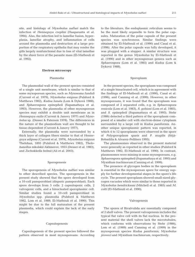
Figs. 7 to 9. Fig. 7. Trans-section through spore containing2 mature polar capsules (PC) with their polar filaments(PF), sporoplasm (Sp) with sporoplasmosomes (white ar-rowheads) and glycogen vacuole (GV), and surrounded byvalvogenic cells (VC) with a sutural ridge joining the valves(arrows). Scale bar = 1 µm. Fig. 8. Trans-section throughspore showing the valvogenic cell (VC) with sutural ridgejoining the valves (arrow) and its nucleus (VN). Scale bar =2 µm. Fig. 9. Electron micrograph showing the fully maturespore with its 2 fully developed polar capsules (PC) andtheir polar filament (PF), sporoplasm (Sp) with its nucleus(SpN), sporoplasmosomes (white arrowheads), glycogenvacuoles (GV), and valvogenic cell (VC). Scale bar = 1 µm
Abdel-Baki et al.: Ultrastructural and histological impacts of Myxobolus naffari
site, and histology of Myxobolus naffari match theinfection of Henneguya creplini (Haaparanta et al.1994). Also, the infection led to lamellar fusion, hyper-plasia, lamellar atrophy, and inflammation of cellsaround the plasmodia and destruction of a large pro-portion of the respiratory epithelia that may render thegills largely nonfunctional due to loss of vital lamellaeby the sheer force of the parasite mass (El-Matbouli etal. 1992).
Electron microscopy
Plasmodia
The plasmodial wall of the present species consistedof a single unit membrane, which is similar to that ofsome myxoporean species, such as Myxosoma funduli(Current et al. 1979), Myxobolus exiguus (Pulsford &Matthews 1982), Kudoa lunata (Lom & Dyková 1988),and Sphaerospora epinepheli (Supamattaya et al.1993). However, the plasmodia of other myxosporeanspecies may exhibit a double unit membrane as inHenneguya exilis (Current & Janovy 1977) and Myxo-bolus sp. (Desser & Paterson 1978). The differences inthe nature of the plasmodium wall are thought to betissue-dependent (Current & Janovy 1977).
Externally, the plasmodia were surrounded by athick layer of collagen fibers similar to that of Henne-guya adiposa (Current et al. 1979), Myxobolus exiguusThelohan, 1895 (Pulsford & Matthews 1982), Thelo-hanellus nikolskii Akhmerov, 1955 (Desser et al. 1983),and Zschokkella helmii (Ali et al. 2003).
Sporogenesis
The sporogenesis of Myxobolus naffari was similarto other described species. The sporogenesis in thepresent study showed that the spore developed froma 10-cell pansporoblast (disporic pansporoblast). Eachspore develops from 5 cells: 2 capsulogenic cells, 2valvogenic cells, and a binucleated sporoplasmic cell.Similar studies found a 10-cell pansporoblast inMyxobolus spp. plasmodia (Pulsford & Matthews1982, Lom et al. 1989, El-Matbouli et al. 1990). Thismight be due to the full maturation of the presentplasmodia, which could explain the lack of the earlystages.
Capsulogenesis
Capsulogenesis of the present species followed thepattern observed in most myxosporeans. According
to the literature, the endoplasmic reticulum seems tobe the most likely organelle to form the polar cap-sules. Maturation of the polar capsule of the presentspecies was synchronous. Similar findings wereobtained by El-Matbouli et al. (1990) and Casal et al.(1996). After the polar capsule was fully developed, itwas plugged with a stopper. A similar structure wasreported in the genus Myxobolus by El-Matbouli etal. (1990) and in other myxosporean genera such asSphaerospora (Lom et al. 1985) and Kudoa (Lom &Dyková 1988).
Sporoplasm
In the present species, the sporoplasm was composedof a single binucleated cell, which is in agreement withthe findings of El-Matbouli et al. (1990), Casal et al.(1996), and Canning et al. (1999). However, in othermyxosporeans, it was found that the sporoplasm wascomposed of 2 separated cells, e.g. in Sphaerosporarenicola (Lom et al. 1983), S. gobioni (Lom et al. 1985),and S. epinepheli (Supamattaya et al. 1993). Stehr(1986) detected a third pattern of the sporoplasm com-posed of a smaller cell with electron-dense cytoplasmsurrounded by a large cell in Kudoa paniformis. An-other unique sporoplasmic pattern was reported, inwhich 4 to 12 sporoplasms were observed in the sporeof Polysporoplasm sparis and P. mugilis (Sitjà-Bobadilla & Alvarez-Pellitero 1995).
The plasmosomes observed in the present materialwere generally as reported in other studies (Pulsford &Matthews 1982, El-Matbouli et al. 1990). In contrast,plasmosomes were missing in some myxosporeans, e.g.Sphaerospora epinepheli (Supamattaya et al. 1993) andMyxidium trachinorum (Canning et al. 1999).
The presence of glycogen bodies in the sporoplasmis essential in the myxosporean spore for energy sup-ply for further developmental stages in the spore’s lifecycle. The present sporoplasm showed small-sized gly-cogen vacuoles which were similar to those reported inMyxobolus hendricksoni (Mitchell et al. 1985) and M.cotti (El-Matbouli et al. 1990).
Valvogenesis
The spores of Bivalvulida are essentially composedof 2 shell valves. The present valvogenesis includes thetypical flat valve cell with its flat nucleus. In the pre-sent material the shell valves lack the microtubules,which conforms with observations by Stehr (1986),Lom et al. (1989) and Canning et al. (1999) in themyxosporean species Kudoa paniformis, Myxobolusjiroveci and Myxidium trachinorum, respectively.
211
Dis Aquat Org 90: 207–213, 2010
Acknowledgements. We are grateful for the support of theCenter of Excellence for Biodiversity Research, College ofScience, King Saud University, Riyadh, Saudi Arabia.
LITERATURE CITED
Abdel-Ghaffar F, El-Shahawi G, Naas S (1995) Myxosporidiainfecting some Nile fishes in Egypt. Parasitol Res 81:163–166
Abdel-Ghaffar F, Ibrahiem EA, Bashter A, Ali MA (1998)Myxosporidia infecting saline and freshwater fishes ofQarun and Wadi El-Raiyan lakes, Egypt (I. Genus:Myxobolus). J Egypt Ger Soc Zool 26(D):209–229
Abdel-Ghaffar F, El-Toukhy A, Al-Quraishy S, Al-Rasheid K,Abdel-Baki AS, Hegazy A, Bashtar AR (2008) Five newmyxosporean species (Myxozoa: Myxosporea) infectingthe Nile tilapia Oreochromis niloticus in Bahr Shebin, NileTributary, Nile Delta, Egypt. Parasitol Res 103:1197–1205
Ali MA, Al-Rasheid AS, Sakran T, Abdel-Baki AS, Abdel-Ghaffar F (2002) Some species of the genus Myxobolus(Myxozoa: Myxosporea) infecting freshwater fishes of theRiver Nile, Egypt. Parasitol Res 88:9–15
Ali MA, Abdel-Baki AS, Sakran T, Entzeroth R, Abdel-Ghaffar F (2003) Light and electron microscopic studiesof Myxobolus stomum n. sp. (Myxosporea: Myxobolidae)infecting the blackspotted grunt Plectorhynicus gaterinus(Forsskal, 1775) in the Red Sea, Egypt. Parasitol Res 91:390–397
Ali MA, Abdel-Baki AS, Sakran Th, Entzeroth R, Abdel-Ghaf-far F (2007) Myxobolus lubati n. sp. (Myxosporea: Myxo-bolidae), a new parasite of haffara seabream Rhabdo-sargus haffara (Forsskal, 1775), Red Sea, Egypt: a lightand transmission electron microscopy. Parasitol Res 100:819–827
Canning EU, Curry A, Anderson CL, Okamura B (1999) Ultra-structure of Myxidium trachinorum sp. nov. from thegallbladder of the lesser weever fish Echiichthys vipera.Parasitol Res 85:910–919
Casal C, Matos E, Azevedo C (1996) Ultrastructure data onthe life cycle stages of Myxobolus braziliensis n. sp., para-site of an Amazonian fish. Eur J Prositol 32:123–127
Cone DK, Stickel RG, Eck GE, Muzzall PM (1996) Myxoboluscognati n.sp. (Myxosporea) from the opercular integumentof Cottus cognatus (Cottidae) in Lake Michigan. J Para-sitol 82:137–139
Current WL, Janovy J Jr, Knight SA (1979) Myxosoma funduliKudo (Myxosporidia) in Fundulus kansae: ultrastructureof the plasmodium wall and of sporogenesis. J Protozool26:574–583
Current WL, Janovy J Jr (1977) Sporogenesis in Henneguyaexilis infecting the channel catfish: an ultrastructuralstudy. Protistologica 13:157–167
Desser SS, Paterson WB (1978) Ultrastructural and cytochem-ical observation on sporogenesis of Myxobolus sp. (Myxo-sporidia: Myxobolidae) from common shiner Notropis cor-nutus. J Protozool 25:314–326
Desser SS, Molnár K, Weller I (1983) Ultrastructure of spo-rogenesis of Thelohanellus nikolskii Akhmerov, 1955(Myxozoa: Myxosporea) from the common carp Cyprinuscarpio. J Parasitol 69:504–518
Eiras JC, Molnár K, Lu YS (2005) Synopsis of the species ofMyxobolus Bütschli, 1882 (Myxozoa: Myxosporea: Myxo-bolidae). Syst Parasitol 61:1–46
El-Matbouli M, Fischer-Scherl T, Hoffmann RW (1990) Lightand electron microscopic studies on Myxobolus cotti El-Matbouli and Hoffmann, 1987 infecting the central ner-
vous system of the bullhead (Cottus gobio). Parasitol Res76:219–227
El-Matbouli M, Fischer-Scherl T, Hoffman RW (1992) Presentknowledge on the life cycle, taxonomy, pathology and thetherapy of some Myxosporea spp. important for freshwaterfish. Annu Rev Fish Dis 2:367–402
Fahmy MA, Mandour AM, El-Naffar MK (1971) Myxobolusniloticus n. sp. in the fish Labeo niloticus from the RiverNile of Assiut. J Egypt Soc Parasitol 1:39–46
Fahmy MA, Mandour AM, El-Naffar MK (1975) A survey ofMyxosporidia of the freshwater fishes collected from theRiver Nile at Assiut province. J Egypt Soc Parasitol 5:93–102
Haaparanta AE, Valtonen T, Hoffmann RW (1994) Patho-genicity and seasonal occurrence of Henneguya creplini(Protozoa, Myxosporea) on the gills of perch Perca fluvi-atilis in central Finland. Dis Aquat Org 20:15–22
Lom J, Arthur JR (1989) A guideline for the preparation ofspecies descriptions in Myxosporea. J Fish Dis 12:151–156
Lom J, Dyková I (1988) Sporogenesis and spore structure inKudoa lunata (Myxosporea, Multivalvulida). Parasitol Res74:521–530
Lom J, Dyková I (1992) Protozoan parasites of fishes. ElsevierScience, Amsterdam
Lom J, Dyková I (2006) Myxozoan genera: definition andnotes on taxonomy, life cycle terminology and pathogenicspecies. Folia Parasitol (Praha) 53:1–36
Lom J, Dyková I, Lhotaková S (1983) Fine structure ofSphaerospora renicola Dykova and Lom, 1982, a myxo-spoean from carp kidney, and comments on the origin ofpansporoblast. Protistologica 18:489–502
Lom J, Pavlaskova M, Dyková I (1985) Notes on kidney infect-ing species of the genus Sphaerospora Thélohan (Myx-osporea), including a new species S. gobionis sp. nov., andon myxosporean life cycle stages in blood of some fresh-water fish. J Fish Dis 8:221–232
Lom J, Feist S, Dyková I, Kepr T (1989) Brain myxoboliasis ofbullhead, Cottus gobio L., due to Myxobolus jiroveci sp.nov.: light and electron microscope observations. J FishDis 12:15–27
Masoumian A, Baska F, Molnár K (1996) Description ofMyxobolus bulbocordis (Myxosporea: Myxobolidae) fromthe heart of Barbus sharpey (Gunther) and histopathologi-cal changes produced by the parasite. J Fish Dis 19:15–21
Mitchell LG (1977) Myxosporidia. In: Kreier JF (ed) Para-sitic protozoa, Vol 4. Academic Press, New York, NY,p 115–154
Mitchell LG, Seymour CL, Gamble JM (1985) Light andelectron microscopy of Myxobolus hendricksoni sp. nov.(Myxozoa: Myxobolidae) infecting the brain of the fatheadminnow, Pimephales promelas Rafinesque. J Fish Dis 8:75–89
Molnár K, Masoumian M, Abasi S (1996) Four new Myxobo-lus spp. (Myxosporea: Myxobolidae) from Iranian barboidfishes. Arch Protistenkd 147:115–123
Negm-Eldim NM, Govedich FR, Davies RW (1999) Gill myxo-sporeans on some Egyptian freshwater fish. Dtsch Tier-arztl Wochenschr 106:459–465
Pulsford A, Matthews RA (1982) An ultrastructure study ofMxyobolus exiguus Thelohan, 1895 (Myxosporea) fromgrey mullet, Crenimugil labrosus (Risso). J Fish Dis 5:509–526
Sitjà-Bobadilla A, Alvarez-Pellitero P (1995) Light and elec-tron microscopic description of Polysporoplasma n. g.(Myxosporea: Bivalvulida), Polysporoplasma sparis n. sp.from Sparus aurata (L.), and Polysporoplasma mugilisfrom Liza aurata L. Eur J Protistol 31:77–89
212
Abdel-Baki et al.: Ultrastructural and histological impacts of Myxobolus naffari
Stehr C (1986) Sporogenesis of the myxosporean Kudoa pan-iformis Kabata and Whitaker, 1981 infecting the musclesof the Pacific whiting, Merlucius productus (Ayres). J FishDis 9:493–504
Su X, White RWG (1994) New myxosporeans (Myxozoa:Myxosporea) from marine fishes of Tasmania, Australia.
Acta Protozool 33:251–259Supamattaya K, Fischer-Scherl T, Hoffmann RW, Boonyarat-
palin S (1993) Light and electron microscopic observationson presporogenic and sporogenic stages of Sphaerosporaepinepheli (Myxosporea) in grouper (Epinephellus mal-abaricus). J Eukaryot Microbiol 40:71–80
213
Editorial responsibility: Dieter Steinhagen,Hannover, Germany
Submitted: April 8, 2010; Accepted: May 6, 2010Proofs received from author(s): June 21, 2010