Morphological, molecular, and ultrastructural...
10
Morphological, molecular, and ultrastructural characterization of Rozella rhizoclosmatii, a new species in Cryptomycota Peter M. LETCHER a, *, Joyce E. LONGCORE b , C. Alisha QUANDT c , Domingos da Silva LEITE d , Timothy Y. JAMES c , Martha J. POWELL a a Department of Biological Sciences, The University of Alabama, Tuscaloosa, AL 35487, USA b School of Biology and Ecology, University of Maine, Orono, ME 04469, USA c Department of Ecology and Evolutionary Biology, University of Michigan, Ann Arbor, MI 48109, USA d Departamento de Gen etica, Evoluc ¸ ~ ao e Bioagentes, Universidade Estadual de Campinas, Campinas, SP, 13082-862, Brazil article info Article history: Received 10 June 2016 Received in revised form 15 August 2016 Accepted 19 August 2016 Available online 4 September 2016 Corresponding Editor: Gordon William Beakes Keywords: Lattice Morphology Nomenclature Parasitism Taxonomy Zoospore abstract Rozella is a genus of unwalled endoparasites of a variety of hosts including Oomycota (Stra- menopiles), Blastocladiomycota and Chytridiomycota (Fungi), and one green alga (Coleo- chaete, Chlorophyceae). It currently includes more than 20 formally described species, and no new species of Rozella have been described since 1987. We discovered a new Rozella species parasitizing Rhizoclosmatium globosum (Chytridiales, Chytridiomycota) and investi- gated its morphology, ultrastructure, and phylogenetic position. Herein named as Rozella rhizoclosmatii sp. nov., the organism induces hypertrophy of the host. Its zoospore is ultra- structurally similar to that of Rozella allomycis, although it has a unique zoospore ultrastruc- tural feature, a lattice of perpendicular rods about the nucleus. The 18S rDNA molecular sequence of R. rhizoclosmatii is similar to that of the previously sequenced ‘Rozella ex Rhizo- closmatium’. This is the first study to inclusively characterize a new species of Rozella with morphological, ultrastructural and molecular data. As this is only the second Rozella spe- cies to be examined ultrastructurally, and because it is parasitic on a member of Chytridio- mycota and not Blastocladiomycota, this research supports the conservative nature of zoospore ultrastructure to help define the genus. ª 2016 British Mycological Society. Published by Elsevier Ltd. All rights reserved. Introduction Cornu (1872) described the genus Rozella and included four species: Rozella monoblepharidis polymorphae Cornu, Rozella rhi- pidii spinosi Cornu, Rozella apodyae brachynematis Cornu, and Rozella septigena Cornu. Clements & Shear (1931) designated R. septigena as the lectotype. The genus is characterized by an endobiotic, holocarpic, unwalled, inoperculate thallus with one or more discharge papillae; zoospores (often elon- gate) with a single posterior flagellum; and thick-walled, smooth or spiny resting spores (Sparrow 1960). At present, more than 20 species have been described, but descriptions * Corresponding author. Tel.: þ1 205 348 8208; fax: þ1 205 348 1786. E-mail addresses: [email protected] (P. M. Letcher), [email protected] (J. E. Longcore), [email protected] (C. Alisha Quandt), [email protected] (D. daS. Leite), [email protected] (T. Y. James), [email protected] (M. J. Powell). journal homepage: www.elsevier.com/locate/funbio fungal biology 121 (2017) 1 e10 http://dx.doi.org/10.1016/j.funbio.2016.08.008 1878-6146/ª 2016 British Mycological Society. Published by Elsevier Ltd. All rights reserved.
Transcript of Morphological, molecular, and ultrastructural...

f u n g a l b i o l o g y 1 2 1 ( 2 0 1 7 ) 1e1 0
journa l homepage : www.e lsev ier . com/ loca te / funb io
Morphological, molecular, and ultrastructuralcharacterization of Rozella rhizoclosmatii, a newspecies in Cryptomycota
Peter M. LETCHERa,*, Joyce E. LONGCOREb, C. Alisha QUANDTc,Domingos da Silva LEITEd, Timothy Y. JAMESc, Martha J. POWELLa
aDepartment of Biological Sciences, The University of Alabama, Tuscaloosa, AL 35487, USAbSchool of Biology and Ecology, University of Maine, Orono, ME 04469, USAcDepartment of Ecology and Evolutionary Biology, University of Michigan, Ann Arbor, MI 48109, USAdDepartamento de Gen�etica, Evoluc~ao e Bioagentes, Universidade Estadual de Campinas, Campinas, SP, 13082-862,
Brazil
a r t i c l e i n f o
Article history:
Received 10 June 2016
Received in revised form
15 August 2016
Accepted 19 August 2016
Available online 4 September 2016
Corresponding Editor:
Gordon William Beakes
Keywords:
Lattice
Morphology
Nomenclature
Parasitism
Taxonomy
Zoospore
* Corresponding author. Tel.: þ1 205 348 8208E-mail addresses: [email protected] (P. M
[email protected] (D. daS. Leite), tyjamhttp://dx.doi.org/10.1016/j.funbio.2016.08.0081878-6146/ª 2016 British Mycological Society
a b s t r a c t
Rozella is a genus of unwalled endoparasites of a variety of hosts including Oomycota (Stra-
menopiles), Blastocladiomycota and Chytridiomycota (Fungi), and one green alga (Coleo-
chaete, Chlorophyceae). It currently includes more than 20 formally described species,
and no new species of Rozella have been described since 1987. We discovered a new Rozella
species parasitizing Rhizoclosmatium globosum (Chytridiales, Chytridiomycota) and investi-
gated its morphology, ultrastructure, and phylogenetic position. Herein named as Rozella
rhizoclosmatii sp. nov., the organism induces hypertrophy of the host. Its zoospore is ultra-
structurally similar to that of Rozella allomycis, although it has a unique zoospore ultrastruc-
tural feature, a lattice of perpendicular rods about the nucleus. The 18S rDNA molecular
sequence of R. rhizoclosmatii is similar to that of the previously sequenced ‘Rozella ex Rhizo-
closmatium’. This is the first study to inclusively characterize a new species of Rozella with
morphological, ultrastructural and molecular data. As this is only the second Rozella spe-
cies to be examined ultrastructurally, and because it is parasitic on a member of Chytridio-
mycota and not Blastocladiomycota, this research supports the conservative nature of
zoospore ultrastructure to help define the genus.
ª 2016 British Mycological Society. Published by Elsevier Ltd. All rights reserved.
Introduction R. septigena as the lectotype. The genus is characterized by
Cornu (1872) described the genus Rozella and included four
species: Rozella monoblepharidis polymorphae Cornu, Rozella rhi-
pidii spinosi Cornu, Rozella apodyae brachynematis Cornu, and
Rozella septigena Cornu. Clements & Shear (1931) designated
; fax: þ1 205 348 1786.. Letcher), longcore@[email protected] (T. Y. Jam
. Published by Elsevier L
an endobiotic, holocarpic, unwalled, inoperculate thallus
with one or more discharge papillae; zoospores (often elon-
gate) with a single posterior flagellum; and thick-walled,
smooth or spiny resting spores (Sparrow 1960). At present,
more than 20 species have been described, but descriptions
ine.edu (J. E. Longcore), [email protected] (C. Alisha Quandt),es), [email protected] (M. J. Powell).
td. All rights reserved.

2 P. M. Letcher et al.
are incomplete for several species. To date, only one species of
Rozella, (Rozella allomycis Foust) has been characterized with
morphological (Foust 1937), ultrastructural (Held 1975) and
molecular data (James et al. 2006). Two strains of Rozella (JEL
347 Rozella ex Rhizoclosmatium and Rozella ex Pythium, from
a gross enrichment culture) have been characterizedwithmo-
lecular data only (James et al. 2006; Lazarus & James 2015).
Rozella is considered a member of the recently erected and
circumscribed phylum Cryptomycota (M.D.M. Jones & T.A.
Richards) emend Karpov& Aleoshin in the superphylumOpis-
thosporidia Karpov, Aleoshin & Mikhailov (Lara et al. 2010;
Jones et al. 2011a, 2011b; Karpov et al. 2014), ¼ Rozellomycota
Doweld (2013). Cryptomycota encompasses numerous envi-
ronmentally derived phylotypes as well as species of Rozella
(Lazarus & James 2015) and the amoebae parasites Paramicro-
sporidium (Corsaro et al. 2014a) and Nucleophaga (Corsaro et al.
2014b, 2016). Recent research (Lazarus & James 2015; Corsaro
et al. 2016) has shown that Rozella strains, the amoebae para-
sites, and related environmental DNA sequences occur as
a monophyletic lineage whose constituents derive primarily
from soil and freshwater habitats but also from some marine
habitats. Karpov et al. (2014) defined boundaries among line-
ages of the clade which they considered sister to the true
fungi, erecting the superphylum Opisthosporidia, which com-
prises the phyla Cryptomycota, Microsporidia, and Aphelidea.
Expanding our knowledge of the diversity of Rozella, herein
we report a strain of Rozella isolated from a fen water sample,
characterize its thallus morphology and zoospore ultrastruc-
ture, place it in a molecular phylogenetic context, and desig-
nate it as a new species, Rozella rhizoclosmatii. This is the first
new species of Rozella described since 1987 (Karling 1987)
and the first new species characterized on the basis of com-
bined morphological, ultrastructural and molecular data. As
this is only the second Rozella species to be examined ultra-
structurally, and because it is parasitic on amember of Chytri-
diomycota and not Blastocladiomycota, this research
supports the conservative nature of zoospore ultrastructure
to help define the genus.
Materials and methods
Isolation and light microscopy
In the summer of 2015 we collected Sphagnum and organic de-
bris monthly from the fen edge of Perch Pond (Penobscot
County, Maine, USA), baited collections with spruce pollen
and chitin, and surveyed for Rozella spp. Rhizoclosmatium globo-
sum, which had previously been found to be a host of Rozella
(James et al. 2006), was consistently present on baits but we
found it infected with Rozella only from our Aug. 10 collection.
We brought the host and parasite into dual culture (desig-
nated as JEL 863) on mPmTG (0.4 g L�1 peptonized milk,
0.4 g L�1 tryptone, 2 g L�1 glucose, 10 g L�1 agar with
200 mg L�1 Penicillin G and 200e500 mg L�1 Streptomycin
added after autoclaving). To facilitate infection of the host in
numbers great enough to harvest for transmission electron
microscopy and molecular analysis we examined infected
material with a stereo microscope (40�) and, with a sterile
needle, picked up and moved mature infected thalli to areas
on nutrient agar plates where we had added discharging
host material. This assured that zoospores of host and para-
site were simultaneously present. We observed and recorded
stages of development with a Nikon Eclipse E400 compound
microscope and photographed developmental stages with
a Spot RT3 digital camera. Attempts to cryopreserve (Boyle
et al. 2003) the dual culture failed with only the host reviving
after thawing.
DNA extraction, purification, and amplification
Using a dissecting pin, approximately 100 thalli of Rhizoclosma-
tium globosum infected with the Rozella parasite were individu-
ally moved to a centrifuge tube. DNA Extraction was
performed using theMO BIO PowerLyzer PowerSoil DNA Isola-
tion Kit (MO BIO Laboratories) following the manufacturer’s
instructions. Amplification of the 18S region of nuclear rDNA
(18S) was obtained using two sets of primers; the Rozella pref-
erential, Rozella 1F and Rozella 1R (Lazarus & James 2015), and
the eukaryotic specific, SSUI and SSU II (Corsaro et al. 2014b).
PCR was conducted with the Rozella primers in 25 mL reactions
with the following recipe: 0.2 mL ExTaq DNA polymerase
(Takara), 10.2 mL H2O, 3.2 mL Bovine Serum Albumin, 3.2 mL
ExTaq Buffer, 2.6 mL ExTaq 2 mM MgCl2, 2.6 mL ExTaq 10 mM
dNTPs, 1 mL each primer, and 1 mL DNA. The thermocycler pro-
tocol for this amplification was 94 �C for 3 min; 35 cycles at
94 �C for 1 min, 55 �C for 30 s, 72 �C for 1 min; 72 �C for
7 min. The iProof high fidelity DNA polymerase (Bio-Rad Lab-
oratories) and modified protocol were used for the SSU I/II
primers with the following recipe for a 12.5 mL reaction:
0.16 mL iProof polymerase, 6.84 mL H2O, 3 mL iProof Buffer,
0.7 mL MgCl2, 0.3 mL 2.5 mM dNTPs (Qiagen), 0.25 mL each
primer, and 1 mL DNA. The PCR conditions were as follows:
98 �C for 3:00 min; 35 cycles at 98 �C for 30 s, 55 �C for 30 s,
72 �C for 1:00; 72 �C for 7min. PCR productswere purified using
ExoSAP (Promega) and sequenced at the University of Michi-
gan DNA Sequencing Core. A consensus sequence of the prod-
ucts from both sets of primerswas then generated and used in
downstream phylogenetic analyses.
Phylogenetic analysis
A sequence of 18S of JEL 863was added to a database including
18S from isolates and sequences included in Fig 2, clade XII in
Lazarus & James (2015) plus the Rozella ex Rhizoclosmatium
clade and additional related isolates. Combined sequences
were aligned with Clustal X (Thompson et al. 1997) and manu-
ally edited in BioEdit (Hall 1999). The combined data had 1267
characters with 219 parsimony informative sites after unin-
formative characters were excluded in PAUP* (Swofford
2002). Maximumparsimony (MP) phylogenetic trees were con-
structed with PAUPRat (Sikes & Lewis 2001) and support
values were generated as heuristic searches with 500 repli-
cates, each with 10 random-addition replicates. Maximum
likelihood (ML) trees were constructed as described in V�elez
et al. (2011). MrModeltest 2.3 (http://mrmodeltest.joydown-
load.com/) was used to determine the best fit model of base
substitution, and GARLI 0.951 (Zwikl 2006) was used to identify
the ML tree. ML branch support was assessed with 500 boot-
strapping replicates. Sequence similarity between isolates of

A new species in Cryptomycota 3
interest was assessed via pairwise alignment in BioEdit.
Inferred trees were rooted with Namako-37 from a clade
near to clade XII (Lazarus & James 2015).
Transmission electron microscopy
Individually infected thalli were harvested 2e5 d following in-
oculation of host with parasite zoospores. Thalli were pro-
cessed for transmission electron microscopy following the
protocol in Letcher et al. (2016). Additionally, embeddedmate-
rial was collected on 300 mesh nickel grids for random sec-
tions, and 30 nm coated slot grids (Luxfilm R TEM Specimen
Support Ni-2.0-30-L-25, Luxel Corp., Friday Harbor, WA, USA)
for serial sections.
Results
Phylogenetic analysis
The alignment was deposited in TreeBASE (http://purl.org/
phylo/treebase/phylows/study/TB2:S19404). All 1005 trees
derived from PAUPRat were equally parsimonious (length
[L] ¼ 733 steps, CI ¼ 0.693, RI ¼ 0.666) and were used to con-
struct a majority rule consensus tree (>50 % branch support).
MrModeltest indicated the most appropriate model of DNA
substitution was the Transversional Substitution Model
(TVM). MP and ML (�lnL ¼ 2367.3) phylogenies were identical
Fig 1 e Phylogenetic analysis. Maximum parsimony phylogeny
related environmental sequences in Cryptomycota (ingroup) an
at nodes. L [ 733 steps, CI [ 0.693, RI [ 0.666, LlnL [ 2367.3
with similar or equal support values. The MP phylogeny
(Fig 1) is presented with bootstrap values (ML/MP) indicated
above branches. Isolate JEL 863 grouped with isolate JEL 347
with�96 % support, in a lineage containing four environmen-
tal sequences, with 100 % support. Sister to that lineage was
a lineage containing two isolates of Rozella allomycis (97 %
support), an isolate of Rozella ex Pythium, and four environ-
mental sequences; that lineage was only marginally sup-
ported (50 %).
Morphology via light microscopy
Infection of Rhizoclosmatium globosum by the Rozella parasite
begins with the simultaneous presence of the larger
(2.5 mm� 3.5 mm)host zoospores (Fig 2A) and themuch smaller
(w1.4 mm � w1.8 mm) parasite zoospores (Fig 2B). In wet
mounts, Rozella zoospores cluster around host zoospores
(Fig 2C) and adhere to swimming host zoospores (Supplemen-
tal Fig 1). Empty parasite zoospore cysts were seen on host
thalli at early stages of development (Fig 2D). Infected thalli
grow and develop for 2e3 d (Fig 2E) before releasing zoospores
(Fig 2F) whereas adjacent, uninfected R. globosum thallimature
and shed zoospores within 24 h (Fig 2G, H arrows). Before, and
during, discharge, the tiny Rozella zoospores rapidly roil (Sup-
plemental Figs 2 and 3) within the host sporangium, a phe-
nomenon that is not observed during discharge of host
sporangia.
derived from SSU rDNA sequences of Rozella strains and
d Nakamo-37 (outgroup). ML/MP bootstrap values indicated
.

Fig 2 e Light microscopic development of host and parasite. (A). Zoospore of host Rhizoclosmatium globosum. (B). Zoospores of
Rozella rhizoclosmatii. (C). Two Rozella zoospores adhering to a Rh. globosum zoospore. (Background structures digitally re-
moved for clarity.) (D). Empty cyst (arrowhead) of Rozella attached to a developing thallus of host. Arrows indicate host rhi-
zoids. (E). Two-day old parasitized thallus. (F). Release of Rozella zoospores through an area that would have served for
release of host zoospores. (G, H). Lowmagnification images illustrate relative size and rate of development of parasitized and
non-parasitized host. (G). At day one Rh. globosum has already released zoospores (arrow); parasitized host (arrowheads) is
still growing. (H). At day two Rh. globosum has formed a colony (arrow) and the parasitized host (arrowhead) has not yet
released zoospores. Scale bars indicate 5 mm for (AeD); 20 mm for (E, F) and 100 m for (G, H). Rh, host rhizoid, Ss, host sub-
sporangial swelling.
4 P. M. Letcher et al.
Supplementary data related to this article can be found
online at http://dx.doi.org/10.1016/j.funbio.2016.08.008.
On agar, uninfected R. globosum thalli were 13e16.5 mm di-
ameter, while infected thalli often were up to 4� the diameter
(37e56 mm) of uninfected thalli (Fig 2G, H), indicating uniform
hypertrophy due to infection by the parasite. At maturity the
parasite occupied the entire hypertrophied host.
Ultrastructure via transmission electron microscopy
Uninfected Rhizoclosmatium globosum sporangia were spheri-
cal to oval, 13e16.5 mm diameter (Fig 3A), and the cytoplasm
contained nuclei w3.5 mm diameter (Fig 3A, B, D), lipid glob-
ules and vacuoles (Fig 3A), and well-developed mitochondria
(Fig 3E). Infected host sporangia were spherical, 37e56 mm di-
ameter (Figs 3B and 5A). During development of the parasite,
the infected host sporangium contained remnants of host cy-
toplasm, aswell as the parasite plasmodium (Fig 3B) withmul-
tiple nucleiw2 mmdiameter (Fig 3B, D) andmitochondria with
poorly developed cristae (Fig 3F). The hosteparasite interface
between the unwalled thallus of Rozella and the host Rhizoclos-
matium consisted of three membranes, the inner of which is
the parasite’s plasma membrane (Fig 3C). We have not
determined developmentally the origin of the outer twomem-
branes of the hosteparasite interface, but it is possibly host
cisterna. Lobes of parasite plasmodium were evident in the
host cytoplasm (Fig 3D), and serial section reconstruction con-
firmed the lobes to be continuous with the main plasmodium
body (Fig 4).
Mature zoospores were dispersed in the sporangium
(Fig 5A). Fixed zoospores were 1.3e1.4 mm wide and
1.8e2.0 mm long (Fig 5B). The anterior end of the zoospore con-
tained a helmet-shaped nucleus (Held 1975) w0.75 mm
wide� 0.6 mmhigh, anteriorly convex and posteriorly concave
(Fig 5BeE). Appressed to the surface of the nucleus was a lat-
tice composed of perpendicular rods, each rod w13 nm diam-
eter (Fig 5CeE). We interpret this as a lattice, based on
tangential sections at a surface of the nucleus (Fig 5E). Serial
sections indicate these are overlapping rods that envelop the
nucleus and not a fenestrated cisterna that is found among
some Chytridiomycota (Letcher & Powell 2014). Posterior to
the nucleus was a single well-developed mitochondrion
(0.54e0.60 mm diameter) nestled into the concave surface of
the nucleus (Fig 5B, C, E). Posterior to themitochondrion, a stri-
ated rhizoplast capped the kinetosome at the anterior end of
the flagellum (Figs 5B and 6E). The flagellum extended from
the kinetosome to the posterior end of the zoospore (Figs 5B

Fig 3 e Ultrastructure of Rozella rhizoclosmatii thallus. (A). Uninfected sporangium of host Rhizoclosmatium globosum, with
a central nucleus and scattered lipid globules. (B). Portion of host sporangium infected with Rozella rhizoclosmatii. A host
nucleus and multiple parasite nuclei are visible; parasite has a central vacuole. (C). Membrane interface between host and
parasite (between arrows); inner membrane (arrowhead) is parasite’s plasma membrane. (D). Host nucleus in residual host
cytoplasm, and parasite nucleus in parasite cytoplasm; asterisks indicate lobes of parasite plasmodium; host and parasite
delineated by membrane interface (arrows). (E). Mitochondrion of host. (F). Mitochondria of parasite. Scale bars in
(A)[ 25.0 mm; in (B)[ 5.0 mm; in (C)[ 0.12 mm; in (D)[ 1.0 mm; in (E), (F)[ 0.25 mm. L, lipid globule; H, host; HN, host nucleus;
P, parasite; PN, parasite nucleus; V, vacuole.
A new species in Cryptomycota 5
and 6E). A complex of 4e5 perikinetosomal microtubules radi-
ated anteriorly from one side of the kinetosome, around the
mitochondrion (Fig 6A, B). A non-flagellated centriole was ad-
jacent and at a slight angle to the kinetosome (Fig 6H, I). The
flagellum was attached to the plasma membrane via flagellar
props (Fig 6C, D). In the central region of the zoospore was
a microbody-lipid globule complex (MLC) composed of two
or more lipid globules, a granular microbody, and a backing
membrane composed of a flat stack of membranes (Fig 6E,
F). Ribosomes were dispersed in the cytoplasm. Vesicles
(0.18e0.2 mm diameter) with or without cores, and vesicles
with electron-opaque contents (0.17e0.2 mm diameter) oc-
curred in the cytoplasm (Fig 6G). A schematic of the zoospore
illustrates its features (Fig 7).
Taxonomy
Rozella rhizoclosmatii Letcher and Longcore, sp. nov.
MycoBank MB817338.
Etymology: The epithet reflects the genus name of the host of
this fungus, Rhizoclosmatium globosum.
Description: Sporangium monocentric, holocarpic, filling host
cell and causing uniform hypertrophy of host as indicated
by larger size of infected host sporangium (45 mmav. diameter)
relative to that of uninfected host sporangium (15 mm av. di-
ameter). Zoospores motile in the sporangium before they re-
lease through a single exit pore. Zoospores oblong or oval,
1e1.3 mm wide � 1.8e2 mm long, with a single posterior flagel-
lum 10e12 mm long. Zoospores have a Rozella-type ultrastruc-
ture (Held 1975), plus a lattice of perpendicular rods
surrounding the nucleus. Resting spores not observed.
Specimen examined: USA, Maine, Penobscot County, Orono,
Perch Pond, 44.2952, �68.7779. Fungus parasitic within the
sporangium of Rhizoclosmatium globosum (Chytridiales) sapro-
phytic on pollen and isolated from a fen water sample con-
taining sphagnum and organic debris, Aug. 2015, J.E.
Longcore 863 (holotype Fig 2E, this paper). Ex-type strain JEL
863 preserved in a metabolically inactive state at �80 �C or
lower in cryoprotectant at UACCC (University of Alabama
Chytrid Culture Collection); GenBank KX354829 (18S),
KX354831 (ITS).
Note: ‘Rozella ex Rhizoclosmatium’ JEL 347 has been included
previously in molecular phylogenies (e.g. James et al. 2006),

Fig 4 e Continuity of parasite plasmodium. (AeD). Serial sections through parasite plasmodium. Arrows (AeD) indicate a lobe
of the plasmodium in host cytoplasm, and arrowhead (D) indicates continuity of lobe of plasmodium to main body of
plasmodium. Scale bars in (AeD) [ 0.5 mm. H, host; P, parasite.
6 P. M. Letcher et al.
although its ultrastructure was not investigated. The 18S
rDNA sequence for our new species JEL 863 R. rhizoclosmatii
is 97.4 % similar to that of JEL 347 ‘Rozella ex Rhizoclosmatium’.
This indicates the two strains are closely related but may not
be the same species.
Discussion
Phylogenetic analysis and ecological considerations
We included in our phylogenetic analysis all sequenced Rozella
strains as well as related environmental sequences. Because
we isolated strain JEL 863 Rozella rhizoclosmatii from the same
fen as strain JEL 347 Rozella ex Rhizoclosmatium, we expected
the two strains to be genetically identical. Both isolates
came from the same habitat, a fen around a small, rural
lake, and w20 m apart. Notably, their ITS1-5.8S-ITS2 rDNA
(ITS) sequences were only 68 % similar, indicating that they
are likely not conspecific, and this agreeswith the 2.6 % 18S di-
vergence, where 1 % divergence is roughly equivalent to 250
million years (Berbee & Taylor 1993). Surprisingly, partial 28S
rDNA sequences of the hosts of JEL 863 R. rhizoclosmatii and
JEL 347 Rozella ex Rhizoclosmatium (designated as JEL 863h
[KX354826] and JEL 347h [DQ273769] respectively) were identi-
cal. This suggests that there are at least two very similar
Rozella parasites of the same host, Rhizoclosmatium globosum.
Rhizoclosmatium globosum is one of themost common chytrids,
and is considered ‘the most ubiquitous of the exuviae-
inhabiting chytrids’ (Sparrow 1960). Unfortunately, the strain
JEL 347 is no longer available for study.
Included in the R. rhizoclosmatii clade was an environmen-
tal sequence (clone BF2-55) recovered from a pharmaceutical
effluent and municipal wastewater treatment plant in China
(Deng et al. 2012), and 18S sequence similarity with R.
rhizoclosmatii was >99 %. Thus, our new Rozella taxon seems
to have distant and global distribution and to occur in highly
diverse habitats. Two additional environmental sequences
(EHM4, EMH5) in the clade were derived from a natural micro-
bial biofilm community that developed in a bioreactor (Nishio
et al. 2010), while a fourth environmental sequence (clone
CES304-34) was recovered from a freshwater sediment sam-
ple from Florida (Lazarus & James 2015). As more environ-
mental sequences are recorded we will be able to see how
widespread this organism (and its close relatives) is. Our
working hypothesis is that these environmental sequences
are also derived from parasites of Rhizoclosmatium or related
monocentric chytrids that are likely to occur in such habitats,
which are almost exclusively a broad array of aquatic
environments.
A sister clade to the R. rhizoclosmatii lineage just discussed
contained two lineages, with one lineage containing two
strains of Rozella allomycis, while the other contained a strain
of Rozella ex Pythium. It is interesting that the 18S sequences
of the two strains of R. allomycis (UCB 47-054 [AY635838] and
CSF55 [KX354828]) were 96 % similar, and, as with the strains
parasitizing Rhizoclosmatium, sequences for the ITS locus for
these R. allomycis strains (UCB 47-054 [AY997087] and CSF55
[KX354827]) were 69 % similar. These data on parasites of Rhi-
zoclosmatium and Allomyces suggest that the phylogenetic re-
latedness is a strong indicator of host affinity, but that the
Rozella species identified on the basis of host are likely to be
comprised of multiple cryptic species. Finally, 18S sequence
similarity among JEL 863 R. rhizoclosmatii (KX3548290), UCB
47-054 R. allomycis (AY635838), and Rozella ex Pythium
(KX354831) is no greater than 88 %. This indicates significant
divergence in what is considered a conservative, slow-
evolving locus, among species in this genus. All of these
data point to ancient, stable associations between host and
parasite over millions of years.
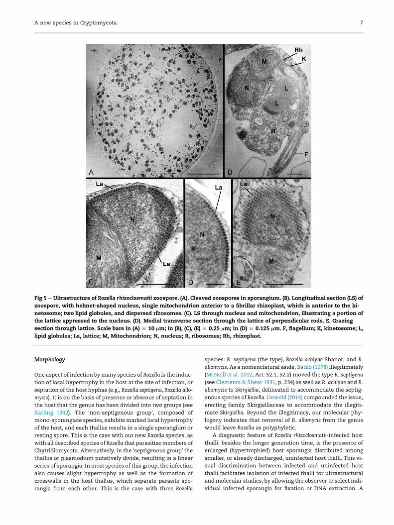
Fig 5 e Ultrastructure of Rozella rhizoclosmatii zoospore. (A). Cleaved zoospores in sporangium. (B). Longitudinal section (LS) of
zoospore, with helmet-shaped nucleus, single mitochondrion anterior to a fibrillar rhizoplast, which is anterior to the ki-
netosome; two lipid globules, and dispersed ribosomes. (C). LS through nucleus and mitochondrion, illustrating a portion of
the lattice appressed to the nucleus. (D). Medial transverse section through the lattice of perpendicular rods. E. Grazing
section through lattice. Scale bars in (A) [ 10 mm; in (B), (C), (E) [ 0.25 mm; in (D) [ 0.125 mm. F, flagellum; K, kinetosome; L,
lipid globules; La, lattice; M, Mitochondrion; N, nucleus; R, ribosomes; Rh, rhizoplast.
A new species in Cryptomycota 7
Morphology
One aspect of infection bymany species of Rozella is the induc-
tion of local hypertrophy in the host at the site of infection, or
septation of the host hyphae (e.g., Rozella septigena, Rozella allo-
mycis). It is on the basis of presence or absence of septation in
the host that the genus has been divided into two groups (see
Karling 1942). The ‘non-septigenous group’, composed of
mono-sporangiate species, exhibits marked local hypertrophy
of the host, and each thallus results in a single sporangium or
resting spore. This is the case with our new Rozella species, as
with all described species of Rozella that parasitizemembers of
Chytridiomycota. Alternatively, in the ‘septigenous group’ the
thallus or plasmodium putatively divide, resulting in a linear
series of sporangia. Inmost species of this group, the infection
also causes slight hypertrophy as well as the formation of
crosswalls in the host thallus, which separate parasite spo-
rangia from each other. This is the case with three Rozella
species: R. septigena (the type), Rozella achlyae Shanor, and R.
allomycis. As a nomenclatural aside, Batko (1978) illegitimately
(McNeill et al. 2012, Art. 52.1, 52.2) moved the type R. septigena
(see Clements & Shear 1931, p. 234) as well as R. achlyae and R.
allomycis to Skirgiellia, delineated to accommodate the septig-
enous species of Rozella. Doweld (2014) compounded the issue,
erecting family Skirgielliaceae to accommodate the illegiti-
mate Skirgiellia. Beyond the illegitimacy, our molecular phy-
logeny indicates that removal of R. allomycis from the genus
would leave Rozella as polyphyletic.
A diagnostic feature of Rozella rhizoclosmatii-infected host
thalli, besides the longer generation time, is the presence of
enlarged (hypertrophied) host sporangia distributed among
smaller, or already discharged, uninfected host thalli. This vi-
sual discrimination between infected and uninfected host
thalli facilitates isolation of infected thalli for ultrastructural
andmolecular studies, by allowing the observer to select indi-
vidual infected sporangia for fixation or DNA extraction. A
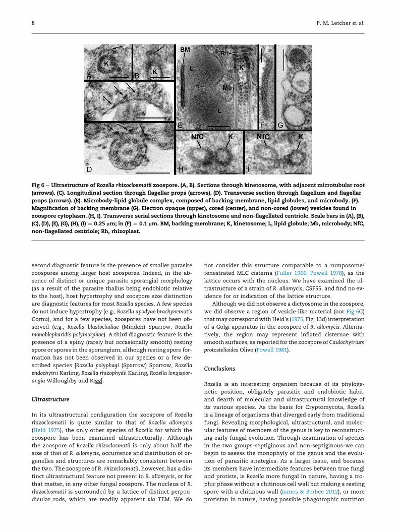
Fig 6 e Ultrastructure of Rozella rhizoclosmatii zoospore. (A, B). Sections through kinetosome, with adjacent microtubular root
(arrows). (C). Longitudinal section through flagellar props (arrows). (D). Transverse section through flagellum and flagellar
props (arrows). (E). Microbody-lipid globule complex, composed of backing membrane, lipid globules, and microbody. (F).
Magnification of backing membrane (G). Electron opaque (upper), cored (center), and non-cored (lower) vesicles found in
zoospore cytoplasm. (H, I). Transverse serial sections through kinetosome and non-flagellated centriole. Scale bars in (A), (B),
(C), (D), (E), (G), (H), (I) [ 0.25 mm; in (F) [ 0.1 mm. BM, backing membrane; K, kinetosome; L, lipid globule; Mb, microbody; NfC,
non-flagellated centriole; Rh, rhizoplast.
8 P. M. Letcher et al.
second diagnostic feature is the presence of smaller parasite
zoospores among larger host zoospores. Indeed, in the ab-
sence of distinct or unique parasite sporangial morphology
(as a result of the parasite thallus being endobiotic relative
to the host), host hypertrophy and zoospore size distinction
are diagnostic features for most Rozella species. A few species
do not induce hypertrophy (e.g., Rozella apodyae brachynematis
Cornu), and for a few species, zoospores have not been ob-
served (e.g., Rozella blastocladiae (Minden) Sparrow, Rozella
monoblepharidis polymorphae). A third diagnostic feature is the
presence of a spiny (rarely but occasionally smooth) resting
spore or spores in the sporangium, although resting spore for-
mation has not been observed in our species or a few de-
scribed species [Rozella polyphagi (Sparrow) Sparrow, Rozella
endochytrii Karling, Rozella rhizophydii Karling, Rozella longispor-
angia Willoughby and Rigg].
Ultrastructure
In its ultrastructural configuration the zoospore of Rozella
rhizoclosmatii is quite similar to that of Rozella allomycis
(Held 1975), the only other species of Rozella for which the
zoospore has been examined ultrastructurally. Although
the zoospore of Rozella rhizoclosmatii is only about half the
size of that of R. allomycis, occurrence and distribution of or-
ganelles and structures are remarkably consistent between
the two. The zoospore of R. rhizoclosmatii, however, has a dis-
tinct ultrastructural feature not present in R. allomycis, or for
that matter, in any other fungal zoospore. The nucleus of R.
rhizoclosmatii is surrounded by a lattice of distinct perpen-
dicular rods, which are readily apparent via TEM. We do
not consider this structure comparable to a rumposome/
fenestrated MLC cisterna (Fuller 1966; Powell 1978), as the
lattice occurs with the nucleus. We have examined the ul-
trastructure of a strain of R. allomycis, CSF55, and find no ev-
idence for or indication of the lattice structure.
Althoughwe did not observe a dictyosome in the zoospore,
we did observe a region of vesicle-like material (our Fig 6G)
thatmay correspondwith Held’s (1975, Fig. 13d) interpretation
of a Golgi apparatus in the zoospore of R. allomycis. Alterna-
tively, the region may represent inflated cisternae with
smooth surfaces, as reported for the zoospore of Caulochytrium
protostelioides Olive (Powell 1981).
Conclusions
Rozella is an interesting organism because of its phyloge-
netic position, obligately parasitic and endobiotic habit,
and dearth of molecular and ultrastructural knowledge of
its various species. As the basis for Cryptomycota, Rozella
is a lineage of organisms that diverged early from traditional
fungi. Revealing morphological, ultrastructural, and molec-
ular features of members of the genus is key to reconstruct-
ing early fungal evolution. Through examination of species
in the two groups-septiginous and non-septiginous-we can
begin to assess the monophyly of the genus and the evolu-
tion of parasitic strategies. As a larger issue, and because
its members have intermediate features between true fungi
and protists, is Rozella more fungal in nature, having a tro-
phic phase without a chitinous cell wall but making a resting
spore with a chitinous wall (James & Berbee 2012), or more
protistan in nature, having possible phagotrophic nutrition

Fig 7 e Schematic of Rozella rhizoclosmatii zoospore. BM,
backing membrane; F, flagellum; FR, fibrillar rhizoplast; K,
kinetosome; L, lipid globule; M, mitochondrion; Mb, micro-
body; Mt, microtubule; N, nucleus; NfC, non-flagellated
centriole; P, flagellar props; R, ribosomes; Ves, vesicle. Lat-
tice surrounds nucleus.
A new species in Cryptomycota 9
(Powell 1984)? Further investigations of Rozella to reveal ul-
trastructural and molecular features of described species
and to discover new species will help put a face on Crypto-
mycota and further our understanding of this enigmatic
group.
Acknowledgments
This study was supported by the National Science Foundation
through MRI DEB-0500766 (The University of Alabama), DEB-
1455611 (P. Letcher and M. Powell), DEB-1354625 (A. Quandt,
T. James), Sao Paulo Research Foundation FAPESP #2015/
05596-4 (D. Leite), and the JEL culture collection at the Univer-
sity of Maine (J. Longcore).
r e f e r e n c e s
Batko A, 1978. Further observations on Nellymyces megaceros andits parasite- Rozellopsis uliginosa sp. nov. Acta Mycologica War-saw 13: 313e324 [1977].
Berbee ML, Taylor JW, 1993. Dating the evolutionary radiations ofthe true fungi. Canadian Journal of Botany 71: 1114e1127.
Boyle DG, Hyatt AD, Daszak P, Berger L, Longcore JE, Porter D,Hengstberger SG, Olsen V, 2003. Cryo-archiving of Batracho-chytrium dendrobatidis and other chytridiomycetes. Diseases ofAquatic Organisms 56: 59e64.
Clements FE, Shear CL, 1931. The Genera of Fungi. H.W. Wilson,New York.
Cornu M, 1872. Monographie des Saprol�egni�ees: �etude physiolo-gique et syst�ematique. Annales de la Soci�et�e Nationale Botanique15: 1e198.
Corsaro D, Walochnik J, Venditti D, Steinmann J, M€uller K-D,Michel R, 2014a. Microsporidia-like parasites of amoebae be-long to the early lineage Rozellomycota. Journal of ParasitologyResearch 113: 1909e1918.
Corsaro D, Walochnik J, Venditti D, M€uller K-D, Haur€oder B,Michel R, 2014b. Rediscovery of Nucleophaga amoebae, a novelmember of the Rozellomycota. Journal of Parasitology Research113: 4491e4498.
Corsaro D, Michel R, Walochnik J, Venditti D, M€uller K-D,Haur€oder B, Wylezich C, 2016. Molecular identification of Nu-cleophaga terricolae sp. nov. (Rozellomycota), and new insightson the origin of the Microsporidia. Journal of Parasitology Re-search. http://dx.doi.org/10.1017/s00436-016-5055-9.
Deng Y, Zhang Y, Gao Y, Li D, Liu R, Liu M, Zhang H, Hu B, Yu T,Yang M, 2012. Microbial community compositional analysisfor series reactors treating high level antibiotic wastewater.Environmental Science and Technology 46: 795e801.
Doweld AB, 2013. Nomenclatural novelties. Rozellomycota IndexFungorum 43: 1.
Doweld AB, 2014. Nomenclatural novelties. Skirgielliaceae IndexFungorum 129: 1.
Foust FK, 1937. A new species of Rozella parasitic on Allomyces.Journal of the Elisha Mitchell Scientific Society 53: 197e204.
Fuller MS, 1966. Structure of the uniflagellate zoospores of aquaticphycomycetes. In: Madelin MF (ed.), The Fungus Spore. Butter-worth’s, London, pp. 67e84.
Hall TA, 1999. BioEdit: a user-friendly biological sequence align-ment editor and analysis program for Windows 95/98/NT.Nucleic Acids Symposium Serial (Oxford) 41: 95e98.
Held AA, 1975. The zoospore of Rozella allomycis: ultrastructure.Canadian Journal of Botany 53: 2212e2232.
James TY, Letcher PM, Longcore JE, Mozley-Standridge SE,Porter D, Powell MJ, Griffith GW, Vilgalys R, 2006. A molecularphylogeny of the flagellated fungi (Chytridiomycota) and de-scription of a new phylum (Blastocladiomycota). Mycologia 98:860e871.
James TY, Berbee ML, 2012. No jacket required- new fungal line-age defies dress code. Bioessays 34: 94e102.
Jones MDM, Forn I, Gadelha C, Egan MJ, Bass D, Massana R,Richards TA, 2011a. Discovery of novel intermediate formsredefines the fungal tree of life. Nature 474: 200e203.
Jones MDM, Richards TA, Hawksworth DL, Bass D, 2011b. Vali-dation and justification of the phylum name Cryptomycotaphyla. nov. IMA Fungus 2: 173e175.
Karling JS, 1942. Parasitism among the chytrids. American Journalof Botany 29: 24e35.
Karling JS, 1987. Ross biological reserve aquatic fungi III. Addi-tional species. Nova Hedwigia 45: 529e535.
Karpov SA, Mamkaeva MA, Aleoshin VV, Nassonova E, Lilje O,Gleason FH, 2014. Morphology, phylogeny, and ecology of theaphelids (Aphelidea, Opisthokonta) and proposal for the new

10 P. M. Letcher et al.
superphylum Opisthosporidia. Frontiers in Microbiology 5 (Arti-cle 112): 1e11.
Lara E, Moriera D, Lopez-Garcia P, 2010. The environmental cladeLKM11 and Rozella form the deepest branching clade of Fungi.Protist 161: 116e121.
Lazarus KL, James TY, 2015. Surveying the biodiversity of theCryptomycota using a targeted PCR approach. Fungal Ecology 14:62e70.
Letcher PM, Lee PA, Lopez S, Burnett M, McBride RC, Powell MJ,2016. An ultrastructural study of Paraphysoderma sedebokerense(Blastocladiomycota), an epibiotic parasite of microalgae.Fungal Biology 120: 324e337.
Letcher PM, Powell MJ, 2014. Hypothesized evolutionary trends inzoospore ultrastructural characters in Chytridiales (Chytri-diomycota). Mycologia 106: 379e396.
McNeill J, Barrie FR, Buck WR, Demoulin V, Greuter W,Hawksworth DL, et al., 2012. International Code of Nomenclaturefor Algae, Fungi, and Plants (Melbourne Code). . In: Regnum Vege-tabile, vol. 154. Koeltz Scientific Books.
Nishio K, Hashimoto K, Watanabe K, 2010. Light/electricity con-version by a self-organized photosynthetic biofilm in a single-chamber reactor. Applied Microbiology and Biotechnology 86:957e964.
Powell MJ, 1978. Phylogenetic implications of the microbody-lipidglobule complex in zoosporic fungi. Biosystems 10: 167e180.
Powell MJ, 1981. Zoospore structure of the mycoparasitic chytridCaulochytrium protostelioides Olive. American Journal of Botany68: 1074e1089.
Powell MJ, 1984. Fine structure of the unwalled thallus of Rozellapolyphagi in itshostPolyphaguseuglenae.Mycologia76: 1039e1048.
Sikes DS, Lewis PO, 2001. Beta Software. Version 1. PAUPRat: PAUP*implementation of the parsimony ratchet. Department EcologyEvolutionary Biology, Univ. Connecticut, Storrs, Distributed bythe authors.
Sparrow FK, 1960. Aquatic Phycomycetes, 2nd edn. University ofMichigan Press, Ann Arbor.
Swofford DL, 2002. PAUP* 4.0b4a: phylogenetic analysis using parsi-mony (*and other methods). Sinauer Associates, Sunderland, MA.
Thompson JD, Gibson TJ, Plewniak F, Jeanmougin F, Higgins DG,1997. The Clustal X windows interface: flexible strategies formultiple sequence alignment aided by quality analysis tools.Nucleic Acids Research 25: 4876e4882.
V�elez CG, Letcher PM, Schultz S, Powell MJ, Churchill PF, 2011.Molecular phylogenetic and zoospore ultrastructural analysesof Chytridium olla establish the limits of a monophyletic Chy-tridiales. Mycologia 103: 118e130.
Zwikl DJ, 2006. Genetic algorithm approaches for the phylogeneticanalysis of large biological sequence datasets under the maximumlikelihood criterion [doctoral dissertation]. University of Texas,Austin, 115 p..