Plant differentiation (organogenesis and somatic embryogenesis)

Revista Brasil. Bot., V.27, n.3, p.429-437, jul.-set. 2004
Ultrastructural analysis of in vitro direct and indirect organogenesisBEATRIZ APPEZZATO-DA-GLÓRIA1 and SILVIA R. MACHADO2
(received: October 9, 2002; accepted: March 18, 2004)
ABSTRACT – (Ultrastructural analysis of in vitro direct and indirect organogenesis). This is a comparative study of theultrastructural characteristics of the cells involved in the organogenesis in vitro of Bauhinia forficata Link (indirect system)and Glycine max (L.) Merrill (direct system). B. forficata calli after 30 days culture and G. max meristemoids after 10 days culturewere prepared for ultrastructural analysis using conventional methods. Concentrically arranged rough endoplasmic reticulum(RER) and plastids containing starch grains were seen during G. max and B. forficata organogenesis. The amitotic process, thepresence of plastids around the nucleus and nuclear envelope with conspicuous pores were found in B. forficata.
Key words - amitosis, leguminous, meristemoids, morphogenesis
RESUMO – (Análise ultra-estrutural da organogênese in vitro direta e indireta). Este é um estudo comparativo das característicasultra-estruturais das células envolvidas na organogênese in vitro de Bauhinia forficata Link (sistema indireto) e Glycine max(L.) Merrill (sistema direto). Calos de B. forficata, após 30 dias de cultura, e meristemóides de G. max, após 10 dias de cultura,foram preparados para as análises ultra-estruturais usando métodos convencionais. Retículo endoplasmático rugoso (RER)arranjado concentricamente e plastídeos contendo grãos de amido foram observados durante a organogênese de G. max eB. forficata. O processo amitótico, a presença de plastídeos ao redor do núcleo e de envelope nuclear com poros conspícuosforam observados apenas em B. forficata.
Palavras-chave - amitose, leguminosas, meristemóides, morfogênese
Introduction
The understanding of plant organogenesis and theinitial developmental stages of meristemoids requireobservation of subcellular level changes and theircorrelation with biochemical alterations (Pihakashi-Maunsbach et al. 1993).
Studies on ultrastructural alterations duringorganogenesis in vitro to characterize the meristemoidalcells responsible for bud formation are scarce (Villaloboset al. 1985, Pihakashi-Maunsbach et al. 1993, Araiet al. 1997). These studies do not make comparativeanalyses between the direct and indirect regenerationsystems or relate the ultrastructural aspects to theorganogenic potential. Some authors, however, haveattempted to relate ultrastructural characteristics toembryogenic potential (Konar et al. 1972, Radojevicet al. 1975, Vujicic et al. 1979).
Histological studies of Bauhinia forficata Link andGlycine max (L.) Merrill have demonstrated that
in vitro plant regeneration occurred throughoutorganogenesis (Vieira & Appezzato-da-Glória 2001).Glycine max regenerated by direct organogenesis, withshoot buds originating from cotyledonary node epidermaland subepidermal tissue, and Bauhinia forficata Linkregenerated by indirect organogenesis. In these species,histological examination revealed that shoots alsodeveloped from superficial tissue layers, and that thepattern of shoot origin and development is very similarto that previously described in literature for otherleguminous species (McClean & Grafton 1989, Jackson& Hobbs 1990, Nauerby et al. 1991, Mohamed et al.1992, Malik & Saxena 1992).
This study is an analysis of the ultrastructuralcharacteristics of the cells involved in the organogenesisin vitro of Bauhinia forficata (indirect system) andGlycine max (direct system).
Material and methods
Tissue culture – Bauhinia forficata Link calli were collectedfrom 1 cm long hypocotyls excised from in vitro growingplants. The explants were cultured in 1/2 MS (Murashige &Skoog 1962) basal medium supplemented with 3.0% (w/v)sucrose, 2.3 g L-1 phytagel (Sigma) and 4.0 mg L-1 BAP(benzylaminopurine). The pH was adjusted to 5.8 prior toautoclaving. The cultures were incubated at 25 ± 2 ºC in a16-hour photoperiod, and with 30 µmolm-2 s-1 irradiance
1. Universidade de São Paulo, Escola Superior de AgriculturaLuiz de Queiroz, Departamento de Ciências Biológicas, CaixaPostal 9, 13418-900 Piracicaba, SP, Brasil.
2. Universidade Estadual Paulista, Instituto de Biociências,Departamento de Botânica, Caixa Postal 510, 18618-000Botucatu, SP, Brasil.
3. Corresponding author: [email protected]

B. Appezzato-da-Glória & S.R. Machado: Ultrastructure of In Vitro Organogenesis430
provided by Philips cool white fluorescent tubes. Glycinemax (L.) Merrill meristemoids were collected fromcotyledonary nodes devoid of axillary buds used as explants.The explants were cultured in 1/2 MS basal mediumsupplemented with 3.0% (w/v) sucrose, 7.0 g L-1 agar (Merck),0.56 mg L-1 BAP (benzylaminopurine). The pH was adjustedto 5.8 prior to autoclaving. The cultures were incubated at27 ± 2 ºC in a 16-hour photoperiod, and with 30 µmolm-2 s-1
irradiance provided by Philips cool white fluorescent tubes.Ultrastructural analysis – Bauhinia forficata Link calli after30 days culture and G. max meristemoids after 10 days culturewere fixed in 2.5% glutaraldehyde, post-fixed in 1% OsO4,both prepared in phosphate buffer 0.1M, pH 7.3. The sampleswere dehydrated in an ethanol alcoholic series and embeddedin Araldite resin (Silva & Machado 1999). Ultra-thin sectionswere stained with uranyl acetate and lead citrate (Reynolds1963) and examined with a Philips TEM 100 operated at 80 kV.
Results
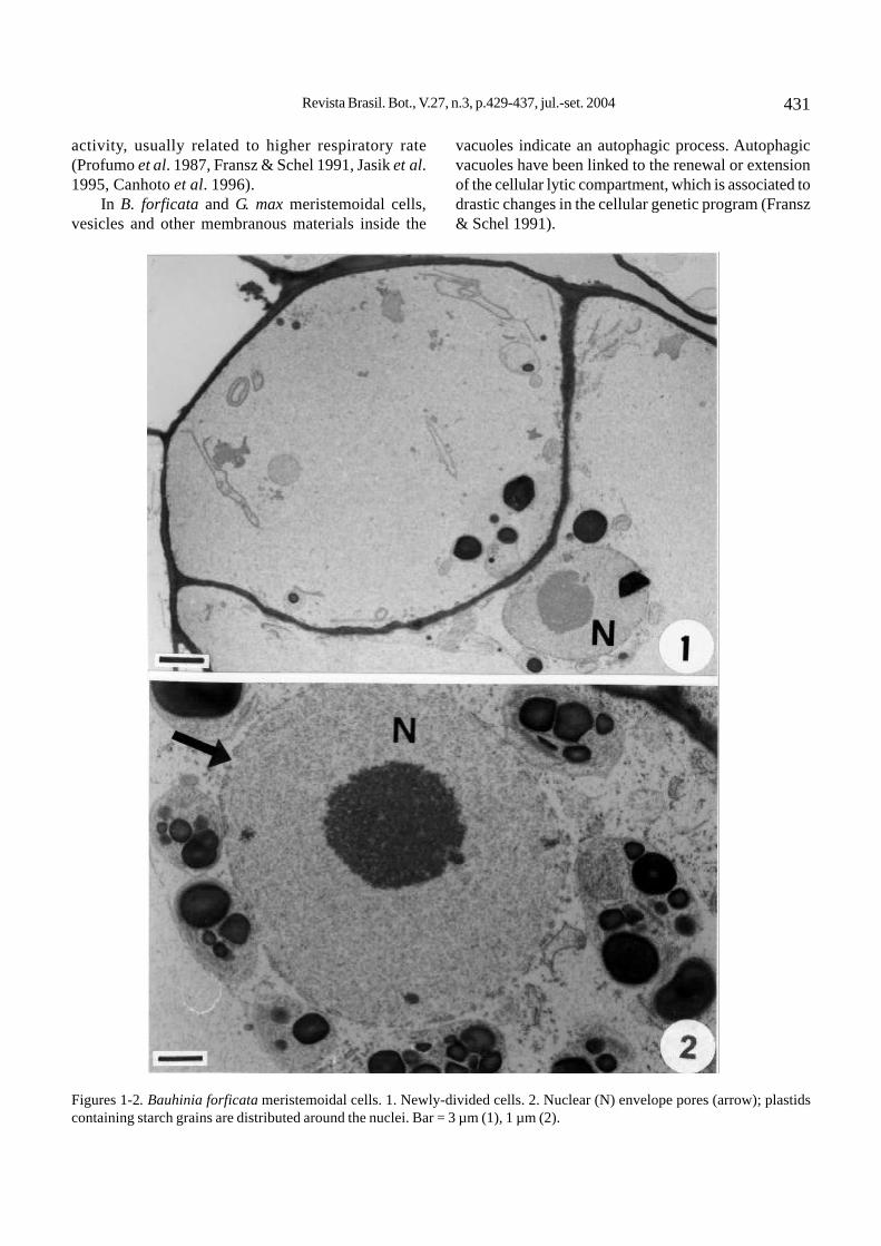
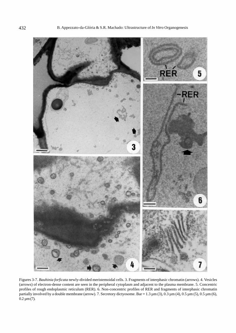
The Bauhinia forficata meristemoidal cells arevacuolated. These generally undergo unequal divisionresulting in different sized daughter cells (figure 1).These cells show a spherical nucleus, a large nucleolus,and a nuclear envelope with conspicuous pores(figure 2). There are abundant amiloplasts usuallylocated around the nucleus (figure 2). Vesicles ofelectron-dense content can be seen in peripheralcytoplasm and adjacent to the plasma membrane (figure4) and tonoplast. The rough endoplasmic reticulum(RER) may be concentrically arranged (figure 5) orextensive (figure 6). The most striking ultrastructuralfeature is the appearance, in newly-divided cells, offragments of interphasic chromatin scattered within thecytoplasm (figures 3, 6). These fragments may be totallyor partially surrounded by a double membrane (figure 6).Another major aspect in some cells is the nuclearenvelope disintegration without simultaneous chromatincondensation. Dictyosome are frequent; each one iscomposed of 8 to 10 dilated cisternae (figure 7).
The Glycine max subepidermal cells, which startdivision resulting in the formation of meristemoids, showa large central vacuole, and nuclei of different shapesand irregular outlines (figure 8). These cells generallyundergo unequal division so that daughter cells are ofdifferent sizes. The wall between the daughter cells isrelatively thinner than the mother cell wall (figure 12).The daughter cells show a central nucleus with onenucleolus and abundant cytoplasm rich in ribosomes(figures 8, 9). The dictyosomes are composed of fourto six cisternae and are active in vesicle production(figures 9, 14). Vesicles and other membraneous
materials can be seen inside the vacuoles (figure 9).Dilated regions of chloroplasts containing material ofdifferent electron-density to stroma and dividingchloroplasts are common in these cells (figure 10). Thechloroplasts have an extensive internal membranesystem, with one or more starch grains (figure 13). Thecytoplasmic connections through plasmodesmata arenumerous in daughter cells (figure 11), indicatingintensive interaction through the symplast. One of themajor ultrastructural features is the RER concentricarrangement (figures 12-14), which seems to be closelyassociated with the dictyosomes (figure 14), plastids,and mitochondria (figure 13). The mitochondria aregenerally oval or round and contain an electron-densematrix and well-developed cristae (figure 13). Manydividing mitochondria can be seen (figure 15).
Discussion
The meristemoids in Bauhinia forficata andGlycine max do not originate from typical meristemoidalcells but from highly vacuolated cells, similar to thoseobserved by Villalobos et al. (1985) during organogenesisin Pinus radiata. In general, these vacuolated cellsdivide unequally, giving rise to daughter cells of differentsizes. This frequently occurs in wounded meristems,where the vacuolated cells show nuclei adjacent to thetrauma area during prophase (Sinnott & Bloch 1941).
In both studied species, the wall between thedaughter cells is extremely thinner than that of themother cell. This has been reported in other organogenic(Villalobos et al. 1985) and embryogenic (Button et al.1974) processes. In G. max, the cytoplasmicconnections, through plasmodesmata, are numerous inthe daughter cells, indicating an intense interaction viasymplast. It was also observed in Zea mays apical regioncells of somatic embryos (Fransz & Schel 1991).
Comparison between the B. forficata and G. maxmeristemoids shows that the nuclear envelope ofB. forficata has numerous and conspicuous pores. Thenuclear pores are dynamic structures, their number beingdirectly proportional to protein production and transport(Garrido et al. 1995).
In G. max, the presence of dividing chloroplastsand mitochondria during the initial phases ofmeristemoid formation allows even distribution of theseorganelles in the daughter cells during cytokinesis. InG. max, the mitochondria are abundant and contain anelectron dense matrix and well-developed cristae.These features have been described in embryogenicsystems as being associated with higher metabolic
Revista Brasil. Bot., V.27, n.3, p.429-437, jul.-set. 2004 431
Figures 1-2. Bauhinia forficata meristemoidal cells. 1. Newly-divided cells. 2. Nuclear (N) envelope pores (arrow); plastidscontaining starch grains are distributed around the nuclei. Bar = 3 µm (1), 1 µm (2).
activity, usually related to higher respiratory rate(Profumo et al. 1987, Fransz & Schel 1991, Jasik et al.1995, Canhoto et al. 1996).
In B. forficata and G. max meristemoidal cells,vesicles and other membranous materials inside the
vacuoles indicate an autophagic process. Autophagicvacuoles have been linked to the renewal or extensionof the cellular lytic compartment, which is associated todrastic changes in the cellular genetic program (Fransz& Schel 1991).
B. Appezzato-da-Glória & S.R. Machado: Ultrastructure of In Vitro Organogenesis432
Figures 3-7. Bauhinia forficata newly-divided meristemoidal cells. 3. Fragments of interphasic chromatin (arrows). 4. Vesicles(arrows) of electron-dense content are seen in the peripheral cytoplasm and adjacent to the plasma membrane. 5. Concentricprofiles of rough endoplasmic reticulum (RER). 6. Non-concentric profiles of RER and fragments of interphasic chromatinpartially involved by a double membrane (arrow). 7. Secretory dictyosome. Bar = 1.3 µm (3), 0.3 µm (4), 0.5 µm (5), 0.5 µm (6),0.2 µm (7).

Revista Brasil. Bot., V.27, n.3, p.429-437, jul.-set. 2004 433
Figures 8-11. Glycine max meristemoidal cells. 8. Newly-divided cells of irregular nucleus shape (arrow). 9. Autophagic vacuoles(V) and dictyosomes (d). 10. Dividing chloroplasts showing dilation-containing material of different electron-density to stroma(arrow). 11. Cytoplasmic connections (arrows) through plasmodesmata between the daughter cells. Bar = 4 µm (8), 0.2 µm (9),0.6 µm (10), 0.2 µm (11).

B. Appezzato-da-Glória & S.R. Machado: Ultrastructure of In Vitro Organogenesis434
Figures 12-15. Glycine max meristemoidal cells. 12. Cell walls between daughter cells are thinner than the mother cell wall.13. Rough endoplasmic reticulum (RER) associated with amiloplastid and mitochondria (M). 14. Concentric profiles of RERsurrounding secretory dictyosome (arrow). 15. Dividing mitochondria. Bar = 2.3 µm (12), 0.6 µm (13), 0.3 µm (14), 0.2 µm (15).
Revista Brasil. Bot., V.27, n.3, p.429-437, jul.-set. 2004 435
In the two systems analyzed in this study,meristemoidal cell differentiation is associated with highermetabolic activity characteristics, such as an increasein the number of polysomes and mitochondrial cristae,and a proliferation of dictyosomes and RER segments(Profumo et al. 1987, Fransz & Schel 1991, Pihakashi-Maunsbach et al. 1993).
In B. forficata and G. max, the dictyosomes areactive in producing many vesicles, which can be seennear the plasma membrane. The increase in number ofdictyosomes, typically involved in the production of cellwall material, may be connected with an acceleratedsynthesis of cell wall compounds after one day ofculture, as seen in Brassica napus by Pihakashi-Maunsbach et al. (1993).
In both species, although the dividing cells of themeristemoids show plastids containing starch grains, itis only in B. forficata that these plastids are usuallylocated around the nucleus. In G. max and in Pinusradiata (Villalobos et al. 1985), both having directregeneration systems, the chloroplasts are randomlydistributed throughout the cytoplasm.
In G. max, chloroplasts with dilations, containingmaterial of a similar electron-density to the cytoplasm,are similar to those observed by Jasik et al. (1995) innon-embryogenic calli. In these calli, some plastidsshowed cytoplasms in cavity. Other ultrastructuralchanges in plastids: secondary dedifferentiation ofmature chloroplasts, thylakoid swelling and disruption,plastid elongation and increase in size were describedin Mammillaria gracillis cultivated in vitro (Poljuhaet al. (2003). According to the authors, plastids areorganelles very sensitive to the artificial environment inthe culture.
Starch grains in the plastids of meristemoidaldividing cells seems to be a common feature oforganogenesis, which has been reported in literaturedescribing ultrastructural changes in this regenerationsystem (Villalobos et al. 1985, Pihakashi-Maunsbachet al. 1993, Ovecka et al. 2000). This feature hasfrequently been related to the acquisition of embryogenicpotential, as embryogenic cells usually contain starchgrains (Maheswaran & Williams 1985, Profumo et al.1987). In this context, why cannot the occurrence ofstarch in the plastids be seen as a feature associatedwith the acquisition of organogenic potential?
However, starch degradation during bud primordiumformation, as described in literature (Arnold 1987,Mangat et al. 1990, Pihakashi-Maunsbach et al. 1993,Arai et al. 1997) as well as in analysis performed onG. max (Vieira & Appezzato-da-Glória 2001) suggest
that starch has a closer association with the highnutritional requirement of cell populations, independentof their regeneration system (Arnold 1987).
The usual RER concentric profile, seen during theinitial stages of G. max and B. forficata meristemoids,may be involved in protein synthesis in response to rapidcellular growth, and not associated with the potential ofa morphogenetic system, as suggested for embryogenicprocesses by Radojevic et al. (1975), Vujicic et al.(1979), and Canhoto et al. (1996). As can be seen inthis study, reporting the occurrence of RER concentricprofiles in organogenetic systems should be morecommon.
Therefore, two ultrastructural characteristicsgenerally associated only with the embryogenic potential,RER in concentric profiles, and plastids containingstarch, have been associated only with the embryogenicpotential. As this has also been observed in B. forficataand G. max organogenesis, these characteristics mayreveal a high synthesis capacity associated with thereorganization of growth pattern during induction ofsomatic morphogenesis.
In B. forficata, which displays indirectorganogenesis, the appearance of interphasic chromatinfragments in the cytoplasm in newly-divided meristemoidcells indicate the occurrence of amitosis (nuclearfragmentation). According to Wilson’s definition (apudBregoli et al. 1997) amitosis is the fragmentation of thenucleus into two parts, but as observed in this study,fragmentation may be multiple. Amitotic divisionapparently occurs at the transition from a differentiatecell state to one of disorganized growth, as theyfrequently appear as an initial response to woundingcaused by excision. In fact, in Vicia faba (Cionini et al.1978), Phaseolus vulgaris (Fasseas & Bowes 1980),and Helianthus tuberosus (Bregoli et al. 1997), amitoticdivisions occur mainly during the initial callus growthstages. Nuclear fragmentation, which occurs during theinitial stages of callogenesis followed by mitosis, maybe a major source of chromosomal variation in tissuecultures (Bayliss 1980). If this chromosomal variationin plant tissue cultures caused by amitotic processescan generate a genetic variability called somaclonalvariation (Larkin & Scowcroft 1981), then directorganogenesis, as seen in G. max in this study, could bethe best indication to obtain transgenic plants regeneratedfrom transformed explants.
Acknowledgments – This research was supported by theFapesp (Fundação de Amparo à Pesquisa do Estado de SãoPaulo) and CNPq (Conselho Nacional de Desenvolvimento

B. Appezzato-da-Glória & S.R. Machado: Ultrastructure of In Vitro Organogenesis436
Científico e Tecnológico). The authors are grateful to staff ofthe Centro de Microscopia Eletrônica, Unesp, Botucatu, forthe use of laboratory facilities.
References
ARAI, M., SAITO, T., KANEKO, Y. & MATSUSHIMA, H.1997. Cellular origin and ultrastructural changes ofregenerating shoots from tobacco (Nicotiana tabacum)internodes cultured in vitro. Physiologia Plantarum99:523-528.
ARNOLD, S. VON. 1987. Effect of sucrose on starchaccumulation in and adventitious bud formation onembryos of Picea abies. Annals of Botany 59:15-22.
BAYLISS, M.W. 1980. Chromosomal variation in plant tissuesin culture. International Review of Cytology 11A:113 -144.
BREGOLI, A.M, CROSTI, P., CAVALLINI, A., CIONINI, G.,DEL LUCA, S., MALERBA, M., NATALI, L., SERAFINI-FRACASSINI, D. & D’AMATO, F. 1997. Nuclear DNAdistribution and amitotic processes in activatedHelianthus tuberosus tuber parenchyma. PlantBiosystems 131:3-12.
BUTTON, J., KOCHBA, J. & BORNMAN, C. H. 1974. Finestructure of embryoid development from embryogenicovular callus of ‘Shamouti’ orange (Citrus sinensisOsb.). Journal of Experimental Botany 25:446-457.
CANHOTO, J.M., MESQUITA, J.F. & CRUZ, G.S. 1996.Ultrastructural changes in cotyledons of pineappleguava (Myrtaceae) during somatic embryogenesis.Annals of Botany 78:513-521.
CIONINI, P.G., BENNICI, A. & D’AMATO, F. 1978. Nuclearcytology of callus induction and development in vitro:I. Callus from Vicia faba cotyledons. Protoplasma96:101-112.
FASSEAS, C. & BOWES, B.G. 1980. Ultrastructuralobservations on proliferating storage cells of maturecotyledons of Phaseolus vulgaris L. cultured in vitro.Annals of Botany 46:143-152.
FRANSZ, P.F. & SCHEL, H.N. 1991. An ultrastructural studyon the early development of Zea mays somatic embryos.Canadian Journal of Botany 69:858-865.
GARRIDO, D., VICENTE, O., HEBERLE-BORS, E. &RODRIGUEZ-GARCÍA, M.I. 1995. Cellular changesduring the acquisition of embryogenic potential inisolated pollen grains of Nicotiana tabacum .Protoplasma 186:220-230.
JACKSON, J.A. & HOBBS, S.L. 1990. Rapid multiple shootproduction from cotyledonary node explants of pea(Pisum sativum L.). In vitro Cellular DevelopmentalBiology 26:835-838.
JASIK, J., SALAJOVA, T. & SALAJ, J. 1995. Developmentalanatomy and ultrastructure of early somatic embryos inEuropean black pine (Pinus nigra Arn.). Protoplasma185:205-211.
KONAR, R.N., THOMAS, E. & STREET, H.E. 1972. Originand structure of embryoids arising from the epidermalcells of Ranunculus sceleratus L. Journal of Cell Science11:77-93.
LARKIN, P. & SCOWCROFT, W.R. 1981. Somaclonalvariation: a novel source of variability from cell culturesfor plant improvement. Theoretical and Applied Genetics60:197-214.
MAHESWARAN, G. & WILLIAMS, E.G. 1985. Origin anddevelopment of somatic embryoids formed directly onimmature embryos of Trifolium repens in vitro. Annalsof Botany 56:619-630.
MALIK, K.A. & SAXENA, P.K. 1992. Somatic embryogenesisand shoot regeneration from intact seedlings ofPhaseolus acutifolius A. Gray, P. aureus (L.) Wilczek,P. coccineus L., and P. wrightii L. Plant Cell Reports1:163-168.
MANGAT, B.S., PELEKIS, M.K. & CASSELLS, A.C. 1990.Changes in the starch content during organogenesis invitro cultured Begonia rex stem explants. PhysiologiaPlantarum 79:267-274.
MCCLEAN, P. & GRAFTON, K.F. 1989. Regeneration of drybean (Phaseolus vulgaris L.) via organogenesis. PlantScience 60:117-122.
MOHAMED, M.F., READ, P.E. & COYNE, D.P. 1992. Darkpreconditioning, CPPU, and thidiazuron promote shootorganogenesis on seedling node explants of commonand faba beans. Journal of the American Society forHorticultural Science 117:668-672.
MURASHIGE, T. & SKOOG, F. 1962. A revised medium forrapid growth and bioassays with tobacco tissuecultures. Physiologia Plantarum 15:473-497.
NAUERBY, B., MADSEN, M., CHRISTIANSEN, J. &WYNDAELE, R. 1991. A rapid and efficient system forpea (Pisum sativum), suitable for transformation. PlantCell Reports 9:676-679.
OVECKA, M., BOBÁK, M. & SAMAJ, J. 2000. A comparativestructural analysis of direct and indirect shootregeneration of Papaver somniferum L. in vitro. Journalof Plant Physiology 157:281-289.
PIHAKASHI-MAUNSBACH, K., NYGAARD, K.B., JENSEN,K.H. & RASMUSSEN, O. 1993. Cellular changes in earlydevelopment of regenerating thin cell layer-explants ofrapeseed analysed by light and electron microscopy.Physiologia Plantarum 87:167-176.
POLJUHA, D., BALEN, B., BAUER, A., LJUBESIC, N. &KRSNIK-RASOL, M. 2003. Morphology andultrastructure of Mammillaria gracillis (Cactaceae) inin vitro culture. Plant Cell, Tissue and Organ Culture75:117-123.
PROFUMO, P., GASTALDO, P. & RASCIO, N. 1987.Ultrastructural study of different types of callus fromleaf explants of Aesculus hippocastanum L. Protoplasma138:89-97.
Revista Brasil. Bot., V.27, n.3, p.429-437, jul.-set. 2004 437
RADOJEVIC, L., VUJICIC, R. & NESKOVIC, M. 1975.Embryogenesis in tissue culture of Coryllus avellanaL. Zeitschrift für Pflanzenphysiologie 77:33-41.
REYNOLDS, E.S. 1963. The use of lead citrate and high pH asan electron-opaque stain in electron microscopy. Journalof Cell Biology 17:208-212.
SILVA, E.M.J. & MACHADO, S.R. 1999. Ultrastructure andcytochemistry of pearl gland in Piper regnellii (Miq.)C. DC.- Piperaceae. Nordic Journal of Botany19:623-634.
SINNOTT, E.W. & BLOCH, R. 1941. Division in vacuolateplant cells. American Journal of Botany 28:225-232.
VIEIRA, M.L.C. & APPEZZATO-DA-GLÓRIA, B. 2001.Fundamentos e aplicações da cultura de tecidos nomelhoramento. In Recursos genéticos e melhoramento –plantas. (L.L. Nass, A.C.C. Valois, I.S. Melo & N.C. Valadares-Inglis, eds.). Fundação MT, Rondonópolis, p.911-938.
VILLALOBOS, V.M., YEUNG, E.C. & THORPE, T.A. 1985.Origin of adventitious shoots in excised radiata pinecotyledons cultured in vitro. Canadian Journal of Botany63:2172-2176.
VUJICIC, R., RADOJEVIC, L. & NESKOVIC, M. 1979. Regularalignment of membrane ribosomes in embryogenic tissueof Paulownia tomentosa. Zeitschrift fürPflanzenphysiologie 91:53-56.