Ultrasonography of the ovary
78
Ultrasonography of the ovary Aboubakr Elnashar Benha university, Egypt ABOUBAKR ELNASHAR
-
Upload
aboubakr-elnashar -
Category
Health & Medicine
-
view
2.626 -
download
0
Transcript of Ultrasonography of the ovary
Ultrasonography of the ovary
Aboubakr Elnashar
Benha university, Egypt ABOUBAKR ELNASHAR
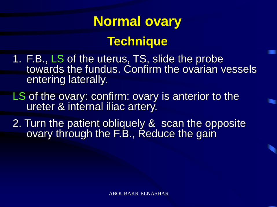
Normal ovary
Technique
1. F.B., LS of the uterus, TS, slide the probe towards the fundus. Confirm the ovarian vessels entering laterally.
LS of the ovary: confirm: ovary is anterior to the ureter & internal iliac artery.
2. Turn the patient obliquely & scan the opposite ovary through the F.B., Reduce the gain
ABOUBAKR ELNASHAR
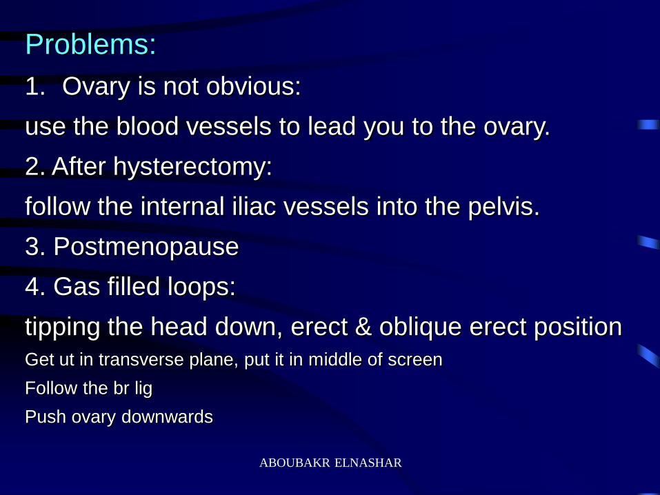
Problems:
1. Ovary is not obvious:
use the blood vessels to lead you to the ovary.
2. After hysterectomy:
follow the internal iliac vessels into the pelvis.
3. Postmenopause
4. Gas filled loops:
tipping the head down, erect & oblique erect position
Get ut in transverse plane, put it in middle of screen
Follow the br lig
Push ovary downwards
ABOUBAKR ELNASHAR
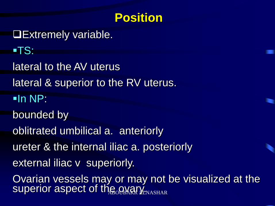
Position
Extremely variable.
TS:
lateral to the AV uterus
lateral & superior to the RV uterus.
In NP:
bounded by
oblitrated umbilical a. anteriorly
ureter & the internal iliac a. posteriorly
external iliac v superiorly.
Ovarian vessels may or may not be visualized at the superior aspect of the ovary. ABOUBAKR ELNASHAR
Echogenecity
Hypoechoic as compared to the uterus
{ multiple follicle in the cortex}.
Follicles:
Do not exceed 25 mm
round or ovoid, sharply marginated & anechoic.
Medulla & capsule:
higher echogenecity.
ABOUBAKR ELNASHAR
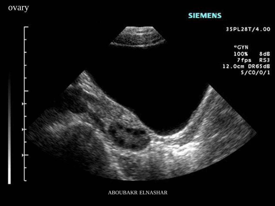
ovary
ABOUBAKR ELNASHAR
ABOUBAKR ELNASHAR
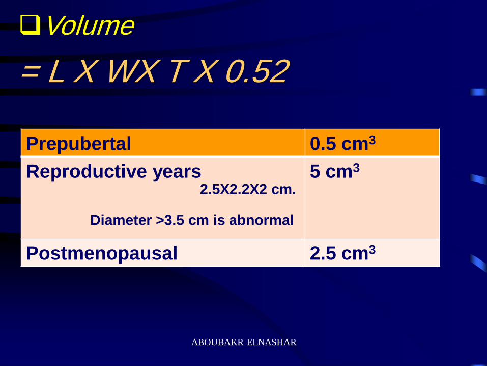
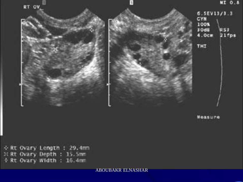
Volume
= L X WX T X 0.52
0.5 cm3 Prepubertal
5 cm3 Reproductive years 2.5X2.2X2 cm.
Diameter >3.5 cm is abnormal
2.5 cm3 Postmenopausal
ABOUBAKR ELNASHAR
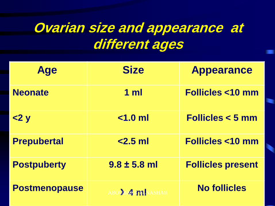
Ovarian size and appearance at different ages
Appearance Size Age
Follicles <10 mm 1 ml Neonate
Follicles < 5 mm <1.0 ml <2 y
Follicles <10 mm <2.5 ml Prepubertal
Follicles present 9.8 ± 5.8 ml Postpuberty
No follicles › 4 ml Postmenopause
ABOUBAKR ELNASHAR
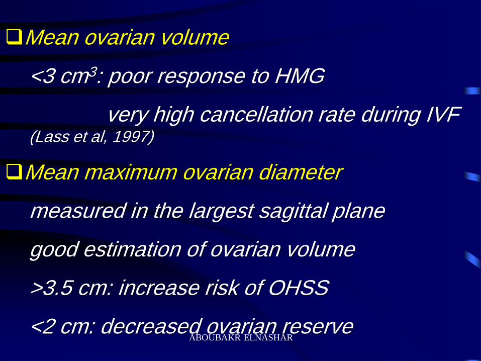
Mean ovarian volume
<3 cm3: poor response to HMG
very high cancellation rate during IVF (Lass et al, 1997)
Mean maximum ovarian diameter
measured in the largest sagittal plane
good estimation of ovarian volume
>3.5 cm: increase risk of OHSS
<2 cm: decreased ovarian reserve ABOUBAKR ELNASHAR

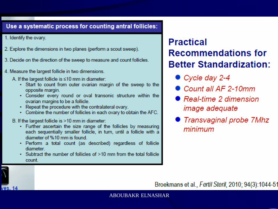
AFC: Resting follicles. Total number of follicles 2–8mm
counted in both ovaries
A threshold of 5 AF (2-5 mm) have the lowest error rate
for the prediction of poor response (Bancsi et al.,2004)
ABOUBAKR ELNASHAR
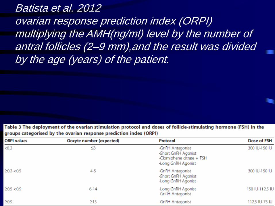
Batista et al. 2012 ovarian response prediction index (ORPI) multiplying the AMH(ng/ml) level by the number of antral follicles (2–9 mm),and the result was divided by the age (years) of the patient.
ABOUBAKR ELNASHAR
ABOUBAKR ELNASHAR
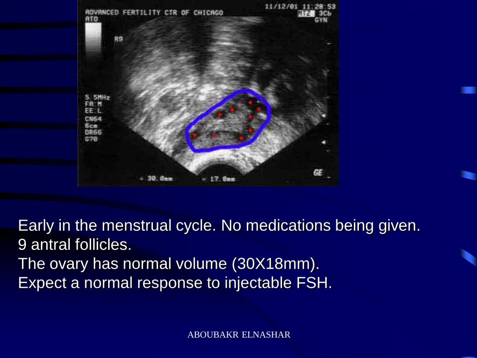
Early in the menstrual cycle. No medications being given.
9 antral follicles.
The ovary has normal volume (30X18mm).
Expect a normal response to injectable FSH.
ABOUBAKR ELNASHAR
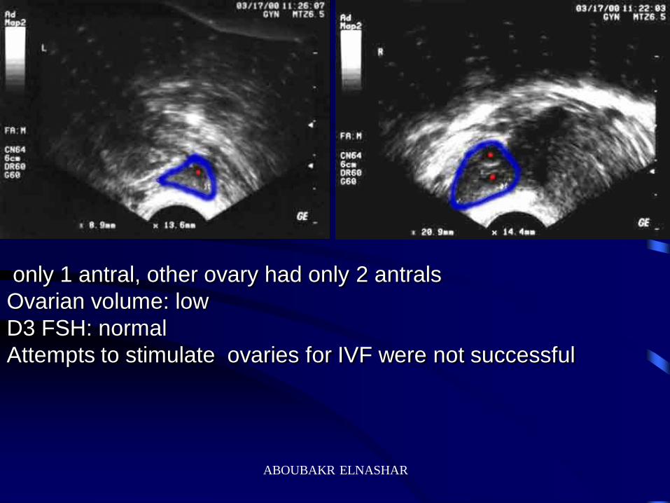
only 1 antral, other ovary had only 2 antrals
Ovarian volume: low
D3 FSH: normal
Attempts to stimulate ovaries for IVF were not successful
ABOUBAKR ELNASHAR

At the beginning of a menstrual cycle, irregular periods, No
medications being given.
Antral follicles:16 are seen in this image. Ovary had a total of 35
antrals (only 1 plane is shown). This is PCO with a high antral
Ovarian volume= 37 X19.5mm
"high responder" to injectable FSH drugs.
ABOUBAKR ELNASHAR
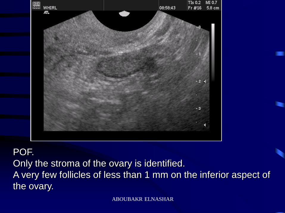
POF.
Only the stroma of the ovary is identified.
A very few follicles of less than 1 mm on the inferior aspect of
the ovary.
ABOUBAKR ELNASHAR
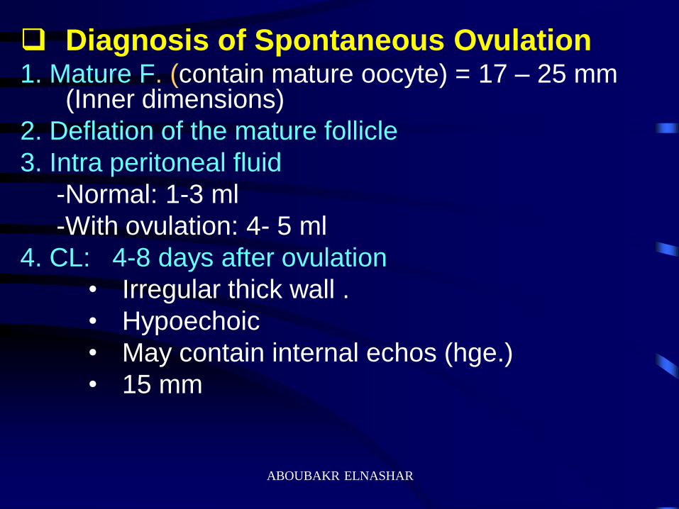
Diagnosis of Spontaneous Ovulation 1. Mature F. (contain mature oocyte) = 17 – 25 mm
(Inner dimensions)
2. Deflation of the mature follicle
3. Intra peritoneal fluid
-Normal: 1-3 ml
-With ovulation: 4- 5 ml
4. CL: 4-8 days after ovulation
• Irregular thick wall .
• Hypoechoic
• May contain internal echos (hge.)
• 15 mm
ABOUBAKR ELNASHAR
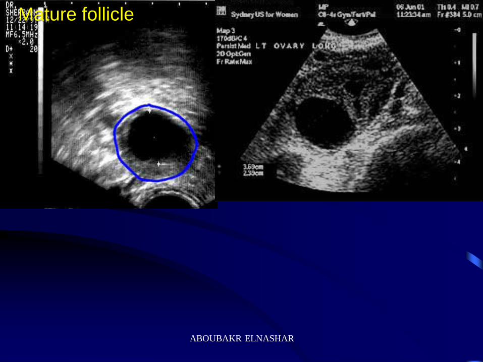
Mature follicle
ABOUBAKR ELNASHAR
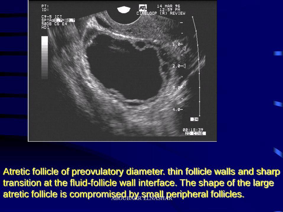
Atretic follicle of preovulatory diameter. thin follicle walls and sharp
transition at the fluid-follicle wall interface. The shape of the large
atretic follicle is compromised by small peripheral follicles. ABOUBAKR ELNASHAR
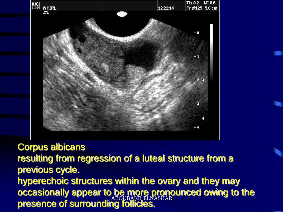
Corpus albicans
resulting from regression of a luteal structure from a
previous cycle.
hyperechoic structures within the ovary and they may
occasionally appear to be more pronounced owing to the
presence of surrounding follicles. ABOUBAKR ELNASHAR
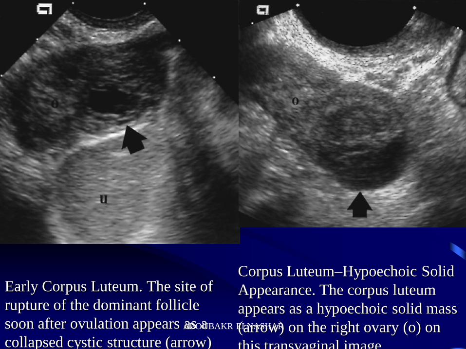
Early Corpus Luteum. The site of
rupture of the dominant follicle
soon after ovulation appears as a
collapsed cystic structure (arrow)
on the ovary (o). u, uterus.
Corpus Luteum–Hypoechoic Solid
Appearance. The corpus luteum
appears as a hypoechoic solid mass
(arrow) on the right ovary (o) on
this transvaginal image.
ABOUBAKR ELNASHAR
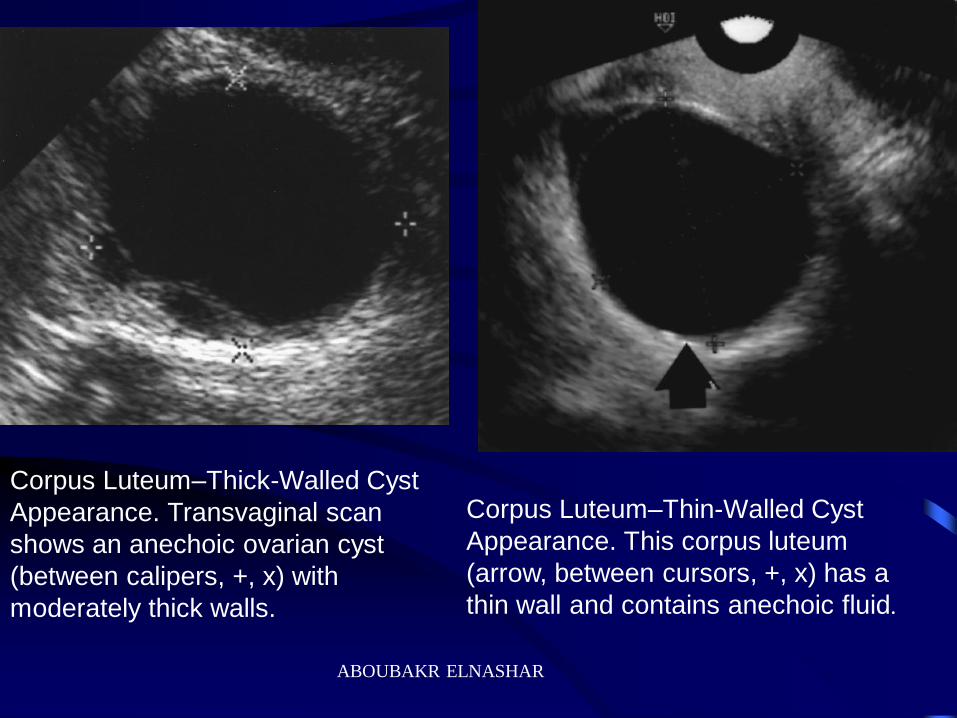
Corpus Luteum–Thick-Walled Cyst
Appearance. Transvaginal scan
shows an anechoic ovarian cyst
(between calipers, +, x) with
moderately thick walls.
Corpus Luteum–Thin-Walled Cyst
Appearance. This corpus luteum
(arrow, between cursors, +, x) has a
thin wall and contains anechoic fluid.
ABOUBAKR ELNASHAR
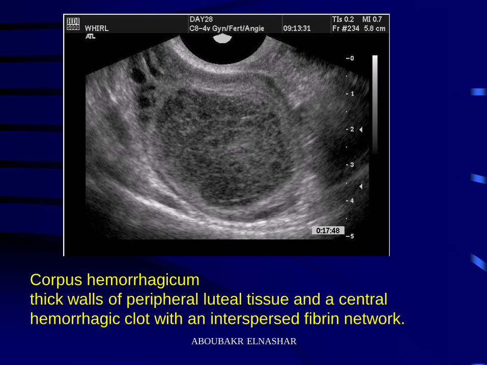
Corpus hemorrhagicum
thick walls of peripheral luteal tissue and a central
hemorrhagic clot with an interspersed fibrin network.
ABOUBAKR ELNASHAR
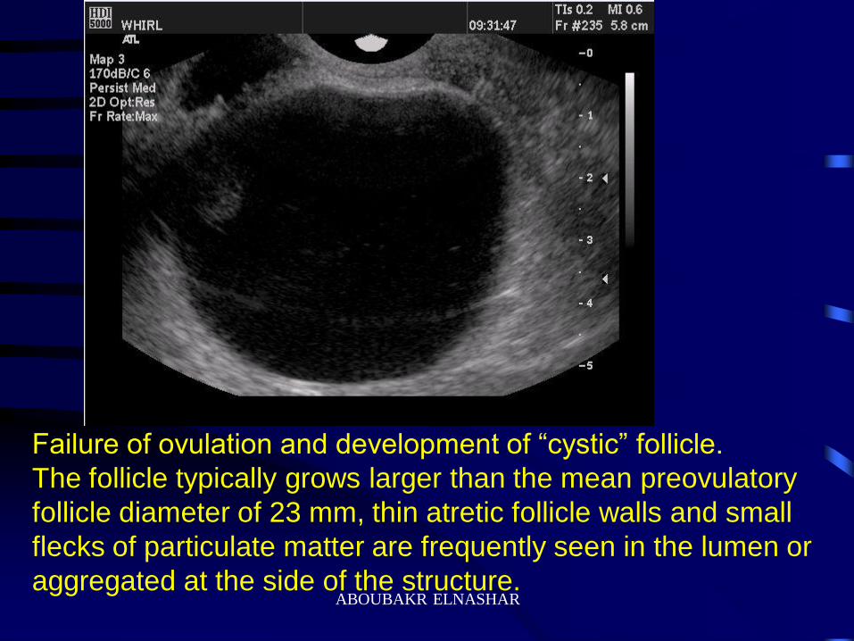
Failure of ovulation and development of “cystic” follicle.
The follicle typically grows larger than the mean preovulatory
follicle diameter of 23 mm, thin atretic follicle walls and small
flecks of particulate matter are frequently seen in the lumen or
aggregated at the side of the structure. ABOUBAKR ELNASHAR
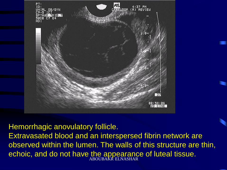
Hemorrhagic anovulatory follicle.
Extravasated blood and an interspersed fibrin network are
observed within the lumen. The walls of this structure are thin,
echoic, and do not have the appearance of luteal tissue.
ABOUBAKR ELNASHAR
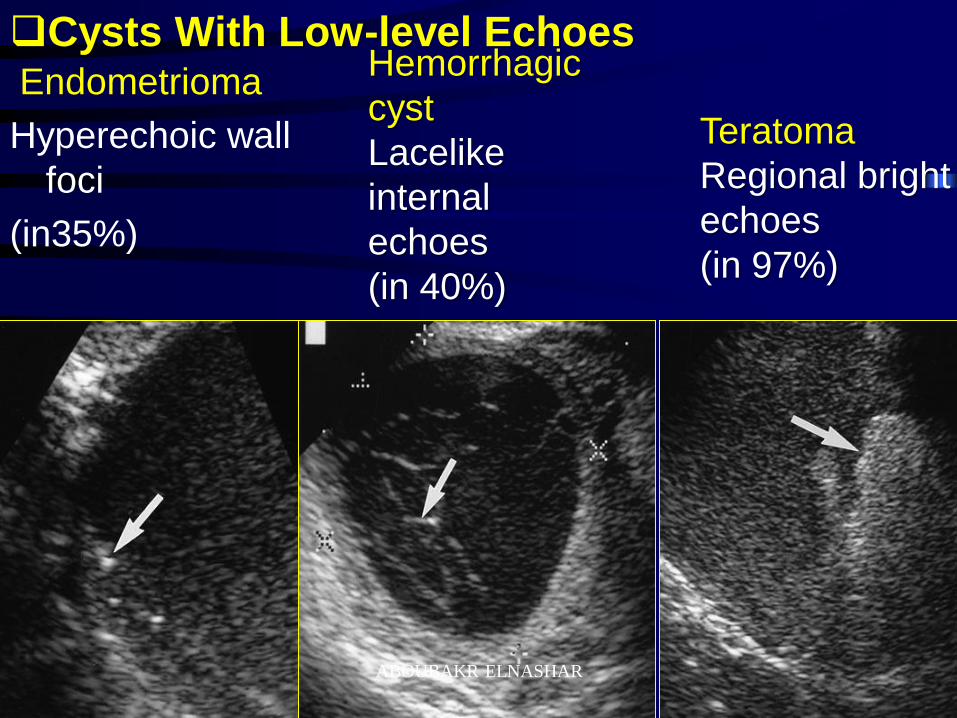
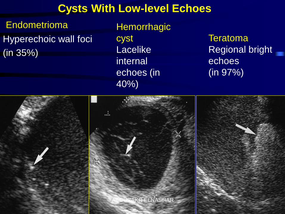
Endometrioma
Hyperechoic wall
foci
(in35%)
Cysts With Low-level Echoes Hemorrhagic
cyst
Lacelike
internal
echoes
(in 40%)
Teratoma
Regional bright
echoes
(in 97%)
ABOUBAKR ELNASHAR
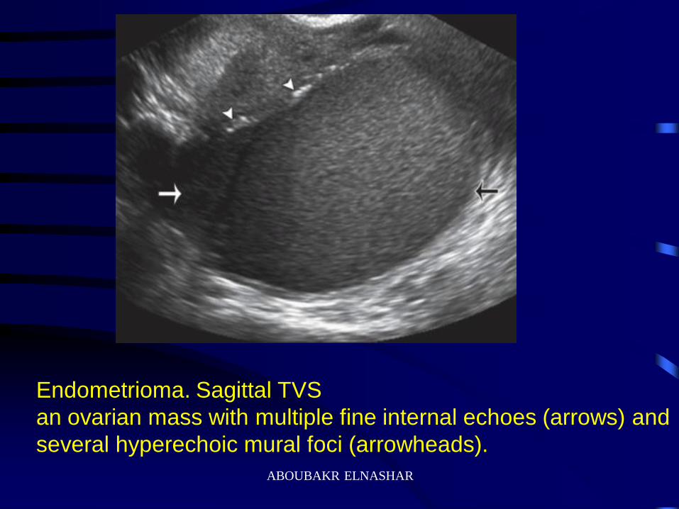
Endometrioma. Sagittal TVS
an ovarian mass with multiple fine internal echoes (arrows) and
several hyperechoic mural foci (arrowheads).
ABOUBAKR ELNASHAR
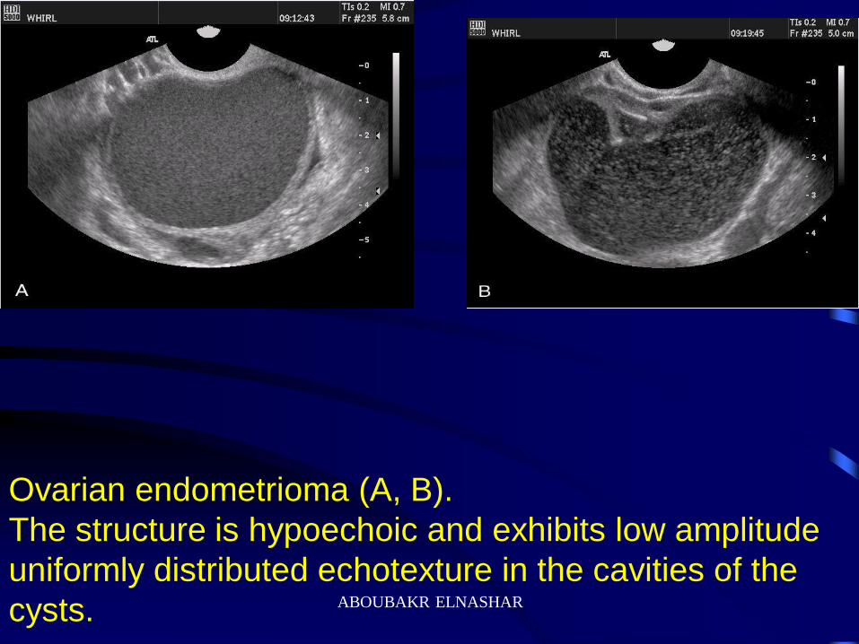
Ovarian endometrioma (A, B).
The structure is hypoechoic and exhibits low amplitude
uniformly distributed echotexture in the cavities of the
cysts. ABOUBAKR ELNASHAR
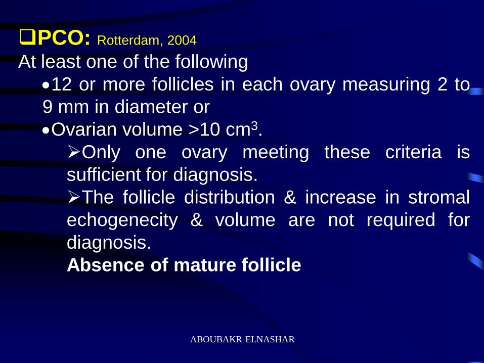
PCO: Rotterdam, 2004
At least one of the following
12 or more follicles in each ovary measuring 2 to
9 mm in diameter or
Ovarian volume >10 cm3.
Only one ovary meeting these criteria is
sufficient for diagnosis.
The follicle distribution & increase in stromal
echogenecity & volume are not required for
diagnosis.
Absence of mature follicle
ABOUBAKR ELNASHAR
ABOUBAKR ELNASHAR

Technical recommendation
1. Regularly menstruating females should be scanned
between days 3-5
Oligo-/ amenorrhoeic should be scanned either at
random or between days 3-5 after progesterone –
induced bleeding
2. If there is evidence of a dominant follicle >10 mm or a
corpus luteum, the scan should be repeated the next
cycle.
3. Ovarian volume= 0.5X length X width X thickness
ABOUBAKR ELNASHAR
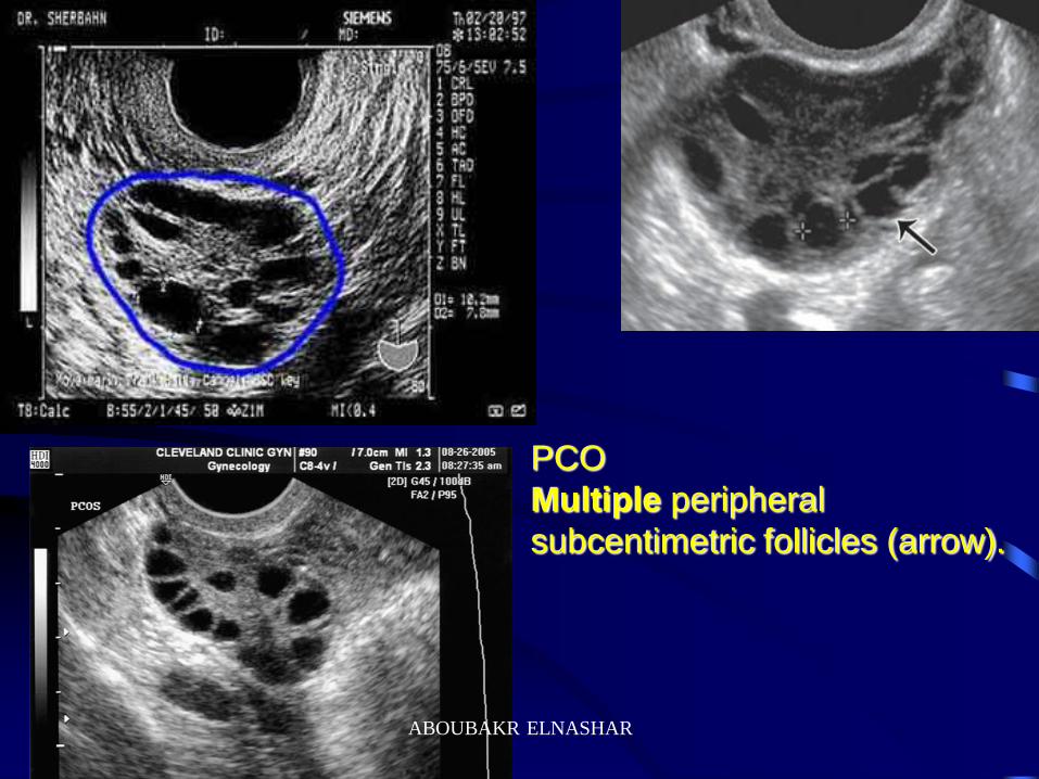
PCO
Multiple peripheral
subcentimetric follicles (arrow).
ABOUBAKR ELNASHAR
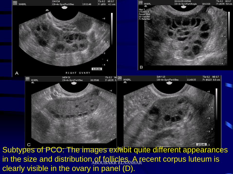
Subtypes of PCO: The images exhibit quite different appearances
in the size and distribution of follicles. A recent corpus luteum is
clearly visible in the ovary in panel (D).
ABOUBAKR ELNASHAR
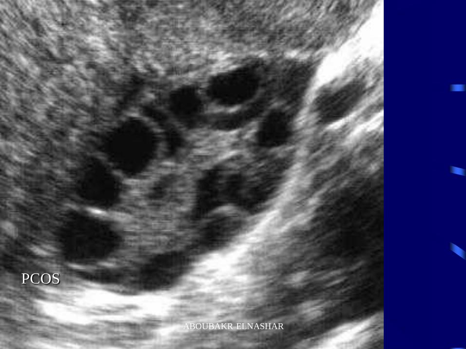
PCOS
ABOUBAKR ELNASHAR
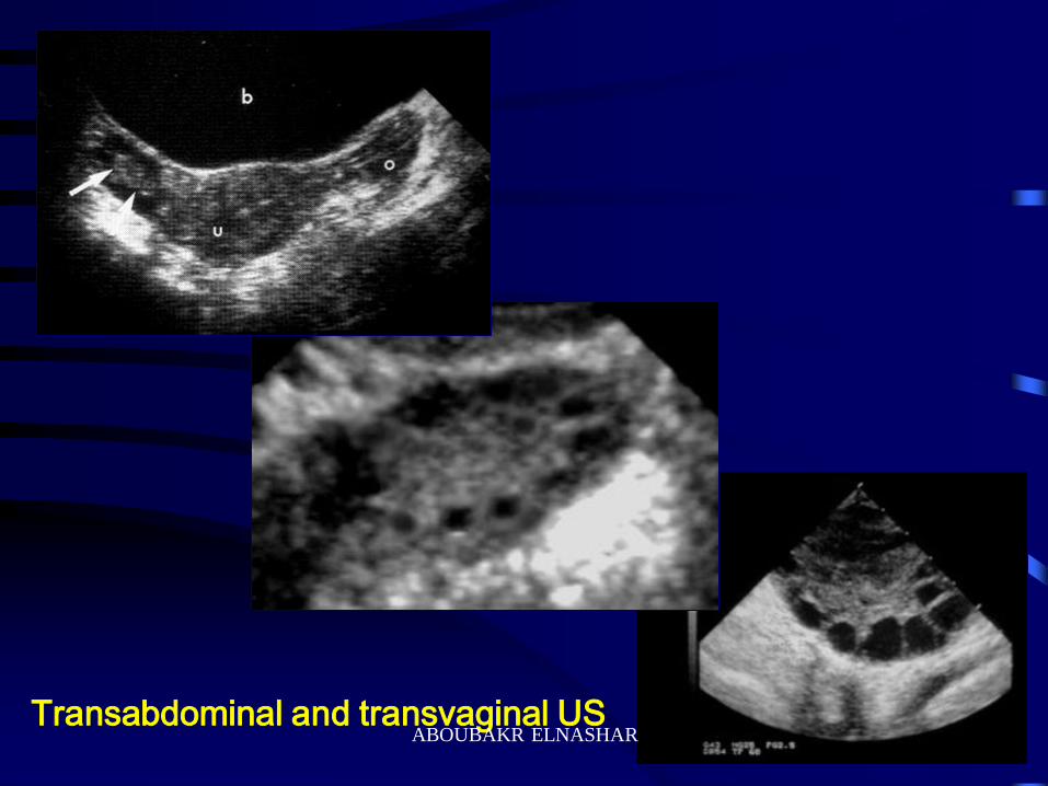
Transabdominal and transvaginal US ABOUBAKR ELNASHAR
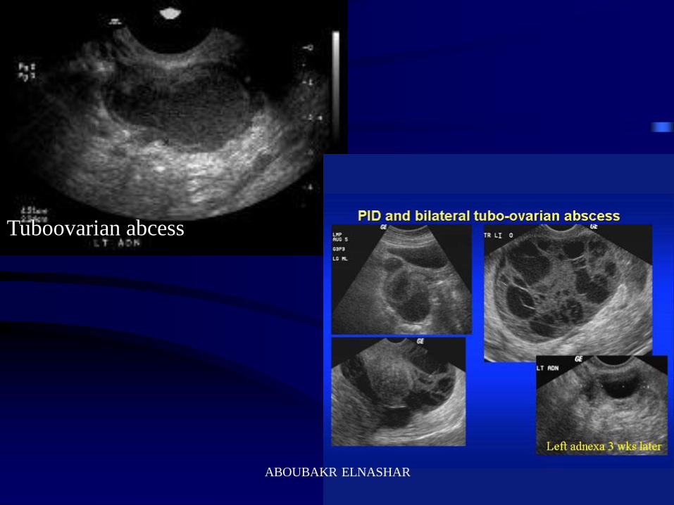
Tuboovarian abcess
ABOUBAKR ELNASHAR
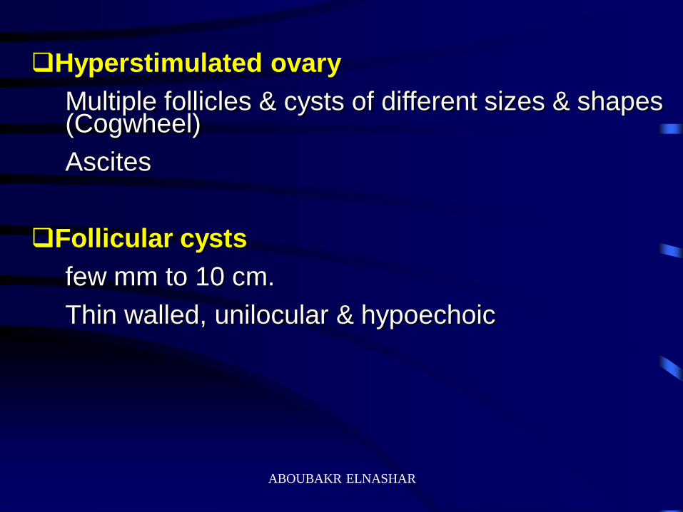
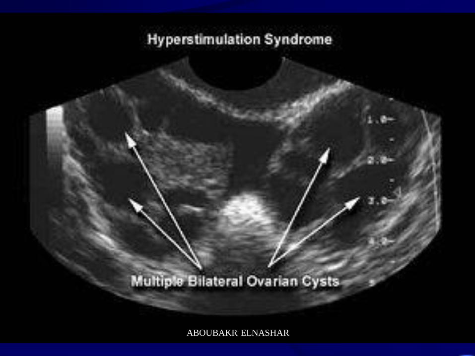
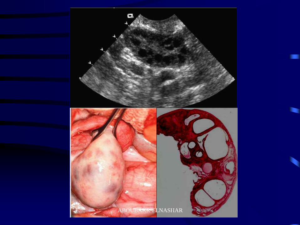
Hyperstimulated ovary
Multiple follicles & cysts of different sizes & shapes (Cogwheel)
Ascites
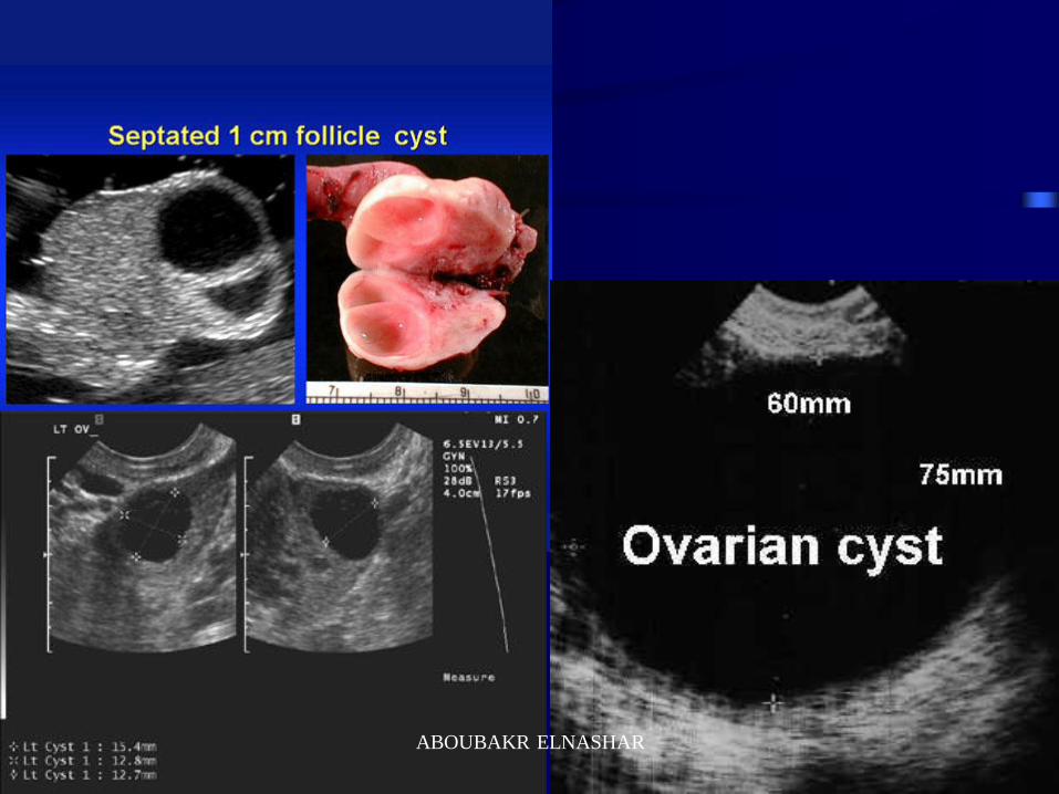
Follicular cysts
few mm to 10 cm.
Thin walled, unilocular & hypoechoic
ABOUBAKR ELNASHAR
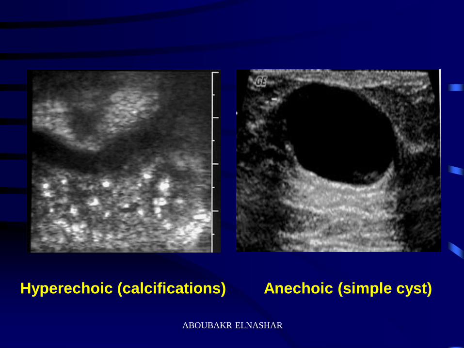
Hyperechoic (calcifications) Anechoic (simple cyst)
ABOUBAKR ELNASHAR
ABOUBAKR ELNASHAR
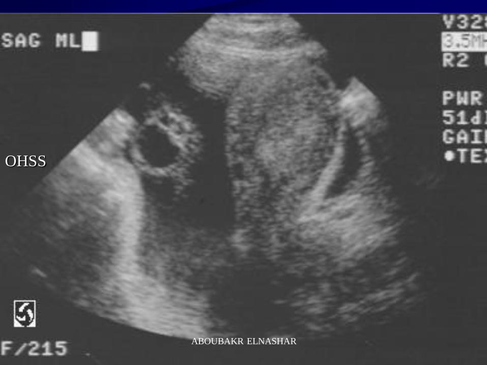
OHSS
ABOUBAKR ELNASHAR
ABOUBAKR ELNASHAR
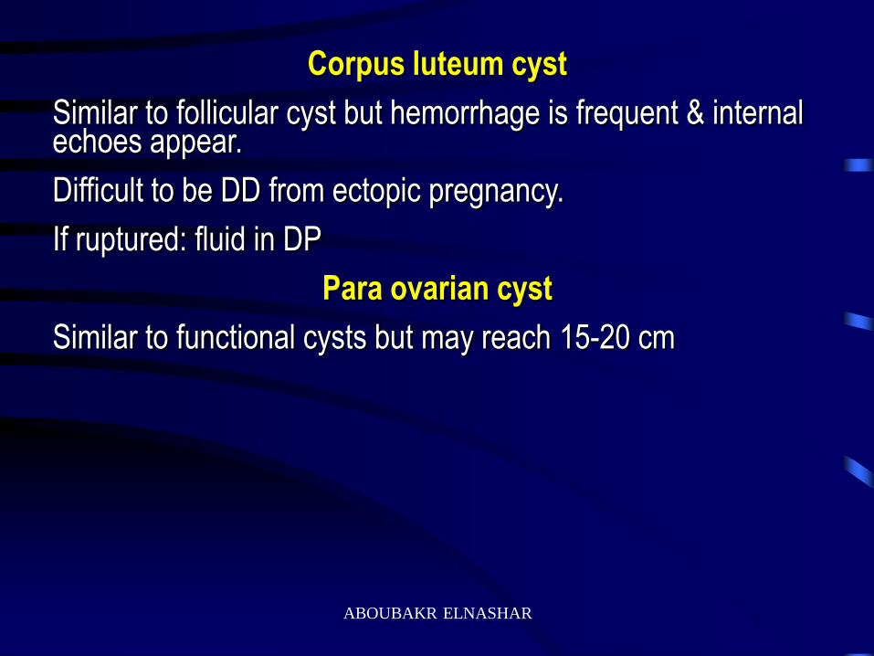
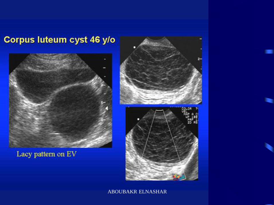
Corpus luteum cyst
Similar to follicular cyst but hemorrhage is frequent & internal echoes appear.
Difficult to be DD from ectopic pregnancy.
If ruptured: fluid in DP
Para ovarian cyst
Similar to functional cysts but may reach 15-20 cm
ABOUBAKR ELNASHAR
ABOUBAKR ELNASHAR
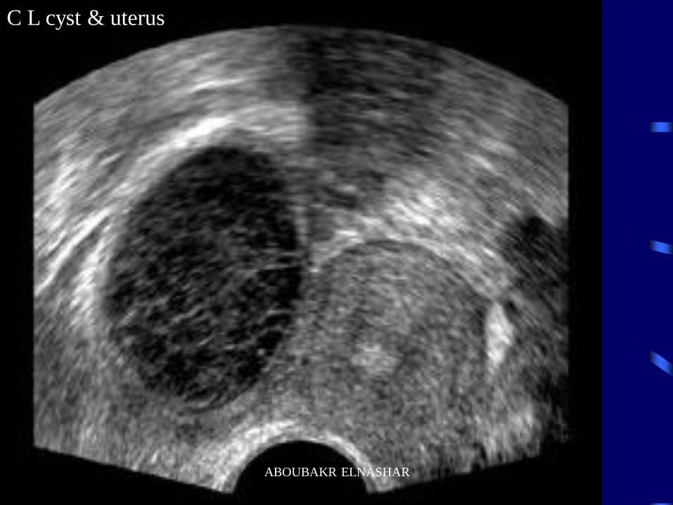
C L cyst & uterus
ABOUBAKR ELNASHAR
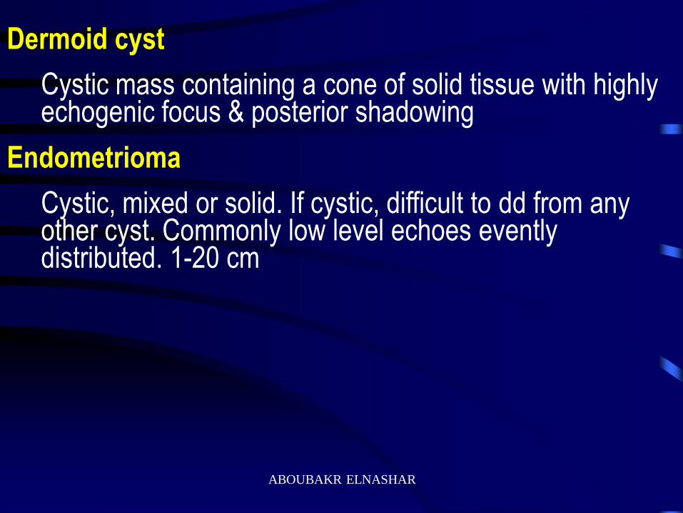
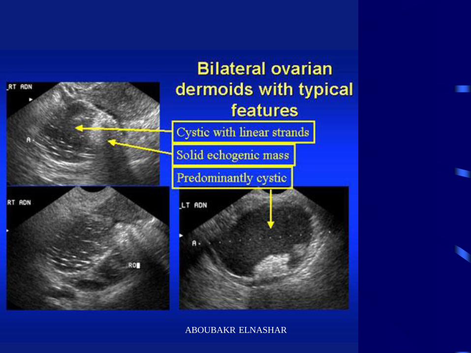
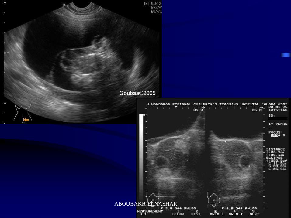
Dermoid cyst
Cystic mass containing a cone of solid tissue with highly echogenic focus & posterior shadowing
Endometrioma
Cystic, mixed or solid. If cystic, difficult to dd from any other cyst. Commonly low level echoes evently distributed. 1-20 cm
ABOUBAKR ELNASHAR
Endometrioma
Hyperechoic wall foci
(in 35%)
Cysts With Low-level Echoes
Hemorrhagic
cyst
Lacelike
internal
echoes (in
40%)
Teratoma
Regional bright
echoes
(in 97%)
ABOUBAKR ELNASHAR
ABOUBAKR ELNASHAR
ABOUBAKR ELNASHAR
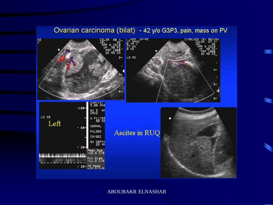
Malignancy 1.Bilaterality
2. Ascites
3. Excrescencies
4. Thick walled cysts
5. Thick septa
6. Solid area within the mass
ABOUBAKR ELNASHAR
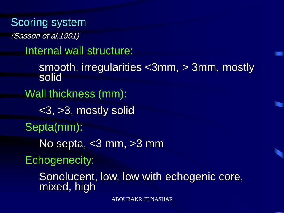
Scoring system (Sasson et al,1991)
Internal wall structure:
smooth, irregularities <3mm, > 3mm, mostly solid
Wall thickness (mm):
<3, >3, mostly solid
Septa(mm):
No septa, <3 mm, >3 mm
Echogenecity:
Sonolucent, low, low with echogenic core, mixed, high
ABOUBAKR ELNASHAR
ABOUBAKR ELNASHAR
ABOUBAKR ELNASHAR

ABOUBAKR ELNASHAR
ABOUBAKR ELNASHAR
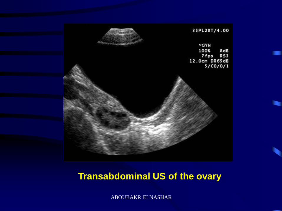
Transabdominal US of the ovary
ABOUBAKR ELNASHAR

Transabdominal Vs Transvaginal US
ABOUBAKR ELNASHAR
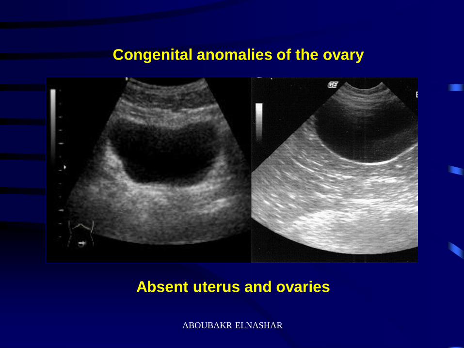
Absent uterus and ovaries
Congenital anomalies of the ovary
ABOUBAKR ELNASHAR
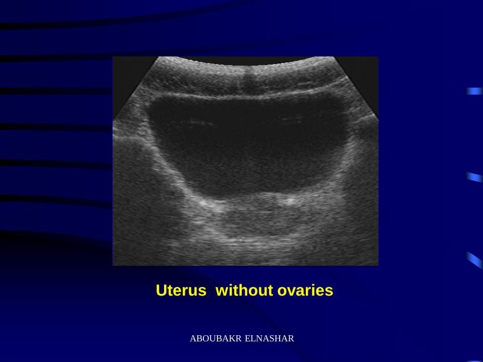
Uterus without ovaries
ABOUBAKR ELNASHAR

Pelvic inflammatory disease (PID)
ABOUBAKR ELNASHAR
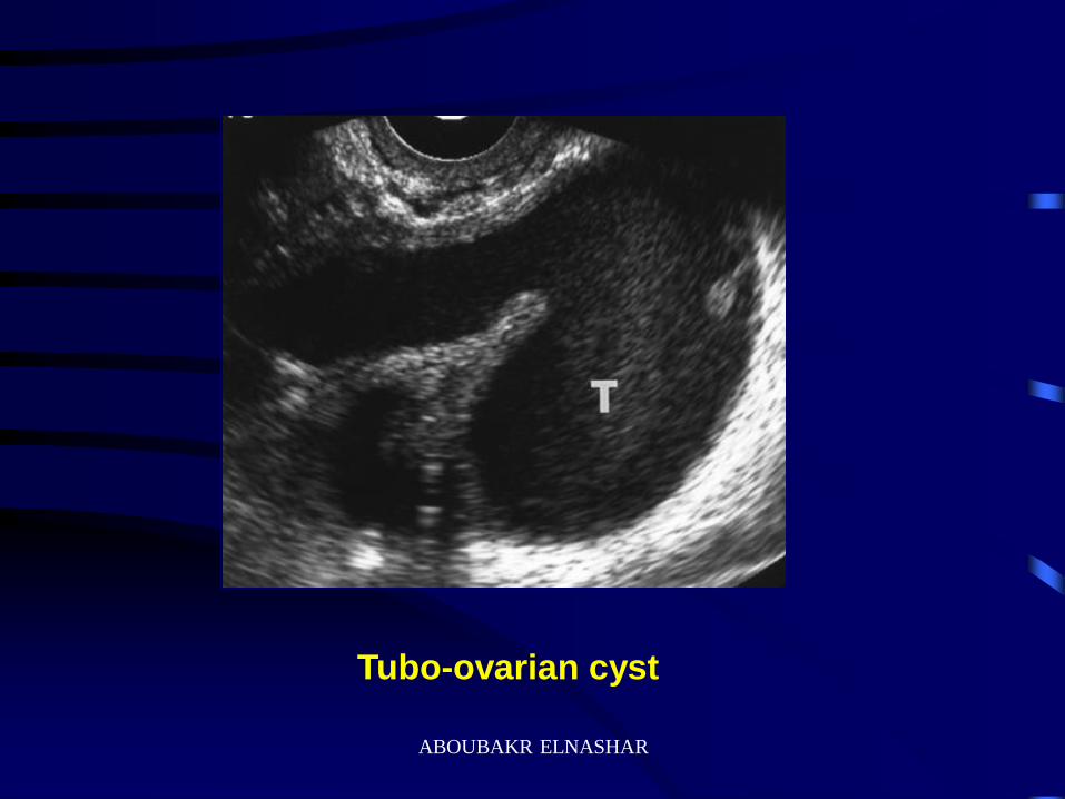
Tubo-ovarian cyst
ABOUBAKR ELNASHAR
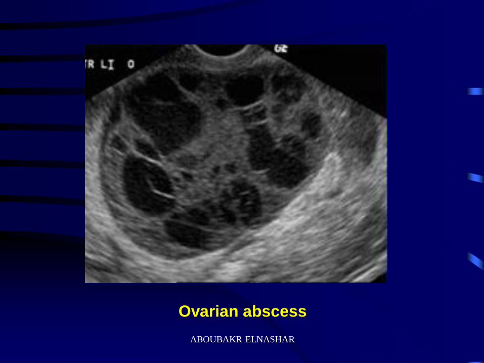
Ovarian abscess
ABOUBAKR ELNASHAR
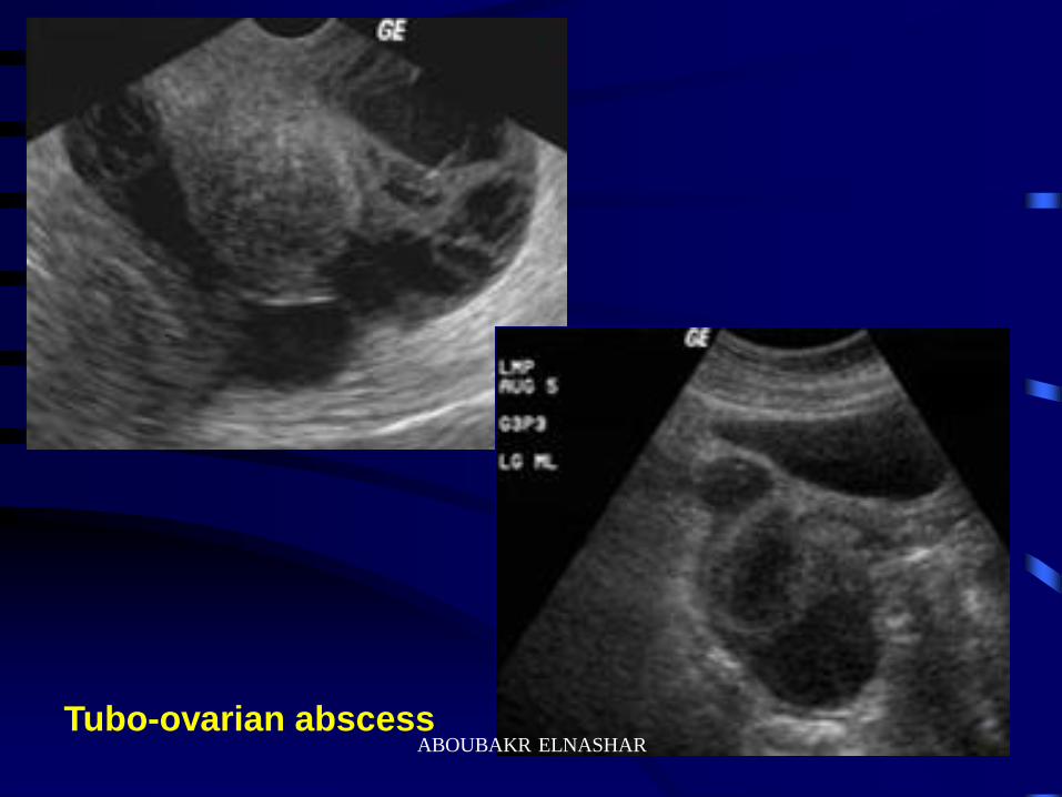
Tubo-ovarian abscess ABOUBAKR ELNASHAR
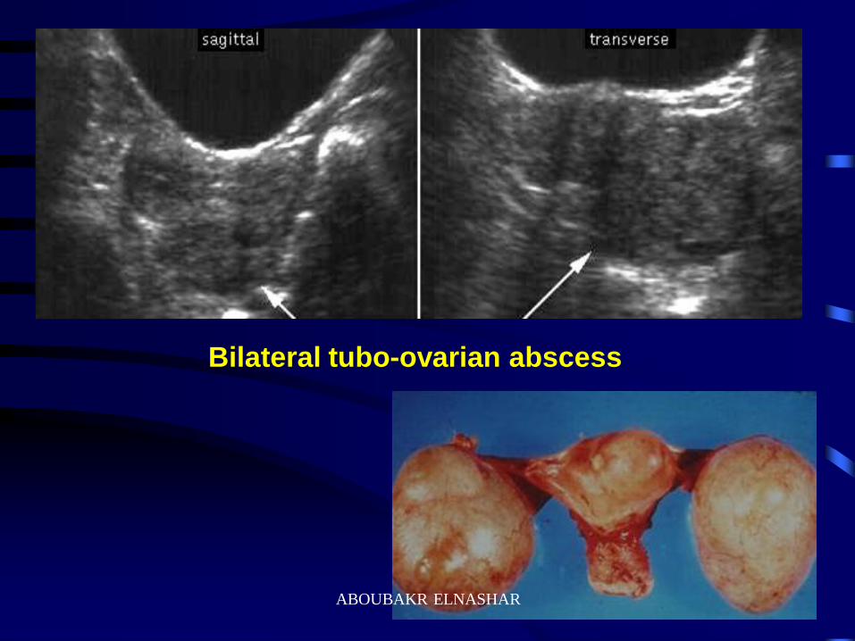
Bilateral tubo-ovarian abscess
ABOUBAKR ELNASHAR
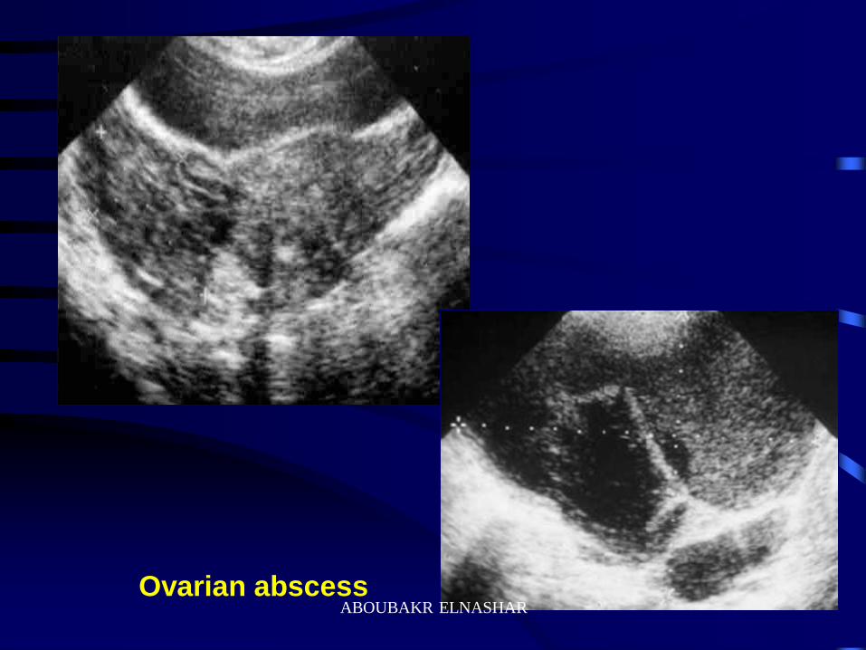
Ovarian abscess ABOUBAKR ELNASHAR
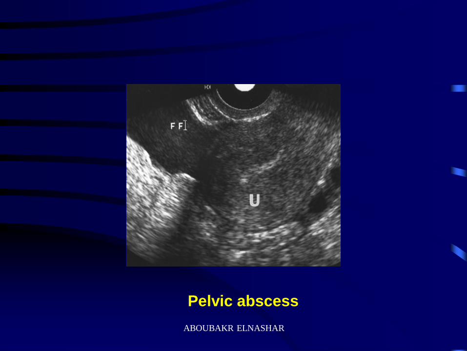
Pelvic abscess
ABOUBAKR ELNASHAR
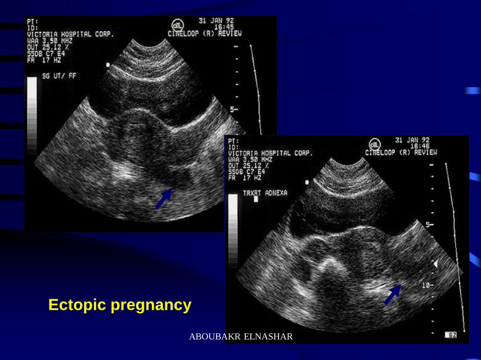
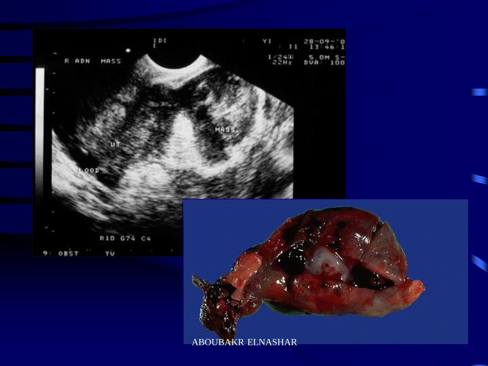
Ectopic pregnancy
ABOUBAKR ELNASHAR
ABOUBAKR ELNASHAR
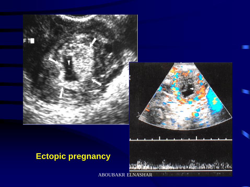
Ectopic pregnancy
ABOUBAKR ELNASHAR
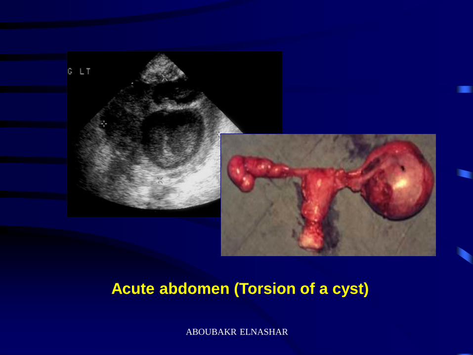
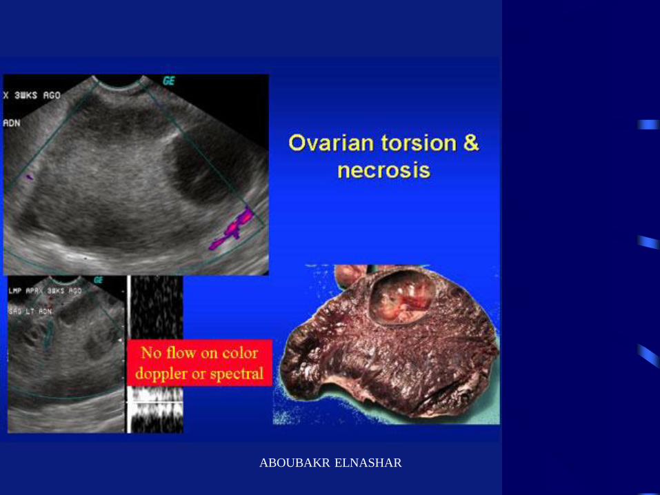
Acute abdomen (Torsion of a cyst)
ABOUBAKR ELNASHAR
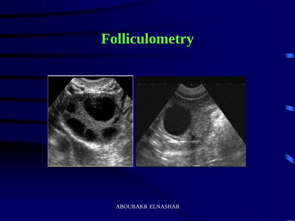
Folliculometry
ABOUBAKR ELNASHAR
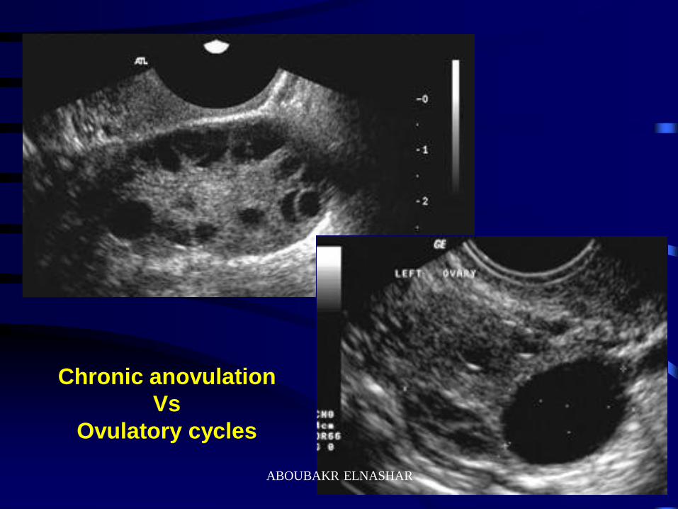
Chronic anovulation
Vs
Ovulatory cycles
ABOUBAKR ELNASHAR
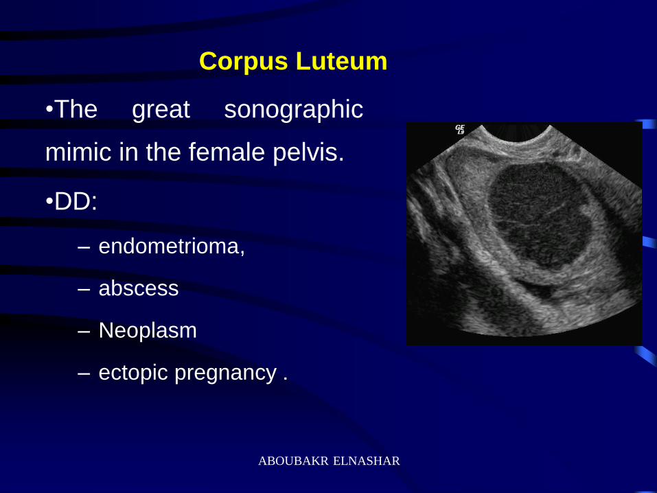
Corpus Luteum
•The great sonographic
mimic in the female pelvis.
•DD:
– endometrioma,
– abscess
– Neoplasm
– ectopic pregnancy .
ABOUBAKR ELNASHAR
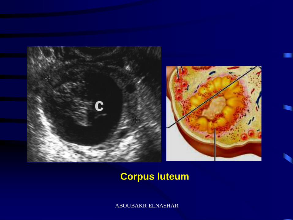
Corpus luteum
ABOUBAKR ELNASHAR
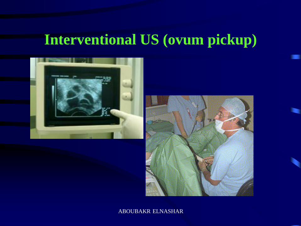
Interventional US (ovum pickup)
ABOUBAKR ELNASHAR
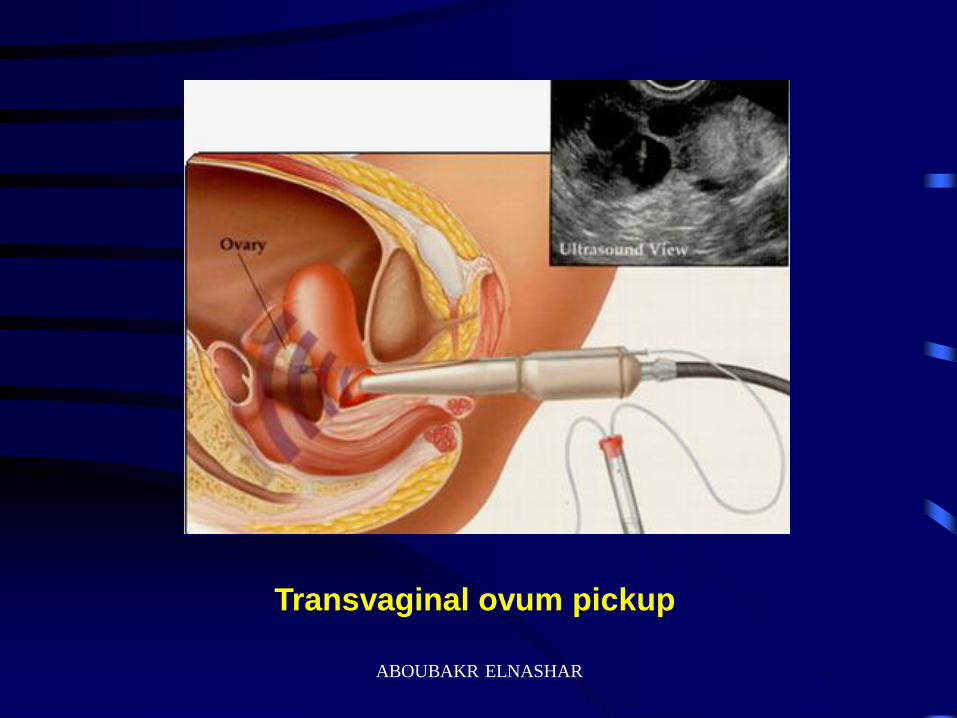
Transvaginal ovum pickup
ABOUBAKR ELNASHAR
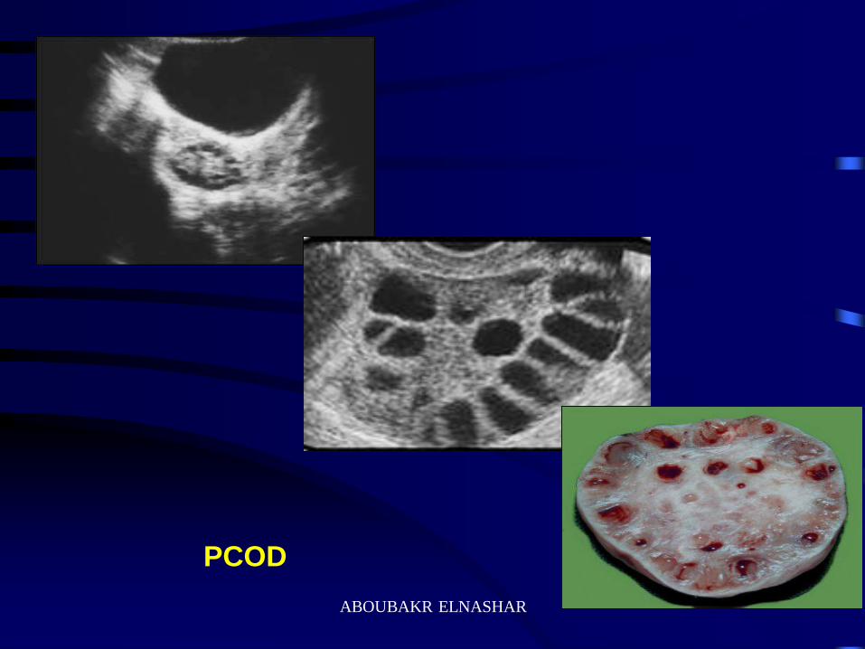
PCOD
ABOUBAKR ELNASHAR
ABOUBAKR ELNASHAR


![The Ovary of the Teleost Fish Xenotoca Eiseni (Goodeidae ... · the ovary, called a gonoduct, connects the ovary to the exterior by a gonopore [7]. These unique features of the ovary](https://static.fdocuments.in/doc/165x107/5f5082c1c1cb78272c63e522/the-ovary-of-the-teleost-fish-xenotoca-eiseni-goodeidae-the-ovary-called-a.jpg)