UCSF Department of Surgery
27
This event is supported by the Department of Surgery’s Howard Naffziger Endowment Fund. UCSF Department of Surgery Education Office Telephone: (415) 476-1239 Email: EducationOffi[email protected] UCSF Department of Surgery 28th Annual Resident Research Symposium Wednesday, March 11, 2015 UCSF Toland Hall 533 Parnassus Avenue, U-142 San Francisco, CA Jennifer R. Grandis, MD J. Englebert Dunphy Professor Associate Vice Chancellor, Clinical and Translational Research Director, Clinical and Translational Science Institute Professor, Otolaryngology-Head & Neck Surgery University of California, San Francisco
Transcript of UCSF Department of Surgery

This event is supported by the Department of Surgery’s Howard Naffziger Endowment Fund.
UCSF Department of Surgery Education OfficeTelephone: (415) 476-1239 Email: [email protected]
UCSF Department of Surgery28th Annual Resident Research Symposium
Wednesday, March 11, 2015 UCSF Toland Hall533 Parnassus Avenue, U-142San Francisco, CA
Jennifer R. Grandis, MDJ. Englebert Dunphy ProfessorAssociate Vice Chancellor, Clinical and Translational Research
Director, Clinical and Translational Science Institute
Professor, Otolaryngology-Head & Neck Surgery
University of California, San Francisco

UCSF Department of Surgery 28th Annual Resident Research Symposium A Tradition of Excellence
J. Englebert Dunphy, M.D. Professor of Surgery and Chairman of the Department from 1964 to 1975
Dr. Dunphy earned his medical degree at Harvard Medical School and completed his surgical residency training at the Peter Bent Brigham Hospital in Boston. He then joined the faculty at
Harvard before accepting the position of the Chair for the Department of Surgery at the University of Oregon. In 1964, Dr. Dunphy was offered and accepted the position of the Chair for
the Department of Surgery at the University of California, San Francisco.
Dr. Dunphy was president of the Society of University Surgeons, the American Surgical Association, and the American College of Surgeons. He received honorary fellowships in six
foreign colleges of surgeons as recognition of his international stature.
Dr. Dunphy was renowned for excellence in many aspects of surgery, with a special interest in the gastrointestinal tract. He was one of the leading surgical educators of his day and was greatly admired and respected by his colleagues and residents. Dr. Dunphy conducted research
in wound healing at a basic level.
Dr. Dunphy strongly believed that prospective academic surgeons should become grounded in basic science, and he was one of the first surgical leaders in the United States to obtain an
NIH training grant supporting residents in the laboratory.

UCSF Department of Surgery 28th Annual Resident Research Symposium A Tradition of Excellence
Jennifer R. Grandis, M.D.
2015 J. Engelbert Dunphy Professor
Jennifer Grandis, M.D., the new Associate Vice Chancellor of Clinical and Translational Science, Director of CTSI and Professor of Otolaryngology‐Head & Neck Surgery comes to UCSF from the University of Pittsburgh, where she was a UPMC Endowed Chair in Head and Neck Cancer Surgical Research and
Distinguished Professor of Otolaryngology and Pharmacology & Chemical Biology. She led the Head and Neck Cancer Program and was Vice Chair for Research in the Department of Otolaryngology.
Dr. Grandis was also the Health Sciences Assistant Vice Chancellor for Research Program Integration.
Her research areas include precision medicine approaches, signal transduction, molecular targeted therapies and mechanisms of resistance to EGFR inhibitors in head and neck cancer. She is responsible
for a number of patents in this area and is primary author to over 250 papers on this topic.
Dr. Grandis has a history of successfully mentoring undergraduates, medical students, otolaryngology residents and fellows in her laboratory. She served as the longstanding PI of two training grants (T32S) from the NIH to train clinicians for careers that combine research and clinical practice. She leads a Specialized Program of Research Excellence (SPORE) focused on
head and neck cancer translational research.
She is an American Cancer Society Clinical Research Professor (since 2008) and is a member of the American Society for Clinical Investigation as well as the American Association of Physicians. In 2012,
she was elected to the Institute of Medicine of The National Academies.

UCSF Department of Surgery 28th Annual Resident Research Symposium A Tradition of Excellence
Past Visiting Professors
Bernard Langer, M.D. Professor and Chairman of Surgery, University of Toronto
February 5‐6, 1988
William Silen, M.D. Professor of Surgery, Harvard Medical School
February 3‐4, 1989
James Thompson, M.D. Professor and Chairman of Surgery, University of Texas, Galveston
February 2‐3, 1990
Murray Brennan, M.D. Professor and Chair of Surgery, Memorial Sloan‐Kettering Cancer Center
February 3‐4, 1991
Richard Simmons, M.D. Professor and Chairman of Surgery, University of Pittsburgh
January 31‐February 1, 1992
Stephen F. Lowry, M.D. Professor of Surgery, Cornell University Medical College
February 4‐5, 1993
Jared Diamond, Ph.D. Professor of Physiology, UCLA School of Medicine
February 4, 1994
Samuel A. Wells, Jr., M.D. Professor and Chairman of Surgery, Washington University
February 17, 1995
Jonathon E. Rhoads Chief of Surgical Oncology, University of Pennsylvania, Philadelphia
February 16, 1996
Patricia K. Donahoe, M.D. Chief, Pediatric Surgical Services, Massachusetts General Hospital
February 27, 1997
David L. Dunn, M.D., Ph.D. Professor and Chairman of Surgery, University of Minnesota
February 27, 1998
Ori D. Rotstein, M.D. Professor of Surgery, Toronto Hospital
February 26, 1999
Olga Jonasson, M.D. Director of Education and Surgical Services Department
American College of Surgeons March 17, 2000
Glenn Steele, Jr., M.D. Ph.D.
Dean, School of Medicine, University of Chicago March 9, 2001
Alexander W. Clowes, M.D.
Professor of Surgery and Chairman, University of Michigan March 7, 2002
Michael Mulholland, M.D., Ph.D.
Professor of Surgery and Chairman, University of Michigan March 7, 2003
Christian Larsen, M.D., Ph.D.
Professor of Surgery, Emory University March 19, 2004
Danny O. Jacobs, M.D., M.P.H.Chair, Department of Surgery, Duke University Medical Center
March 4, 2005
Steven D. Leach, M.D. Chief of Surgical Oncology, Johns Hopkins University
March 3, 2006
M. Judah Folkman, M.D. Professor of Pediatric Surgery & Cell Biology, Harvard Medical School
Director, Vascular Biology Program, Children’s Hospital, Boston February 15‐16, 2007
Sir Peter Morris, AC, FRS, FRCS
Director, Centre for Evidence in Transplantation Royal College of Surgeons of England
April 4, 2008
George K. Gittes, M.D.
Chair of Pediatric Surgery, University of Pittsburgh April 3, 2009
Joseph P. Vacanti, M.D. Chief, Pediatric Surgery, Massachusetts General Hospital
March 12, 2010
Maria Bertagnolli, M.D. Professor of Surgery, Harvard
Chief, Surgical Oncology, Brigham and Women’s Hospital April 1, 2011
Michael Harrison, M.D.
Director Emeritus, Fetal Treatment Center, Professor of Pediatric Surgery, University of California, San Francisco
April 13, 2012
Martin Elliott, M.D. Professor of Cardiothoracic Surgery, University College London
April 5, 2013
Clifford Ko, M.D. Director, Division of Research and Optimal Patient Care
Director, National Surgical Quality Improvement Program (ACS NSQIP) American College of Surgeons
April 25, 2014

UCSF Department of Surgery 28th Annual Resident Research Symposium A Tradition of Excellence
2014SymposiumWinners
From left to right: Clifford Ko, Nancy Ascher, Carolyn Seib, Chris Derderian, Cerine Jeanty, Adam Laytin, James Gardner, Emily Huang, Peter Stock
Pancreatic Antigen Expression in Extrathymic Aire‐Expressing BEST BASIC SCIENCE PRESENTATION: Cells Prevents Autoimmune Diabetes, James Gardner MD PhD
From Novice to Master Surgeon: Improving Feedback with a BEST CLINICAL SCIENCE PRESENTATION: Descriptive Approach to Intraoperative Assessment. Emily Huang MD
In Utero Depletion of Fetal Host Hematopoietic Stem OUTSTANDING BASIC SCIENCE PRESENTATION: Cells Improves Engraftment Following Neonatal Transplantation in Mice. Chris Derderian MD
Prognostic Utility of Lymph Node Size, Number, OUTSTANDING CLINICAL SCIENCE PRESENTATION: Histology and Extranodal Extension for Papillary Thyroid Cancer Nodal Metastases Following Selective Lymph Node Dissection. Carolyn Seib MD
Precocious T Cell Maturation In Patients With Gastroschisis. BEST BASIC SCIENCE “QUICK‐SHOT”: Cerine Jeanty MD
Choice of Injury Scoring System in a Resource Poor Setting: BEST CLINICAL SCIENCE “QUICK‐SHOT”: Lessons from Mumbai. Adam Laytin MD

UCSF Department of Surgery 28th Annual Resident Research Symposium March 11, 2015
9:00 am Opening Remarks Nancy Ascher MD, PhDProfessor & Chair
SESSION 1: "Trauma, Inflammation & Cancer" Moderator: Jason Pomerantz, MD
9:05 am Variation in Surgeon Payment for Cancer Resection Tasce Bongiovanni, MD, MPP
2nd year Research Fellow
9:20 am The Tissue Factor Pathway Mediates Both Activation of Coagulation
and Coagulopathy After Injury
Benjamin Howard, MD, MPH
2nd year Research Fellow
9:35 am MammaPrint Accurately Predicts Long‐term Survival and Endocrine
Therapy Benefit in Lymph Node Negative Breast Cancer Patients
Carlie Thompson, MD
2nd year Research Fellow
9:50 am The Impact of Radiation, Lymph Node Dissection, and Hormonal
Therapy on Outcomes of Total Skin‐Sparing Mastectomy and
Immediate Tissue Expander‐Based Breast Reconstruction
Frederick Wang, MD
2nd year Research Fellow
10:05 am Development of a Novel Gli Inhibitor and its Combined Effect With
Erlotinib for Treatment of Non‐small Cell Lung Cancer and Pancreatic
Ductal Adenocarcinoma
Gavitt Woodard, MD
1st year Research Fellow
10:20 am Targeted Antibiotic Prophylaxis Associated With Increased Microbiota
and Decreased Experimental Necrotizing Enterocolitis
Joanna Lim, MD
1st year Research Fellow
10:25 am A Comparison of Mammographic Findings Following Oncoplastic
Mammoplasty and Lumpectomy Without Reconstruction
Merisa Piper, MD
1st year Research Fellow
10:30 am 20‐minute break
SESSION 2: "Global Surgery, Public Health, Outcomes & Education" Moderator: Catherine Juillard, MD
10:50 am Impact of Opioid Sparing Pain Regimens on Elective Colorectal Surgery
Outcomes
Jenny Kaplan, MD
1st year Research Fellow
11:05 am Improving Breast Cancer Survivors’ Knowledge Using a Patient‐
Centered Intervention
Jesus Ulloa, MD
1st year Research Fellow
11:10 am A Kinesthetic Surgical Skills Curriculum and Validated Assessment Tool Carolyn Vaughn, MD
1st year Research Fellow
11:15 am In Their Own Words: Improving Trauma Services for Young Men of
Color
Vincent Chong, MD, MS
2nd year Research Fellow
11:30 am A Surgical Decision Aid for Patients With Ulcerative Colitis: A Pilot Study Jessica Cohan, MD
2nd year Research Fellow
11:45 am Patterns of Injury at a Trauma Center in Mumbai: Opportunities for
Injury Prevention
Adam Laytin, MD
2nd year Research Fellow

UCSF Department of Surgery 28th Annual Resident Research Symposium March 11, 2015
12:00 pm Buffet Lunch Faculty Alumni House
745 Parnassus
SESSION 3: "Stem Cells, Transplantation, Vascular & Innovation” Moderator: Tammy Chang, MD, PhD
1:05 pm Splenic Vein Thrombosis Following Pancreas Transplantation: Identification of Factors That Support Conservative Management
Jack Harbell, MDChief Resident, General Surgery
1:20 pm Wound Class Does Not Alter Recurrence in Complex Ventral Hernia Repair With Mesh: A Single‐center Retrospective Review
Victoria Lyo, MD1st year Research Fellow
1:35 pm Serotonin Signaling Promotes Growth of Postnatal‐derived Enteric
Neuronal Stem Cells
Lily Cheng, MD
1st year Research Fellow
1:40 pm SmartDerm: Real‐Time Prediction and Prevention of Pressure Ulcers Isabelle Chumfong, MD, MEng
1st year Research Fellow
1:45 pm
Renal Artery Embolization for Minimally Invasive Induction of Renal
Failure
Willieford Moses, MD
1st year Research Fellow
1:50 pm Lower Extremity Amputations in Patients With SIRS/Sepsis: Surgical
Specialty and 30 Day Outcomes
Joseph Patterson, MD
PGY1 Orthopedic Surgery
1:55 pm A Biodegradable Device for Perivascular Delivery of Pro‐resolving Lipid
Mediators
Bian Wu, MD
1st year Research Fellow
2:00 pm 30‐minute break
2:30 pm Keynote Presentation: “Precision Head and Neck Cancer Medicine” Jennifer Grandis, MD
2015 Dunphy Professor
3:30 pm Closing Remarks & Awards Presentation Peter Stock, MD, PhD
Research Committee Chair
= Quick Shot presentations
= Standard presentations
4:00 PM Reception for Residents and Faculty
Social Kitchen & Brewery1326 9th Ave., San Francisco

Title: Variation in surgeon payment for cancer resection Authors: Tasce Bongiovanni1,2,3 MD MPP, Simon P. Kim4 MD, MPH, Anthony W. Kim5 MD, Brigid K Killelea5 MD, MPH, FACS, Cary P. Gross,2,6 MD MHS Author Affiliations: 1Surgical Resident, University of California San Francisco School of Medicine, Department of Surgery, San Francisco, CA 2Robert Wood Johnson Foundation Clinical Scholars Program, Yale University School of Medicine, New Haven, CT 3U.S. Department of Veterans Affairs, Connecticut Healthcare System, West Haven, CT 4Department of Surgery, Case Western Reserve University School of Medicine, Cleveland, OH 5Department of Surgery, Yale University School of Medicine, New Haven, CT 6Department of Internal Medicine, Yale University School of Medicine, New Haven, CT Introduction: Demand for price transparency in healthcare is growing, empowering employers and beneficiaries to better understand what they pay for services. Variation in surgeon payments for oncologic resection has not been well studied, especially in the private-payer sector. We therefore assessed (1) geographic variation of private payer expenditures to surgeons for cancer resection, (2) variation within geographical regions for these expenditures. Methods: We use data based on fee-for-service allowed amounts, billed by a mix of private-payer providers for the year 2013, obtained in partnership with Fair Health, Inc. FAIR Health (FH) is an independent, not-for-profit corporation that collects and compiles claims data from insurers nationwide. We selected surgical resections frequently used for treating breast, lung and prostate cancer with CPT codes for simple mastectomy (SM), open lobectomy (LB), and robotic radical prostatectomy (RP). We evaluated surgeon reimbursement, as derived by FH through an ‘allowed medical benchmark’ (AMB) based on the maximum allowable payments for the submitted procedure codes and relative value units of the procedure. The data is grouped into 491 geographical regions based on zip code. To assess variation across regions, we compared the median AMB between regions. To assess intra-region variability, we evaluated regional inter-quartile ranges of AMB. Results: For each surgical procedure, the surgeon’s median AMB varied substantially across regions: For SM, the median AMB ranged from $487 in the least expensive region to $1,364 in the costliest region. For LB the median AMB ranged from $498 to $5,047; for RP from $1,716 to $5,775. Even standard deviations for the 50th percentile of AMB were high (LB $1,450, RP $1,173). There was also substantial variation within geographic regions. The AMB means of the 25th & 75th percentile within regions were as follows: for SM $552 & $1,088, LB $997 & $3,061, and RP $2,350 & $3,567. Conclusions: We found a wide range of variation both across and within geographic regions in the allowed reimbursement for these common surgical procedures. Transparency about surgical payment may have a profound impact on patient and employer choice and facilitate future assessments of value in cancer care.

The Tissue Factor Pathway Mediates Both Activation of Coagulation and Coagulopathy After Injury
BM Howard, BY Miyazawa, W Dong, W Cedron, RF Vilardi, W Ruf, MJ Cohen
Introduction: The initiation of coagulation in trauma is thought to originate from exposed tissue factor (TF); recent data have led to the alternative hypothesis that DAMPs may contribute to post-injury coagulation. In acute traumatic coagulopathy (ATC), aberrant coagulation is mediated via the activated protein C (aPC) pathway; the upstream regulators of this process, and its relation to TF, remain uncharacterized. To examine the role of the TF pathway in mediating ATC, we employed specific antibody blockades in an established murine model of traumatic hemorrhagic shock, hypothesizing that both coagulation activation after injury and aPC-mediated coagulopathy are driven by TF via thrombin. Methods: Mice underwent an established model of trauma and hemorrhage, and were subjected to either sham (vascular cannulation), or trauma-hemorrhage (cannulation, laparotomy, shock to MAP 35mmHg); they were monitored for 60 min prior to sacrifice. Mice in each group were pre-treated with either targeted anti-TF antibody to block the TF pathway, or hirudin for specific blockade of thrombin. Plasma was assayed for thrombin-antithrombin (TAT) and aPC by ELISA. Results: Compared to controls, trauma-hemorrhage mice treated with anti-TFAb had significantly reduced levels of TAT (2.3 vs. 5.7 ng/mL, p=0.016, Figure 1), and corresponding decreases in aPC (16.3 vs. 31.6 ng/mL, p=0.034, Figure 2), with reductions to levels seen in sham mice. Direct inhibition of thrombin yielded similar results, with reduction in aPC to levels below those seen in sham mice (Figures 3 & 4). Conclusions: In this study, blockade of the TF pathway led to attenuation of both thrombin production and aPC activation observed in traumatic shock. Specific thrombin inhibition achieved similar results, indicating that aPC-related coagulopathy is mediated via thrombin activated by the TF pathway. The near-complete blockade of TAT and aPC observed in this model argues for a dominant role of the TF-thrombin pathway in both coagulation activation after injury and traumatic coagulopathy.
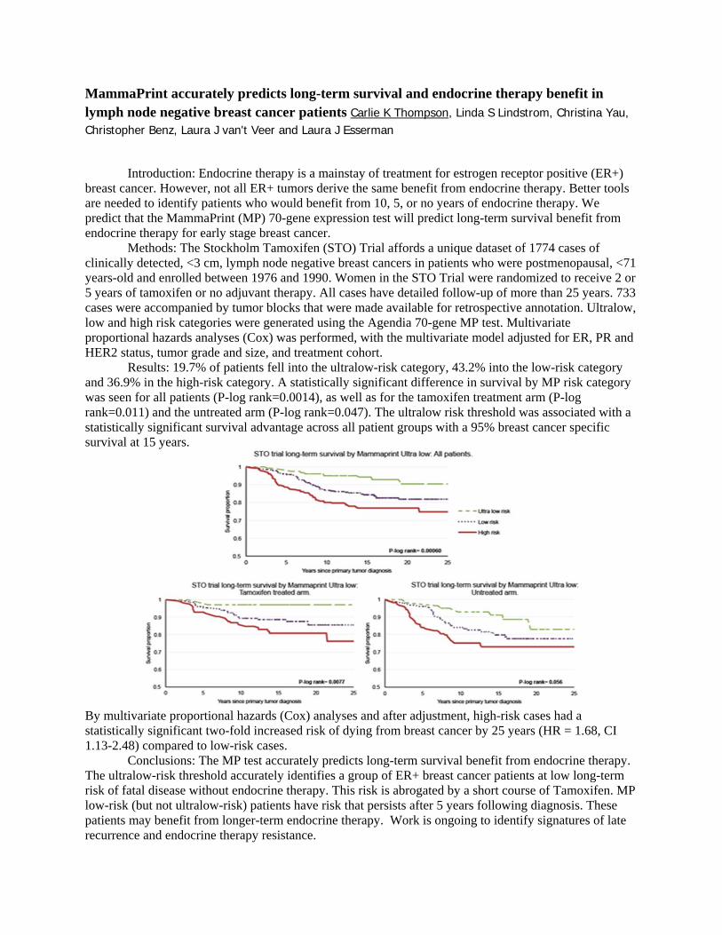
MammaPrint accurately predicts long-term survival and endocrine therapy benefit in lymph node negative breast cancer patients Carlie K Thompson, Linda S Lindstrom, Christina Yau, Christopher Benz, Laura J van't Veer and Laura J Esserman
Introduction: Endocrine therapy is a mainstay of treatment for estrogen receptor positive (ER+)
breast cancer. However, not all ER+ tumors derive the same benefit from endocrine therapy. Better tools are needed to identify patients who would benefit from 10, 5, or no years of endocrine therapy. We predict that the MammaPrint (MP) 70-gene expression test will predict long-term survival benefit from endocrine therapy for early stage breast cancer.
Methods: The Stockholm Tamoxifen (STO) Trial affords a unique dataset of 1774 cases of clinically detected, <3 cm, lymph node negative breast cancers in patients who were postmenopausal, <71 years-old and enrolled between 1976 and 1990. Women in the STO Trial were randomized to receive 2 or 5 years of tamoxifen or no adjuvant therapy. All cases have detailed follow-up of more than 25 years. 733 cases were accompanied by tumor blocks that were made available for retrospective annotation. Ultralow, low and high risk categories were generated using the Agendia 70-gene MP test. Multivariate proportional hazards analyses (Cox) was performed, with the multivariate model adjusted for ER, PR and HER2 status, tumor grade and size, and treatment cohort.
Results: 19.7% of patients fell into the ultralow-risk category, 43.2% into the low-risk category and 36.9% in the high-risk category. A statistically significant difference in survival by MP risk category was seen for all patients (P-log rank=0.0014), as well as for the tamoxifen treatment arm (P-log rank=0.011) and the untreated arm (P-log rank=0.047). The ultralow risk threshold was associated with a statistically significant survival advantage across all patient groups with a 95% breast cancer specific survival at 15 years.
By multivariate proportional hazards (Cox) analyses and after adjustment, high-risk cases had a statistically significant two-fold increased risk of dying from breast cancer by 25 years (HR = 1.68, CI 1.13-2.48) compared to low-risk cases.
Conclusions: The MP test accurately predicts long-term survival benefit from endocrine therapy. The ultralow-risk threshold accurately identifies a group of ER+ breast cancer patients at low long-term risk of fatal disease without endocrine therapy. This risk is abrogated by a short course of Tamoxifen. MP low-risk (but not ultralow-risk) patients have risk that persists after 5 years following diagnosis. These patients may benefit from longer-term endocrine therapy. Work is ongoing to identify signatures of late recurrence and endocrine therapy resistance.

The Impact of Radiation, Lymph Node Dissection, and Hormonal Therapy on Outcomes of Total Skin-Sparing Mastectomy and Immediate Tissue Expander-Based Breast Reconstruction Frederick Wang, MD, Anne Warren Peled, MD, Robin Chin, BA, Barbara Fowble, MD, Michael Alvarado, MD, Cheryl Ewing, MD, Laura Esserman, MD, MBA, Robert Foster, MD, Hani Sbitany, MD Introduction: As total skin-sparing mastectomy (TSSM) with immediate breast reconstruction has increased in popularity, reconstructive surgeons are managing more patients who are still undergoing adjuvant therapies for their breast cancer, including chemotherapy, hormonal therapy, and radiation therapy. We aimed to assess the impact of ongoing adjuvant treatments on postoperative outcomes after tissue expander-implant exchange. Methods: We identified all TSSM cases that had completed tissue expander-implant exchange with at least 3 months of follow-up. Patients who had started on hormonal therapy at the time of exchange were classified as receiving hormonal therapy. Multivariate generalized estimating equation (GEE) models for relative risks were used to assess the risks of radiation therapy, axillary nodal dissection, and hormonal therapy. Results: We identified 776 TSSM cases performed in 489 patients with median follow-up of 26 (IQR 10-48) months. In our multivariate GEE model, radiation was associated with a significantly higher risk of wound breakdown (RR 3.3, 95% CI 2.0-5.7), infections requiring PO antibiotics (RR 2.2, 95% CI 1.3-3.6), infections requiring IV antibiotics (RR 6.4, 95% CI 3.9-10.7), infections requiring procedures (RR 8.9, 95% CI 4.5-17.5), implant exposure (RR 3.9, 95% CI 1.9-8.3), and implant loss (RR 2.2, 95% CI 0.9-5.1). Axillary lymph node dissection (ALND) was associated with an increased risk of wound breakdown (RR 2.3, 95% CI 1.0-5.0), infections requiring oral antibiotics (RR 2.7, 95% CI 1.3-5.7), implant exposure (RR 4.4, 95% CI 0.98-19.5), and implant loss (RR 4.0, 95% CI 1.5-10.9) compared to cases without node dissections (Table 8b). ALND was also associated with an increased risk of implant loss (RR 2.1, 95% CI 1.1-3.9) relative to sentinel lymph node biopsy (SLNB) alone. Hormonal therapy was not independently associated with an increased risk of complications, but there was a significant interaction between hormonal therapy and radiation therapy. In cases with radiation prior to exchange, those with hormonal therapy had 4.5 (95% CI 1.0-21.1) times the risk of implant loss compared to those who had not been started on hormonal therapy. Conclusions: In this retrospective cohort study, we demonstrate that ALND is a significant risk factor for wound breakdown, infections requiring oral antibiotics, and implant loss after controlling for radiation exposure. Radiation remains a major risk factor for wound breakdown, infections, implant exposure, and implant loss. In patients who have had radiation, hormonal therapy may increase the risk of implant loss, and this should be further evaluated with prospective studies.

Development of a novel Gli inhibitor and its combined effect with Erlotinib for treatment of non-small cell lung cancer and pancreatic ductal adenocarcinoma Gavitt A. Woodard, MD; Bhairavi Tolani, PhD; Jane Crockard, BS; Csaba Peto, PhD; Biao He, PhD; David M. Jablons, MD Background: Gli1 is a transcription factor that is the final effector of the Sonic Hedgehog pathway where it drives cancer cell growth. We have previously shown that Gli1 expression is increased in non-small cell lung cancer (NSCLC) and that increased Gli1 expression is correlated with resistance to Erlotinib, an EGFR tyrosine kinase inhibitor commonly used to treat NSCLC and pancreatic ductal adenocarcinoma (PDAC). A novel Gli inhibitor was developed in our lab and we hypothesize that this Gli inhibitor has potential as a small molecule targeted chemotherapy drug and may work synergistically with Erlotinib. Methods: A Gli inhibitor was tested in vitro in a panel of NSCLC cell lines A549, H1703, H2170 and PDAC cell lines Panc1, Panc8.13, and Colo357. Cell Titer-Glo cell viability assay was performed after 72 hours of drug treatment to determine the IC50 of the Gli inhibitor and Erlotinib in these cell lines alone and as a two-drug combination. Western blots were performed on whole cell lysates after 48 hours of drug treatment to examine the effect of the Gli inhibitor on RAS and PI3K pathways downstream of EGFR. Results: The Gli inhibitor demonstrated excellent potency against NSCLC and PDAC cell lines in vitro with IC50 in the nanomolar range in all cell lines. Combination of the Gli inhibitor with Erlotinib resulted in synergistic effects with more cell death and lower IC50 with a two-drug combination than with either drug alone. Cells treated with the Gli inhibitor alone and in combination with Erlotinib showed decreased AKT phosphorylation and decreased mTOR phosphorylation downstream of EGFR. Conclusion: A novel Gli inhibitor kills NSCLC and PDAC cells in vitro and appears to work synergistically with Erlotinib by acting on the PI3K pathway via a mechanism of decreased AKT phosphorylation. This Gli inhibitor shows promise as a small molecule targeted chemotherapy drug for NSCLC and PDAC either alone or in combination with Erlotinib.

Targeted Antibiotic Prophylaxis Associated with Increased Microbiota and Decreased Experimental Necrotizing Enterocolitis Joanna C. Lim, MD1,2, Gene E. Jang, BA2, Anatoly V. Grishin, PhD2, and Henri R. Ford, MD, MHA2
1UCSF-East Bay, Oakland, CA 2Children’s Hospital Los Angeles, Los Angeles, CA Introduction: Necrotizing enterocolitis (NEC) is the most lethal gastrointestinal disorder in the neonatal intensive care unit, whose risk factors include a susceptible host, enteral feeding, and bacterial colonization. Through clinical outbreaks of NEC, the concept of opportunistic pathogens has come into the study of this disease. We hypothesize that antibiotic prophylaxis targeted to a specific opportunistic pathogens would decrease the incidence of NEC. Methods: We utilized a known neonatal rat model of NEC induction, consisting of thrice daily formula feeding and hypoxia. Ampicillin-resistant or ampicillin-sensitive versions of an opportunistic pathogen, Cronobacter muytjensii, 107 CFU was introduced with formula with every feeding; control animals received no bacteria. Ampicillin treatment (20 mg/kg PO thrice daily) was started on day 1 or day 3. Animals were sacrificed on day 4. The terminal ileum was scored histologically; score ≥2 is NEC. Microbiota of the terminal ileum and stool were characterized by culture-based 16S rRNA sequencing. Results: The neonatal rat model produced an NEC incidence of 29%. C. muytjensii increased incidence to 59% (p=0.0013). Early ampicillin with C. muytjensii decreased incidence to 25% (p=0.54 compared to baseline, p=0.018 to C. muytjensii). Late ampicillin appeared to have no effect on the C. muytjensii-exposed rats, incidence 71% (p=0.018 compared to baseline, p=0.40 to C. muyjtensii). Ampicillin without C. muytjensii, regardless of timing, increased NEC: early with 88% incidence and late with 50% incidence. Microbiota profiling showed overall paucity of bacteria in animals with NEC compared to those without NEC. Utilizing Shannon’s diversity index, no significant trends were found between treatment groups or NEC scores. Conclusions: Antibiotics showed positive impact when given early and in the presence of a sensitive opportunistic pathogen, associated with overall increased quantity of bacteria. Antibiotics caused negative impact when not targeting a specific pathogen or when given late, correlating with paucity of bacteria. Therefore, we suggest that at-risk neonates should be screened for opportunistic pathogens to guide antibiotic therapy. They also may benefit from startegies to augment the microbiota such as probiotics or fecal microbiota transplant.

A Comparison of Mammographic Findings Following Oncoplastic Mammoplasty and Lumpectomy Without Reconstruction
Merisa Piper, MD*; Anne Warren Peled, MD*; Robert D. Foster, MD*; Laura J. Esserman,
MD˚, MBA; Elissa R. Price, MD^; Hani Sbitany, MD* Introduction: Reconstruction of lumpectomy defects with reduction mammoplasty techniques can improve aesthetic outcomes and patient satisfaction. It can also allow for larger resections without compromising cosmesis. However, one concern with the substantial tissue rearrangement required is the possible difficulty with mammographic follow up and/or increased recommendations for future biopsies. We report on the post-operative mammographic findings and subsequent recommendations for biopsy in patients undergoing oncoplastic reduction mammoplasty compared to patients undergoing lumpectomy alone. Methods: We performed a retrospective review of 49 patients who underwent oncoplastic reduction mammoplasty between 2001 and 2009 and compared them to an age-matched cohort of 49 patients who underwent lumpectomy without reconstruction. Mammography reports at 6 months, 1, 2, and 5 years post-operatively were reviewed for predominant findings, Breast Imaging Reporting and Data System (BI-RADS) final assessments, and recommendations for biopsy. Results: The overall cancer recurrence risk at 5 years in the cohort of 98 women in this study was 10.2%, with no significant difference between the lumpectomy and oncoplastic reduction groups (p = 0.35). There was no significant difference in abnormal mammographic findings prompting biopsy between the oncoplastic and lumpectomy cohorts at 6 months, 2 years and 5 years post-operatively (p > 0.05, Fisher exact test). At 1 year, the oncoplastic mammoplasty group had more abnormal findings for which biopsy was recommended (p < 0.05, Fisher exact test); however, observation was chosen over biopsy except in two cases. Biopsy rates over the five-year period did not differ significantly between the two cohorts [9 (18%) in the lumpectomy cohort, 12 (24%) in the oncoplastic cohort, p = 0.46, Wilcoxon analysis]. Overall cancer-to-biopsy ratio was 33% (3 of 9) in the lumpectomy cohort and 42% (5 of 12) in the oncoplastic cohort.
Conclusions: Although substantial tissue rearrangement is performed at the time of oncoplastic reduction mammoplasty, our results overall demonstrate no increased incidence of post-operative mammographic abnormalities or unnecessary biopsies compared to lumpectomy alone. These results demonstrate that fear of increasing mammographic abnormalities and biopsies after reduction mammoplasty is unfounded and should not be a barrier to the use of this technique if it can optimize cosmetic outcomes and enable breast conservation.

Impact of opioid sparing pain regimens on elective colorectal surgery outcomes Kaplan, Jennifer; Finlayson, Emily; Auerbach, Andrew Introduction: Single center trials of enhanced recovery programs suggest that opioid sparing pain regimens improve outcomes after colorectal surgery. Whether these studies have changed clinical practice and outcomes on a population level is poorly understood. We hypothesized that patients receiving opioid sparing pain regimens after surgery would have shorter lengths of stay and lower hospital costs without an associated increase in readmission rate as compared to those receiving opioid-based pain regimens. Methods: This is a retrospective study of patients who underwent elective colorectal surgery between January 1, 2006 and December 31, 2012 in a national network of hospitals participating in Premier Perspective. Patients were grouped into opioid sparing and non-opioid sparing based on specific postoperative medication charges. Primary outcome measures included length of stay and 30-day readmission rate, and secondary outcomes included 30-day mortality, discharge destination, total cost, and days to oral pain medication. Results: Among 91,936 patients who underwent surgery, 35,608 (38.7%) were in the opioid sparing group and 56,328 (61.3%) were in the non-opioid sparing group. Patients in the non-opioid sparing group tended to be older (age 66.1 vs. 61.1 years, p<0.0001), more likely to have Medicare insurance (55.9% vs. 40.6%, p<0.0001), and had more medical comorbidities. After adjustment for age, gender, comorbidities, hospital characteristics, surgery approach, and diagnosis, length of stay was 10% shorter, cost and time to oral pain medications were 10% higher, odds of mortality were 0.72, and odds of readmission were 1.2, and odds of being discharged home were 1.2 in the opioid sparing group compared to non-opioid sparing (table).
Outcome
# Events (%) or Mean ± SD,
Opioid sparing
# Events (%) or Mean ± SD,
Non-opioid sparing Adjusted measure of association† (95% CI)
Length of stay (days) 6.4 ± 4.6 6.9 ± 5‡ 9.86% (9.80%-9.92%)‡ 30-day readmission 2,472 (9.6%) 4,950 (8.8%)‡ 1.20 (1.15-1.26) ‡ 30-day mortality 197 (0.6%) 635 (1.1%)‡ 0.72 (0.61-0.84) ‡ Discharge to home 33,543 (94.8%) 51,058 (91.7%)‡ 1.19 (1.12-1.27) ‡ Total cost (dollars) 14,408 ± 11,244 14,827±11,974 ‡ 10.13% (10.07%-10.19%)‡ Time to oral pain medication (days)
3.4 ± 2.3 3.4 ± 2.2‡ 10.15% (10.04%-10.27%)‡
† Percent change: Length of stay, total cost, days to oral pain medication; Odds ratio: 30-day readmission, 30-day mortality, discharge to home ‡P value <0.05 Conclusion: Despite established evidence than opioid-sparing pain management improves outcomes after colorectal surgery, fewer than half of patients undergoing elective colorectal surgery in this cohort received opioid sparing pain regimens. Opioid sparing regimens in this study were associated with shorter lengths of stay and 30-day mortality, 30-day readmission rate and total cost, however, were increased in this group. Whether our findings are related to opioid sparing strategies themselves or unmeasured confounding, cannot be discerned from our data.

Improving Breast Cancer Survivors’ knowledge using a Patient-Centered Intervention Jesus G. Ulloa MD, MBA, Marian Hemmelgarn, MPH, Lori Viveros, MPH, Patience Odele, MD, Patricia A. Ganz, MD, Melinda M. Gibbons, MD, MSHS
Background: Low income minority women with breast cancer can experience a range of barriers to quality care including poor access, treatment adherence, and follow-up care. Some hospitals adopted navigators to improve patient experience and delivery of care; however studies suggest communication of health information may still be limited, possibly related to low health literacy. Our objective was to test a novel patient-centered survivorship card (language appropriate) containing treatment and survivorship care plans targeted to improve health information communication. Methods: Breast cancer survivors were enrolled over 8 months at a public safety net hospital. All patients completed active therapy 2 years prior and were provided standardized educational information throughout treatment. After completion of treatment, patients were surveyed on their health literacy (SILS-Single Item Literacy Screener), knowledge of long term care and recall of stage, node status and treatment plan. Patients were given an individualized survivorship card (reviewed by a navigator or community health worker [CHW]). The card included information on cancer stage, treatment received, and recommendations from the American Society of Clinical Oncology regarding need for annual physical exam. A follow-up survey to assess retention was completed within 1 week. Z-test of proportions was used to assess knowledge differences pre and post-survivorship card. Results: 130 women completed the baseline survey; 104 completed post-survivorship card survey (80% retention). 34.6% had stage II disease and 15.4% had stage III; mean age was 54.6 (+/-) 9.1 years. 25.0% completed 6th grade education or less. 42.3% needed help reading health related information; 64.4% were Spanish speakers. Responses between baseline and post-survivorship card intervention were respectively: 66.3% to 93.3% for correctly knowing cancer stage (P<.05), 72.1% to 84.6% node status (P<.05) and 80.8% to 93.3% (P<.05) for past treatment. Knowledge of risk for cancer recurrence increased from 69.2% to 85.6% (P<.05) and knowledge of symptoms of recurrence increased from 44.2% to 91.3% (P<.05). 39.8% did not know when they needed a physical exam at baseline, which was reduced to 13.6% (P<.05) post-survivorship card. 38.5% of patients rated the card as very easy to understand. Conclusion: Use of a patient-centered survivorship information wallet card improves short-term recall of disease specific knowledge and survivorship care. This approach further reduces barriers of communication and is effective in ethnic minorities that demonstrate difficulty with provider communication. We are collecting 3 month post-intervention data for assessment of longer term retention and will explore the effectiveness of CHW versus a navigator.

AKinestheticSurgicalSkillsCurriculumandValidatedAssessmentToolCarolynJ.VaughnMD,EmilyHuangMD,HueylanChernMD,PatriciaO’SullivanEdD,BrianCookBS,ErikMcDonaldBS,BarnardPalmerMD,TerrenceLiuMD,EdwardKimMDIntroduction:Knottyingandsuturingarefundamentalandcrucialsurgicalskills.Wedevelopedakinestheticpedagogicalapproachthatincreasesprecisionandeconomyofmotionbyexplicitlyteachingsuturehandlingmaneuversandstudieditseffectsonnoviceperformance.Tomakethiscurriculumfeasibleforaskillslabsetting,wethendevelopedacheck‐liststyleassessmenttoolforknot‐tyingandsuturing,andsoughttodetermineifthechecklistcouldprovideavalidscore,andifitcanbeusedbynovicesurgeonsinareliablemanner.Methods:Firstyearmedicalstudentsparticipatinginasurgicalskillselectivewererandomizedtobetaughtknot–tyingbytraditionalmethodsfollowingprinciplesoutlinedintheAmericanCollegeofSurgeonsSurgicalSkillsCurriculumforResidents,orbyakinestheticcurriculumdevelopedatUCSF.Bothgroupsreceived4hourstotalofin‐personteachingwiththesame1:6instructortostudentratio.Bothweregivenaninstructionalvideo,whichreinforcedtheprinciplesofthein‐personteachingsessiontowatchathomeandwererequiredtopracticeoverthenextweekandbring8cmofknots.After1weekofindependentpractice,studentswerevideotapedperforming4knot‐tyingtasks.Threeratersscoreddeidentifiedvideosusingavalidatedvisualanalogscale.Thegroupswerecomparedusinganalysisofcovariancewithpracticeknotsasacovariateandvisualanalogscore(range0to100)asthedependentvariable.Partialeta‐squarewascalculatedtoindicateeffectsize.Results:74studentswererandomizedtolearnknottyingviathetraditionalorkinestheticmethods.Overallraterreliabilitywas.92.Thekinestheticgroupscoredsignificantlyhigherthanthetraditionalgroupforindividualtasksandoverall,controllingforpractice(P,.004).Thekinestheticoverallmeanwas64.15(SD=16.72)vstraditional46.31(SD=16.20;P,.001;effectsize=0.28).Fortheknot‐tyingchecklist,bothnoviceandexperiencedsurgeonscanusetheknottyingchecklistwithacceptablereliabilities(>0.8with3raters).Thechecklistisabletodifferentiatebetweennoviceandexperiencedsurgeons,whenusedbybothnoviceandexperiencedraters.Theexpertknot‐tyingscorecorrelatedwiththeglobalscoreoverall(r=0.88)andforeachtask(r=0.82fortask1,r=0.85task2,r=0.80task3,r=0.81task4).Discussion/Conclusion:Fornovices,emphasizingkinestheticsuturehandlingsubstantivelyimprovedperformanceonknottying.Webelievethiseffectcanbeextrapolatedtomorecomplexsurgicalskills.Theknot‐tyingchecklistprovidesavalidscoreforbasicsurgicalknot‐tyingandcanbeusedbynoviceandexperiencedraters.Itsusesupportspeerassessmentofperformanceinasurgicalskillslaboratorysetting.
IN THEIR OWN WORDS: IMPROVING TRAUMA SERVICES FOR YOUNG MEN OF COLOR
Vincent E. Chong, MD, MS; Randi Smith, MD, MPH; Linnea Ashley, MPH; Anne Marks, MPP; Theodore Corbin, MD, MPP; John Rich, MD, MPH; Gregory P. Victorino, MD – UCSF-East Bay
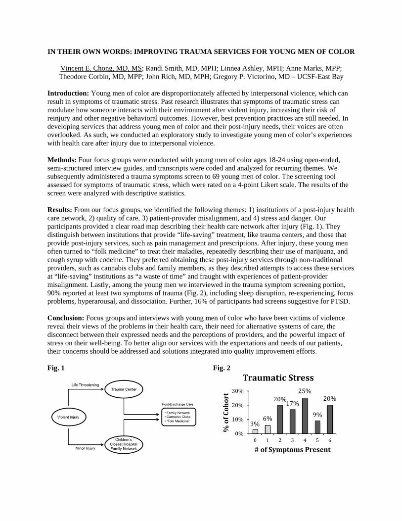
Introduction: Young men of color are disproportionately affected by interpersonal violence, which can result in symptoms of traumatic stress. Past research illustrates that symptoms of traumatic stress can modulate how someone interacts with their environment after violent injury, increasing their risk of reinjury and other negative behavioral outcomes. However, best prevention practices are still needed. In developing services that address young men of color and their post-injury needs, their voices are often overlooked. As such, we conducted an exploratory study to investigate young men of color’s experiences with health care after injury due to interpersonal violence. Methods: Four focus groups were conducted with young men of color ages 18-24 using open-ended, semi-structured interview guides, and transcripts were coded and analyzed for recurring themes. We subsequently administered a trauma symptoms screen to 69 young men of color. The screening tool assessed for symptoms of traumatic stress, which were rated on a 4-point Likert scale. The results of the screen were analyzed with descriptive statistics. Results: From our focus groups, we identified the following themes: 1) institutions of a post-injury health care network, 2) quality of care, 3) patient-provider misalignment, and 4) stress and danger. Our participants provided a clear road map describing their health care network after injury (Fig. 1). They distinguish between institutions that provide “life-saving” treatment, like trauma centers, and those that provide post-injury services, such as pain management and prescriptions. After injury, these young men often turned to “folk medicine” to treat their maladies, repeatedly describing their use of marijuana, and cough syrup with codeine. They preferred obtaining these post-injury services through non-traditional providers, such as cannabis clubs and family members, as they described attempts to access these services at “life-saving” institutions as “a waste of time” and fraught with experiences of patient-provider misalignment. Lastly, among the young men we interviewed in the trauma symptom screening portion, 90% reported at least two symptoms of trauma (Fig. 2), including sleep disruption, re-experiencing, focus problems, hyperarousal, and dissociation. Further, 16% of participants had screens suggestive for PTSD. Conclusion: Focus groups and interviews with young men of color who have been victims of violence reveal their views of the problems in their health care, their need for alternative systems of care, the disconnect between their expressed needs and the perceptions of providers, and the powerful impact of stress on their well-being. To better align our services with the expectations and needs of our patients, their concerns should be addressed and solutions integrated into quality improvement efforts. Fig. 1 Fig. 2
3%6%
20%17%
25%
9%
20%
0%
10%
20%
30%
0 1 2 3 4 5 6
%ofCohort
#ofSymptomsPresent
TraumaticStress

A surgical decision aid for patients with ulcerative colitis: a pilot study
Jessica N. Cohan, Elissa M. Ozanne, Justin L. Sewell, Uma Mahadevan, Daniel Dohan, Madhulika G. Varma, and Emily Finlayson
Introduction: Up to a third of ulcerative colitis patients require surgery, leaving many patients faced with the complex decision between end ileostomy and ileal pouch-anal anastomosis (IPAA). We developed a decision aid to encourage shared decision making between patients and surgeons.
Methods: We created a decision aid and knowledge questionnaire using literature review and interviews with patients, gastroenterologists, colorectal surgeons, and enterostomal therapists. Consecutive adult patients with ulcerative colitis were identified prior to surgical consultation at three colorectal surgery clinics and the decision aid was self-administered at home. We measured changes in knowledge, procedure preference, and stage of decision making before and after the decision aid, as well as patient satisfaction, preparation for decision making, and surgeon satisfaction.
Results: We analyzed data from 18 patients with ulcerative colitis who used the decision aid. Median age was 49 years (range 22-60), 10 were male, and 5 had previously undergone subtotal colectomy. Knowledge scores improved significantly after using the decision aid (range 0-13, median score 9 vs 12, p=0.003). Before using the decision aid, 12 patients preferred IPAA, 1 preferred end ileostomy, and 5 were unsure. After using the decision aid, 4 patients changed their preferred procedure (Table). Seven patients reported a change in the stage of decision making after using the decision aid, including 2 who were less confident in their procedure choice, and 5 who were more confident. The median preparation for decision making score was 77.8 (range 0-100) and patient satisfaction was generally high, with 88.9% patients reporting they would recommend the decision aid to others. Surgeons reported that the decision aid complemented their usual approach 87.5% of the time. All surgeons reported that they would like to incorporate the decision aid into their practice, although only one felt that the decision aid shortened the time of the clinical encounter.
Conclusions: These preliminary results of a decision aid for surgical patients with ulcerative colitis show promise in increasing patient knowledge and preparing patients for the discussion with their surgeon. In addition, it was well received by surgeons. This suggests that the decision aid is feasible and effective and warrants further study.
Table: Procedure preference in patients with ulcerative colitis before and after using the decision aid. Before Decision Aid After Decision Aid Number of Patients Unsure Unsure 1 Unsure Ileostomy 1 Unsure IPAA 2 IPAA Ileostomy 1 Ileostomy Ileostomy 1 IPAA IPAA 12 IPAA: Ileal Pouch-Anal Anastomosis

Patterns of injury at a trauma center in Mumbai: opportunities for injury prevention Adam Laytin, Catherine Juillard, Vineet Kumar, Bhakti Sarang, Nobhojit Roy, Rochelle Dicker Introduction: In India, the WHO estimates that 10% of deaths and 13% of disability-adjusted life years lost are due to injury. The purpose of this study is to describe patterns of injury among patients treated at a tertiary trauma center in Mumbai in order to identify opportunities for targeted injury prevention strategies. Methods: This is an analysis of data from an institutional trauma registry. All patients presenting to the hospital with life- or limb-threatening injuries over a 16 month period were included. Univariate and bivariate analyses were performed for demographic characteristics, injury mechanisms and clinical outcomes. Results: A total of 1115 patients met inclusion criteria. Mean age was 31 years and 88% were male. In-hospital mortality in this severely injured cohort was 32%. The most common mechanisms of injury were blunt assault (33%), road traffic injuries (32%) and falls (24%). Among victims of road traffic injuries, 52% were pedestrians struck by vehicles, 30% were injured while riding motorcycles or bicycles, and 18% were injured in motor vehicle collisions. Falls accounted for 73% of injuries among children under 10 and 45% of injuries among adults over 65. Mortality was highest among pedestrians struck by automobiles (38%) and blunt assault victims (37%), and lowest among victims of penetrating injuries (4%). Conclusions: Pedestrians struck by automobiles and blunt assault victims are the most common groups of trauma victims this setting, and have the highest mortality. Falls disproportionately affect children and the elderly. Pedestrian safety and fall prevention are important targets for injury prevention efforts, and an investigation into potential risk factors for interpersonal violence is warranted.

Splenic vein thrombosis following pancreas transplantation: identification of factors that support conservative management
Authors: Jack Harbell1, Garrett Roll1,2, Tara Morgan3, Vickie A. Feldstein3, Andrew Posselt1,2, Sang-Mo Kang1,2, Sandy Feng1,2, Ryutaro Hirose1,2, Chris E. Freise1,2, Peter Stock1,2
1Department of Surgery, 2Division of Transplantation, University of California San Francisco, San Francisco, CA
3Department of Radiology, University of California San Francisco, San Francisco, CA
Introduction: Prophylaxis for graft venous thrombosis following pancreas transplant varies between institutions. Similarly, treatment of venous thrombosis ranges from urgent thrombectomy to conservative management with anticoagulation. In this study we wished to determine the prevalence of graft splenic vein (SV) thombosis, as well as the clinical significance of non-occlusive thrombus observed on routine imaging.
Methods: Records of 112 pancreas transplant recipients from January 1st, 2008 to December 31st, 2012 at a single center were reviewed. All patients received aspirin prior to surgery, and aspirin and dipyridimole post-operatively. Non-dialysis dependent patients also received a bolus of intravenous (IV) heparin (2000-4000 units) at the time of vascular anastomosis. Post-operative anticoagulation was surgeon specific, but usually included low dose heparin infusion (200-400 units/hr) for 24-48h after surgery. Venous thrombosis was defined as absence of flow or presence of thrombus identified in any part of the graft SV on ultrasound. Patients with SV thrombus were anticoagulated with IV heparin in addition to anti-platelet therapy, then transitioned to warfarin for 3-6 months or until documented thrombus resolution.
Results: 30 patients (27%) had thrombus or absence of flow in the SV on post-operative ultrasound. There were 4 graft losses in this group. All were due to arterial and venous thrombosis, and all occurred within 20 days of transplant. Graft losses occurred in dialysis dependent and independent recipients (2 SPK, 1 PAK, 1 PTA). Patients identified to have partial SV thrombus but normal arterial signal on ultrasound were all successfully treated conservatively with IV heparin followed by warfarin for 3-6 months, and remained insulin independent. Seven patients with ultrasound findings of graft swelling and abnormal arterial waveforms with reversal of diastolic flow were taken emergently to the operating room for thrombectomy. Two of those patients were found to have no thrombus at exploration, one patient underwent successful venous thrombectomy, one patient had a successful venous thrombectomy initially but lost the graft 19 days later due to arterial and venous thrombosis, and 3 were found to have graft necrosis and underwent graft pancreatectomy.
Conclusion: In the absence of graft swelling and arterial signal abnormalities on ultrasound, non-occlusive thrombosis of the pancreas graft SV can be successfully treated conservatively with anticoagulation. Findings of graft swelling and arterial signal abnormalities, such as reversal of diastolic flow within the graft, require urgent operative intervention, since this finding can be associated with more extensive thrombus that may lead to graft loss.

Wound class does not alter recurrence in complex ventral hernia repair with mesh: A single-center retrospective review
Victoria Lyo, Michael Tufaga, Uk-Sok Shin, Sophia Fong, Frank Primus and Hobart W. Harris
Introduction: Ventral hernias occur following 11-23% of laparotomy incisions. With 5 million laparotomies performed annually in the United States alone, an estimated 400,000 ventral hernia repairs are performed annually, adding over 8 billion dollars in healthcare costs. Permanent prosthetic mesh repair during a clean case is the standard of care, yielding the best long-term results and reducing the hernia recurrence rate by 50%. However, the management of ventral hernias with wound contamination remains controversial due to insufficient evidence to support a specific treatment course. Placing a permanent synthetic prosthetic into a contaminated field is generally thought to result in an unacceptably high rate of complications, namely surgical site infection, enterocutaneous fistula and recurrent hernia formation. We sought to determine the degree to which wound contamination contributes to recurrence and complications after complex ventral hernia repair. Methods: We performed a retrospective record review of an accumulated database of open incisional hernia repair cases performed by a single surgeon at a large, tertiary care hospital. We identified 188 operations on 163 patients from April 19, 2004 through May 13, 2013. Postoperative outcomes, including hernia recurrences were recorded as were surgical site occurrences, defined as all wound complications, including cellulitis, skin/fat necrosis or fluid collections that required antibiotics, debridement or drainage. Clean cases were denoted as wound class 1 and contaminated cases were denoted as wound class 2-4. Statistical analysis was completed using Chi Square tests (GraphPad Prism Software). Results: A total of 188 cases in 163 patients (age range: 19-87 years, median age 55, female to male ratio of 2.2) met our criteria. Average follow-up time was 18 months with zero 30-day mortality. Of our patient population, 65% (n=122) had recurrent hernias. There were 139 (73.9%) clean cases and 49 (26%) contaminated cases. The rate of surgical site occurrences was significantly lower in clean versus contaminated classes (25.2% versus 44.1%, p=0.03). Synthetic mesh was used in 94.5% of clean cases and 70.6% of contaminated cases. Biologic mesh was used in 5.5% of clean cases and 29.4% of contaminated cases. Wound contamination was not significantly associated with recurrence in complex ventral hernia repair: recurrence in clean cases was 24.4% compared to 35.3% in contaminated cases (p=0.19). Interestingly, type of mesh used did not alter recurrence rates in clean versus contaminated cases. The recurrence rate did not differ significantly when synthetic mesh was used in clean versus contaminated cases (23% vs 21% respectively, p=0.86) or when biologic mesh was used in clean versus contaminated cases (57% vs 70% respectively, p=0.59). The use of synthetic mesh also did not result in more wound events in contaminated cases compared to clean cases (39% versus 24%, p=0.59). Moreover, regardless of wound class, the use of synthetic mesh was associated with a lower overall recurrence rate than biologic mesh (22% versus 65%, p=0.002). Conclusions: The management and treatment of ventral hernias in the setting of bacterial contamination remains a major clinical challenge due to inconsistent and controversial current practices. Our results confirm that wound contamination is associated with increased surgical site occurrences. However, contrary to conventional wisdom, we found that wound contamination was not a risk factor for hernia recurrence, irrespective of the type of mesh used. Moreover, since the use of biologic mesh is associated with a higher recurrence rate than synthetic mesh, our results indicate that the use of synthetic mesh in a one-stage repair of contaminated cases can be performed without an unacceptable rate of complications. Due to the limitations of this retrospective study, future prospective trials are necessary to generate definitive treatment recommendations.

Serotonin Signaling Promotes Growth of Postnatal-Derived Enteric Neuronal Stem Cells Lily S. Cheng, M.D.; Ryo Hotta, M.D., Ph.D.; Hannah K. Graham, B.S.; Allan M. Goldstein, M.D. Introduction: Transplantation of enteric neuronal stem cells (ENSCs) offers an innovative approach for treating enteric neuropathies, including Hirschsprung disease. However, postnatal-derived cells, a potential autologous source, are less proliferative than embryonic precursors. Since serotonin (5-HT) promotes enteric neuronal growth during development, we hypothesized that nanoparticles expressing a 5-HT receptor agonist would augment growth of neurons derived from postnatal ENSCs. Methods: Postnatal ENSCs were isolated from 2-4 week-old mouse colon and cultured 7 days without additive (n=3) or with nanoparticles loaded with 5-HT4 receptor agonist (RS67506; n=3). ENSCs were cultured ex vivo with colon explants for 7 days in the presence of RS67506-loaded (n=3) or empty nanoparticles (n=3). ENSCs were also transplanted into mouse rectum in vivo for 14 days with RS67506-loaded (n=4) or empty nanoparticles (n=3). Neuronal density, proliferation, and neurite extension were analyzed immunohistochemically. Results were compared statistically using Chi-square and Student’s t-test. Results: Cultured ENSCs and co-cultured explants both contained more neurons in the presence of 5-HT4 agonism than without (34.4±5.8 vs. 8.2±0.2%, p<0.05; and 74.6±3.3% vs. 53.1±15.5%, p<0.01, respectively). Neurite length was also greater with 5-HT4 agonism than without both in vitro and in vivo (425.9±27.4µm vs. 223.7±33.4µm and 116.7±14.2µm vs. 74.3±7.0µm, respectively; p<0.05). ENSCs cultured in vitro with RS67506-loaded nanoparticles had significantly more neuronal proliferation than controls (17.5±5.1% vs 3.8±0.8%, p<0.05). Similarly, ENSCs co-cultured with colon explants and RS67506-loaded nanoparticles exhibited more neuronal proliferation than controls (28.0±7.7% vs. 15.1±2.9%, p<0.05). Importantly, ENSCs transplanted in vivo with RS67506-loaded nanoparticles had significantly more neuronal proliferation than control transplants (20.8±6.4% vs. 5.0±2.1%, p<0.01). Conclusion: Co-transplantation of ENSCs with nanoparticles expressing a 5-HT4 receptor agonist led to significant increases in neuronal density, proliferation, and neurite extension. Optimization of postnatal ENSCs supports their potential use in cell-based therapies for Hirschsprung and other neurointestinal diseases.

SmartDerm: Real-Time Prediction and Prevention of Pressure Ulcers Isabelle Chumfong, M.D. M.Eng.; Michael Hemati M.T.M..; Sachin Rangarajan, M.T.M.; Katie Mo; Sandra Thao; Ian Tran; Hanmin Lee, M.D. Introduction: Pressure ulcers occur in 2.5 million patients annually, resulting in 60,000 deaths and a $30 billion burden to the healthcare system. As of 2007, neither Medicare nor Medicaid reimburses hospitals for the treatment of hospital-acquired pressure ulcers, which can cost as much as $43,000 per treatment. This creates a significant incentive for hospitals to actively prevent pressure ulcer formation and reduce related expenditures. Preventative measures today have limited efficacy; nurses monitor and reposition patients every two hours. These practices require significant personnel time and resources. Furthermore, the earliest visual indication of pressure ulcer formation occurs after tissue damage has already taken place. This reality demonstrates an unmet clinical and market need for a low-cost technology that can continuously monitor at-risk patients and preempt pressure ulcer formation well ahead of the initial visual indication. To meet this need, we have developed the SmartDerm system, which consists of four components: (1) a wireless network of focal pressure sensors placed on high-risk areas, (2) software that employs personalized prevention risk algorithms, (3) a pressure relief device that automatically repositions patients to reduce risk at specific locations based on real-time data, and (4) management software to monitor patients and to collect data/metrics. We hypothesize that patients who ultimately develop pressure ulcers have a predictable pattern of exposure(s) to pressure over a certain threshold that can be characterized using a series of pressure measurements over time. To this end, we are planning an observational clinical study to collect pressure data with the SmartDerm sensor and generate pressure profiles that can be combined with other patient characteristics to predict risk for pressure ulcer formation. Methods: We plan to record pressure measurements at the sacrum and heels in n=50 ICU patients and patients undergoing surgical procedures greater than 2.5 hours. Patients will have the SmartDerm device placed on admission to the ICU and/or before they are positioned for their surgical procedure. These devices will be left on during the hospital stay and during the intra-operative period with pressure measurements recorded into the patient’s study file. Patients will be routinely assessed for pressure ulcer formation and monitored until hospital discharge. Data collected with the device will not be used to modify patient care. Progress: Presently, our product is in the pre-clinical and prototyping stages. We anticipate beginning patient enrollment in Spring 2015. Data from the pilot will be used to revise our prediction model and sensor device. We predict that patients who develop pressure ulcers will have stereotypical pressure profiles (exceeding a threshold over a certain length of time) we can use to direct intervention. Follow-on experiments will incorporate measurement-directed intervention, first by providers and then by our automated-repositioning system. Conclusion: There is an unmet clinical need in pressure ulcer risk detection and prevention, for which we have developed a prototype of a closed-feedback loop system intended to detect where and when patients are at risk for developing pressure ulcers and automatically intervene to minimize risk. We anticipate that data from the SmartDerm sensors and algorithm in combination with active intervention from the repositioning system will result in significant reduction in pressure ulcer incidence and cost-savings to healthcare systems.

Renal Artery Embolization for Minimally Invasive Induction of Renal Failure
Willieford Moses MD, Steven Kim MD, Elisabeth Leeflang MD, Clarence Chow BS, Zohora Iqbal BS, Mark Wilson MD, Shuvo Roy PhD Introduction New treatments for renal replacement therapy are being developed such as the BioArtificial Kidney; however, a large animal model of renal failure is needed to investigate their effectiveness. Embolization agents have been used to treat a variety of medical issues, e.g., tumors and aneurysms. We propose a minimally invasive technique to initiate complete renal failure in a swine model using a combination of nanoparticles and coils to embolize both renal arteries. Methods A 5Fr catheter was placed in the right femoral artery of a 55-kg female Yorkshire pig. After the renal arteries were identified with radiopaque contrast (Conray™), particle embolization was performed with polyvinyl alcohol flakes (Contour™, 250 - 700nm nanoparticles), followed by coil embolization (Tornado® Coils). After complete occlusion was confirmed, the sheath was removed and the pig was allowed to recover. Meloxicam (15mg IM) and buprenorphine (0.12mg/kg IM) were administered once for post-procedural pain. Daily blood draws and clinical assessments were performed until euthanasia, which occurred based on the veterinarian’s assessment of severe metabolic derangement and/or signs of decompensation. Results The procedure was well tolerated with no significant rise in inflammatory markers (white cell count and C-reactive protein) nor need for further pain medications. Immediate renal failure resulted from the embolization as evidenced by complete cessation of urine output. There was a progressive rise in urea and creatinine from a baseline of 8 mg/dL and 1.35 mg/dL to 95 mg/dL and 21.62 mg/dL, respectively. This also coincided with worsening uremic symptoms including progressive lethargy and decreased oral intake. On day 5, the animal was euthanized due to severe lethargy and metabolic derangements (e.g., potassium: 8.6 mEq/L). Conclusion A combination of nanoparticle and coil embolization of the renal arteries can induce immediate renal failure in a pig. This minimally invasive technique can serve as a viable large animal model for investigating the feasibility of the next generation of renal replacement devices such as the BioArtificial Kidney.

Lower extremity amputations in patients with SIRS/sepsis: surgical specialty and 30 day outcomes Joseph T. Patterson1, MD; Richard Coughlin, MD; Saam Morshed, MD, MPH, PhD 1 Department of Orthopaedic Surgery, University of California – San Francisco, San Francisco, CA 94122 Introduction. Patients with systemic inflammatory response syndrome (SIRS), sepsis, or septic shock may require amputation as a life-saving intervention for infectious source control or as salvage after ischemic complications of prolonged hypoperfusion. Technical training and expertise in lower extremity amputation varies among surgical specialists and may be associated with adverse outcomes in patients with SIRS/sepsis. Methods. A retrospective review of the prospectively collected ACS-NSQIP database was conducted. Patients with preoperative SIRS, sepsis, or septic shock undergoing a lower extremity amputation at the index surgery were identified by Common Procedure Terminology codes. Multivariate regression analyses to investigate the association of surgical specialty (general, orthopaedic, or vascular) with any adverse event, serious adverse event, or death within 30-days of amputation were conducted controlling for baseline demographics, comorbidities, sepsis severity, amputation level, and type of anesthesia. Results: 7,945 lower extremity amputations were identified in 202,402 patients meeting SIRS criteria from 2005-2013 in the ACS-NSQIP database. Patients treated by general, orthopaedic, or vascular surgeons were significantly different with regard to age, sex, functional dependence, severity of SIRS/sepsis, cardiovascular disease, diabetes, disseminated cancer. Anesthesia, amputation level, delay from admission to surgery, and operative time were significantly different between groups. Vascular surgeons performed lower extremity amputations in an older, less male population with greater incidence of cardiovascular comorbidity, milder severity of SIRS/sepsis criteria, and longer delays from admission to surgery than those treated by orthopaedic or general surgeons. SIRS patients undergoing amputation by an orthopaedic surgeon were more obese, less functionally dependent, more likely to have diabetes or disseminated cancer, received more distal and fewer guillotine amputations, and experienced longer operative times. On multivariate analysis controlling for the differences above, no difference in mortality was observed between surgical specialties. Lower extremity amputations by orthopaedic surgeons in SIRS/sepsis patients were associated with a significantly lower risk of any adverse event (RR = 0.90, 95% CI [0.81-0.99]) and superficial surgical site infection (RR = 0.29, 95% CI [0.12-0.75]) within 30 days of surgery relative to those by general surgeons. Amputations by orthopaedic surgeons were also associated with lower risk of serious adverse events (RR = 0.87, 95% CI [0.81-0.99]) and reoperation (RR = 0.59, 95% CI [0.44-0.79]) relative to procedures performed by both vascular and general surgeons. Those treated by vascular surgeons were more likely to experience an unplanned reintubation and discharge to a facility other than home relative to those treated by a general surgeon. Conclusions: General, orthopaedic, and vascular surgeons perform lower extremity amputation in heterogeneous populations of SIRS/sepsis patients. Controlling for these differences, orthopaedic surgeons perform slower lower extremity amputations with fewer complications or reoperations within 30 days.

A Biodegradable Device for Perivascular Delivery of Pro-Resolving Lipid Mediators Bian Wu, Kevin D Lance, Anuran Chatterjee, Giorgio Mottola, Mian Chen, Sevan R Komshian, Tejal A Desai, Michael S Conte Introduction: Persistent inflammation following vascular injury leads to excessive scarring, limiting the success of vascular interventions. Recent work has identified that endogenous specialized proresolving lipid mediators (SPM) such as resolvin D1 (RvD1) actively orchestrate the process of resolution, exerting vasculo-protective effects without associated toxicity. We propose local vascular delivery of SPM through a biodegradable perivascular wrap. Methods: RvD1 (200 ng) was heat-sealed between thin layers of polycaprolactone (PCL) or poly-lactic-co-glycolic acid (PLGA). PLGA membranes differed in their composition of lactic versus glycolic acid (%-lactide). Directional drug release was measured via EIA in a cell-free system in vitro and into rabbit aortas exposed to pulsatile flow ex vivo. Bioactivity in vitro was confirmed on human vascular smooth muscle cells (VSMC) using molecular (NFkB activation) and cellular (migration, proliferation) assays. Safety and efficacy were tested in vivo using a rat model of carotid angioplasty. Results: Of the constructs tested, a 3-layered PLGA wrap consisting of 85%/75%/50%-lactide provided the most favorable drug elution (Fig1), with sustained release of >800 pg/day for at least 14 days and nearly all elution occurring from the 50%-lactide side. Perivascular application of this wrap ex vivo with the 50%-lactide side facing “in” demonstrated uptake into rabbit aorta at 8 hours (0.4 ± 0.1 pg/mg). VSMC exposed to drug-loaded wraps in vitro showed inhibition of NFkB activation (60%, Fig2a), migration (40%, Fig2b) and proliferation (70%, Fig2c). Perivascular application of control 3-layered PLGA wraps in vivo caused a mild perivascular fibroinflammatory (foreign body) response at both 4 days and 14 days (Fig3). However, there was no evidence of infection, thrombosis or neointimal hyperplasia associated with the wrap at either 4 or 14 days (n=3 and n=7, respectively). Conclusion: We demonstrate sustained and directional elution of therapeutically relevant amounts of biologically-active RvD1 through a biodegradable perivascular wrap, providing opportunity for translational studies of SPM in vascular injury. We hypothesize that perivascular application of SPM in vivo will attenuate neointimal hyperplasia in a rat model of carotid angioplasty.
1.
2b.
3.
2c.2a.