Vascular Lesions of the Breast - UCSF CME€¦ · morphology clinical presentation definitive...
22
5/26/2016 1 Vascular Lesions of the Breast UCSF 32 nd Annual Current Issues in Anatomic Pathology and Cytology Sandra J Shin, MD Professor of Pathology and Laboratory Medicine Chief of Breast Pathology Weill Cornell Medicine MORPHOLOGY CLINICAL PRESENTATION DEFINITIVE SURGERY (MASTECTOMY) DIAGNOSIS MORPHOLOGY CLINICAL PRESENTATION MAMMOGRAPHIC PRESENTATION DEFINITIVE SURGERY CORE NEEDLE BIOPSY MORPHOLOGY MORPHOLOGY IHC MOLECULAR
Transcript of Vascular Lesions of the Breast - UCSF CME€¦ · morphology clinical presentation definitive...

5/26/2016
1
Vascular Lesions of the Breast
UCSF 32nd AnnualCurrent Issues in Anatomic Pathology and Cytology
Sandra J Shin, MDProfessor of Pathology and Laboratory MedicineChief of Breast PathologyWeill Cornell Medicine
MORPHOLOGY
CLINICAL PRESENTATION
DEFINITIVE SURGERY
(MASTECTOMY)
DIAGNOSIS
MORPHOLOGY
CLINICAL PRESENTATION
MAMMOGRAPHIC PRESENTATION
DEFINITIVE SURGERY
CORE NEEDLE BIOPSY
MORPHOLOGYMORPHOLOGY
IHCMOLECULAR

5/26/2016
2
BREAST LESIONS
ARE SMALLER
MAMMOGRAPHIC PRESENTATION
SAMPLING IS SMALLER
CORE BIOPSY
IHC AND MOLECULAR CAN HELP OR
CONFUSE!
MORPHOLOGYIHC
MOLECULAR
For Pathologists• The most important goal when encountering a
mammary vascular lesion is to identify (or exclude) ANGIOSARCOMA
• Two most difficult diagnostic challenges– Identify low grade primary angiosarcoma in core
needle biopsy material– Distinguish atypical vascular lesion from post
radiation angiosarcoma in skin punch biopsy material
AngiosarcomaPrimary: Breast +/- cutaneous involvement
Secondary: Mammary skin +/- subadjacent breast1. Radiation-induced ( )2. Lymphedema-associated (Stewart-Treves) ( )
– Arm, may extend to chest wall
Primary angiosarcoma• Rare (<0.05% breast malignancies)• Constitutes about 10-25% of all mammary sarcomas• No known risk factors• Younger patients (20s-50s) than those with breast
carcinoma or radiation-induced angiosarcomas• Presents with a breast mass• Mastectomy • Aggressive clinical behavior with high propensity to
metastasize and high risk of dying from disease

5/26/2016
3
Grading of primary angiosarcomas• Rosen’s three tiered grading system
– Grade I (low grade)– Grade II (intermediate grade)– Grade III (high grade)
• Individual tumors can exhibit the full spectrum of grades
• Grading should be done only on the excised tumor specimen
Low grade angiosarcoma• Complex anastomosing vascular channels• Minimal endothelial nuclear atypia• No cellular stratification• Low mitotic rate• No necrosis• Dissects into adipose tissue and breast elements

5/26/2016
4
Intermediate grade angiosarcoma• More cellular than low grade • Moderate cytologic atypia• Multilayering of lesional endothelial cells• Papillary structures can be seen but not solid
areas• Moderate mitotic rate

5/26/2016
5
High grade angiosarcoma• Solid areas with poorly formed vascular
channels; spindled tumor cells• Blood lakes (hemorrhage)• Necrosis• Marked cytologic atypia including
hyperchromasia• Brisk mitotic rate
Solid growth
Spindled
Blood lakes
Necrosis
Grading and prognosis• Rosen et al reported a similar estimated survival
probability for angiosarcomas grades I, II and III at year 1 but much worse outcome at 5 and 10 years as well as worse recurrence-free survival for grade III
• Study limitations: small cohort, short follow-up• More recently, the prognostic value of histologic
grading in primary angiosarcomas has been challenged

5/26/2016
6
Nascimento AF, et al. AJSP 2008• Similar tumor sizes and numbers of cases for
each histologic grade (n=49)• No statistically significant correlation between
– tumor size and likelihood of local recurrence or dying of disease
– histologic grade and the rate of local recurrence or distant mets
– tumor grade and death owing to disease
DIAGNOSTIC CHALLENGE #1 • Know the differential
diagnosis • Correlate with clinical and
radiologic findings• Know the diagnostic
pitfalls• Know when/how ancillary
studies can help
Identify primary low grade angiosarcomain core biopsy material
DIAGNOSTIC CHALLENGE #1 • Hemangioma
• Papillary endothelial hyperplasia
• Angiolipoma (if lesion is involving the subcutaneous fat)
Differential Diagnosis

5/26/2016
7
Clinical and radiologic correlation• Angiosarcoma
• Hemangioma, papillary endothelial hyperplasia, angiolipoma
• >2cm or radiographically occult
• <2 cm; nodule/MRI enhancement/ rarely calcs; well circumscribed
DIAGNOSTIC CHALLENGE #1 • Hemangioma
• Papillary endothelial hyperplasia
• Angiolipoma (if lesion is involving the subcutaneous fat)
Differential Diagnosis
HemangiomaVarious morphologies
– capillary – cavernous– venous
Location-parenchymal vs non-parenchymal (adipose tissue)-if parenchymal, extralobular vs perilobular Capillary
hemangioma
5/26/2016
8
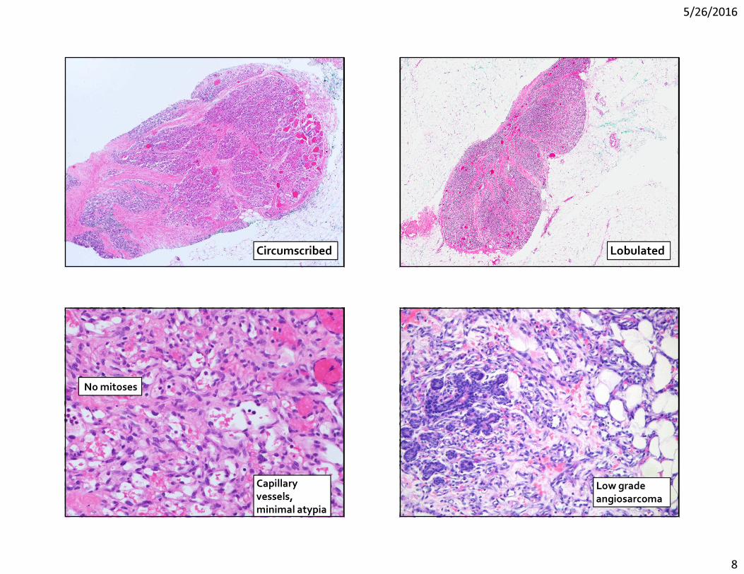
Circumscribed
Lobulated
Lobulated
No mitoses
Capillary vessels, minimal atypia
Low grade angiosarcoma

5/26/2016
9
Hemangioma – focal infiltrative borders
Extralobularhemangioma

5/26/2016
10
Perilobularhemangioma
Perilobularhemangioma
Angiosarcoma

5/26/2016
11
Ki-67 Ki-67

5/26/2016
12
Ki-67 and mammary vascular lesions• Best used in the setting of confirming a lesion
with morphologic features highly supportive of the diagnosis (angiosarcoma versus hemangioma)
• Prior biopsy changes in the lesion can lead to a falsely elevated Ki-67 index
DIAGNOSTIC CHALLENGE #1 • Hemangioma
• Papillary endothelial hyperplasia
• Angiolipoma (if lesion is involving the subcutaneous fat)
Differential Diagnosis
Papillary endothelial hyperplasia
Mass
Circumscribed
Associated with thrombus, hematoma, hemangioma

5/26/2016
13
Angiosarcoma
DIAGNOSTIC CHALLENGE #1 • Hemangioma
• Papillary endothelial hyperplasia
• Angiolipoma (if lesion is involving the subcutaneous fat)
Differential Diagnosis
Angiolipoma

5/26/2016
14
Hyaline thrombi
Angiosarcoma

5/26/2016
15
Ki-67 DIAGNOSTIC CHALLENGE #2 • Know the differential
diagnosis • Correlate with clinical and
radiologic findings• Know the diagnostic
pitfalls• Know when/how ancillary
studies can help
Distinguish between AVL and post radiation AS in a skin biopsy
Post radiation angiosarcoma• First documented case after breast conserving
surgery in 1987; >200 cases reported• Constitutes about 40% of all radiation induced
sarcomas• Older patients than those with primary AS (70s) • Presents with a rash/bruise +/- ulceration• Latency after RT is 7-10 years

5/26/2016
16
Post radiation angiosarcoma
High or intermediate nuclear gradeAny growth pattern
CD 31
FLI-1
ERG
Atypical vascular lesion (AVL)• First described in 1994 by Fineberg and Rosen• Spectrum of vascular proliferations that
develop in previously irradiated skin• Latency from time of RT is shorter (2-6 years)
than for post-radiation angiosarcoma (7+ years)• Can be multiple• Benign clinical course

5/26/2016
17
Atypical vascular lesion (AVL)• Localized superficial proliferation composed of well-formed
empty vascular spaces lined by plump endothelial cells• Lack multilayering, significant cytologic atypia, significant
infiltrative growth• Two patterns
– Dilated vessels resembling lymphangioma(lymphatic-type) – more common
– Slit-like vascular spaces with hobnail endothelium (vascular-type) – less common but more easily confused with AS
Atypical vascular lesion (AVL)Lymphatic type
Atypical vascular lesion (AVL)Vascular type Atypical vascular lesion (AVL)
• Overlap with post radiation angiosarcoma– clinically (age at presentation, latency from RT
and lesion duration before bx)– histomorphologically (prominent nucleoli, mitotic
figures, cytologic atypia, infiltrative growth)

5/26/2016
18
Post-radiation angiosarcoma
AVL – like areas in angiosarcoma
Diagnostic pitfall
Angiosarcomas can exhibit AVL-like areasMay be impossible to distinguish
in small biopsy material
Ki-67
Ki-67 proliferation index of AVLs has not been studied

5/26/2016
19
c-MYC is a proto-oncogene located on chromosome 8q24-21 that encodes a transcription factor involved in cell growth, proliferation, apoptosis and other cancer processes such as angiogenesis
MYC amplification
• High level MYC amplification (5-20 fold) limited to post radiation AS and rare primary AS; absent in all reported AVLs and almost all primary AS
• One study found MYC amplified post-radiation AS to have worse prognosis than those without MYC amplification
• Suggests different pathogenetic pathways of – primary and secondary (post radiation) AS– AVL and secondary (post radiation) AS

5/26/2016
20
CD 31
Ki-67
C-myc
Utility of anti-myc IHC in mammary vascular lesions
• Used to discriminate between post-radiation AS and AVL• Highly concordance with MYC gene amplification• Positive staining is strong and diffuse (>80%) in lesional
cells; important in small biopsy samples• Can also stain lymphocytes• Benign vessels are negative and should not be mistaken
for non-immmunoreactive lesional vessels
FLT4 and mammary vascular lesions• FLT4 gene found on chromosome 5q35.3 which
encodes VEGFR-3 and belongs to tyrosine kinase receptor family
• FLT4 co amplifies with MYC• FLT4 protein expression by IHC in benign and
malignant neoplasms including up to 80% of AS• Possible therapeutic target – multi-kinase
inhibitors; anti-VEGFR inhibitors

5/26/2016
21
Copy number alterations and gene expression were analyzed in 7 primary AS and 18 post radiation AS
Single nucleotide polymorphism (SNP) analysis revealed a single locus which was considered significant.18/18 post-rad AS and 1/7 primary AS showed amplification in 8q24region – all contained MYC
53 gene signature that discriminated post rad AS and primary AS. MYC was not listed confirming that its expression is not a marker of radiation tumorigenesis
• Two distinct transcriptome signatures co-exist in radiation-induced breast AS and both discriminate the tumors as a function of their etiology
• One signature specific to breast AS. The deregulation of marker genes suggests that post rad AS develop from radiation-stimulated lymphatic vessel endothelial cells
• High rate of MYC amplification in post rad AS , likely a consequence of genome instability initiated by ionizing radiation, is NOT a marker of radiation tumoriogenesissince it is also observed at a low rate in primary AS

5/26/2016
22
PTPRB• Negative regulator of angiogenesis; Inhibits VEGFR2,
VE-cadherin and angiopoietin signalling• Expressed exclusively in vascular endothelium• Study showed 14 PTPRB mutations in 10/39 (26%) AS• All PTPRB mutations identified in tumors that were
either post-rad AS and/or have MYC amp; 45% in this subgroup (10/22)
• Possible clinical utility as biomarker of radiation assocdisease and/or novel therapeutic targets in AS
MORPHOLOGY&
IHC(molecular)
Radiologic Presentation
Core biopsy
DIAGNOSIS
Excision or Mastectomy or
survelliance
Thank You