
Ubiquitin–proteasome-rich cytoplasmic structures in … heads: V. Necchi et al ... Mutation...
27
Ubiquitin–proteasome-rich cytoplasmic structures in neutrophils of patients with Shwachman–Diamond syndrome by Vittorio Necchi, Antonella Minelli, Patrizia Sommi, Agostina Vitali, Roberta Caruso, Daniela Longoni, Maria R. Frau, Cristina Nasi, Fabiola De Gregorio, Marco Zecca, Vittorio Ricci, Cesare Danesino, and Enrico Solcia Haematologica 2012 [Epub ahead of print] Citation: Necchi V, Minelli A, Sommi P, Vitali A, Caruso R, Longoni D, Frau MR, Nasi C, De Gregorio F, Zecca M, Ricci V, Danesino C, and Solcia E. Ubiquitin–proteasome-rich cytoplasmic structures in neutrophils of patients with Shwachman–Diamond syndrome. Haematologica. 2012; 97:xxx doi:10.3324/haematol.2011.048462 Publisher's Disclaimer. E-publishing ahead of print is increasingly important for the rapid dissemination of science. Haematologica is, therefore, E-publishing PDF files of an early version of manuscripts that have completed a regular peer review and have been accepted for publication. E-publishing of this PDF file has been approved by the authors. After having E-published Ahead of Print, manuscripts will then undergo technical and English editing, typesetting, proof correction and be presented for the authors' final approval; the final version of the manuscript will then appear in print on a regular issue of the journal. All legal disclaimers that apply to the journal also pertain to this production process. Haematologica (pISSN: 0390-6078, eISSN: 1592-8721, NLM ID: 0417435, www.haemato- logica.org) publishes peer-reviewed papers across all areas of experimental and clinical hematology. The journal is owned by the Ferrata Storti Foundation, a non-profit organiza- tion, and serves the scientific community with strict adherence to the principles of open access publishing (www.doaj.org). In addition, the journal makes every paper published immediately available in PubMed Central (PMC), the US National Institutes of Health (NIH) free digital archive of biomedical and life sciences journal literature. Official Organ of the European Hematology Association Published by the Ferrata Storti Foundation, Pavia, Italy www.haematologica.org Early Release Paper Support Haematologica and Open Access Publishing by becoming a member of the Europe Hematology Association (EHA) and enjoying the benefits of this membership, which inc participation in the online CME?program Copyright 2012 Ferrata Storti Foundation. Published Ahead of Print on March 14, 2012, as doi:10.3324/haematol.2011.048462.
-
Upload
nguyenthuy -
Category
Documents
-
view
215 -
download
2
Transcript of Ubiquitin–proteasome-rich cytoplasmic structures in … heads: V. Necchi et al ... Mutation...

Ubiquitin–proteasome-rich cytoplasmic structures in neutrophils of patients with Shwachman–Diamond syndrome
by Vittorio Necchi, Antonella Minelli, Patrizia Sommi, Agostina Vitali, Roberta Caruso, Daniela Longoni, Maria R. Frau, Cristina Nasi, Fabiola De Gregorio,Marco Zecca, Vittorio Ricci, Cesare Danesino, and Enrico Solcia
Haematologica 2012 [Epub ahead of print]
Citation: Necchi V, Minelli A, Sommi P, Vitali A, Caruso R, Longoni D, Frau MR, Nasi C,De Gregorio F, Zecca M, Ricci V, Danesino C, and Solcia E. Ubiquitin–proteasome-richcytoplasmic structures in neutrophils of patients with Shwachman–Diamond syndrome.Haematologica. 2012; 97:xxx doi:10.3324/haematol.2011.048462
Publisher's Disclaimer. E-publishing ahead of print is increasingly important for the rapid dissemination of science.Haematologica is, therefore, E-publishing PDF files of an early version of manuscripts thathave completed a regular peer review and have been accepted for publication. E-publishingof this PDF file has been approved by the authors. After having E-published Ahead of Print,manuscripts will then undergo technical and English editing, typesetting, proof correction andbe presented for the authors' final approval; the final version of the manuscript will thenappear in print on a regular issue of the journal. All legal disclaimers that apply to the journal also pertain to this production process.
Haematologica (pISSN: 0390-6078, eISSN: 1592-8721, NLM ID: 0417435, www.haemato-logica.org) publishes peer-reviewed papers across all areas of experimental and clinicalhematology. The journal is owned by the Ferrata Storti Foundation, a non-profit organiza-tion, and serves the scientific community with strict adherence to the principles of openaccess publishing (www.doaj.org). In addition, the journal makes every paper publishedimmediately available in PubMed Central (PMC), the US National Institutes of Health (NIH)free digital archive of biomedical and life sciences journal literature.
Official Organ of the European Hematology AssociationPublished by the Ferrata Storti Foundation, Pavia, Italy
www.haematologica.org
Early Release Paper
Support Haematologica and Open Access Publishing by becoming a member of the EuropeHematology Association (EHA) and enjoying the benefits of this membership, which inc
participation in the online CME?program
Copyright 2012 Ferrata Storti Foundation.Published Ahead of Print on March 14, 2012, as doi:10.3324/haematol.2011.048462.

1
Ubiquitin–proteasome-rich cytoplasmic structures in neutrophils of patients
with Shwachman–Diamond syndrome Running heads: V. Necchi et al. PaCS in SDS neutrophils
Vittorio Necchi1,2, Antonella Minelli1, Patrizia Sommi1,3, Agostina Vitali3, Roberta Caruso4,
Daniela Longoni5, Maria Rita Frau6, Cristina Nasi7, Fabiola De Gregorio8, Marco Zecca9,
Vittorio Ricci3, Cesare Danesino,1 and Enrico Solcia1
1Department of Human Pathology and Genetics, University of Pavia and Fondazione IRCCS
Policlinico S. Matteo, Pavia, Italy; 2Centro Grandi Strumenti, University of Pavia, Pavia, Italy; 3Department of Physiology, University of Pavia, Pavia, Italy; 4Department of Pediatric
Hematology/Oncology and Transfusion Medicine, IRCCS Pediatric Hospital Bambino Gesù, Rome 5Department of Pediatrics, University of Milano Bicocca, Monza, Italy; 6Azienda Sanitaria ASL
Nuoro, Division of Pediatrics, Nuoro, Italy; 7Azienda Sanitaria ASL 17, Division of Pediatrics,
Savigliano, Italy; 8Department of Pediatrics-Federico II University, Napoli, Italy,
and 9Pediatric Hematology/Oncology, Fondazione IRCCS Policlinico San Matteo, Pavia, Italy
Correspondence
Enrico Solcia, Department of Human Pathology and Genetics, University of Pavia Medical School,
Via Forlanini 16, 27100 Pavia, Italy. Phone: international +39.0382.503057.
Fax: international +39.0382525866. E-mail: [email protected]
Funding
This study was supported in part by grants from the Italian Ministry of Health to Fondazione
IRCCS Policlinico San Matteo (RF PSM 2006 401345), and from AISS, Associazione Italiana
Sindrome di Shwachman, and Regione Lombardia (Progetto SAL-45).
DOI: 10.3324/haematol.2011.048462

2
Abstract
Background. Shwachman–Diamond syndrome is an autosomal recessive disorder with severe
bone marrow dysfunction causing neutropenia and increased leukemia risk. Recently, novel
particulate cytoplasmic structures, named PaCSs, rich in ubiquitinated and proteasomal proteins,
have been detected in epithelial cells and neutrophils from Helicobacter pylori gastritis and several
epithelial neoplasms.
Design and Methods. Blood neutrophils from 13 Shwachman–Diamond syndrome cases, 10
with and three without SBDS gene mutation, and 10 controls were investigated by confocal
microscopy and ultrastructural immunocytochemistry using antibodies against ubiquitinated
proteins, proteasomes, p62 protein, and Helicobacter pylori VacA, urease and outer membrane
proteins.
Results. Many extensively disseminated PaCSs, accounting for 22.78±5.57% (mean ± SD) of the
total cytoplasm, were found in blood neutrophils from mutated Shwachman–Diamond syndrome
patients. PaCSs showed immunoreactivity for polyubiquitinated proteins and proteasomes, but no
reactivity for Helicobacter pylori products, which are present in PaCSs of Helicobacter pylori-
positive gastritis. Neutrophils from Shwachman–Diamond syndrome patients frequently showed
p62-positive autophagic vacuoles and apoptotic changes in 5% of cells. No PaCSs were observed in
most control neutrophils; however, in a few cells from two cases, we noted focal development of
minute PaCSs, accounting for 0.74±0.56% of total cytoplasm (P<0.001 vs PaCSs from mutated
Shwachman–Diamond syndrome patients). Neutrophils from non-mutated Shwachman–Diamond-
syndrome-like patients resembled controls in two cases, and a third case showed PaCS patterns
intermediate between those in control and mutated Shwachman–Diamond syndrome patients.
Conclusions . PaCSs are a prominent feature of neutrophils from Shwachman–Diamond syndrome
patients. They may help us to understand the mechanism of granulocyte dysfunction and the
neoplastic risk of the disease.
DOI: 10.3324/haematol.2011.048462

3
Key words: ubiquitin, proteasome, particulate cytoplasmic structure, Shwachman–Diamond
syndrome, autophagy, apoptosis
Introduction
Shwachman–Diamond syndrome (SDS; OMIM 260400) is an autosomal recessive disorder,
characterized by exocrine pancreatic insufficiency, skeletal abnormalities and bone marrow
dysfunction, resulting in variable degrees of neutropenia, thrombocytopenia and anemia, and an
increased risk of developing myelodysplastic syndrome and/or acute myeloid leukemia
(MDS/AML). It is a rare disorder with an estimated incidence of 1/76,000.1 Bone marrow failure is
associated with reduced number and abnormally accelerated apoptosis of hematopoietic progenitor
cells.2
Approximately 90% of the patients have a mutation in the SBDS gene,3 coding for a
multifunctional protein implicated in ribosome biogenesis, as well as in DNA metabolism and
repair.4,5 Of special interest is the observation that the SBDS protein plays a role in cellular stress
responses. Indeed, SBDS depletion causes cellular hypersensitivity to a variety of stress conditions,
including endoplasmic reticulum stress and DNA damage, which may help explain patient
predisposition to MDS/AML.5 In fact, SBDS protein deficiency leads to markedly increased
intracellular reactive oxygen species (ROS) in epithelial HeLa and myeloid TF1 cells, with
accelerated apoptosis and reduced cell growth; both of which are rescued by antioxidants.6 ROS are
known to cause protein damage, with intracellular accumulation of misfolded and partially
denatured proteins, which must be rapidly eliminated through hyperfunction of the ubiquitin–
proteasome system (UPS) to preserve cell homeostasis and function.7,8 UPS failure leads to
accumulation, aggregation and precipitation of ubiquitinated proteins to form aggresome-like
induced structures (ALISs; p62 protein-reactive sequestosomes), autophagy activation and
increased sensitivity to apoptosis.9–12 Thus, investigation of SDS neutrophils for any sign of UPS
change, inclusion body formation and autophagy or apoptosis might contribute to explaining the
pathogenesis of the disorder.
DOI: 10.3324/haematol.2011.048462

4
During ultrastructural investigation of endoscopic biopsies with Helicobacter pylori chronic
gastritis, in human foveolar cells, we found well-defined particulate cytoplasmic structures (PaCSs)
filled with barrel-like particles, 13 nm thick and 15–40 nm long, which were selectively reactive for
polyubiquitinated proteins and proteasomes, as well as for bacterial virulence products.13 H. pylori
infection is known to cause markedly increased intracellular ROS,14 which may enhance formation
and ubiquitination of misfolded or partially degraded proteins, leading to proteasome stimulation,
especially at sites of bacterial toxin concentration.13 In preliminary investigations, PaCSs were also
found in some neutrophilic granulocytes of H. pylori-infected mucosa (V.N. and E.S., unpublished
data). This is of particular interest because bacterial virulence factors such as lipopolysaccharide
(LPS) are known to induce ALIS/sequestosome formation with autophagy activation in various cell
lines,11,12 and to trigger oxygen-dependent apoptosis in human neutrophils.15
Therefore, we decided to investigate blood neutrophils from SDS patients with known
SBDS gene status for the occurrence of ubiquitin–proteasome reactive PaCSs, p62-reactive
sequestosomes and autophagic and apoptotic changes.
Design and Methods
We investigated 13 patients, 10 of pediatric age (3–13 years old) and three young adults (18–38
years old), in whom a diagnosis of SDS was suspected on the basis of clinical symptoms of
pancreatic insufficiency, growth retardation and neutropenia, as suggested by Kuijpers and
coworkers;16 in addition, 10 age-matched healthy controls were studied (Table 1). Mutation analysis
of the SBDS gene was performed as previously described.17 The presence of two of the common
mutations ([c.258+2T>C], [c.183_184TA>CT], [c.183_184TA>CT+ 258+2T>C]) was considered
diagnostic for SDS; when no or only one of the common mutations was found, sequencing was
extended to all exons of the gene. The study was approved by the Ethics Committee of the
University of Pavia and, for each patient, written informed consent was obtained.
DOI: 10.3324/haematol.2011.048462

5
Blood samples were centrifuged at 75 × g for 5 min, and then the upper layer, enriched in
white blood cells, was transferred to a new tube, washed twice in 0.9% NaCl buffer and re-
centrifuged to obtain a pellet. White blood cell pellets were fixed in 2.5% glutaraldehyde and 2%
paraformaldehyde in cacodylate buffer for 4 h at 4°C, post-fixed in 1% osmium tetroxide for 1 h at
room temperature, washed in cacodylate buffer for 10 min, and embedded in Epon–Araldite resin
mixture. For light microscopy, semithin sections (0.5–1 µm) were stained with toluidine blue, and
uranyl acetate–lead citrate contrasted ultrathin sections (60 nm) were prepared for transmission
electron microscopy (TEM). Biopsies from H. pylori-infected gastric mucosa available from a
previous study,13 served as controls.
For ultrastructural immunolocalization, we used the colloidal gold technique, as previously
reported.13,18 Ultrathin sections collected on 300 mesh nickel grids were pretreated with saturated
water solution of sodium metaperiodate for 10 min, washed with buffer A (0.45 M NaCl, 1% Triton
X-100, and 0.05 M Tris–HCl, pH 7.4), and incubated in non-immune goat serum at room
temperature for 1 h, to prevent non-specific binding of immunoglobulins. The sections were then
incubated at 4°C overnight with antibodies directed against polyubiquitinated proteins (mouse
monoclonal, FK1 clone; Enzo Life Sciences International, Plymouth Meeting, PA, USA); 20 S
proteasome, αβ or β5i (immunoproteasome) subunits, and 19S proteasome, S2 subunit (all rabbit
polyclonal; Calbiochem, La Jolla, CA, USA); p62 protein (rabbit polyclonal H-290; Santa Cruz
Biotechnology, Santa Cruz, CA, USA); and H. pylori urease (UreA subunit; mouse monoclonal;
Austral Biological, San Ramon, CA, USA), VacA (rabbit polyclonal; Austral Biological) or outer
membrane proteins18 (rabbit polyclonal; Biomeda, Forster City, CA, USA) diluted in buffer B
(0.45 M NaCl, 1% bovine serum albumin, 0.5% sodium azide, and 0.05 M Tris–HCl, pH 7.4). After
washing in buffer B, primary immunoglobulin binding was revealed by appropriate gold-labeled
anti-mouse or anti-rabbit immunoglobulins (EM GAMM 15; British Biocell International, Cardiff,
UK) diluted in buffer B. The sections were stained with uranyl and lead and then analyzed by TEM
using a Jeol 1200 EX II microscope.13,18 Morphometric observations were carried out with the
DOI: 10.3324/haematol.2011.048462

6
ITEM soft imaging system (Olympus Soft Imaging Solutions GmbH, Münster, Germany).
Numerical data were expressed as means ± SD; the statistical significance of the differences was
evaluated by Student’s t test, or ANOVA followed by Newman–Keuls’ Q test, or linear regression
analysis.
For confocal microscopy, 0.5-µm sections from aldehyde–osmium-fixed Epon–Araldite
blocks were processed for immunofluorescence with antibodies directed against 20S proteasome
(rabbit polyclonal PW 8155: Enzo Life Sciences International), mono- and polyubiquitinated (FK2
clone) or polyubiquitinated only (FK1 clone) proteins (both mouse monoclonal; Enzo Life Sciences
International), and p62 protein (Santa Cruz Biotechnology), followed by Alexa488-labeled anti-
mouse or anti-rabbit antibodies as appropriate. A TCS SP2 confocal laser scanning microscope
(Leica, Heidelberg, Germany) equipped with a 63× oil-immersion objective was used. For
correlative confocal and TEM13,19 the two faces of an ultrathin resin section collected on a 200-mesh
gilder finder grid (Electron Microscopy Sciences, Hatfield, PA, USA) were processed separately.
First, one face was immunostained and viewed by confocal microscopy, as above, then the reverse
face of the section was processed for immunogold labeling and observed by TEM after uranyl–lead
staining. The resulting confocal and TEM images of the same area were then overlapped using
Adobe Photoshop.
For SDS-PAGE and immunoblotting, peripheral blood collected in a heparinized syringe
was centrifuged on a Ficoll–Hypaque gradient at 400 × g for 35min. A granulocyte pellet was
recovered, resuspended in 90% NH4Cl2 in PBS, and incubated on ice for 15min to obtain complete
lysis of residual red blood cells. Cells were then rinsed with PBS and recovered at 690 g for 10
min.20 Granulocyte purity >98% was obtained with this protocol. The granulocyte pellet was then
lysed with lysis buffer (1.5 M Tris–HCl, pH 6.8, 8% SDS, and 40% glycerol) supplemented with
20% 2-mercaptoethanol. Volumes equivalent to a total of 50,000 cells for healthy controls or SDS
patients were subjected to SDS-PAGE in 4–20% polyacrylamide gel. Proteins were then blotted
onto nitrocellulose membranes (Bio-Rad Laboratories, Richmond, CA, USA) and the
DOI: 10.3324/haematol.2011.048462

7
immunological analyses were performed using antibodies directed against 20S proteasome (rabbit
polyclonal PW 8155; Enzo Life Sciences International), polyubiquitinated proteins (mouse
monoclonal FK1 clone; Enzo Life Sciences International), p62 protein (SQSTM1, rabbit
polyclonal; Santa Cruz Biotechnology), and β-actin (clone AC-74, mouse monoclonal; Sigma–
Aldrich, St Louis, MO, USA), followed by secondary anti-mouse or anti-rabbit horseradish-
peroxidase-labeled antibodies as appropriate.
For in vitro investigation of the role of ROS, purified human granulocytes (5×10⁵ cells/mL)
were incubated at 37°C for 5 h in RPMI cell culture medium, not supplemented with fetal calf
serum, in the absence or presence of an ROS generating system, as described previously21 (i.e. 50
mU/mL xanthine oxidase plus 1 mM xanthine; both from Sigma–Aldrich). After incubation, cells
were processed for TEM or SDS-PAGE as described above.
Results
SBDS gene analysis was carried out in 13 patients with neutropenia, pancreatic insufficiency and
growth retardation, the characteristic clinical signs of SDS.16 They showed the common mutations
of the disease in 10 cases, one of which also showed a rare mutation [c.92_93GC>AG], as detailed
in Table 1.
PaCSs filled with barrel-like particles, 13 nm thick and 15–40 nm long, were found by TEM
to be extensively distributed throughout the cytoplasm in all blood neutrophils taken from the 10
SDS patients with SBDS gene mutations (Figure 1A). High-resolution analysis of PaCS particles
showed a regularly arranged, aligned, punctuate substructure like that found in H. pylori-infected
gastric epithelium.13 In 30 representative cells (three from each case), PaCSs showed a mean
diameter of 373±116 nm and accounted for 22.78±5.57% (range: 16.04±1.54 to 27.12±2.05%) of
the total cytoplasmic area. No PaCSs were seen in erythrocytes, lymphocytes, monocytes,
eosinophils or basophils.
DOI: 10.3324/haematol.2011.048462

8
In healthy control neutrophils, minute, clear “areolae” (112±39 nm in diameter, P<0.001 vs
PaCSs of neutrophils from mutated SDS cases) with poorly defined content were seen scattered in
the cytoplasm (Figure 1B). No particle-rich PaCSs were detected in 125 of 127 neutrophils
analyzed (10–15 for each case) from the 10 healthy controls during systematic TEM analysis.
However, in two neutrophils from two separate control cases, focal development of small PaCSs
with their usual particulate content was observed (Figure 1C). Further analysis of an additional 40
neutrophils from the same two cases yielded three more such cells, giving a total of 5 out of 64
(8%) investigated from the two patients. Their PaCSs showed a mean diameter of 159.35±77.66 nm
and accounted for 0.74±0.56% of the total cytoplasmic area (P<0.001 vs mutated SDS PaCSs).
Cytochemical tests for polyubiquitinated proteins using the FK1 antibody showed a high
concentration of immunogold particles inside the PaCSs in mutated SDS neutrophils, with only
sparse distribution in non-PaCS cytoplasm; intermediate concentrations were seen in the PaCS-free
cytoplasm of control neutrophils (Table 2 and Figure 1A and B). Mitochondria, endoplasmic
reticulum cisternae and secretory granules remained unreactive. Total, PaCS-inclusive, cytoplasmic
concentration of FK1 reactivity in SDS neutrophils increased by about 100% compared to that in
the cytoplasm of control cells. In mutated SDS neutrophils, we also found a selective PaCS
concentration of 20S, 20S β5i and 19S proteasome immunoreactivities, although less intense than
that of ubiquitinated proteins. Proteasome reactivities were scarce (<1 gold particle/µm2 cytoplasm)
and scattered in the PaCS-free cytoplasm of control neutrophils (Figure 1A and B), whereas their
total cytoplasm concentration increased substantially in SDS neutrophils (by 150–200%) (Table 2).
Immunoblotting analyses on whole cell lysates confirmed an increase in both 20S proteasome and
FK1 reactive polyubiquitinated proteins in SDS neutrophils with SBDS gene mutations, compared
to control cells (Online Supplementary Figure S1).
In two of the three SDS-like cases lacking mutations, blood neutrophils resembled those of
healthy controls in showing either no or only small focal PaCS development (Figure 1D). In the
DOI: 10.3324/haematol.2011.048462

9
third case, larger and polarized PaCSs were observed in most cells (Figure 1E), accounting for
7.97±3.29% of total cell cytoplasm.
Under confocal microscopy, many intensely fluorescent cytoplasmic spots were detected in
granulocytes from SBDS-mutated patients tested with FK2, FK1 or 20S proteasome antibodies
(Figure 2A). Correlative, confocal immunofluorescence/immunogold electron microscopy, allowed
us to overlap and compare directly light and electron microscopy findings on the same cell section
using the same or different antibodies, thus confirming the PaCS localization of most of such
immunoreactivity in mutated SDS neutrophils (Figure 2A). We did not identify membrane-free
p62-reactive fibrillary/amorphous cytoplasmic bodies (sequestosomes, ALISs) of the type induced
in various cell lines by stressful conditions (e.g. oxidative stress, LPS, or puromycin),10–12
In SDS neutrophils from mutated patients, we frequently observed membrane-delimited
p62-reactive vesicles, some of which were formed by a double membrane enclosing degenerating
cytoplasmic organelles and lysosome-like dense bodies, thus closely resembling autophagic vesicles
(Figure 2B and C). Immunoblotting analysis of whole cell lysates showed increased p62 protein in
mutated SDS compared to control granulocytes (Online Supplementary Figure S1). As a rule, the
autophagic vesicles remained physically separated from PaCSs, which were p62-unreactive; direct
contact between the two structures, occasionally with apparent discharge of PaCS content into the
vesicle, was observed only rarely (Figure 2B).
Light microscopy of toluidine-blue-stained semithin resin sections showed round, dense
pyknotic nuclei suggestive of apoptosis in 3–8% of neutrophils from mutated SDS cases, and in 0–
1% of those from control or non-mutated SDS-like cases. In accordance with the available
literature,22,23 TEM of consecutive ultrathin sections allowed us to confirm the apoptotic nature of
most of such cells in seven mutated SDS cases, from which enough material remained for extensive
ultrastructural investigation. Among the more prominent and distinctive features were chromatin
compaction, often forming dense crescents abutting the inner nuclear membrane, or round, target-
like bodies surrounded by residual karyoplasm, with or without nuclear membrane retention (Figure
DOI: 10.3324/haematol.2011.048462

10
2D–F). Membrane-delimited cytoplasmic vesicles, a possible remnant of autophagic vesicles, were
frequently found in such cells, whose cytoplasm was usually dense and homogeneous, often in the
absence of recognizable organelles or PaCSs, and with sharply reduced UPS immunoreactivity.
Intermediate patterns showing a frankly apoptotic nucleus coexisting with partly preserved
cytoplasmic structures and immunoreactivities were also seen in some mutated SDS neutrophils
(Figure 2D d1). When blood neutrophils from healthy subjects were incubated with the ROS-
generating xanthine/xanthine oxidase system, we found a large increase of apoptotic features
compared with those incubated with culture medium only (Online Supplementary Figure S2).
However, neither PaCSs nor sequestosomes were a consistent finding of such ROS-exposed cells.
In a parallel investigation of H.pylori-infected gastric mucosa, extensive PaCSs were found
in neutrophilic granulocytes, but not in lymphocytes, macrophages or fibroblasts of the inflamed
lamina propria (Figure 2G). Similarly to PaCSs in epithelial cells of the same biopsies, PaCSs of
gastric mucosal neutrophils showed prominent reactivity for H. pylori products such as VacA,
urease (Figure 2G g1) or outer membrane proteins, which, on the contrary, lacked reactivity on
PaCSs of blood SDS neutrophils. Interestingly, in both neutrophils and epithelial cells, PaCSs of H.
pylori-infected gastric mucosa, the concentration of proteasome immunogold (63.23±25.30 particles
for 20S in epithelial PaCSs) was of the same order as that of FK1-reactive polyubiquitinated
proteins (56.46±10.27 in consecutive sections of the same PaCS). This can be compared with a
much lower (P<0.001) FK1/20S or FK1/20Sβ1 ratio in PaCSs of SDS blood neutrophils (Table 2),
even when both gastric biopsies and blood neutrophils tests were performed simultaneously using
the same procedures and reagents.
Discussion
In blood neutrophils from SDS patients with SBDS gene mutations, we showed the presence of
extensive PaCSs, that is, cytoplasmic structures characterized by barrel-like particles resembling
both in size and in high-resolution ultrastructure those of the proteasome unit.13 In SDS neutrophils,
DOI: 10.3324/haematol.2011.048462

11
PaCSs were found to resemble closely the morphology and UPS reactivity of those first observed in
gastric epithelium, and here described in neutrophils from H. pylori-infected gastric mucosa. H.
pylori-dependent PaCSs were believed to result from focal UPS overstimulation at sites of
accumulation of bacterial products, either as an attempt to degrade them or in response to infection-
elicited cellular stress. Indeed, H. pylori virulence factors such as VacA, urease and outer
membrane proteins were found to be selectively concentrated inside PaCSs of H. pylori-infected
gastric mucosa, both in epithelial cells13 and in neutrophils (this investigation). However, no
evidence of H. pylori infection was seen in blood SDS neutrophils and no H. pylori products were
detected in their PaCSs, which also differed from gastric mucosa PaCS with regard to their lower
concentration of proteasomes compared to polyubiquitinated proteins. Proteasomes and
polyubiquitinated proteins were both increased quantitatively in SDS compared to control
neutrophils, and were selectively concentrated in the PaCSs. This has already been seen in both H.
pylori-infected mucosa cells and in a variety of, mostly H. pylori-unrelated, epithelial
neoplasms.13,19
SBDS gene mutations of the type commonly reported in patients with SDS3,17 were found in
10 of the 13 cases investigated in the present study. Such mutations are known to result in severe
loss of the corresponding protein,24 whereas SBDS protein deficiency in epithelial and myeloid cell
lines has been shown to increase ROS levels, leading to decreased cell growth and accelerated
apoptosis, which are rescued by antioxidants.6 Thus, it seems likely that mutation-induced SBDS
protein deprivation causes neutropenia in SDS patients, possibly through oxidative-stress-induced
apoptosis.2,5,6 Indeed, increased apoptotic figures were observed in mutated SDS neutrophils and in
control neutrophils treated with the ROS-generating xanthine/xanthine oxidase system, compared to
untreated control neutrophils. In addition, the high concentration of polyubiquitinated proteins that
we found in PaCSs of SDS neutrophils, in apparent excess of their proteasome content, suggests a
relative insufficiency of proteasome degradative function as a probable cause of disproportionate
accumulation of polyubiquitinated proteins, which in turn may activate autophagy and
DOI: 10.3324/haematol.2011.048462

12
apoptosis.8,9,11 The known role of SBDS protein in ribosome biogenesis and translation
activation,4,25 coupled with the free ribosomal origin of proteasomal proteins13 and the translational
burst usually associated with the formation of ubiquitinated protein aggregates,10 may also support
the hypothesis of a ribosome-dependent insufficient proteasome response in SBDS-deprived SDS
neutrophils.
The considerable amount of polyubiquinated proteins found scattered in the cytoplasm of
control neutrophils, in the absence of or with very scarce PaCSs, sometimes with preferential
distribution in minute clear areas (areolae) of amorphous to poorly structured content is noteworthy.
Our ultrastructural findings suggest that the areolae may work as a starting point for PaCS
formation when most of the ubiquitinated proteins apparently shift from the cytosol to developing
PaCSs, where the proteasomes also concentrate and barrel-like particles appear. The origin and
functional role of the high content of polyubiquitinated proteins in healthy human circulating
neutrophils (a novel finding to the best of our knowledge) remains unexplained. It may be recalled
that blood neutrophils are short-lived cells whose programmed death by apoptosis, central to their
homeostasis as well as to the resolution of inflammation, is known to involve special ROS-
dependent mechanisms.15,26,27
Unequivocal ultrastructural signs of apoptosis21,22 were a prominent finding in a limited
fraction of circulating neutrophils of SDS patients, but not in those of healthy controls. Accelerated
apoptosis, both Fas-dependent and -independent, has already been documented in bone marrow
cells and suggested to be a crucial cause of SDS patient neutropenia,2 a major clinical sign of the
disease. Autophagy-related programmed cell death has also been proposed to occur in neutrophils,28
which may fit with the p62-positive autophagic vesicles that we observed in well-preserved, PaCS-
positive neutrophils, as well as with the frequent membrane-delimited vacuoles that we found in the
cytoplasm of otherwise classically apoptotic cells. Despite these associations and the known
regulatory role of the UPS on several crucial factors of the apoptotic process,9 at present, there is no
direct evidence for a role of the PaCSs in accelerated SDS neutrophil apoptosis. In particular, no
DOI: 10.3324/haematol.2011.048462

13
PaCS development was seen in granulocytes in the presence of ROS-generating treatment that
caused apoptosis.
Direct ultrastructural and correlative confocal/TEM analysis proved that, in most SDS
neutrophils, polyubiquitinated protein spots corresponded essentially to proteasome-positive, p62-
negative PaCSs. In particular, no p62-reactive, free cytoplasmic bodies resembling ALISs or
sequestosomes of the type found in vitro in macrophages or epithelial cells under various stress
conditions,11,29,30 have been consistently detected in our preparations. This underlines a distinct
cellular biology of PaCSs compared to ALISs/sequestosomes, two structures that also differ in their
high-resolution ultrastructure and cytochemistry.13,19
The few patients showing SDS-like disease in the absence of SBDS gene mutation may
form a subgroup whose genetic background remains to be clarified.21,31 The poor or reduced PaCS
development that we observed in blood neutrophils from three such patients discloses some
differences in cell biology compared to that in their mutated counterparts, and outlines the need for
further molecular and cellular investigation to characterize them positively as a distinct
clinicopathological entity.
In conclusion, extensive development of UPS-rich PaCSs is a prominent, novel finding in
neutrophils from SDS patients carrying SBDS gene mutations, which may offer a new opportunity
to investigate cellular mechanisms underlying the disease, including its predisposition to neoplasia.
In particular, given the widespread occurrence of PaCSs in neoplastic and preneoplastic lesions,19 a
search for their presence in leukemia, especially of myeloid type, is needed.
Authorship and Disclosures
ES was the principal investigator and takes primary responsibility for the paper. VN, AM, PS, AV
and VR performed the laboratory work and analyzed the data. RC, DL, MRF, CN, FDG and MZ
recruited and clinically evaluated the patients. VR, CD and ES coordinated the research. ES wrote
the paper. The authors report no potential conflicts of interest.
DOI: 10.3324/haematol.2011.048462

14
DOI: 10.3324/haematol.2011.048462
15
References
1. Rommens JM, Durie PR. Shwachman-Diamond Syndrome In: Pagon RA, Bird TD, Dolan
CR Stephens K editors. GeneReviews, Seattle (WA): University of Washington, Seattle;
1993-2008 Jul 17.
2. Dror Y, Freedman. Shwachman-Diamond syndrome marrow cells show abnormally
increased apoptosis mediated through the Fas pathway. Blood 2001;97(10):3011-6.
3. Boocock GR, Morrison JA, Popovic M, Richards N, Ellis N, Durie PR, et al. Mutations in
SBDS are associated with Shwachman-Diamond syndrome. Nat Genet 2003;33(1):97-101.
4. Menne TF, Goyenechea B, Sanchez-Puig N, Wong CC, Tonkin LM, Ancliff PJ, et al. The
Shwachman-Bodian-Diamond syndrome protein mediates translational activation of
ribosomes in yeast. Nat Genet 2007;39(4):486-95.
5. Ball HL, Zhang B, Riches JJ, Gandhi R, Li J, Rommens JM, et al. Shwachman-Bodian
Diamond syndrome is a multi-functional protein implicated in cellular stress responses.
Hum Mol Gen 2009;18(19):3684-95.
6. Ambekar C, Das B, Yeger H, Dror Y. SBDS-deficiency results in deregulation of reactive
oxygen species leading to increased cell death and decreased cell growth. Pediatr Blood
Cancer 2010;55(6):1138-44.
7. Goldberg AL. Protein degradation and protection against misfolded or damaged proteins.
Nature 2003;426(6968);895-9.
8. Seifert U, Bialy LP, Ebstein F, Bech-Otschir D, Voigt A, Schroter F, et al.
Immunoproteasomes preserve protein homeastasis upon interferon-induced oxidative stress.
Cell 2010;142(4):613-24.
9. Jesenberger V, Jentsch S. Deadly encounter: ubiquitin meets apoptosis. Nat Rev Mol. Cell
Biol 2002;3(2):112-21.
10. Lelouard H, Ferrand V, Marguet D, Bania J, Camosseto V, David A, et al. Dendritic cell
aggresome-like induced structures are dedicated areas for ubiquitination and storage of
newly synthesized defective proteins. J Cell Biol 2004;164(5):667-75.
11. Szeto J, Kanuk NA, Canadien V, Nisman R, Mizushima N, Yoshimori T, et al. ALIS are
stress-induced protein storage compartments for substrates of the proteasome and
autophagy. Autophagy 2006;2(3):189-99.
12. Pankiv S, Clausen TH, Lamark T, Brech A, Bruun JA, Outzen H, et al. p62/SQSTM1 binds
directly to Atg8/LC3 to facilitate degradation of ubiquitinated protein aggregates by
autophagy. J Biol Chem 2007;282(33):24131-45.
DOI: 10.3324/haematol.2011.048462

16
13. Necchi V, Sommi P, Ricci V, Solcia E. In vivo accumulation of Helicobacter pylori
products, NOD1, ubiquitinated proteins and proteasome in a novel cytoplasmic structure.
PLoS ONE 2010;5(3):e9716.
14. Kawahara T, Kohjima M, Kuwano Y, Mino H, Teshima-Kondo S, Takeya R, et al.
Helicobacter pylori lipopolysaccharide activates Rac1 and transcription of NADPH oxidase
Nox 1 and its organizer NOXO1 in guinea pig gastric mucosal cells. Am J Physiol Cell
Physiol 2005;288(2):C450-7.
15. Blomgran R, Zheng L, Stendahl O. Uropathogenic Escherichia coli triggers oxygen-
dependent apoptosis in human neutrophils through the cooperative effect of type 1 fimbriae
and lipopolysaccharide. Infect Immun 2004;72(8):4570-8.
16. Kuijpers TW, Alders M, Tool AT, Mellink C, Roos D, Hennekam RC. Hematologic
abnormalities in Shwachman Diamond syndrome: lack of genotype-phenotype
relationship. Blood 2005;106(1):356-61.
17. Minelli A, Maserati E, Nicolis E, Zecca M, Sainati L, Longoni D, et al. The isochromosome
i(7)(q10) carrying c.258+2t>c mutation of the SBDS gene does not promote development of
myeloid malignancies in patients with Shwachman syndrome. Leukemia 2009;23(4):708-11.
18. Necchi V, Candusso ME, Tava F, Luinetti O, Ventura U, Fiocca R, et al. Intracellular,
intercellular, and stromal invasion of gastric mucosa, preneoplastic lesions, and cancer by
Helicobacter pylori. Gastroenterology 2007;132(3):1009-23.
19. Necchi V, Sommi P, Vanoli A, Manca R, Ricci V, Solcia E. Proteasome particle-rich
structures are widely present in human epithelial neoplasms. Correlative light, confocal and
electron microscopy study. PLoS ONE 2011;6(6):e21317.
20. Pecci A, Canobbio I, Balduini A, Stefanini L, Cisterna B, Marseglia C, Noris P, Savoia A,
Balduini CL, Torti M. Pathogenetic mechanisms of hematological abnormalities of patients
with MYH9 mutations. Hum Mol Genet 2005;14(21):3169-78.
21. Hiraishi H, Terano A, Ota S, Mutoh H, Razandi M, Sugimoto T, Ivey KJ. Role for iron in
reactive oxygen species-mediated cytotoxicity to cultured rat gastric mucosal cells. Am J
Physiol 1991;260(4 Pt 1):G556-63.
22. Lambertenghi Deliliers G, Caneva L. Images in clinical medicine. Apoptosis in
myelodysplastic syndromes. N Engl J Med 1999;340(21):1639.
23. Galluzzi L, Maiuri MC, Vitale I, Zischka H, Castedo M, Zitvogel L, Kroemer G. Cell death
modalities: classification and pathophysiological implications. Cell Death Differ
2007;14(7):1237-43.
DOI: 10.3324/haematol.2011.048462

17
24. Woloszyneck JR, Rothbaum RJ, Rawls AS, Minx PJ, Wilson RK, Mason PJ, et al.
Mutations of the SBDS gene are present in most patients with Shwachman-Diamond
syndrome. Blood 2004;104(12):3588-90.
25. Ganapathi KA, Austin KM, Lee CS, Dias A, Malsch MM, Reed R, et al. The human
Shwachman-Diamond syndrome protein, SBDS, associates with ribosomal RNA. Blood
2007;110(5):1458-65.
26. Scheel-Toellner D, Wang K, Craddock R, Webb PR, McGettrick HM, Assi LK, et al.
Reactive oxygen species limit neutrophil life span by activating death receptor signaling.
Blood 2004;104(8):2557-64.
27. Blomgran R, Zheng L, Stendahl O. Cathepsin-cleaved bid promotes apoptosis in human
neutrophils via oxidative stress-induced lysosomal membrane permeabilization. J Leuk Biol
2007;81(5):1213-22.
28. van Gunten S, Simon HU. Autophagic-like cell death in neutrophils induced by
autoantibodies. Autophagy 2007;3(1):67-8.
29. Bjorkoy G, Lamark T, Brech A, Outzen H, Perander M, Overvatn A, e al. p62/SQSTM1
forms protein aggregates degraded by autophagy and has a protective effect on huntingtin-
induced cell death. J Cell Biol 2005;171(4):603-14.
30. Fujita K, Maeda D, Xiao Q, Srinivasula S. Nrf2-mediated induction of p62 controls Toll-
like receptor-4-driven aggresome-like induced structure formation and autophagic
degradation. Proc Natl Acad Sci USA 2011;108(4):1427-32.
31. Austin KM, Leary RJ, Shimamura A. The Shwachman-Diamond SBDS protein localizes to
the nucleolus. Blood 2005;106(4):1253-8.
DOI: 10.3324/haematol.2011.048462

18
Table 1 . Clinical, molecular and cellular findings in patients and controls.
No. of cases
investigated
SDS
diagnosis*
Patients genotype
With PaCS
Extensiveº
FocalΔ
3 3 [c.258+2T>C]/[c.183_184TA>CT] 3
2 2 [c.258+2T>C]/[c.258+2T>C] 2
4 4 [c.258+2T>C]/[c.183_184TA>CT+
258+2T>C]
4
1 1 [258+2T>C]/[c.92_93GC>AG] 1
3 3 No mutations 0 3
10† 0 No mutations 0 2
*Based on neutropenia, pancreatic insufficiency and growth retardation, according to Kuijpers et al.16
º, Involving all neutrophils and >15% of cell cytoplasm Δ, Involving a few neutrophils and <2% of cell cytoplasm in affected cells, except one of non-mutated SDS
cases, which involved most neutrophils and around 8% of their cytoplasm †, Healthy controls
DOI: 10.3324/haematol.2011.048462

19
Table 2. Cellular distribution of polyubiquitinated proteins, proteasome and immunoproteasome in
five SDS patients with SBDS gene mutations, and five age-matched controls.
Gold particles /µm2 Ratios
SDS
Control
PaCS/non-PaCS in
SDS
SDS /Control
(total cytoplasm)
PaCS non-PaCS cytoplasm
total cytoplasm
total cytoplasm
Polyubiquitinated
proteins (FK1)
144.66±24.01*
6.32±1.33
43.76±3.49#
21.68±4.66
22.89
2.02
Proteasome
(20S)
14.10±4.59*
0.97±0.70
3.52±2.00
1.38±0.22
14.54
2.55
Immunoproteasome
(20Sβ5i)
9.55±4.07*
0.76±0.29
2.56±1.26º
0.83±0.14
12.57
3.08
*, p<0.001 vs non-PaCS cytoplasm; #, p<0.001 vs Control; º, p<0.05 vs Control.
DOI: 10.3324/haematol.2011.048462

20
Figure Legends
Figure 1. (A) Electron microscopy (4,000×) of blood neutrophils from an SDS patient with SBDS
gene mutation showing several PaCSs scattered in the cytoplasm. The boxed area is enlarged in a1
(20,000×) to show PaCS-concentrated FK1 immunogold reactivity for polyubiquitinated proteins.
In the inset (a2, 200,000×) from the same specimen as a1, the finely punctuate ultrastructure and
barrel-like morphology of PaCSs are shown at high resolution. Preferential PaCS reactivity of 20S
(a3, 25,000×) and 19S (a4, 14,000×) proteasome antibodies is also seen. Black asterisk: PaCS; g:
secretory granule. (B) PaCS-free control neutrophil (4,000×) enlarged in b1 (12,000×) to show thin
areolae (white arrows) with preferential FK1 immunoreactivity. Scarce 20S proteasome
immunoreactivity is seen in another section of the same control cell, with scattered areolae (b2,
12,000×). (C) Control neutrophil with focal development of small PaCSs (boxed in C; 3,000×)
enlarged in c1 (10,000×) and c2 (40,000×), with PaCS-concentrated FK1 immunogold. White
asterisk: ribosomes. (D) One of the few neutrophils from an SDS-like syndrome without SBDS
gene mutation showing PaCS. Note focal development of small PaCSs, boxed in D (6,000×) and
enlarged in d1 (35,000×) to recognize their particulate substructure and FK1 immunoreactivity. (E,
3,000×) A representative neutrophil from another SDS-like case shows on the left large, polarized
PaCSs, one of which is enlarged in e1 (30,000×) to illustrate its barrel-like particles and FK1
immunoreactivity; note PaCS-free cytoplasm on the right.
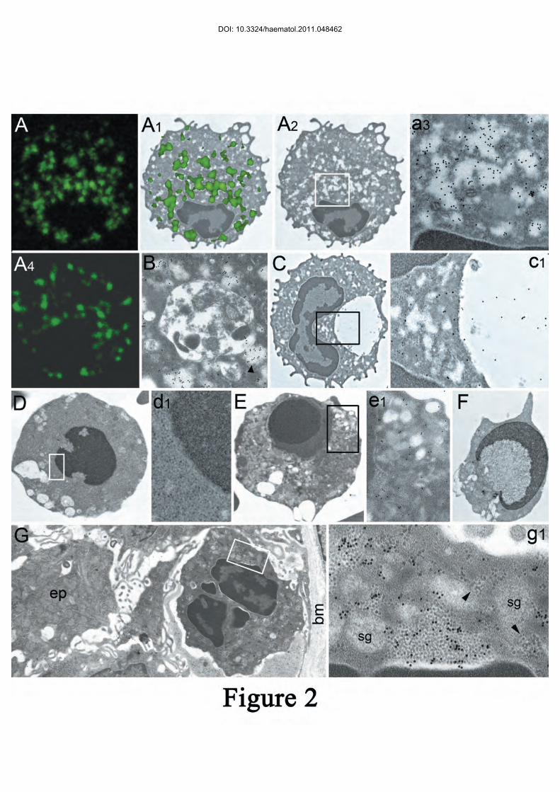
Figure 2. Correlative confocal and electron microscopy of PaCSs in SDS neutrophils. (A, 3,000×)
Confocal microscopy of an SDS neutrophil immunostained with FK2 antibody against ubiquitinated
proteins. Note intensely immunofluorescent areas which, when projected on a TEM micrograph at
the same enlargement (a1, 3,000×), show substantial overlapping with PaCSs (a2, 3,000×) as
confirmed by the intense FK1 immunogold reactivity at higher enlargement (a3, 20,000×). (A4,
DOI: 10.3324/haematol.2011.048462

21
3,000×) 20S proteasome immunofluorescence of cytoplasmic areas in another neutrophil from the
same SDS patient. (B and C) Autophagic vacuoles and PaCSs in SDS neutrophils. Note in B
(10,000×) the FK1 reactivity of PaCSs, one of which (arrowhead) is apparently discharging its
content into the vacuole, and in C (3,000×; enlarged in c1, 20,000×), the p62 reactivity of the
vacuole and cytoplasm with, however, no reactivity of PaCSs. (D–F, 3,000×) Three apoptotic
neutrophils with typical chromatin condensation and segregation (target-type in E and crescentic in
F) from SDS patients with SBDS gene mutations. In D, enlarged in d1 (10,000×), cytoplasmic
homogenization with disappearance of organelles structure can be seen, whereas in E, enlarged in
e1 (10,000×), secretory granules and some FK1 immunoreactivity are still recognizable. Note
cytoplasmic vacuoles (of autophagic origin?) in all apoptotic cells. (G, 2,500×) Intraepithelial
neutrophil in a gastric biopsy specimen with H. pylori gastritis, enlarged in g1 (36,000×) to show
several PaCSs intensely reactive for H. pylori urease. ep: epithelial cell; bm: basal membrane; sg:
secretory granules; arrowheads: ribosomes.
DOI: 10.3324/haematol.2011.048462

DOI: 10.3324/haematol.2011.048462
DOI: 10.3324/haematol.2011.048462

1
Online Supplementary Data
Figure S1. Representative western blot of lysed blood granulocytes from a healthy control subject
and an SDS patient with SBDS gene mutation. Lysates were resolved by SDS-PAGE (4–20%) and
immunoblotted for FK1 (polyubiquitinated proteins), 20S proteasome (Prt) and p62 protein. β-Actin
was used as a loading control. The graph on the right, obtained by densitometry and normalized to
β-actin, shows the increment, expressed as a percentage, of FK1, Prt and p62 in SDS patients
compared to that in control granulocytes.
Figure S2. Aldehyde-osmium fixed blood neutrophils from a healthy control, either untreated (a1,
1,000×) or treated with the xanthine/xanthine oxidase system (a2, 1,000×; B, 4,000×; b1, 20,000×).
In toluidine-blue-stained semithin resin sections a single cell (black arrow) in a1, and several cells
in a2, show round, pyknotic nuclei. The apoptotic nature of these cells was confirmed by electron
microscopy of an adjacent thin section (B, enlarged in b1) showing a round dense, chromatin
aggregate, leaving a thin crescentic remnant of karyoplasm in direct contact with a mostly
homogeneous cytoplasm, at a thin borderline (white arrowheads) devoid of a nuclear membrane
envelop. A few secretory granules, dense bodies and several membrane-delimited vesicles, but no
PaCSs, can still be recognized in the cytoplasm.
DOI: 10.3324/haematol.2011.048462

DOI: 10.3324/haematol.2011.048462

DOI: 10.3324/haematol.2011.048462


![[Vierstra, 2003 TIPS]. Ubiquitin/26S proteasome pathway Ub + ATP E1 E3 E2 Target Ub Target 26S proteasome UbiquitinationProteolysis + ATP Simplified.](https://static.fdocuments.in/doc/165x107/56649c7d5503460f94932c85/vierstra-2003-tips-ubiquitin26s-proteasome-pathway-ub-atp-e1-e3-e2-target.jpg)


