Types of C.T.Fibers There are three(3) types of C.T. 1-white collagennous fibers 2-yellow elastic...
55
Types of C.T.Fibers Types of C.T.Fibers There are three(3) types of There are three(3) types of C.T C.T . . 1 1 - - white collagennous fibers white collagennous fibers 2 2 - - yellow elastic fibers yellow elastic fibers 3 3 - - reticular fibers reticular fibers
-
Upload
felix-rose -
Category
Documents
-
view
234 -
download
1
Transcript of Types of C.T.Fibers There are three(3) types of C.T. 1-white collagennous fibers 2-yellow elastic...

Types of C.T.FibersTypes of C.T.Fibers
There are three(3) types of C.TThere are three(3) types of C.T..11--white collagennous fiberswhite collagennous fibers
22--yellow elastic fibersyellow elastic fibers33--reticular fibersreticular fibers

11--White Collagenous FibersWhite Collagenous Fibers
ShapeShape::colourless wavy branching bundles,the colourless wavy branching bundles,the
fibers run paralled to each otherfibers run paralled to each otherCharcterCharcter::
soft,strong&flexible but not elastic fiberssoft,strong&flexible but not elastic fibers

StructuresStructures::formed of protein known as (collagen) formed of protein known as (collagen)
which can be digested by pepsin&trypsin which can be digested by pepsin&trypsin enzymesenzymes
StainingStaining::collagenous fibers are acidophiliccollagenous fibers are acidophilic,,
stain pink with eosin,red instain pink with eosin,red in van-Gison&blue with Mallory stainvan-Gison&blue with Mallory stain

Types of collagenTypes of collagen
There are five(5) types of collagenThere are five(5) types of collagen11--Type I collagenType I collagen::
PresentPresent:: losse C.T.,White fibro-cartilage,bone&teeth this losse C.T.,White fibro-cartilage,bone&teeth this
type is formed by type is formed by fibroblast,osteoblast,odontoblastfibroblast,osteoblast,odontoblast
22--Type II collagenType II collagen:: hyaline&elastic cartilage&formed by hyaline&elastic cartilage&formed by
chondroblastchondroblast

33--Type III collagenType III collagen : :skin ,smooth muscle&reticular fibers formed by skin ,smooth muscle&reticular fibers formed by
fibroblast&by smooth muscle cellsfibroblast&by smooth muscle cells44--Type IV collagen: Type IV collagen: present in the basementpresent in the basementmembrane of epithelial tissue&lenes of eye,It membrane of epithelial tissue&lenes of eye,It
formed by fibroblast&by endothelial cellsformed by fibroblast&by endothelial cells55--Type V collagenType V collagen::
in placenta,it is formed by fibroblastin placenta,it is formed by fibroblast

22--Elastic fibersElastic fibers
ShapeShape : :fine,straight branching fibersfine,straight branching fibers
CarachterCarachter::the fibers branched&anastomose with the fibers branched&anastomose with
each othereach otherThey run singly¬ in bundlesThey run singly¬ in bundles
Appear yellow in fresh stateAppear yellow in fresh state

StructuresStructures:: formed of protein known as elastin which formed of protein known as elastin which
is resistant to chemical and to boilingis resistant to chemical and to boilingCan digested by the pancreatic elastase Can digested by the pancreatic elastase
enzymeenzymeStainingStaining::
brown with orcein,black with verhoeff and brown with orcein,black with verhoeff and yellow with van-Gisonyellow with van-Gison

Orecin Stain for Elastin – purple “ribbon candy”

33--Reticular FibersReticular Fibers
ShapeShape:: they are thin,branched anastomose to they are thin,branched anastomose to
form anetwork or reticulumform anetwork or reticulumStrucuresStrucures::
--formed of protein known as collagen type formed of protein known as collagen type IIIIII
--Staining: Black with silverStaining: Black with silver

Sliver Stain for Reticular Fibers

PAS stain of epithelial basement membranes (BM)

Masson’s Trichrome Stain – blue = collagen

Areolar Tissue
Pink = collagen
Purple = elastin

following 6 types of C.T. properfollowing 6 types of C.T. proper
11--Areolar C.TAreolar C.T..22--Adipose tissueAdipose tissue
33--Yellow elastic C.TYellow elastic C.T..44--White Collagenous C.TWhite Collagenous C.T..
55--Reticular C.TReticular C.T..66--Micoid C.TMicoid C.T..

11--Areolar C.TAreolar C.T..
Commone type in the human body contain Commone type in the human body contain all types of C.T.fibers&C.T. cellsall types of C.T.fibers&C.T. cells
FunctionFunction::packaging material for other tissuepackaging material for other tissue

StructuresStructures : :formed of aloose matrix formed of formed of aloose matrix formed of
mucopolysacchids contain areolar(spaces)filled mucopolysacchids contain areolar(spaces)filled with air or fluidwith air or fluid
C.T. cells&C.T. fibers are embedded in the loose C.T. cells&C.T. fibers are embedded in the loose matrixmatrix
C.T. cells are mainly fibroblasts,macrophages,fat C.T. cells are mainly fibroblasts,macrophages,fat &mast cells&mast cells
C.T. fibers are mainly collagenous which are C.T. fibers are mainly collagenous which are found in dermis of skinfound in dermis of skin

SitesSites::11--submucosa of digestive tractsubmucosa of digestive tract
22--Present in all over the body except brain Present in all over the body except brain tissue&under dermis of skintissue&under dermis of skin
33--pleura,peritoneum&pericardiumpleura,peritoneum&pericardium44--under epithelial lining of organsunder epithelial lining of organs
55--around the organs&blood vesselsaround the organs&blood vessels

22--Adipose C.TAdipose C.T
..The fat cells develop from(UMC) The fat cells develop from(UMC)
transformed in lipoblast then into fat transformed in lipoblast then into fat cellscells

There are 2 types of adipose There are 2 types of adipose C.TC.T..
A.White Adipose TissueA.White Adipose TissueB.Brown Adipose tissueB.Brown Adipose tissue

A-White adipose tissueA-White adipose tissue
Composed of large fat cellsComposed of large fat cellsThese fat cells have eccentric flat nucleiThese fat cells have eccentric flat nuclei
--the rim of cytoplasm around the nuclei contain the rim of cytoplasm around the nuclei contain few cell organellesfew cell organelles
These fat cells can form fatty acids from These fat cells can form fatty acids from glucprotien processes is regulate by hormones glucprotien processes is regulate by hormones as insulinas insulin
This type affect by hormones&by the restriction This type affect by hormones&by the restriction of diet(regime)of diet(regime)

Function of White Adipose Function of White Adipose C.TC.T..
--Heat insulator&fat storge areasHeat insulator&fat storge areasGives the body normal shapeGives the body normal shape
SitesSites::11--under the skin(in female)present in under the skin(in female)present in
mammary glands&gluteal regionmammary glands&gluteal region22--Around the kidney&blood vesselsAround the kidney&blood vessels
33--in mesentry,omentum,abdominal wallin mesentry,omentum,abdominal wall

B-Brown adipose C.TB-Brown adipose C.T
..Formed of small fat cells filled with many Formed of small fat cells filled with many
droplets of pigmented lipiddroplets of pigmented lipidDevelops mainly in the emrbyo from Develops mainly in the emrbyo from
UMC.persist for few months after UMC.persist for few months after birth,supplies new born infant with heat to birth,supplies new born infant with heat to protect them from coldprotect them from cold

FunctionFunction::Heat generator,gives,heat to the different Heat generator,gives,heat to the different
body organsbody organsSiteSite::
Interscapular region,axillary Interscapular region,axillary region,mediastinal around the thoracic aortregion,mediastinal around the thoracic aort
PresentPresent::Eyelid,lungEyelid,lung

33--Elastic C.TElastic C.T..
Appears yellow when present in afresh conditionAppears yellow when present in afresh condition--stained brown with orecin stainstained brown with orecin stain
--elastic fibers separated with areolar C.Telastic fibers separated with areolar C.T..--elastic tissue stretchableelastic tissue stretchable
--elastic tissue is present in the form of elastic elastic tissue is present in the form of elastic membranes as in aortamembranes as in aorta

SiteSite::--aorta&large arteriesaorta&large arteries
--bronchi,bronchioles&around alveoli of bronchi,bronchioles&around alveoli of lunglung
--ligment nuchae(in the back of the neck)to ligment nuchae(in the back of the neck)to facilitate movments of trunk&neckfacilitate movments of trunk&neck

44--White CollagenousWhite Collagenous
Very dense type of C.T.form collagenous fibersVery dense type of C.T.form collagenous fibers--seperated from each other by areolar C.Tseperated from each other by areolar C.T..
Arrangement of the collagenous bundlesArrangement of the collagenous bundlesWhite fibrous C.T.may be regular or irregularWhite fibrous C.T.may be regular or irregular
Fibroblast===tendon cellsFibroblast===tendon cellsThe fibroblast are triangular in shape with The fibroblast are triangular in shape with
basophilic cytoplasm cell&their nuclei are oval in basophilic cytoplasm cell&their nuclei are oval in shapeshape

Type of white collagenous C.TType of white collagenous C.T
..11--regular white collagenous C.T. regular regular white collagenous C.T. regular
collagenous bundles present collagenous bundles present cornea,eye,tendons of musclecornea,eye,tendons of muscle
22--Irregular white collagenous:irregular Irregular white collagenous:irregular collagenous bundlescollagenous bundles

PresentPresent::
11--sclerasclera22--capsulecapsule
33--dura materdura mater44--perichondrium,periosteumperichondrium,periosteum
55--Reticular C.TReticular C.T..From the stroma or background of glandsFrom the stroma or background of glands

55--Reticular C.T.is formed ofReticular C.T.is formed of
a-reticular fibers which are thin fibersa-reticular fibers which are thin fibersb-reticular cells which are stellate shape b-reticular cells which are stellate shape
cellscellsc-mononuclear phagocitic cellsc-mononuclear phagocitic cells
the reticular cells&fibers form anetworkthe reticular cells&fibers form anetworkStainingStaining::
Brown with silver stainBrown with silver stain

SiteSite::11--present in the stroma of bone marrowpresent in the stroma of bone marrow
22--stroma or framework of the stroma or framework of the spleen,lymph spleen,lymph nodese,liver,testis,ovary&endocrine nodese,liver,testis,ovary&endocrine glandsglands
33--kidney,lung&gastro-intestinal tractkidney,lung&gastro-intestinal tract

66--Mucoid C.TMucoid C.T..
Fromed ofFromed of11--mucoid cells which are young fibroblastmucoid cells which are young fibroblast
22--matrix formed of hyaloronic matrix formed of hyaloronic acid&collagenous fibersacid&collagenous fibers

Site of mucoid C.TSite of mucoid C.T
..11--umbilical cord(between the blood umbilical cord(between the blood
vessels of umbilical cord where it is calles vessels of umbilical cord where it is calles wartons jelly)wartons jelly)
22--in adult it is present in vitreous of the in adult it is present in vitreous of the eye ball&pulp of growing teetheye ball&pulp of growing teeth
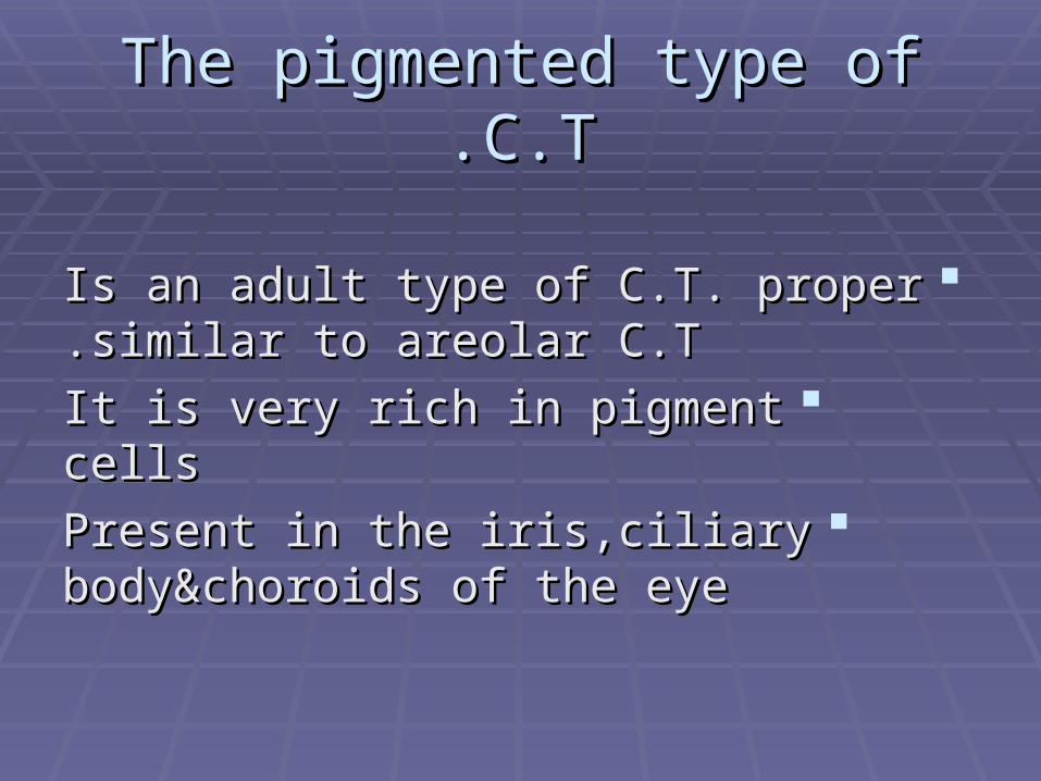
The pigmented type of C.TThe pigmented type of C.T..
Is an adult type of C.T. proper similar to Is an adult type of C.T. proper similar to areolar C.Tareolar C.T..
It is very rich in pigment cellsIt is very rich in pigment cellsPresent in the iris,ciliary body&choroids of Present in the iris,ciliary body&choroids of
the eyethe eye

Function of C.T. properFunction of C.T. proper
Support&connects organs&tissue togetherSupport&connects organs&tissue togetherC.T.plasma cells secret antbodies,mast C.T.plasma cells secret antbodies,mast
cells secrete histamine&heprinecells secrete histamine&heprineC.T.is important in regeneration&healing C.T.is important in regeneration&healing
of woundsof woundsDefency&immune responseDefency&immune response

CartilageCartilage
DefinitionDefinition::it it is firm rigid flexible dense C.T poor in blood is firm rigid flexible dense C.T poor in blood
supplysupplyStructuresStructures::
11--cartilage cell:young&mature chondrocytescartilage cell:young&mature chondrocytes22--C.T. Fibers:collagenous&elastic C.T.FibersC.T. Fibers:collagenous&elastic C.T.Fibers
33--Matrix:formed of collagen glycoproteinsMatrix:formed of collagen glycoproteins

Cartilage CellsCartilage Cells
11--Young Chondrocytes== Young Chondrocytes== Condroblast==Chondrogenic cellsCondroblast==Chondrogenic cells
--flat cells with flat nuclei they can divideflat cells with flat nuclei they can divide--cytoplasm is basophilic contains all cytoplasm is basophilic contains all
organoid&inclusionsorganoid&inclusions--present mainly under the perichondriumpresent mainly under the perichondrium

22--Mature ChondrocytesMature Chondrocytes
Rounded cells with round nucleiRounded cells with round nuclei--cytoplasm is basophilic contain all cytoplasm is basophilic contain all
organoids&inclusion,rich in glucogen organoids&inclusion,rich in glucogen fat&phosphatase enzymesfat&phosphatase enzymes
Condrocytes can divid present in group Condrocytes can divid present in group called cell nestscalled cell nests
--the groups of cartilage cells are the groups of cartilage cells are surrounded with aspaces called lacunasurrounded with aspaces called lacuna

Function of mature chondrocytesFunction of mature chondrocytes::
synthesis type II collagen synthesis type II collagen proteoglycans,hyaluronic proteoglycans,hyaluronic acid&chondroprotein of the matrixacid&chondroprotein of the matrix

Matrix of cartilageMatrix of cartilage
--rubbery in consistencyrubbery in consistency--formed of proteins called formed of proteins called
proteoglcans,hyaluronic acidproteoglcans,hyaluronic acid--glycoprotein&type II collagenglycoprotein&type II collagenBasophilic in staining,blue with Basophilic in staining,blue with
hematoxlinehematoxline

Type of CartilageType of Cartilage
Carilage cells&the C.T. fibers are Carilage cells&the C.T. fibers are embeded in rubbery matrix in embeded in rubbery matrix in order,following 3 types of cartilageorder,following 3 types of cartilage
11--Hyaline cartilage(appears glassy)Hyaline cartilage(appears glassy)22--Elastic fibro-cartilage(contain elastic Elastic fibro-cartilage(contain elastic
fibers)fibers)33--white fibro-cartilage(white collagenous white fibro-cartilage(white collagenous
bundles)bundles)

Hyaline CartilageHyaline Cartilage
Commonest type of cartilage,translucent&pale blue in Commonest type of cartilage,translucent&pale blue in colorcolor
The matrix is poor in blood supplyThe matrix is poor in blood supply--covered by avascular membrane or perichondriumcovered by avascular membrane or perichondrium
--perichondrium is formed ofperichondrium is formed ofa-outer fibrous layer of collagenous bundles:rich in a-outer fibrous layer of collagenous bundles:rich in
B.V.&fibroblastB.V.&fibroblastb-Inner chondrogenic layer formed of chondroblast which b-Inner chondrogenic layer formed of chondroblast which
can be change into chondrocytes can divid&can secrete can be change into chondrocytes can divid&can secrete new matrixnew matrix

Function of perichondriumFunction of perichondrium
11--supplies cartilage with bloodsupplies cartilage with blood22--chondroblast form matrix of cartilagechondroblast form matrix of cartilage33--provides an attachment for musclesprovides an attachment for muscles

Embeded in matrix are 2 types of cartilage Embeded in matrix are 2 types of cartilage cells cells
11--Young chondrocytes: flat cells surround Young chondrocytes: flat cells surround by lacunne they have flat by lacunne they have flat nuclei&basophilic cytoplasm present nuclei&basophilic cytoplasm present single cells under the perichondriumsingle cells under the perichondrium

22--Mature ChondrocytesMature Chondrocytes
Spherical cells with round Spherical cells with round nuclei&basophilic cytoplasm rich in nuclei&basophilic cytoplasm rich in phosphatase enzymesphosphatase enzymes
Each cells is present in aspace called Each cells is present in aspace called LacuneLacune

Site of hyline cartilageSite of hyline cartilage
11--costal cartilages present in thoraciccagecostal cartilages present in thoraciccage22--cartilage of respiratory cartilage of respiratory
passages,nose,trachea,bronchi,larnxpassages,nose,trachea,bronchi,larnx33--articular surface of joints(cartlage is not articular surface of joints(cartlage is not
carred with perichondrium)carred with perichondrium)

22--Yellow elastc fibro-cartilageYellow elastc fibro-cartilage
This type of carilage is similar structures to This type of carilage is similar structures to hyaline cartilage buthyaline cartilage but
a-the matrix is rich in elastic fibers a-the matrix is rich in elastic fibers surround by cartilage cell surround by surround by cartilage cell surround by perichondriumperichondrium
b-this cartilage is flexible&yellow in color b-this cartilage is flexible&yellow in color due to presence of elastic fibersdue to presence of elastic fibers

site of elastic-fibro cartilagesite of elastic-fibro cartilage
11--Epiglotitis,artetenoid,corniculateEpiglotitis,artetenoid,corniculate22--External ear&Eustachian tubeExternal ear&Eustachian tube

33--White fibro-cartilageWhite fibro-cartilage
Characteristic of fibro-cartlageCharacteristic of fibro-cartlage11--simillar to hyaline cartilage very rich in type I simillar to hyaline cartilage very rich in type I
collagen fiberscollagen fibers22--matrix is acidophilic due to presence of type I matrix is acidophilic due to presence of type I
collagencollagen33--formed of chondrocytesformed of chondrocytes
44--cartilage cells arranged in rows or in columnscartilage cells arranged in rows or in columns55--the white F-C. is not covered by chondriumthe white F-C. is not covered by chondrium
66--cartlage cells are separated by acidophilic cartlage cells are separated by acidophilic collagenous bundlescollagenous bundles

Site of W.F.C. in the bodySite of W.F.C. in the body
11--presence in the intervertebral discpresence in the intervertebral disc22--semilunar cartilage of knee jointssemilunar cartilage of knee joints
33--symphysis pubis,acetabulumsymphysis pubis,acetabulum44--discs between sterno-discs between sterno-
clavicular&mandibular jointclavicular&mandibular joint55--terminal parts of the muscle tendonterminal parts of the muscle tendon

FunctionFunction
11--maintaining the patency of respiratory maintaining the patency of respiratory passagespassages
22--cartilage&bone form the skeleton of bodycartilage&bone form the skeleton of body33--cartilage forms asmooth firm surface for the cartilage forms asmooth firm surface for the
articular jointsarticular joints33--cartilage is essential for growth of bone before cartilage is essential for growth of bone before
birthbirth44--cartilage&bone protect essential organs as cartilage&bone protect essential organs as
lung,brain&bone marrowlung,brain&bone marrow

Young cartilage can grow out by Young cartilage can grow out by the following 2 different methodsthe following 2 different methods
11--Interstitial growthInterstitial growth : :from groups of young chondrocytes secret from groups of young chondrocytes secret
the matrix resulting in growth of cartilagethe matrix resulting in growth of cartilage22--Appositional growthAppositional growth::
chondroblast of the perichondrium become chondroblast of the perichondrium become transformed into chondrocytes which transformed into chondrocytes which secret the matrixsecret the matrix

(resorcin-fuchsin stain) Elastic Fibers
Elastic Fibers- silver stain
Elastic Cartilage- pinnae of ear Elastic Cartilage
Matrix
hyaluronic acid chondroitin sulfate kertatin sulfate
Fibers
elastic (elastin)
Typical Locations
external ear walls of external auditory canal and eustachian tubes epiglottis & larynx bridge of nose
Properties
resiliency and pliability

Fibroblasts Collagen
Perichondrium
Blood vessels
Chondroblasts
Hyaline matrix
Isogenous groups
(nests of chondrocytes)

Hyaline cartilageHyaline cartilage
Chondrocytes nucleus
Matrix
Lacuna
EM : hyaline cartilage