TUMORSPHERE CELL CULTURE · 2019-03-06 · Incubate the culture for 4–10 days, depending on the...
1
TUMORSPHERE CELL CULTURE METHOD AND ITS EFFICIENCY promocell.com | promocell-academy.com For more information, see the posters and application notes available on the PromoCell website: www.promocell.com/downloads/ • 3D Tumorsphere Culture Model With Cancer Cell Lines and Human Primary Cells • Tumorsphere Culture of CSC; Determination of the TFE 07/2017 Fig. 4: Quantification of tumorsphere formation efficiency (TFE). A, cumulative population doublings of MCF-7 cells during 3D tumorsphere culture. Serial passages were performed every 9 days. Tumorsphere formation and proliferation were maintained during the culture, which was discontinued after passage 10 with no signs of growth rate inhibition. The MCF-7 tumorsphere culture achieved approximately 2.5 population doublings per passage. B, serial passage of MCF-7 cells in the 3D Tumorsphere Medium XF results in a significant increase of TFE from 2% in P1 to 28% in P9. Population doublings Tumorsphere formation efficiency [%] 10 Passage 0 1 3 4 5 6 7 8 9 0 5 10 15 20 25 30 2 Passage 1 4 9 0 10 20 30 A B MCF-7 Fig. 1: Examples of cancer cell lines cultured as 3D tumorspheres. A, MCF-7 mammary carcinoma cells after 10 serial passages. B, HT-29 colon carcinoma cells (passage 2). C, HT1080 fibrosarcoma cells after 10 serial passages. Robust tumorsphere formation was maintained during serial culture. See Figs. 2 and 4 for proliferation data. A B C Cancer stem cells (CSCs) can be enriched and cultured as extended serial 3D tumorsphere cultures (Fig. 1), thus permitting the establishment of a more physiological 3D microenvironment. In contrast to adherent 2D culturing of cancer cells, this type of 3D culture selectively exploits inherent biologic fea- tures of CSC such as anoikis resistance and self-re- newal. CSCs are highly responsive and therefore require a reliable and reproducible standardized cell culture without any stimuli of uncharacterized origin. This can be achieved, for example, with a chemically defined medium. The PromoCell 3D Tumorsphere Medium XF has been successfully tested for the most commonly used cancer stem cell lines in tumorsphere/mammosphere culture (Tab. 1). Continuous proliferation and enrichment of CSCs are also supported over serial passages (Fig. 2). Tissue Tested cell line Cell line origin Brain U-87 MG Grade IV glioblastoma/ astrocytoma of the human brain Breast Breast MCF-7 MDA-MB-231 Colon HT-29 Human colon adenocarcinoma Human fibrosarcoma Pleural effusion of metastasic human breast adenocarcinoma (triple-negative) Pleural effusion of metastasic human breast adenocarcinoma Connective tissue HT1080 Liver HepG2 Hepatocellular carcinoma of the human liver Lung A-549 Human lung carcinoma Pancreas Panc-1 Epithelioid carcinoma of the human pancreatic duct Prostate LNCaP Lymph node metastasis of human prostate adenocarcinoma Skin A-431 Epidermoid carcinoma of the human skin Tab. 1: List of cell types successfully tested for serial passage with the PromoCell 3D Tumorsphere Medium XF. Fig. 2: Proliferation curves of various established cancer cell lines expanded by serial passage of 3D tumorsphere cultures. Population doublings 10 Passage 0 4 6 8 0 10 20 30 2 40 U-87 MG PanC-1 HT-29 Hep-G2 A-431 A-549 MCF-7 HT-1080 Dissociate Check for sphere formation Incubate Single-cell suspension Plate 1 cell/well 3D sphere culture 96-well plate Fig. 3: Schematic flowchart of the TFE assay. Single cancer cells obtained by enzymatic dissociation of 3D tumorsphere cultures are plated by limiting dilution to one cell per well on 96-well plates. After an adequate incubation period, the wells originally containing the cells are analyzed for tumorsphere formation. Cells with stemlike properties are capable of forming a new tumorsphere derived from a single cell, while more restricted or differentiated tumor cells undergo anoikis. See the protocol on the right. TUMORSPHERE CULTURE PROTOCOL TUMORSPHERE CELL CULTURE TFE PROTOCOL TUMORSPHERE FORMATION EFFICIENCY (TFE) I. Materials • Tumorspheres generated with the tumorsphere culture protocol (see protocol above: II. Initiation of the Tumorpshere Culture) • 3D Tumorsphere Medium XF (C-28070) • Phosphate Buffered Saline w/o Ca++/Mg++ (PBS, C-40232) • Detach-Kit (C-41210) • 96-well u-bottom suspension plates (e.g. Greiner Bio One, No. 650 161) • 40 μm cell strainer • Optional: multichannel pipet (100 μl) II. Generation of a Single-Cell Suspension This procedure largely corresponds to the subculture protocol of tumorsphere cultures, which routinely requires generation of a single cell suspension (see proto- col above and perform step 1–5 as stated in III. Serial Passage of Tumorsphere Cul- ture, then proceed with the following step 6. 6. Determine the cell number and cell viability Make up to 5 ml with fresh PromoCell 3D Tumorsphere Medium XF and centri- fuge cells for 5 minutes at 300 x g. Discard the supernatant and resuspend the cells in 5 ml of the fresh medium. Pass the cell suspension through a 40 μm cell strainer to obtain a singel-cell suspension. Then determine the cell count and viability. III. Tumorsphere Formation Efficiency (TFE) Assay 1. Dilute single-cell suspension (day 0) Dilute an aliquot of the single-cell suspension obtained in step 6 of II. Generation of a Single-Cell Suspension with an appropriate amount of PromoCell 3D Tum- orsphere Medium XF in order to obtain a concentration of 10 viable cells per ml of this medium. Prepare at least 50 ml of this diluted cell suspension. 2. Plate the single cells (day 0) Distribute the diluted cell suspension at 100 μl per well in 96-well u-bottom suspension culture plates. On the same day, check the single wells for the presence of a single cell using a microscope (see Fig. 5). Mark wells without a cell. 3. Let the tumorsphere grow Incubate the plates in the incubator at 37°C and 5% CO 2 . 4. Add fresh medium (day 4–6) On day 4 to 6, add 100 μl of fresh PromoCell 3D Tumorsphere Medium XF to each well. Note: Do not change the medium, only add the fresh medium. 100 μm A B Fig. 5: Setup for the tumorsphere formation efficiency (TFE) detection assay. A, a single cell seeded in an individual well of a 96-well u-bottom suspension plate. B, an empty well without a cell, to be excluded from analysis. Fig. 6: Expected results of the TFE assay. A, a tumorsphere derived from a single MCF-7 cell larger than the cutoff size ≥150 μm: ”positive for tumorsphere formation“. B, a cell aggregate smaller than the cutoff size rated as ”negative for tumorsphere formation“. The dark center indicates the onset of degeneration. 100 μm A B I. Materials • 3D Tumorsphere Medium XF (C-28070) • Phosphate Buffered Saline w/o Ca++/Mg++ (PBS, C-40232) • Detach-Kit (C-41210) • 6-well Suspension Culture Plates (e.g. Greiner Bio One, No. 657 185) • Adherently growing human cancer cells (for initial tumorsphere culture set-up) II. Initiation of the Tumorsphere Culture 1. Harvest the adherent cells Detach the cells of a human CSC-containing adherently growing cancer cell line using your standard procedures. The cells should be 80–90% confluent and in good condition. Centrifuge the cell suspension for 5 minutes at 300 x g and aspi- rate the supernatant. Resuspend the cells in a small volume, e.g. 3–5 ml, of the PromoCell 3D Tumorsphere Medium XF. 2. Count the cells Count the cells using your routine method and adjust the volume with PromoCell 3D Tumorsphere Medium XF to obtain a concentration of 1 million cells/ml. 3. Set up the tumorsphere culture Seed the cells in appropriate suspension culture vessels at 10,000 cells/ml, e.g. 40,000 cells in 4 ml of PromoCell 3D Tumorsphere Medium XF in each well of a 6-well suspension culture plate. 4. Allow the tumorspheres to grow Incubate the culture for 4–10 days, depending on the cell type used. Add one half of the culture volume of fresh PromoCell 3D Tumorsphere Medium XF every 3–4 days. Do not change the medium. 5. Passage of the tumorsphere culture The tumorspheres should be passaged (section B below) before they start to develop a dark center. Depending on the cell type used optimal passage should occur after 4–10 days. III. Serial Passage of Tumorsphere Cultures 1. Collect the tumorspheres Transfer the PromoCell 3D Tumorsphere Medium XF containing the tumor- spheres into 15 ml conical tubes using a serological pipet. 2. Gravity sedimentation of the tumorspheres Allow the spheres to settle by gravity sedimentation for 10 minutes at room tem- perature. Aspirate the supernatant, but leave approximately 200 μl in the conical tube. Do not aspirate the tumorspheres. 3. Wash the tumorspheres Repeat the sedimentation (step 2) with an equal volume of PBS. Gently aspirate the PBS leaving approximately 200 μl in the conical tube. 4. Enzymatic digestion of the tumorspheres Add 1 ml of Trypsin-EDTA to the tumorspheres and incubate for 2–4 minutes at room temperature. Keep the spheres resuspended in the trypsin solution by pipetting up and down once every 30 seconds. Avoid sedimentation of the spheres. Note: The optimal incubation time required to achieve complete dissociation for step III. 5. (below) must be determined empirically by the user for each cell type. While 2–3 minutes will be optimal in most cases, tumorspheres of some cell types, e.g. MCF-7, may be used as an alternative dissociation reagent according to the supplier‘s instructions. 5. Break down remaining cell aggregates Pipet the spheres up and down 10–20 times using a 1000 μl pipet tip to generate a single-cell suspension. Aspirate the cell suspension as normal, but tilt the pipet tip slightly at the bottom of the tube when expelling the cells. The shear forces generated facilitate the break up of any residual cell aggregates. Perform a visual check to confirm that no large cell aggregates remain. Immediately after trituration, add twice the volume of Trypsin Neutralization Solution (TNS). Note: Do not over-triturate as cell viability will be compromised. If in doubt, monitor the dissociation process microscopically. Non-dissociated cell aggregates may be removed by passaging the cell suspension through a 40 μm cell strainer. When using recombinant trypsin use fresh PromoCell 3D Tumorsphere Medium XF for inactivation instead of TNS. 6. Determine the cell number and viability Make up to 5 ml with fresh PromoCell 3D Tumorsphere Medium XF and deter- mine the cell number and viability. Centrifuge the cells for 5 minutes at 300 x g. Discard the supernatant and resuspend the cells in fresh 3D Tumorsphere Medium XF at 1 million cells/ml. Note: Alternatively, the cells may be resuspended in buffer, e.g. PBS w/o Ca ++ /Mg ++ plus 0,5% albumin plus 2 mM EDTA, and used for furhter experi- ments and/or analytical procedures. 7. Plate the cells Reseed the cells at 10,000 cells/ml in new suspension culture vessels. Typically, 6-well plates with 40,000 cells in 4 ml of medium per well are used. The tumorsphere formation efficiency (TFE) indi- cates the percentage of cells within a culture that are capable of forming a sphere from a single cell (Fig. 3). Since only stemlike cells have this property, the TFE assay is a valuable qualitative and quantita- tive tool based on exclusively functional features of cells under selective culture conditions. It provides a method for exploring and characterizing unidenti- fied CSC subpopulations without the need to rely on unassured markers. Depending on the culture conditions, the TFE can be either fairly static or distinctly dynamic. For example, in adherent serum-containing cultures of established cancer cell lines – standard culture con- ditions that favor differentiation – the TFE remains relatively stable at a low level. By contrast, the TFE may behave dynamically, especially after being switched from a standard culture to an environ- ment that is more selective for stem cells. Under these conditions, sphere-forming cells can be grad- ually enriched (Fig. 4b). Consequently, the TFE assay is a significant, cost-effective and reliable in vitro method of indi- rectly measuring the CSC content of cultured cancer cells at a specific point of time and/or in a certain culture state. By way of example, the repeated concomitant determination of TFE at different passages during serial culturing of cancer cells in the PromoCell 3D Tumorsphere Medium XF can indirectly quantify the effects of this medium with regard to the culture’s functional CSC content. 5. Evaluate the results (day 7–12+) Check the wells of the seeded 96-well plates for sphere formation (see Fig. 6). Omit wells marked as ”w/o cell“ from analysis. Divide the number of spheres counted by the number of total cell-containing seeded wells and multiply by 100. The result is the TFE percentage (% TFE). Note: To ensure reproducible results, it is important to establish a specific defini- tion for ”positive sphere formation“. It is recommended to use the mean diame- ter of the cell aggregate as a readout parameter, which can be easily measured using the measure/ruler function of the microscope image capture software or on an optical grid. Ideally, the median sphere diameter of the specific cell line will already be known from preceeding 3D cultures. Intact MCF-7 spheres, for example, exhibit a mean diameter of >150 μm after 10–12 days. Smaller aggregates are therefore not counted as positive. If the corresponding sphere diameter is unknown, set your own cutoff value by measuring the size of some ”nice spheres“ within the assay. Aggregates <80 μm should never be considered as spheres. While most cell lines will form tumorspheres from a single cell within 7–12 days, it may be necessary to adjust the duration of the growth phase for very fast-growing or very slowly proliferating cell lines in order to obtain unambiguously evaluable results.
Transcript of TUMORSPHERE CELL CULTURE · 2019-03-06 · Incubate the culture for 4–10 days, depending on the...
TUMORSPHERE CELL CULTUREMETHOD AND ITS EFFICIENCY
promocell.com | promocell-academy.comFor more information, see the posters and application notes available on the PromoCell website:www.promocell.com/downloads/
• 3D Tumorsphere Culture Model With Cancer Cell Lines and Human Primary Cells• Tumorsphere Culture of CSC; Determination of the TFE
07/2
017
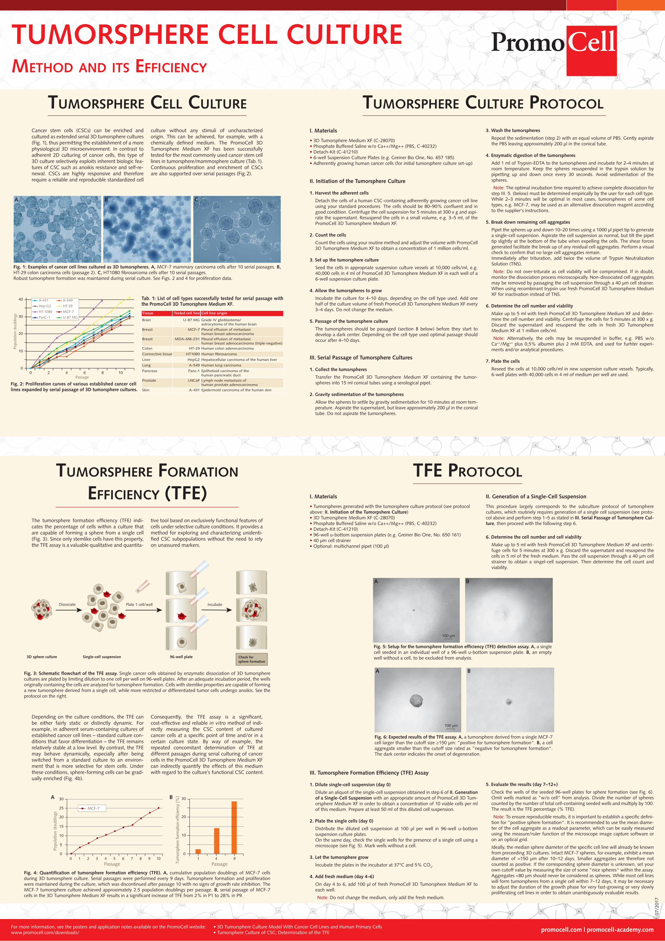
Fig. 4: Quantification of tumorsphere formation efficiency (TFE). A, cumulative population doublings of MCF-7 cells during 3D tumorsphere culture. Serial passages were performed every 9 days. Tumorsphere formation and proliferation were maintained during the culture, which was discontinued after passage 10 with no signs of growth rate inhibition. The MCF-7 tumorsphere culture achieved approximately 2.5 population doublings per passage. B, serial passage of MCF-7 cells in the 3D Tumorsphere Medium XF results in a significant increase of TFE from 2% in P1 to 28% in P9.
Popu
latio
n do
ublin
gs
Tum
orsp
here
form
atio
n ef
ficie
ncy
[%]
10Passage
0 1 3 4 5 6 7 8 90
5
10
15
20
25
30
2Passage
1 4 90
10
20
30A B
MCF-7
Fig. 1: Examples of cancer cell lines cultured as 3D tumorspheres. A, MCF-7 mammary carcinoma cells after 10 serial passages. B, HT-29 colon carcinoma cells (passage 2). C, HT1080 fibrosarcoma cells after 10 serial passages.Robust tumorsphere formation was maintained during serial culture. See Figs. 2 and 4 for proliferation data.
A B C
Cancer stem cells (CSCs) can be enriched and cultured as extended serial 3D tumorsphere cultures (Fig. 1), thus permitting the establishment of a more physiological 3D microenvironment. In contrast to adherent 2D culturing of cancer cells, this type of 3D culture selectively exploits inherent biologic fea-tures of CSC such as anoikis resistance and self-re-newal. CSCs are highly responsive and therefore require a reliable and reproducible standardized cell
culture without any stimuli of uncharacterized origin. This can be achieved, for example, with a chemically defined medium. The PromoCell 3D Tumorsphere Medium XF has been successfully tested for the most commonly used cancer stem cell lines in tumorsphere/mammosphere culture (Tab. 1). Continuous proliferation and enrichment of CSCs are also supported over serial passages (Fig. 2).
Tissue Tested cell line Cell line origin
Brain U-87 MG Grade IV glioblastoma/astrocytoma of the human brain
Breast
Breast
MCF-7
MDA-MB-231
Colon HT-29 Human colon adenocarcinoma
Human fibrosarcoma
Pleural effusion of metastasic human breast adenocarcinoma (triple-negative)
Pleural effusion of metastasic human breast adenocarcinoma
Connective tissue HT1080
Liver HepG2 Hepatocellular carcinoma of the human liver
Lung A-549 Human lung carcinoma
Pancreas Panc-1 Epithelioid carcinoma of the human pancreatic duct
Prostate LNCaP Lymph node metastasis of human prostate adenocarcinoma
Skin A-431 Epidermoid carcinoma of the human skin
Tab. 1: List of cell types successfully tested for serial passage with the PromoCell 3D Tumorsphere Medium XF.
Fig. 2: Proliferation curves of various established cancer cell lines expanded by serial passage of 3D tumorsphere cultures.
Popu
latio
n do
ublin
gs
10Passage
0 4 6 80
10
20
30
2
40
U-87 MGPanC-1
HT-29Hep-G2
A-431 A-549
MCF-7HT-1080
Dissociate
Check for sphere formation
Incubate
Single-cell suspension
Plate 1 cell/well
3D sphere culture 96-well plate
Fig. 3: Schematic flowchart of the TFE assay. Single cancer cells obtained by enzymatic dissociation of 3D tumorsphere cultures are plated by limiting dilution to one cell per well on 96-well plates. After an adequate incubation period, the wells originally containing the cells are analyzed for tumorsphere formation. Cells with stemlike properties are capable of forming a new tumorsphere derived from a single cell, while more restricted or differentiated tumor cells undergo anoikis. See the protocol on the right.
TUMORSPHERE CULTURE PROTOCOLTUMORSPHERE CELL CULTURE
TFE PROTOCOLTUMORSPHERE FORMATION
EFFICIENCY (TFE) I. Materials
• Tumorspheres generated with the tumorsphere culture protocol (see protocol above: II. Initiation of the Tumorpshere Culture)• 3D Tumorsphere Medium XF (C-28070)• Phosphate Buffered Saline w/o Ca++/Mg++ (PBS, C-40232)• Detach-Kit (C-41210)• 96-well u-bottom suspension plates (e.g. Greiner Bio One, No. 650 161)• 40 µm cell strainer• Optional: multichannel pipet (100 µl)
II. Generation of a Single-Cell Suspension
This procedure largely corresponds to the subculture protocol of tumorsphere cultures, which routinely requires generation of a single cell suspension (see proto-col above and perform step 1–5 as stated in III. Serial Passage of Tumorsphere Cul-ture, then proceed with the following step 6.
6. Determine the cell number and cell viability
Make up to 5 ml with fresh PromoCell 3D Tumorsphere Medium XF and centri-fuge cells for 5 minutes at 300 x g. Discard the supernatant and resuspend the cells in 5 ml of the fresh medium. Pass the cell suspension through a 40 µm cell strainer to obtain a singel-cell suspension. Then determine the cell count and viability.
III. Tumorsphere Formation Efficiency (TFE) Assay
1. Dilute single-cell suspension (day 0)
Dilute an aliquot of the single-cell suspension obtained in step 6 of II. Generation of a Single-Cell Suspension with an appropriate amount of PromoCell 3D Tum-orsphere Medium XF in order to obtain a concentration of 10 viable cells per ml of this medium. Prepare at least 50 ml of this diluted cell suspension.
2. Plate the single cells (day 0)
Distribute the diluted cell suspension at 100 µl per well in 96-well u-bottom suspension culture plates.On the same day, check the single wells for the presence of a single cell using a microscope (see Fig. 5). Mark wells without a cell.
3. Let the tumorsphere grow
Incubate the plates in the incubator at 37°C and 5% CO2.
4. Add fresh medium (day 4–6)
On day 4 to 6, add 100 µl of fresh PromoCell 3D Tumorsphere Medium XF to each well.
Note: Do not change the medium, only add the fresh medium.
100 µm
A B
Fig. 5: Setup for the tumorsphere formation efficiency (TFE) detection assay. A, a single cell seeded in an individual well of a 96-well u-bottom suspension plate. B, an empty well without a cell, to be excluded from analysis.
Fig. 6: Expected results of the TFE assay. A, a tumorsphere derived from a single MCF-7 cell larger than the cutoff size ≥150 µm: ”positive for tumorsphere formation“. B, a cell aggregate smaller than the cutoff size rated as ”negative for tumorsphere formation“. The dark center indicates the onset of degeneration.
100 µm
A B
I. Materials
• 3D Tumorsphere Medium XF (C-28070)• Phosphate Buffered Saline w/o Ca++/Mg++ (PBS, C-40232)• Detach-Kit (C-41210)• 6-well Suspension Culture Plates (e.g. Greiner Bio One, No. 657 185)• Adherently growing human cancer cells (for initial tumorsphere culture set-up)
II. Initiation of the Tumorsphere Culture
1. Harvest the adherent cells
Detach the cells of a human CSC-containing adherently growing cancer cell line using your standard procedures. The cells should be 80–90% confluent and in good condition. Centrifuge the cell suspension for 5 minutes at 300 x g and aspi-rate the supernatant. Resuspend the cells in a small volume, e.g. 3–5 ml, of the PromoCell 3D Tumorsphere Medium XF.
2. Count the cells
Count the cells using your routine method and adjust the volume with PromoCell 3D Tumorsphere Medium XF to obtain a concentration of 1 million cells/ml.
3. Set up the tumorsphere culture
Seed the cells in appropriate suspension culture vessels at 10,000 cells/ml, e.g. 40,000 cells in 4 ml of PromoCell 3D Tumorsphere Medium XF in each well of a 6-well suspension culture plate.
4. Allow the tumorspheres to grow
Incubate the culture for 4–10 days, depending on the cell type used. Add one half of the culture volume of fresh PromoCell 3D Tumorsphere Medium XF every 3–4 days. Do not change the medium.
5. Passage of the tumorsphere culture
The tumorspheres should be passaged (section B below) before they start to develop a dark center. Depending on the cell type used optimal passage should occur after 4–10 days.
III. Serial Passage of Tumorsphere Cultures
1. Collect the tumorspheres
Transfer the PromoCell 3D Tumorsphere Medium XF containing the tumor-spheres into 15 ml conical tubes using a serological pipet.
2. Gravity sedimentation of the tumorspheres
Allow the spheres to settle by gravity sedimentation for 10 minutes at room tem-perature. Aspirate the supernatant, but leave approximately 200 µl in the conical tube. Do not aspirate the tumorspheres.
3. Wash the tumorspheres
Repeat the sedimentation (step 2) with an equal volume of PBS. Gently aspirate the PBS leaving approximately 200 µl in the conical tube.
4. Enzymatic digestion of the tumorspheres
Add 1 ml of Trypsin-EDTA to the tumorspheres and incubate for 2–4 minutes at room temperature. Keep the spheres resuspended in the trypsin solution by pipetting up and down once every 30 seconds. Avoid sedimentation of the spheres.
Note: The optimal incubation time required to achieve complete dissociation for step III. 5. (below) must be determined empirically by the user for each cell type. While 2–3 minutes will be optimal in most cases, tumorspheres of some cell types, e.g. MCF-7, may be used as an alternative dissociation reagent according to the supplier‘s instructions.
5. Break down remaining cell aggregates
Pipet the spheres up and down 10–20 times using a 1000 µl pipet tip to generate a single-cell suspension. Aspirate the cell suspension as normal, but tilt the pipet tip slightly at the bottom of the tube when expelling the cells. The shear forces generated facilitate the break up of any residual cell aggregates. Perform a visual check to confirm that no large cell aggregates remain.Immediately after trituration, add twice the volume of Trypsin Neutralization Solution (TNS).
Note: Do not over-triturate as cell viability will be compromised. If in doubt, monitor the dissociation process microscopically. Non-dissociated cell aggregates may be removed by passaging the cell suspension through a 40 µm cell strainer. When using recombinant trypsin use fresh PromoCell 3D Tumorsphere Medium XF for inactivation instead of TNS.
6. Determine the cell number and viability
Make up to 5 ml with fresh PromoCell 3D Tumorsphere Medium XF and deter-mine the cell number and viability. Centrifuge the cells for 5 minutes at 300 x g. Discard the supernatant and resuspend the cells in fresh 3D Tumorsphere Medium XF at 1 million cells/ml.
Note: Alternatively, the cells may be resuspended in buffer, e.g. PBS w/o Ca++/Mg++ plus 0,5% albumin plus 2 mM EDTA, and used for furhter experi-ments and/or analytical procedures.
7. Plate the cells
Reseed the cells at 10,000 cells/ml in new suspension culture vessels. Typically, 6-well plates with 40,000 cells in 4 ml of medium per well are used.
The tumorsphere formation efficiency (TFE) indi-cates the percentage of cells within a culture that are capable of forming a sphere from a single cell (Fig. 3). Since only stemlike cells have this property, the TFE assay is a valuable qualitative and quantita-
tive tool based on exclusively functional features of cells under selective culture conditions. It provides a method for exploring and characterizing unidenti-fied CSC subpopulations without the need to rely on unassured markers.
Depending on the culture conditions, the TFE can be either fairly static or distinctly dynamic. For example, in adherent serum-containing cultures of established cancer cell lines – standard culture con-ditions that favor differentiation – the TFE remains relatively stable at a low level. By contrast, the TFE may behave dynamically, especially after being switched from a standard culture to an environ-ment that is more selective for stem cells. Under these conditions, sphere-forming cells can be grad-ually enriched (Fig. 4b).
Consequently, the TFE assay is a significant, cost-effective and reliable in vitro method of indi-rectly measuring the CSC content of cultured cancer cells at a specific point of time and/or in a certain culture state. By way of example, the repeated concomitant determination of TFE at different passages during serial culturing of cancer cells in the PromoCell 3D Tumorsphere Medium XF can indirectly quantify the effects of this medium with regard to the culture’s functional CSC content.
5. Evaluate the results (day 7–12+)
Check the wells of the seeded 96-well plates for sphere formation (see Fig. 6). Omit wells marked as ”w/o cell“ from analysis. Divide the number of spheres counted by the number of total cell-containing seeded wells and multiply by 100. The result is the TFE percentage (% TFE).
Note: To ensure reproducible results, it is important to establish a specific defini-tion for ”positive sphere formation“. It is recommended to use the mean diame-ter of the cell aggregate as a readout parameter, which can be easily measured using the measure/ruler function of the microscope image capture software or on an optical grid.
Ideally, the median sphere diameter of the specific cell line will already be known from preceeding 3D cultures. Intact MCF-7 spheres, for example, exhibit a mean diameter of >150 µm after 10–12 days. Smaller aggregates are therefore not counted as positive. If the corresponding sphere diameter is unknown, set your own cutoff value by measuring the size of some ”nice spheres“ within the assay. Aggregates <80 µm should never be considered as spheres. While most cell lines will form tumorspheres from a single cell within 7–12 days, it may be necessary to adjust the duration of the growth phase for very fast-growing or very slowly proliferating cell lines in order to obtain unambiguously evaluable results.