Tumors of the Choroid Plexus - Dergisineurosurgery.dergisi.org/pdf/pdf_JTN_447.pdf · Tumors of the...
7
Tllrkislr Nellro;;lIrgay 12: 26 - 32, 2002 Tumors of the Dng(lllnr: Tllillor, "1 tire CIr"roid Plex",' Choroid Plexus Koroid Pleksiis Tiimorleri ADNAN DAG<;INAR, M. MEMET OZEK, HAKAN KARABAGLI, TURKER KILI<;, iLHAN ELMACI, AYDIN SA V, M NECMETTiN P AMiR Marmara University School of Medicine, Department of Neurosurgery (AD, MMO, iE, MNP) istanbul Haydarpa~a Public Hospital (HK) istanbul Marmara University School of Medicine, Department of Pathology (AS) istanbul Received: 30.10.2001 ~ Accepted: 26.11.2001 Abstract: Objective: Choroid plexus neoplasms originate from choroid plexus epithelium. They are classified as papillomas and carcinomas histopathologically. The aim of this study is to discuss the signs, symptoms, treatment modalities and surgical results of this entities. Method: Ten cases of choroid plexus tumors operated on in Neurosurgery Department of Marmara University Medical School - istanbul between 1992 and 2000, are presented with the review of literature. Radiological investigations of the patients are reevaluated. Patients operative, histopathological and postoperative follow up records are detected. Filldillgs: One of the tumors was choroid plexus carcinoma and the others were choroid plexus papillomas. The youngest patient was two months old, the oldest one was 39 years old and the average age was 12,4. Female to male ratio was (5:5)one. Three patients were adults and the others were children. Major symptoms were headache, alteration in conscious level and enlargement of head. Most encountered neurological signs were papilla edema, alterations in conscious level, incrased head circumference and sixth nerve palsy. Five of the tumors (50%) were localized in the trigon of the lateral ventricle, three were in the fourth ventricle(30%) and one each in the third ventricle(lO%) and in cerebellopontine angle (CPA) 00%). All of the tumors were excised totally except two. One of the residual mass was excised with reoperation and the gamma knife surgery was applied to the other one. Two patient died in early postoperative period. The others are still alive. Average postoperative follow up is 25 months. COIlc1usioll: Total surgical excision is the most important prognostic factor for choroid plexus tumors. Key words: Choroid plexus, papilloma, carcinoma 26 Ozet: Amllf: Koroid pleksus h.imorleri koroid pleksus epitelinden koken alan neoplazmlardlr. Histopatolojik olarak papillomlar ve karsinomlar olarak slmflandmhrlar. Bu <;ah~mamn amaci bu tiimorlerin belirti, bulgu, tedavi yontemleri ve cerrahi sonu<;lanm tartl~maktlr. Metot: 1992-2000 Yilian arasmda Marmara Universitesi Tip Fakultesi Noro~irurji BolUmii'nde opere edilen 10 koroid pleksus rumorii olgusuna ait sonu<;lar literatiirle beraber tartl~llml~tlr. Olgulann radyolojik incelemeleri tekrar gozden ge<;irilmi~tir. Ameliyat bulgulan, histopatolojik ve ameliyat somasl izlem kaYltian yeniden degerlendirilmi~tir. Buiguilll': Bir olgu koroid pleksus karsinomu digerleri koroid pleksus papillomu olarak rapor edilmi~tir. Olgulann en kii<;iigu2 ayhk , en biiyiigii 39 ya;;mdCl,ortalama ya 12,4 duro Kadm/ erkek oram (5:5) birdir. U<;olgu eri~kin, digerleri <;ocuktur. Ba;; agnsl, bilin<;bulamkhgl ve ba~da biiyume en slk rastlamlan ~ikayetlerdir. Norolojik muayenlerinde, staz papiller, bilin<; bulamkhgl, ba~ <;evresinde biiyiime ve 6. sinir parezisi en slk gorlilen bulgulardlr. Be~olguda lateral ventrikiil trigonunda ('7u50), 3 olguda 4. ventrikul i<;inde(%30), 1 olguda 3. ventrikul i<;inde (%10) ve bir olguda serebellopon tin a<;lda ('i" 10) rumor tespit edilmi~tir. Iki olgu haricinde tiim lezyonlarda total rumor eksizyonu uygulanml;;hr. Subtotal eksizyon yapilan rumorlerin biri daha soma tekrar opere edilerek rezidu rumor eksize edilmi;; diger olguda ise gamaknife cerrahisi uygulanml~tlr. iki olgu erken postoperatif donemde eksitus olmu~, diger hastalar ya;;amaktadlr. Ortalama takip suresi 25 aydlr. SOIllIF Koroid pleksus tiimorlerinde total cerrahi eksizyon en onemli prognostik faktordur. Anahtar kelimeler: Koroid pleksus, papilloma, karsinoma
Transcript of Tumors of the Choroid Plexus - Dergisineurosurgery.dergisi.org/pdf/pdf_JTN_447.pdf · Tumors of the...

Tllrkislr Nellro;;lIrgay 12: 26 - 32, 2002
Tumors of the
Dng(lllnr: Tllillor, "1 tire CIr"roid Plex",'
Choroid Plexus
Koroid Pleksiis Tiimorleri
ADNAN DAG<;INAR, M. MEMET OZEK, HAKAN KARABAGLI, TURKER KILI<;,
iLHAN ELMACI, AYDIN SA V, M NECMETTiN PAMiR
Marmara University School of Medicine, Department of Neurosurgery (AD, MMO, iE, MNP) istanbulHaydarpa~a Public Hospital (HK) istanbul
Marmara University School of Medicine, Department of Pathology (AS) istanbul
Received: 30.10.2001 ~ Accepted: 26.11.2001
Abstract: Objective: Choroid plexus neoplasms originatefrom choroid plexus epithelium. They are classified aspapillomas and carcinomas histopathologically. The aimof this study is to discuss the signs, symptoms, treatmentmodalities and surgical results of this entities.Method: Ten cases of choroid plexus tumors operated onin Neurosurgery Department of Marmara UniversityMedical School - istanbul between 1992 and 2000, arepresented with the review of literature. Radiologicalinvestigations of the patients are reevaluated. Patientsoperative, histopathological and postoperative follow uprecords are detected.Filldillgs: One of the tumors was choroid plexus carcinomaand the others were choroid plexus papillomas. Theyoungest patient was two months old, the oldest one was39 years old and the average age was 12,4. Female tomale ratio was (5:5)one. Three patients were adults and theothers were children. Major symptoms were headache,alteration in conscious level and enlargement of head. Mostencountered neurological signs were papilla edema,alterations in conscious level, incrased head circumferenceand sixth nerve palsy. Five of the tumors (50%) werelocalized in the trigon of the lateral ventricle, three were inthe fourth ventricle(30%) and one each in the thirdventricle(lO%) and in cerebellopontine angle (CPA) 00%).All of the tumors were excised totally except two. One ofthe residual mass was excised with reoperation and thegamma knife surgery was applied to the other one. Twopatient died in early postoperative period. The others arestill alive. Average postoperative follow up is 25 months.COIlc1usioll: Total surgical excision is the most importantprognostic factor for choroid plexus tumors.
Key words: Choroid plexus, papilloma, carcinoma
26
Ozet: Amllf: Koroid pleksus h.imorleri koroid pleksusepitelinden koken alan neoplazmlardlr. Histopatolojikolarak papillomlar ve karsinomlar olarak slmflandmhrlar.Bu <;ah~mamn amaci bu tiimorlerin belirti, bulgu, tedaviyontemleri ve cerrahi sonu<;lanm tartl~maktlr.Metot: 1992-2000 Yilian arasmda Marmara UniversitesiTip Fakultesi Noro~irurji BolUmii'nde opere edilen 10koroid pleksus rumorii olgusuna ait sonu<;lar literatiirleberaber tartl~llml~tlr. Olgulann radyolojik incelemeleritekrar gozden ge<;irilmi~tir. Ameliyat bulgulan,histopatolojik ve ameliyat somasl izlem kaYltian yenidendegerlendirilmi~tir.Buiguilll': Bir olgu koroid pleksus karsinomu digerlerikoroid pleksus papillomu olarak rapor edilmi~tir. Olgulannen kii<;iigu2 ayhk , en biiyiigii 39 ya;;mdCl,ortalama ya12,4 duro Kadm/ erkek oram (5:5) birdir. U<;olgu eri~kin,digerleri <;ocuktur. Ba;;agnsl, bilin<;bulamkhgl ve ba~dabiiyume en slk rastlamlan ~ikayetlerdir. Norolojikmuayenlerinde, staz papiller, bilin<; bulamkhgl, ba~<;evresinde biiyiime ve 6. sinir parezisi en slk gorlilenbulgulardlr. Be~olguda lateral ventrikiil trigonunda ('7u50),3 olguda 4. ventrikul i<;inde(%30), 1 olguda 3. ventrikuli<;inde (%10) ve bir olguda serebellopon tin a<;lda ('i" 10)rumor tespit edilmi~tir. Iki olgu haricinde tiim lezyonlardatotal rumor eksizyonu uygulanml;;hr. Subtotal eksizyonyapilan rumorlerin biri daha soma tekrar opere edilerekrezidu rumor eksize edilmi;; diger olguda ise gamaknifecerrahisi uygulanml~tlr. iki olgu erken postoperatifdonemde eksitus olmu~, diger hastalar ya;;amaktadlr.Ortalama takip suresi 25 aydlr.SOIllIF Koroid pleksus tiimorlerinde total cerrahi eksizyonen onemli prognostik faktordur.
Anahtar kelimeler: Koroid pleksus, papilloma, karsinoma
Turkish Neurosurgery 11: 16 - 31, 2001
INTRODUCTION
Tumors of the choroid plexus originate fromchoroid plexus epithelium. (9,21,26,36).Papillomas constitute 0,3-0,5% of all intracranialtumors. Carcinomas are less enf=ountered thanpapillomas 05-30% of papillomas) (9,21,26,33,36).In childhood, overall ratio of choroid plexustumors is higher than adults and it makes the 3%of all intacranial tumors of children (22,27). Theyare usually localized in lateral ventricles but bythe increasing age, fourth ventricular tumors areseen more frequently (4,21,33,46). Increasedintracranial pressure signs and symptoms areprominent features of these tumors (7,21,26,27,46).Hydrocephalus is considered to be a primarycause of this clinical picture (5,17,21,26,27,29,34,46). Total surgical excision leads to cure forpapillomas. Eventhough the carcinomas are highlymalignant tumors, gross total resection of thetumor is the most important part of therapy(7,20,26,32,36,40,46) .
PATIENTS AND METHOD
Ten cases of choroid plexus tumors arepresented here which were treated in NeurosurgeryDepartment of University of Marmara-Istanbulbetween 1992-2000. Average age of the patients is12,4.The youngest patient was 2 months old and theoldest one was 39 years old. Female to male ratio is5:5.Seven patients were children (70%)and the otherswere adults (30%).
All of the patient"s were operated on andfollowed up in our clinic.
FINDINGS
The complaints of the patients were headache(60%), alterations of conscious level (30%),diplopia (30%), increased head circumference(20%), nausea-vomiting (20%), tinnitus (%10) andweakness 00%). Neurological examinationsrevealed; Papilledema (50%), decreased consciouslevel (40%), sixth nerve palsy (30%), enlargementof head circumference (20%), sunset appearance(20%), cerebellar findings (20%), third nerve palsy(20%), visiual field deficits 00%), hemiparesis00%) and spasticityOO%).
Diagnosises were all made by computed braintomography (CT) and magnetic resonance imaging(MR). Tumors were seen in the lateral ventricle
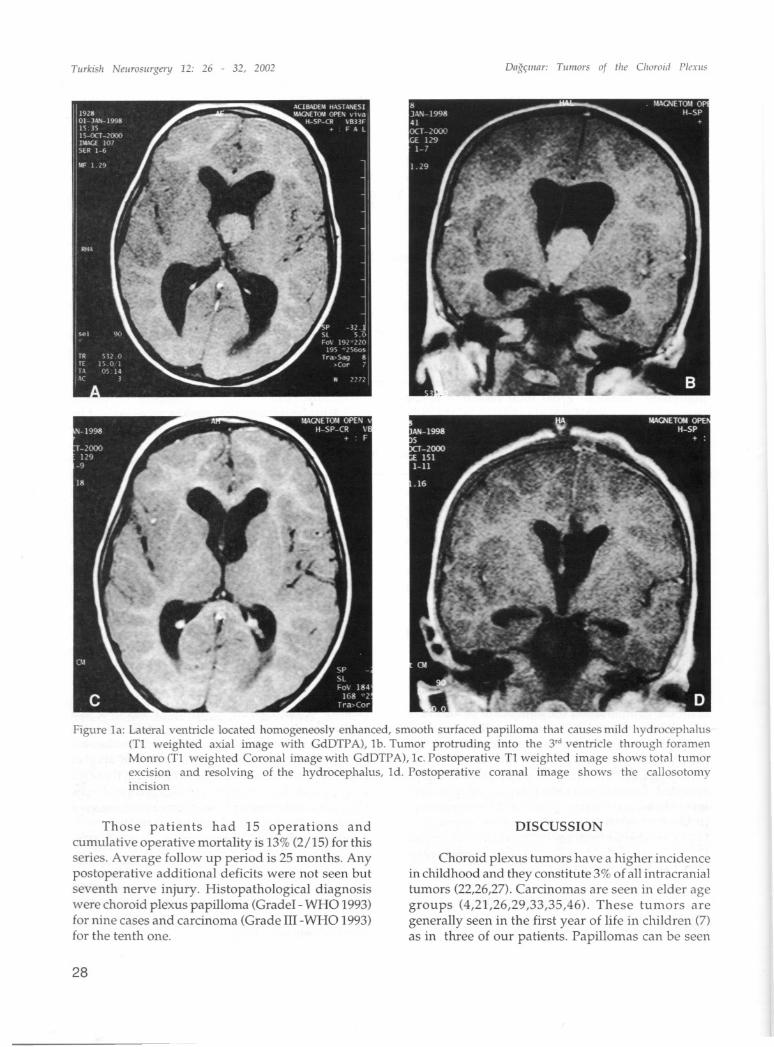
(Figure 1a,b) in five patients (50'1;-,),in the fourthventricle (Figure 2a,b) in three patients (30%) andone each in the third ventricle 00%) and in CPA. Inadults two of the tumors were in fourth ventricle andthe other one was in CPA. While all of the adult
tumors were localized in the posterior fossa only oneout of seven childhood tumors was in so. Our
papilloma cases except the tumor in CPA were allintraventricular and homogenousl)' enhancingmasses. In carcinoma case MR revealed a
homogenously enhanced, intraventricular mass with8 em radius.
First imaging studies revealed hydrocephalusin six cases. Ventricles of the patient having a CPAtumor was found normal as a result of previousshunt operation had done in other institution. Thepreoperative hydrocephalus rate was 70 % for ourcases.
Four lateral ventricle tumors were excised bytranscallosal (Figure1c,d), the fifth was by posteriortranscortical. While fourth ventricular tumors were
operated on by standart suboccipital route (Figure2c), tumor of CPA was excised by transpetrosal andthe third ventricle tumor by pterional way. In twoof the tumors, residual masses were left aftersurgery, one in left lateral ventricle and one in fourthventricle. Five months after first operation 10 mmresidual mass was excised from lateral ventricle.
Gamma knife surgery was applied for 5mm residualtumor mass in the fourth ventricle. One of the
patients that had a lateral ventricular tumor(carcinoma) and the patient with third ventricularpapilloma had died in early postoperative coursebecause of hypovolemic status due to the massiveperi operative bleeding.
After tumor excision, one of the patient withlateral ventricular tumor needed a
ventriculoperitoneal shunt. Because of the increasedsubdural cerebrospinal fluid (CSF) accumulation asubduroperitoneal shunts were apllied in anothertwo cases with lateral ventricular tumor. In the other
three patients having hydrocephalus preoperatively,ventricles became normal after tumor excision. As a
result two (%25) ventriculoperitoneal and two (%25)subduroperitoneal shunts were needed afteroperation.
The seventh nerve was injuredperioperatively in CPA tumor case. Four monthsafter primary surgery seven to twelve nerveanostomosis was done.
27
Turkislz Neurosurgery 12: 26 - 32, 2002 Dngqlllnr: TUn/aI's of the Choroid Plexlls
Figure 1a: Lateral ventricle located homogeneosly enhanced, smooth surfaced papilloma that causes mild hydrocephalus(Tl weighted axial image with GdDTPA), lb. Tumor protruding into the 3,d ventricle through foramenMonro (Tl weighted Coronal image with GdDTPA), lc. Postoperative Tl weighted image shows total tumorexcision and resolving of the hydrocephalus, ld. Postoperative coranal image shows the callosotomyincision
Those patients had 15 operations andcumulative operative mortality is 13% (2/15) for thisseries. Average follow up period is 25 months. Anypostoperative additional deficits were not seen butseventh nerve injury. Histopathological diagnosiswere choroid plexus papilloma (Gradel - WHO 1993)for nine cases and carcinoma (Grade III -WHO 1993)for the tenth one.
28
DISCUSSION
Choroid plexus tumors have a higher incidencein childhood and they constitute 3% of all intracranialtumors (22,26,27). Carcinomas are seen in elder agegroups (4,21,26,29,33,35,46). These tumors aregenerally seen in the first year of life in children (7)as in three of our patients. Papillomas can be seen

Turkish Neurosurgenj 12: 26 - 32, 2002
congenitally like teratomas, astrocytomas, primitiveneuroectodermal tumors and craniopharyngiomas(8,37). Some cases diagnosed as choroid plexusintrauterin were reported too 0,13). Choroid plexustumors are epithelial tumors (9,21,26,36,39)and areclassified as papillomas and carcinomas. Due to theclassification of WHO in 1993 papillomas areclassified as Grade 1 and carcinomas are grade 3-4tumors (9,21,26,39). Nine of the tumors of this seriesare papillomas and the remaning one is carcinoma.Mitosis, atipy and necrosis were demonstrated in
Dag~lIIar: TUlIIors of the Choroid Pin",;
carcinoma case but lack of vascular endothelial
proliferartion put this tumor into the grade 3cathegory.
Focal neurological findings like hemiparesis,visual field deficits, cerebellar, brain stem and cranialnerve symptoms due to the localization of the tumorcan be detected with these patients, but as in ourseries the increased intracranial pressure signs aremore prominent in diagnosis (7,21,26,27,46). Slowlygrowing tumor mass, accompanying hydrocephalusand the intraventricular localization of tumors are
causes of this clinical picture.
In children choroid plexus tumors are usuallylocalized in the trigonum of the lateral ventricles. Bythe increased age and in adults, localization of thetumor shifts to the infra tentorial space and mostlyinto the fourth ventricle (4,9,20,21,26,46). Butcarcinomas are generally originate from lateralventricle even in adults (21). The third ventriculartumors are very rare 02,21,28,29,42,43). In adults thepontocerebellar angle is another localization wheretumors can be observed (3,21,44). Rarely, tumors insuprasellar space (22), in lateral cerebellomedullarysistern (which lead to glossopharyngeal neuralgia)(9) and double sided lateral venticles have beenreported (4,21,46).
All of the cases presented here werediagnosed with both MR and CT. In CT papillomas
Figure 2a. Fourth ventricular, enhanced papilloma (Tl weighted axial image with GdDTPA), 2b. Sagittal image of thetumor filling all of the fourth ventricle eTl weighted image with GdDTP A), 2c. Postoperative Tl weightedimage shows total tumor excision.
29

Turkish Neurosurgery 12: 26 - 32, 2002
are generally lobular, intraventricular, hyperdenseor isodense masses and hydrocephalus usuallyaccompanies to the tumor. Calcification can be seenand tumors enhance homogenously (4,5,21,25,26,31,46). In MR they appear as a isointense masses withbrain tissue and enhancement with Gd-DTPA is veryobvious (4,5,21,25,26,31,46). Carcinomas may havecystic and hemorhagic portions. The contour of thetumor is usually irregular and the peritumoral edemasecondary to brain invasion is very important. Cysticchanges and hemorrhages are usually considered asmalignant features (4,5).Some papillomas dissect theependymal surface and show a malignantappearance (4,5,24) by penetrating the brain tissue.This can be observed intraoperatively and it'srecommended that choroid plexus tumors with abenign cellular appearance but evidence of localparenchymal invasion and loss of the normal villusarchitecture at site of invasion be classified as
papillomas. Patients with these tumors respond tosurgical therapy alone without the need of adjuvanttreatment (24).
Choroid plexus papillomas are benign tumors.Total surgical excision is the primary way oftreatment (20,26,34,36,46). Partial resection causesthe symptoms continue and increases the risk ofmalignant transformation 00,11,21). For resolvinghydrocephalus, releaving compresssion symptomsand avoidance of the recurrence and malignanttransformation, total surgical resection is necessary01,16,21,33). In contrast to the papillomas, thecarcinomas are malign lesions but total surgicalexcision is the first step in their treatment too(6,20,21,32,46). Pognosis is worse than papillomas.There are series reporting 100% 5 years survivalrates for papillomas (35). This rate decrease to 2640% for carcinomas (6,35). But it has been reportedthat if the total excision is achieved, 5 years survivalrate of 85 % may be estabilished (35). Adjuvanttherapies are advocated for carcinomas specially forresidual or recurrent lesions (20,30,40), but someauthors suggest reoperation for carcinomas (6) too.
As a surgical approaches interhemispherictranscallosal or transcortical (transsulcal)transventricular routes can be use for lateralventricular tumors. Decreased exposure for formerand increased risk of seizure and neurological deficitsfor latter are disadvantages (21,36). Transcortical ortranscallosal approaches give way CSF to reachsubdural space easily and Nagib et all (27) offerssulcal dissection for lateral ventricular tumor. By thisway after tumor resection, closed lips of sulcus
30
prevents compressing subdural accumulation bylimiting the CSF escape to the subdural space. It'smore easy to reach and coagulate feeding vessels bythe transcortical (transsulca1) way. In bigger tumors,narrow operative corridor and bulk of the tumormakes this manoeuvre very hard in transcallosalapproaches third ventricular tumors can be resectedfrom transcallosal or transcortical viay too(20,21,36,38,46). By the way some authors advocatecombined approaches for giant third ventriculartumors (42).
Generally fourth ventricular choroid plexuspapillomas adhere to the choroid plexus at theinferior of the ventricle (31). Although thepontocerebellar tumors usually grow into thesubarachnoid space, some time they may invade thebrain stem too (44). While the exploration of thislocation lower cranial nerves must be protected09,44). In order to increase the operative view,transpetrosal approach was used for CPA tumor ofthis series.
Cerebrospinal seedings in papillomas and morehigher rates in carcinomas have been reported inchoroid plexus tumors (21,33,45). Because of thisfinding if new neurological signs appeared in follo'wup, all parts of the central nervous system must beexamined again. There are no cranial or spinal spreadin this series.
Another problem of this patients ishydrocephalus. It is related to the overproductionof the CSF by tumor tissue (17,20,26,34,46). Gha tak(17) et al observed secretuar organellas andhistological structure in a papilloma case just likethe normal choroid plexus, studied with electronmicroscopy. In some cases eventhough the totaltumor excision is achieved, hydrocephaluspersists. Inflammatory ependimitis, arachnoiditis,and CSF pathway obstruction have all beenconsidered to be contributing factors (5,7,26,27,36,46). Sometime intraventricu lar localizationof the tumors may lead to obstructive typehydrocephalus but it usually resolves after tumorexcision (5,21,46). As mentioned before there is a riskof subdural bleeding or CSF accumulation in patientswith very large ventricles (27,45). After tumorexcision progressive subdural CSF accum.ulation isobserved in two patients with lateral ventricularpapillomas of this series and subduroperitonealshunt operations were performed. In our series 50%of patients needed VIP or SIP shunts and becauseof this high ratio, detection of postoperative CSF

Turkish Neurosurgery 12: 16 - 31, 2001
circulation problems are the most important partof follow up.
There are some alternative treatment modalities
in the literature. Gaab et al (16) offers an endoscopicexcision for small tumors and Duke (15) reported atumor control in one papilloma case with streotacticradiosurgery. After one year follow up, the tumorrest disappeared of our gamma knife applied case.
Adjuvant therapy is very dispute in carcinomabut there is consensus for total surgical excision isthe primary condition for long survival(20,21,32,40,46). But chemotherapy is advocated ininfants or residual or recurrent tumors and
craniospinal radiotherapy for patients older than twoyears 0Id(6,14,21,35,40,46). Because the carcinomasare very fragile and vascular tumors, some studieshave been focused on preoperative chemotherapythat serves to tumor shrinkage and more comfortableoperative status (18,41).
CONCLUSION
1. Total surgical excision is the first conditionthat improves the survival rate in choroid plexustumors.
2. It is possible to resect the tumors with minimalmorbidity if the appropraite surgical approaches areused.
Correspondence: Adnan DAC<;:INAR MDMarmara Umversity School OfMedicine Division of NeurosurgeryAltunizade istanbulPhone: 02.163264559Fax: 02163275249
REFERENCES
1. Adra AM, Mejides AA, Salman FA, Landy HJ, HelfgottAW: Prenatal sonographic diagnosis of a third ventriclechoroid plxus papilloma. Prenat Diagn 14(9): 865-867,199-1
2. Agamanolis DP, Malone JM: Chromosomalabnormalities in 47 pediatric brain tumors. CancerGenet Cytogenet 81(2): 125-134, 1995
3. Aksoy FG, Gomori JM: Choroid plexus papilloma offoramen of Luschka with multiple recurrences andcystic features. Neuroradiology 41(9): 654-656, 1999
4. Barkovich AJ, Edwards MSB: Brain tumors ofchildhood. Barkovich AJ (editor) ContemporaryNeuroimaging 1" ed, vol 1 (Pediatric neuroimaging)Raven Press- New York 1990:149-204
5. Barkovich AJ: Neuroimaging of pediatric brain tumors.Winn HR, Mayberg MR (ed). Neurosurgery Clinics ofNorth America - Pediatric Neurooncology VoI3(4) WB
Dag~lIIar: TUlllors of thl! Choroid Plt'.ms
Saunders 1992:739-7796. Berger C, Thiesse P, Lellouch-Tubiana A, Kalifa C,
Pierre Kahn A, Bouffet E: Choroid plexus carcinomasin childhood. Clinical features and prognostic factors.Neurosurgery 42(3): 470-475,1998
7. Bruce DA, Schut L, Sutton LN : Supratentorial braintumors in children Youmans JR (editor). NeurologicalSurgery, 3'd ed, vol 5. WB Saunders Philadelphia 1990:3000-3016
8. Buetow PC, Smirniotopoulos JG, Done S: Congenitalbrain tumors: A review of 45 cases. AJR 155(3):587593, 1990
9. Burger PC: Classification and biology of brain tumors.Youmans JR (editor). Neurological Surgery, 3'.1ed, vol5. WB Saunders Philadelphia 1990: 2967-2999
10. Chow E, Jenkins JJ, Burger PS, Reardon DA, LanstonJW, Sanford RA, Heideman RL, Kun LE, Merchant TE:Malignant evaluation of choroid pleus papilloma.Pediatr Neurosurg 31(3): 127-130,1999
11. Chow E, Reardon DA, Shah AB, Jenkins JJ, Langston J,Hedeman RL, Sanford RA, Kun LE, Merchant TE:Pediatric choroid plexus neoplasms. int J Radiat OncolBioi Phys 44(2);249-254,1999
12. Costa JM, Ley L, C1aramunt E, Lafuente J: Choroidplexus papillomas of the III ventricle in infants. Reportof three cases. Child's Nerv Syst 13(5): 244-249, 1997
13. 0'Addario V, Pinto V, Meo F, Resta M: The specificityof ultrasound in the detection of fetal intracranialtumors. J Perinat Med 26(6): 480-485,1998
14. Duffner PK, Kun LE, Burger PC, Horowitz ME, CohenME, Sanford RA, Krischer JP, Mulhern RK, James HE;Rekate HL: Postoperative chemotherapy and delayedradiation in infants and very young children withchoroid plexus carcinomas. The Pediatric onkologicgroup. Pediatr Neurosurg 22(4):189-196,1995
15. Duke BJ, Kindt GW, Breeze RE: Pineal region choroidplexus papilloma treated with stereotacticradiosurgery. Com put Aided Surg 2(2): 135-138, 1997
16. Gaab MR, Schroeder HW: Neuroendoscopic approachto intraventricular lesions. J Neurosurg 88(3): 496505,1998
17. Ghatak NR, McWhorter JM: Ultrastructural evidencefor CSF production by a choroid plexus papilloma. JNeurosurg 45(4):409-415,1976
18. Greenbeg ML: Chemotherapy of choroid plexuscarcinoma. Child's Nerv Syst 15(10): 571-577, 1999
19. Greene KA, Karahalios DG, Spetzler RF:Glossopharyngeal neuralgia associated with vascularcompression and choroid plexus papilloma. Br JNeurosurg 9(6): 809-814, 1995
20. Haroun RI, Khan WL, Carson BS, Brem H: Primarytumors of the choroid plexus. ContemporaryNeurosurg 22(3):2-8,2000
21. Harsh GR, Wilson BC:Neuroepithelial tumors of adultbrain. Youmans JR (ed). Neurological Surgery, 3'ded.,vol5. WB Saunders Philadelphia 1990: 3040-3136
22. Kimura M, Takayasu M, Suzuki Y, Negoro M,Nagasaka T, Nakashima N, Sugita K: Primary choroidplexus papilloma located in the suprasellar region:Case report. Neurosurgery 31(3): 563-566, 1992
23. Leblanc R, Bekhor S, MelansonD, CarpenterS: Diffu~ecraniospinal seeding from a benign fourth ventriclechoroid plexus papilloma. Case repor. J Neurosurg88(49): 757-760, 1998
31

TlIrkish Nellrosurgery 12: 26 - 32, 2002
24. Levy ML, Goldfarb A, Hyder OJ, Gomez IG, NelsonM, Gilles FH, McComb JG: Choroid Plexus tumors inchildren: Significance of stromal invasion. Neurosurg48(2); 303-309,2001
25. Li S, Savolaine ER: Imaging of choroid plexuspapillomas. Clin Imaging 20(2): 85-90, 1996
26. Mapstone TB: Supratentorial hemispheric non glialneoplasms. Albright AC, Pollack IF, Adelson PO (ed),Principles and practice of pediatric neurosurgery 1st
ed: Thieme - Newyork 1999:515-524 p27. Nagib MG, O'Fallon T: Lateral ventricle choroid plexus
papilloma in childhood: Management andcomplications. Surg Neurol 54; 366-372,2000
28. Nakano I, Kondo A, Iwasaki K: Choroid plexuspapilloma in the posterior third ventricle: case report.Nuerosurgery 40(6): 1279-1282, 1997
29. Namer IJ, Pamir MN, Benli K, Bertan V, Ruacan $:papilloma du plexus choroide du 3e ventricule.Neurochirurgie 30:401-405 ,1984
30. Namer IJ, Pamir MN, Benli K, Erbengi A, Ruacan $:Carcinome primitif du plexus choride. Presentation dedeux cas et revue de la litterature. Neurochirurgie 31:429-434,1985
31. Okuyama T, SohmaT, Tsuchita H, Kitami K, KohamaI, Saito K: Magnetic resonance imaging characteristicsof choroid plexus papillomas in the fourth ventricle.Neurol med chir 35(7): 442-444, 1995
32. Packer R], Prilongo G, Sutton LN, Vezina G,Zimmerman RA, Ryan J, Reaman G, Schut L: Choroidplexus carcinoma of childhood. Cancer 69(2):580585,1992
33. Pamir MN, Benli ~! Ozgen T, Erbengi A, Bertan V,Saglam S, Giir<;ay 0: Tumors of the choroid plexus.Kanser 1082): 83-92,1980
34. Parkkila AK, Herva R, Parkkilla S, Rajaniemi H:tmmunohistochemical demonstration of humancarbonic anhydrase isoenzyme II in brain tumors.Histochem J 27(2).974-982,1995
35. Pencalet P, Saint Rose C, Lellouch-Tubiana A, KalifaC, Brunelle F, Sgoures S, Meyer P, Cinalli G, Zerah M,Pierre Kahn A, Renier 0: Papillomas and carcinomasof choroid plexus in children: J Neurosurg 88(3): 521528,1998
Dngqmnr: Tilmors of the Choroid Plexils
36. Piepmeier JM, Spencer DO, Kimberlee JS, George TM:Lateral ventricular masses. Apuzzo MLJ, Litofsky LS(ed). Brain Surgery Complication, avoidence andmanagement 1st ed, vol 1. Churchill Livingstone New York 1993: 581-589 p
37. Raisanen JM, Davis RL: Congenital brain tumors.Pathology(Phila) 2(1).103-116,1993
38. Schijman E, Monges J, Raimondi AJ;Tomita T:Choroidplexus papillomas of the III ventricle in childhood.Their diagnosis and surgical management. Child'sNerv Syst 6(6): 331-334, 1990
39. Schofield DE: Diagnostic histopathology, cytogeneticsand molecular markers of pediatric brain hlmors. WinnHR, Mayberg MR (ed). Nerosurgery Clinics of NorthAmerica - Pediatric Neurooncology VoI3(4) WBSaunders 1992:723-738 p
40. Shinoda J, Kawaguchi M, Matsuhisa T, Deguchi K,Sakai N: Choroid plexus carcinomas in infants: Reportsof two cases and review of the literature. ActaNeurochir (Wien) 140(6): 557-563,1998
41. Souweidane MM, Johnson JH Jr, Lis E: Volumetricreduction of a choroid plexus carcinoma usingpreoperative chemotherapy. J NeurooncoI43(2): 167171,1999
42. Sutton LN, Phillips P, Lange B:Midline supratentorialtumors. Winn HR, Mayberg MR (editbrler).Nerosurgery Clinics of North America - PediatricNeurooncology VoI3(4) WB Saunders 1992:821-837 p
43. Tahta K, Bavbek M, Bertan V, Ankan U: Third ventriclechoroid plexus papillomas in children - Report oftwo cases and review of the literature. RomNeurosurg, new series 4(1-2):43-47,1997
44. Talacchi A, De Micheli E, Lombardo C, Turazzi S,Bricolo A: Choroid plexus papilloma of thecerebellopontine angle:A twelve patient series. SurgNeuroI5l(6) 621-629, 1999
45. Vaquero J, Cabezudo J, Leunda G, Carrillo R, GarciaUJ: Primary carcinoma of the choroid plexus withmetastatic dissemination within the central nervoussystem. Acta nuerochir (Wien) 510-2)105-111,1979
46. Zuccaro G, Sosa F, Cuclia V, Lubreniecky F, Manges J:Lateral ventricular tumors in children; A series of 54cases. Child's Nerv Syst 15: 774-785, 1999
Am J Pathol 2002 Mari160(3):1105-13
Chromosomal imbalances in choroid plexus tumors.
Rickert CH, Wiestler OD, Paulus W.
Among choroid plexus papillomas, children more often showed +8q,+14q, +12, and +20q; adults mainly presented with +Sq, +6q, +1Sq,+18q, and -22q. Although the number of aberrations overall as wellas of gains and losses on their own bore no significance onsurvival among choroid plexus tumors, a significantly longer survivalamong patients with choroid plexus carcinomas was associated with+9p and -10q.
32


![Infratentorial choroid plexus tumors in children · 2020-07-12 · plexus carcinomas have a poorer prognosis thought to be due to increased local invasion [ 6]. Many patients present](https://static.fdocuments.in/doc/165x107/5fb981415693b60a881c6cec/infratentorial-choroid-plexus-tumors-in-children-2020-07-12-plexus-carcinomas.jpg)