Choroid Plexus Papillomas of the Foramen of Luschka: MR ... · 1201 Choroid Plexus Papillomas of...
3
1201 Choroid Plexus Papillomas of the Foramen of Luschka: MR Appearance James G. Ken,1.2 David F. Sobel, 1 Brian Copeland, 3 James Davis 111, 4 and Keith E. Kortman 5 Choroid plexus papillomas are rare intracranial neoplasms that are most commonly found in the trigone of the lateral ventricles or in the fourth ventricle. Rare cases of extraven- tricular choroid plexus papillomas have been reported within the cerebellopontine (CP) angle cistern [1-6]. Such cases have been described as either extending from the fourth ventricle via the foramen of Luschka or arising directly from the CP angle cistern. We report MR findings in two unusual cases of choroid plexus papillomas that appeared to arise from within the foramen of Luschka without an associated intraventricular component. In the first case, the tumor presented as an expansile mass of the foramen of Luschka, whereas in the second case the tumor arose from the foramen of Luschka and extended to the CP angle cistern. Case Reports Case 1 A 52-year-old woman presented with a 2-month history of pro- gressive headaches and ataxia. Physical examination was unremark- able. T1-weighted MR images revealed a mass of low signal intensity expanding the right foramen of Luschka (Fig. 1 ). The mass appeared as inhomogeneous high signal intensity on intermediate and T2- weighted images and enhanced homogeneously on T1-weighted images obtained after administration of gadopentatate dimeglumine. Moderate hydrocephalus was present without evidence of ventricular obstruction. At surgery, a choroid plexus papilloma was found ex- panding the foramen of Luschka and was completely resected . The patient's hydrocephalus resolved postoperatively, but recurred sev- eral months later, necessitating shunt placement. Case 2 A 55-year-old woman presented with a 2-year history of progres- sive dizziness and clumsiness in writing with her right hand. Physical examination was normal except for right finger-to-nose difficulty. T1- weighted MR images showed a multilobulated mass of low signal intensity expanding the right foramen of Luschka and extending to the right CP angle cistern (Fig. 2) . The mass appeared inhomogene- ously bright on intermediate and T2-weighted images. On T1 - weighted images obtained after administration of gadopentatate di- meglumine, the mass enhanced markedly except for persistent com- ponents of low signal intensity. Subsequent CT scans showed the mass to contain multiple foci of calcifications of variable size . At surgery, a choroid plexus papilloma was found arising from within the foramen of Lu schka. Th e tumor was very vascular. Discussion Choroid plexus papillomas are rare, histologically benign intracranial neoplasms that typically arise from an intraventric- ular location. They occur commonly in children and less frequently in adults. They represent 3-5% of intracranial tumors in children and 0.5% in adults [7-9] . These tumors occur most frequently in the lateral ventricles followed by the fourth and third ventricles in order of decreas- ing frequency [7 -9]. In children they are found most often in the trigone of the lateral ventricles, whereas in adults they are more likely to be found in the fourth ventricle. Involvement of the CP angle cistern occurs in up to 9% of choroid plexus papillomas, either from direct extension of a fourth ventricular papilloma, from distant CSF seeding, or as a primary CP angle lesion possibly arising de novo from embryonic choroidal remnants [1-5 , 10]. In a recent review of the literature, Martin et al. [1] found 20 reported cases of primary unconnected papillomas . Most of these were thought to have developed from the small choroidal tuft that normally protrudes from the foramen of Luschka. Both our cases appear to have arisen from this foramina! tuft. In the first case the tumor remained within the foramen as an expansile mass, whereas in the second case the tumor extended to the CP angle. There have been two recent case reports of choroid plexus papillomas of the CP angle at MR imaging [1 , 6]. In the first report, the tumor extended to the CP angle cistern from the fourth ventricle via the anterolateral recess and the foramen of Luschka [6]. This tumor was described as isointense with brain on T1-weighted MR images and as homogeneously enhancing on contrast-enhanced CT scans. T2-weighted im- Received March 11 , 1991; revision requested April 17, 1991 ; revision received May 21 , 1991 ; accepted May 28, 1991 . ' Department of Radiology, Division of Neuroradiology, Scripps Clinic and Research Foundation, 10666 N. Torrey Pines Rd ., 100C, La Jolla, CA 92037. Address reprint requests to D. F. Sobel. 2 Department of Radiology, University of California, San Diego, CA 92103. 3 Department of Neurosurgery, Scripps Clinic and Research Foundation, La Jolla, CA 92037 . ' Department of Radiology, Scripps Memorial Hospital, La Jolla, CA 92038-0028. 5 Department of Radiology, Sharp Memorial Hospital, San Diego, CA 92123. AJNR 12:1201-1203, November/December 1991 0195-6108/91/1206-1201 © American Society of Neuroradiology
Transcript of Choroid Plexus Papillomas of the Foramen of Luschka: MR ... · 1201 Choroid Plexus Papillomas of...

1201
Choroid Plexus Papillomas of the Foramen of Luschka: MR Appearance James G. Ken,1.2 David F. Sobel,1 Brian Copeland, 3 James Davis 111,4 and Keith E. Kortman5
Choroid plexus papillomas are rare intracranial neoplasms that are most commonly found in the trigone of the lateral ventricles or in the fourth ventricle. Rare cases of extraventricular choroid plexus papillomas have been reported within the cerebellopontine (CP) angle cistern [1-6]. Such cases have been described as either extending from the fourth ventricle via the foramen of Luschka or arising directly from the CP angle cistern.
We report MR findings in two unusual cases of choroid plexus papillomas that appeared to arise from within the foramen of Luschka without an associated intraventricular component. In the first case, the tumor presented as an expansile mass of the foramen of Luschka, whereas in the second case the tumor arose from the foramen of Luschka and extended to the CP angle cistern.
Case Reports
Case 1
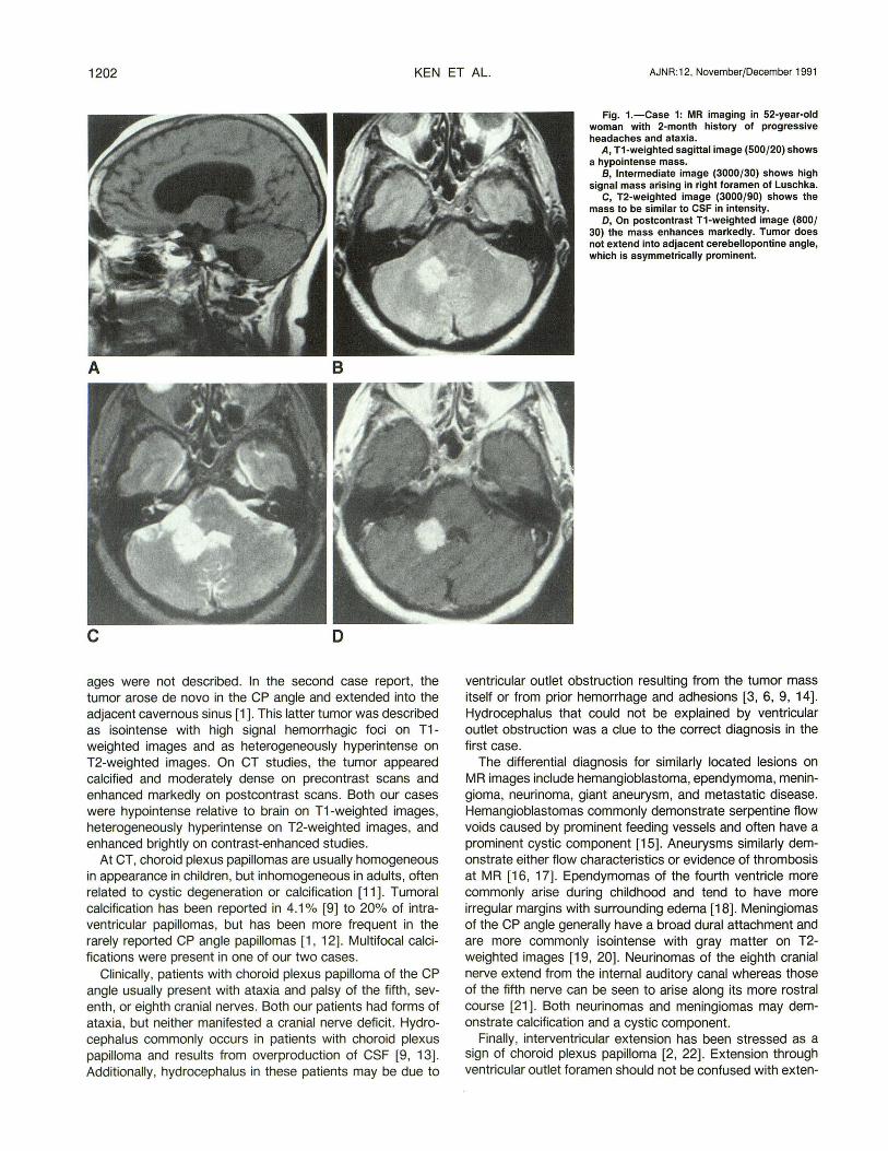
A 52-year-old woman presented with a 2-month history of progressive headaches and ataxia. Physical examination was unremarkable. T1-weighted MR images revealed a mass of low signal intensity expanding the right foramen of Luschka (Fig. 1 ). The mass appeared as inhomogeneous high signal intensity on intermediate and T2-weighted images and enhanced homogeneously on T1-weighted images obtained after administration of gadopentatate dimeglumine. Moderate hydrocephalus was present without evidence of ventricular obstruction. At surgery, a choroid plexus papilloma was found expanding the foramen of Luschka and was completely resected . The patient 's hydrocephalus resolved postoperatively, but recurred several months later, necessitating shunt placement.
Case 2
A 55-year-old woman presented with a 2-year history of progressive dizziness and clumsiness in writing with her right hand. Physical examination was normal except for right finger-to-nose difficulty. T1-weighted MR images showed a multilobulated mass of low signal intensity expanding the right foramen of Luschka and extending to the right CP angle cistern (Fig. 2). The mass appeared inhomogene-
ously bright on intermediate and T2-weighted images. On T1 -weighted images obtained after administration of gadopentatate dimeglumine, the mass enhanced markedly except for persistent components of low signal intensity. Subsequent CT scans showed the mass to contain multiple foci of calcifications of variable size.
At surgery, a choroid plexus papilloma was found arising from within the foramen of Luschka. The tumor was very vascular.
Discussion
Choroid plexus papillomas are rare, histologically benign intracranial neoplasms that typically arise from an intraventricular location. They occur commonly in children and less frequently in adults. They represent 3-5% of intracranial tumors in children and 0.5% in adults [7-9] .
These tumors occur most frequently in the lateral ventricles followed by the fourth and third ventricles in order of decreasing frequency [7 -9]. In children they are found most often in the trigone of the lateral ventricles, whereas in adults they are more likely to be found in the fourth ventricle. Involvement of the CP angle cistern occurs in up to 9% of choroid plexus papillomas, either from direct extension of a fourth ventricular papilloma, from distant CSF seeding, or as a primary CP angle lesion possibly arising de novo from embryonic choroidal remnants [1-5 , 1 0]. In a recent review of the literature, Martin et al. [1] found 20 reported cases of primary unconnected papillomas. Most of these were thought to have developed from the small choroidal tuft that normally protrudes from the foramen of Luschka. Both our cases appear to have arisen from this foramina! tuft. In the first case the tumor remained within the foramen as an expansile mass, whereas in the second case the tumor extended to the CP angle.
There have been two recent case reports of choroid plexus papillomas of the CP angle at MR imaging [1 , 6]. In the first report , the tumor extended to the CP angle cistern from the fourth ventricle via the anterolateral recess and the foramen of Luschka [6]. This tumor was described as isointense with brain on T1-weighted MR images and as homogeneously enhancing on contrast-enhanced CT scans. T2-weighted im-
Received March 11 , 1991; revision requested April 17, 1991 ; revision received May 21 , 1991 ; accepted May 28, 1991 . ' Department of Radiology, Division of Neuroradiology, Scripps Clinic and Research Foundation, 10666 N. Torrey Pines Rd., 100C, La Jolla, CA 92037. Address
reprint requests to D. F. Sobel. 2 Department of Radiology, University of California, San Diego, CA 92103. 3 Department of Neurosurgery, Scripps Clinic and Research Foundation, La Jolla, CA 92037 . ' Department of Radiology, Scripps Memorial Hospital , La Jolla, CA 92038-0028 . 5 Department of Radiology, Sharp Memorial Hospital , San Diego, CA 92123.
AJNR 12:1201-1203, November/December 1991 0195-6108/91/1206-1201 © American Society of Neuroradiology
1202 KEN ET AL. AJNR :12, November/December 1991
A B
c D
ages were not described. In the second case report, the tumor arose de novo in the CP angle and extended into the adjacent cavernous sinus [1]. This latter tumor was described as isointense with high signal hemorrhagic foci on T1-weighted images and as heterogeneously hyperintense on T2-weighted images. On CT studies, the tumor appeared calcified and moderately dense on precontrast scans and enhanced markedly on postcontrast scans. Both our cases were hypointense relative to brain on T1 -weighted images, heterogeneously hyperintense on T2-weighted images, and enhanced brightly on contrast-enhanced studies.
At CT, choroid plexus papillomas are usually homogeneous in appearance in children, but inhomogeneous in adults, often related to cystic degeneration or calcification [11). Tumoral calcification has been reported in 4.1% [9] to 20% of intraventricular papillomas, but has been more frequent in the rarely reported CP angle papillomas [1, 12]. Multifocal calcifications were present in one of our two cases.
Clinically, patients with choroid plexus papilloma of the CP angle usually present with ataxia and palsy of the fifth, seventh, or eighth cranial nerves. Both our patients had forms of ataxia, but neither manifested a cranial nerve deficit. Hydrocephalus commonly occurs in patients with choroid plexus papilloma and results from overproduction of CSF [9, 13]. Additionally , hydrocephalus in these patients may be due to
Fig. 1.-Case 1: MR imaging in 52-year-old woman with 2-month history of progressive headaches and ataxia.
A, T1-weighted sagittal image (500/20) shows a hypointense mass.
8, Intermediate image (3000/30) shows high signal mass arising in right foramen of Luschka.
C, T2-weighted image (3000/90) shows the mass to be similar to CSF in intensity.
D, On postcontrast T1-weighted image (800/ 30) the mass enhances markedly. Tumor does not extend into adjacent cerebellopontine angle, which is asymmetrically prominent.
ventricular outlet obstruction resulting from the tumor mass itself or from prior hemorrhage and adhesions [3, 6, 9, 14). Hydrocephalus that could not be explained by ventricular outlet obstruction was a clue to the correct diagnosis in the first case.
The differential diagnosis for similarly located lesions on MR images include hemangioblastoma, ependymoma, meningioma, neurinoma, giant aneurysm, and metastatic disease. Hemangioblastomas commonly demonstrate serpentine flow voids caused by prominent feeding vessels and often have a prominent cystic component [15]. Aneurysms similarly demonstrate either flow characteristics or evidence of thrombosis at MR [16, 17). Ependymomas of the fourth ventricle more commonly arise during childhood and tend to have more irregular margins with surrounding edema [18). Meningiomas of the CP angle generally have a broad dural attachment and are more commonly isointense with gray matter on T2-weighted images [19, 20]. Neurinomas of the eighth cranial nerve extend from the internal auditory canal whereas those of the fifth nerve can be seen to arise along its more rostral course [21). Both neurinomas and meningiomas may demonstrate calcification and a cystic component.
Finally, interventricular extension has been stressed as a sign of choroid plexus papilloma [2, 22]. Extension through ventricular outlet foramen should not be confused with ex ten-
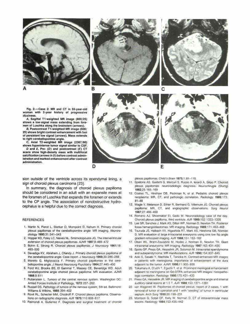
Fig. 2.-Case 2: MR and CT in 55-year-old woman with 2-year history of progressive dizziness.
A, Sagittal T1-weighted MR image (800/25) shows a low-signal mass extending from foramen of Luschka along the brainstem (arrows).
B, Postcontrast T1-weighted MR image (600/ 25) shows bright contrast enhancement with foci of persistent low signal (arrows). Mass extends to right cerebellopontine angle.
C, Axial T2-weighted MR image (2367/80) shows hyperintense tumor signal similar to CSF.
0 and E, Pre- (0) and postcontrast (E) CT scans show high-density mass with multifocal calcification (arrows in 0) before contrast adminIstration and marked enhancement after contrast administration.
sion outside of the ventricle across its ependymal lining, a sign of choroid plexus carcinoma [23] .
In summary, the diagnosis of choroid plexus papilloma should be considered in an adult with an expansile mass at the foramen of Luschka that expands the foramen or extends to the CP angle. The association of nonobstructive hydrocephalus is a helpful clue to the correct diagnosis.
REFERENCES
1. Martin N, Pierot L, Sterker 0 , Mompoint D, Nahum H. Primary choroid plexus papillomas of the cerebellopontine angle: MR imaging. Neuroradiology 1990;31 :541 - 543
2. Hopper KD, Foley LC, Nieves NL. Smirniotopoulos JG. The interventricular extension of choroid plexus papillomas. AJNR 1987;8:469-472
3. Bohm E, Strong R. Choroid plexus papillomas. J Neurosurg 1961;18: 493-500
4. Devadiga KV, Abraham J, Chandy J. Primary choroid plexus papilloma of the cerebellopontine angle. Case report . J Neurosurg 1969;30 : 286-288
5. Morello G, Migliavacca F. Primary choroid papillomas in the cerebellopontine angle. J Neurol Neurosurg Psychiatry 1964;27:445-450
6. Ford WJ, Brooks BS, El Gamma! T, Massey CE, Beveridge WD. Adult cerebellopontine angle choroid plexus papilloma: MR evaluation. AJNR 1988;9:611
7. Rubenstein L. Tumors of the central nervous system. Washington DC: Armed Forces Institute of Pathology, 1972 :257-262
8. Russell OS. Pathology of tumors of the nervous system, 5th ed. Baltimore: Williams & Wilkins, 1989:394- 402
9. Rovit RL, Schecter MM, Chodroff P. Choroid plexus papilloma. Observations on radiographic diagnosis. AJR 1970;110:608-617
10. Raimondi A, Gutierrez F. Diagnosis and surgical treatment of choroid
plexus papillomas. Child 's Brain 1975;1 :81-115 11. Spallone AS, Guidetti G, Mercuri S, Russo A, Ierardi A, Silipo P. Choroid
plexus papillomas: neuroradiologic diagnosis. Neurochirugia (Stuttg) 1982;25: 165-169
12. Coates TL, Hinshaw DB, Peckman N, et al. Pediatric choroid plexus neoplasms: MR, CT, and pathologic correlation. Radiology 1989;173: 81-88
13. Wagle V, Melanson D, Ethier R, Bertrand G, Villemure JG. Choroid plexus papilloma: MR, CT, and angiographic observations. Surg Neurol 1987;27:466-468
14. Romano AJ, Shoemaker El, Gado M. Neuroradiology case of the day. Choroid plexus papilloma, third ventricle. AJR 1989;152: 1333-1335
15. Lee SR, Sanches J, Mark AS, Dillon WP, NormanD, Newton TH. Posterior fossa hemangioblastomas: MR imaging. Radiology 1989;171 :463-468
16. Tsuruda JS, Halbach VV, Higashida AT, Mark AS, Hieshima GB, Norman D. M R evaluation of large intracranial aneurysms using cine low fl ip angle gradient-refocused imaging. AJR 1988;151: 153-162
17. Olsen WL, Brant-Zawadzki M, Hodes J, Norman 0 , Newton TH . Giant intracranial aneurysms: MR imaging. Radiology 1987;163:431-435
18. Spoto GP, Press GA, Hesselink JR, Solomon M. Intracranial ependymoma and subependymoma: MR manifestations. AJR 1990;154 :837-845
19. Aoki S, Sasaki Y, Machida T, Tanioka H. Contrast-enhanced MR images in patients with meningioma: importance of enhancement of the dura adjacent to the tumor. AJNR 1990;11 :935-938
20. Tokumaru A, O'uchi T, Eguchi T, et al. Prominent meningeal enhancement adjacent to meningioma on Gd-DTPA-enhanced MR images: histopathologic correlation. Radiology 1990;175:431 - 433
21 . Press GA, Hesselink JR. MR imaging of cerebellopontine angle and internal auditory canal lesions at 1.5 T. AJR 1988;150: 1371-1381
22. van Wagenen W. Papillomas of choroid plexus: report of 2 cases, 1 with removal of tumor at operation and 1 with "seeding" of tumor in ventricular septum. Arch Surg 1930;20 : 199-231
23. Morrison G, Sobel OF, Kelly W, Norman D. CT of intraventricular mass lesions. Radiology 1984;153:435-442