Tuberculosis Bálint Beatrix MD, PhD SZTE, Dpt. of Pulmonology Deszk 2014.
44
Tuberculosis Bálint Beatrix MD, PhD SZTE, Dpt. of Pulmonology Deszk 2014.
-
Upload
kellie-french -
Category
Documents
-
view
221 -
download
4
Transcript of Tuberculosis Bálint Beatrix MD, PhD SZTE, Dpt. of Pulmonology Deszk 2014.

Tuberculosis
Bálint Beatrix MD, PhD
SZTE, Dpt. of Pulmonology
Deszk
2014.

Tuberculosis
TB a chronic bacterial infection, causes more deaths worldwide than any other infectious disease.
TB is spread through the air and usually infects the lungs, although other organs are sometimes involved.
Some 2 billion people - one-third of the world's population - are infected with the TB organism,
Mycobacterium tuberculosis.
History 1.
Germ theory: -Robert Koch (1882)-Pathogenicity of Mycobacterium tuberculosis -Konrad Röntgen (1892)- X ray
Paleopathological evidences- skeletal TB, bone TB
Ancient greek physisians used the word PHTYSIS 8th-9th century ¼ of the european adults died from TB.

TB in the World (number of TB cases)

Tbc incidencia Európában az elmúlt években
0,0
20,0
40,0
60,0
80,0
100,0
120,0
140,0
160,0
Své
dors
zág
Dán
ia
Ola
szor
szág
Finn
orsz
ág
Hol
land
ia
Ném
etor
szág
Fran
ciao
rszá
g
Cse
hors
zág
Bel
gium
Aus
ztri
a
Szl
ovák
ia
Nag
y-B
rita
nnia
Szl
ovén
ia
Spa
nyol
orsz
ág
Mag
yaro
rszá
g
Leng
yelo
rszá
g
Sze
rbia
Por
tugá
lia
Oro
szor
szág
Rom
ánia
%00
0
2001 2002 2003 2004 2005 2006
TB in EUROPE

Causes of death of HIV positive patients
Mycobacterium tuberculosis
• The causative agents for tuberculosis
• Discovered by Robert Koch in 1882
• ~25 % of world’s population infected
• 25 million is infected in USA
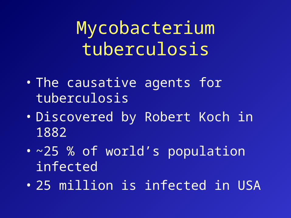
MicrobiologyMycobacterium tuberculosis
obligate, aerobic parazite, acid-fastslow growthvisible colonial growth: 4-6 weeksINH resistant and sensitive strains are different
Direct examinationZiehl-Neelsen stain:4 m long and 0,2-0,5 m wide10 000 organism/ml of sputumsmear positive
Culture of sputum/fluidM. tuberculosis: growths slowly, lack of pigment, produces niacinM. bovis: niacine negativeDrug sensitivity test.
Quick test: PCR, Bactec
Mode of spread
• TB is spread from in microscopic droplets person to person — droplet nuclei — expelled from the lungs when a TB sufferer coughs, sneezes, speaks, sings, or laughs. Only people with active disease are contagious.
• People are most likely to be contagious when their sputum contains bacilli, when they cough frequently and when the extent of their lung disease, as revealed by a chest x-ray, is great.
* People who have been treated with appropriate drugs for at
least two weeks usually are not infectious.

Predisposing Factors• Babies and young children
• HIV infection
• substance abuse
• diabetes mellitus
• silicosis
• cancer
• leukemia or Hodgkin's disease
• severe kidney disease
• low body weight
• certain medical treatments – corticosteroid treatment
– organ transplants
– chemotherapy

HOW DOES TB DISEASE DEVELOP? There are two possible ways a person can become sick with TB
disease:
1.A person who may have been infected with TB for years and has been perfectly healthy. The time may come when this person suffers a change in health. The cause may be another disease like AIDS or diabetes. Or it may be drug or alcohol abuse or a lack of health care because of homelessness. Whatever the cause, when the body's ability to protect itself is damaged, the TB infection can become TB disease. In this way, a person may become sick with TB disease months or even years after they first breathed in the TB germs.
2. A person first breathes in the TB germs the body is unable to protect itself against the disease. The germs then develop into active TB disease within weeks. (This way TB disease develops happens much more quickly.)

Pathogenesis
The site of initial infection alveoli macrophages ingest the inhaled M. tuberculosis.
Some bacilli may be killed immediately; others may multiply within the macrophages.
During the 2 to 8 weeks after initial infection in people with intact immune systems,
macrophages present pieces of the bacilli, displayed on their cell surfaces, to the T cells
release an elaborate array of chemical signals
cell-mediated hypersensitivity T cells responds tuberculin skin test (PPD test)
cell signals inflammatory reactions;
recruit and activate specialized cells to kill bacilli and
In HIV-infected people and in children, the bacilli spread to other sites in the body
dissemination life-threatening meningitis and other problems.

Pathogenesis 2.
The body's immune system maintains a standoff with the infection,
sometimes for years.
TB bacilli may persist within macrophages, but further multiplication
and spread of M. tuberculosis are confined.
Most people undergo complete healing of their initial infection,
and the tubercles calcify and lose their viability.
A positive TB skin test, and in some cases a chest x-ray,
may provide the only evidence of the infection.
If, the body's resistance is because of aging, infections (HIV),
malnutrition,or other factors, the bacilli may break out of the tubercles
in the alveoli and cause active disease.

Pathogenesis 3.(X ray)
Simon foci: The initial infection leaves nodular scars in the apices of one or both lungs, called which are the most common seeds for later active TB.
Ghon foci: calcified scars of primary infection and residual calcified hilar lymph nodes.
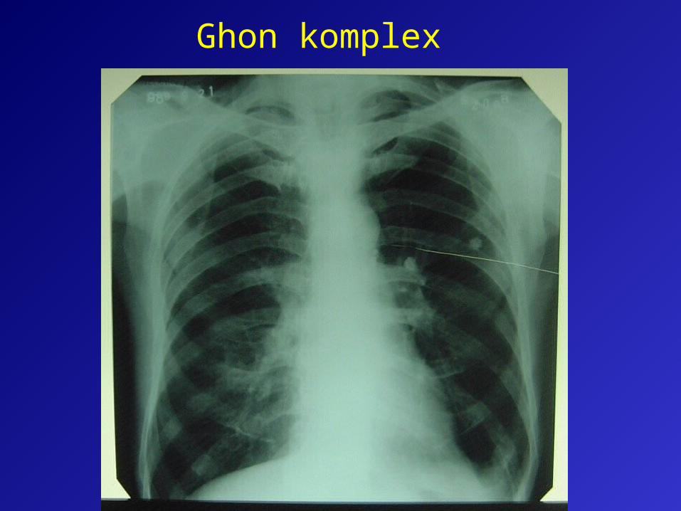
Ghon komplex
Symptoms
Early TB (single or multiple nodule, caseous lesion)- no symptomes
Progresszive TB (cavitation, pneumonitis)- nonspecific symptomes: anorexia, fatigue, weight loss,
remittent fever, night sweets
- cough, sputum (mucopurulent)- haemoptysis- chest pain (inflammation of parietal pleura)

Laboratory findings
IIn advanced TB!
- RBC - Se albumin - WBC - Sodium - Calcium
Characteristic X-ray findings
• Apical, subapical patchy infiltration• Bilateral upper lobe infiltration• Dissemination: miliary tb• Lower lobe TB
– cavitation or infiltration– atelectesis, mass leasions, large cavitation with fluid,
pneumonic-like infiltration
• Non-specific• Pleural effusion
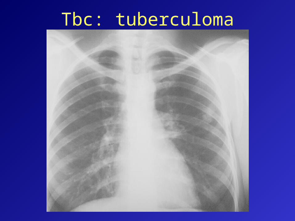
Tbc: tuberculoma

Tb pneumonia

Tb hilar adenopathy

Miliary tb
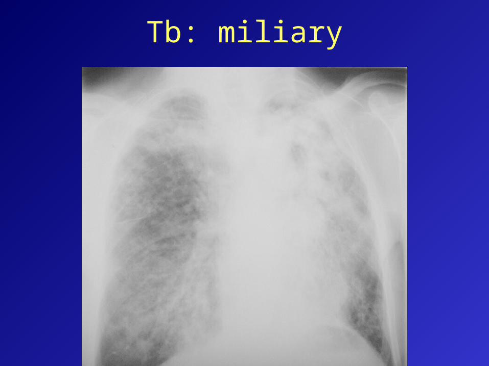
Tb: miliary
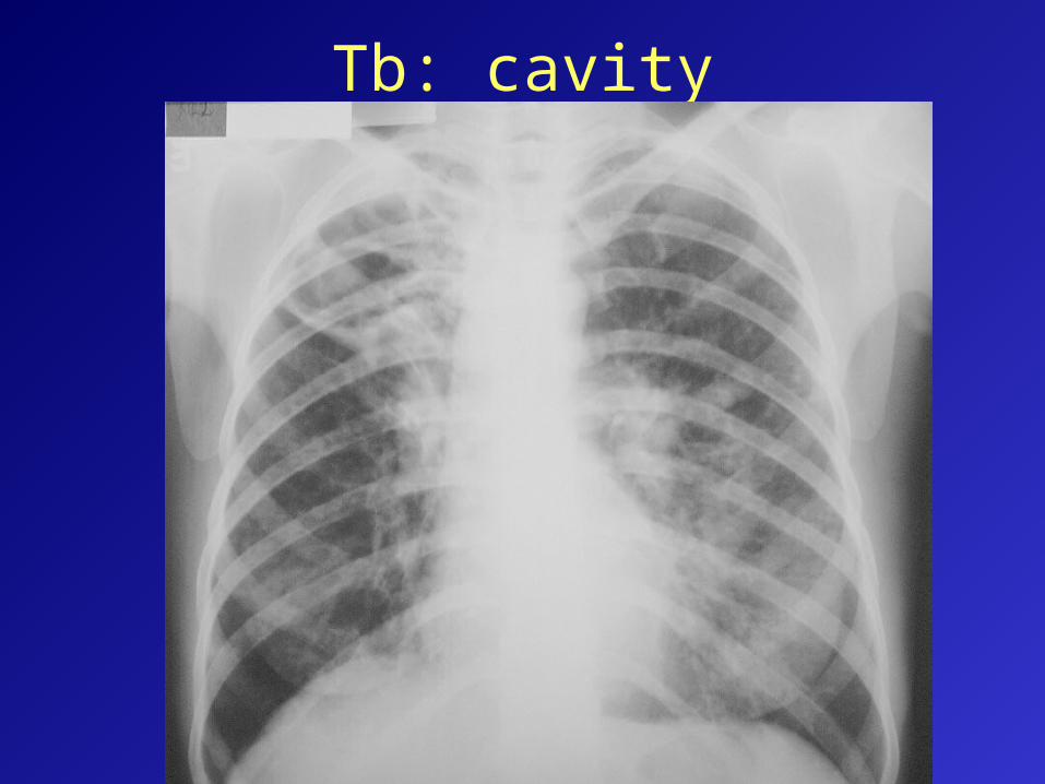
Tb: cavity

Tb: cavity
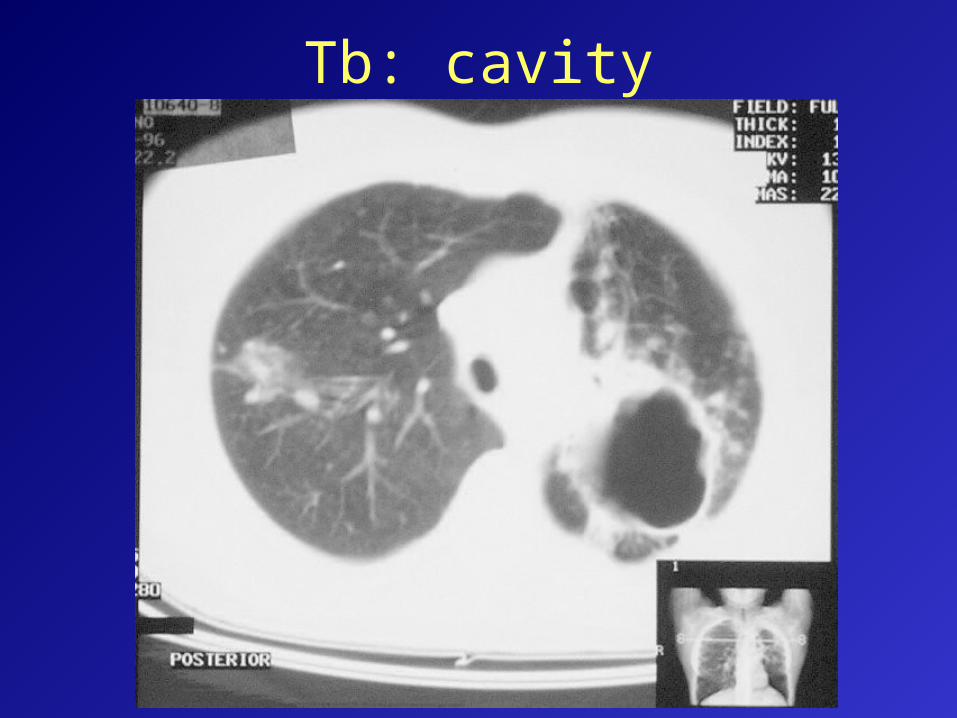
Tb: cavity

Tb:progressive

Callus pleurae, residuum
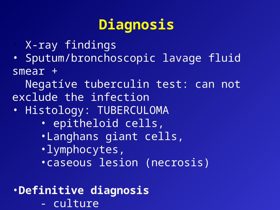
Diagnosis• X-ray findings• Sputum/bronchoscopic lavage fluid smear + Negatíve tuberculin test: can not exclude the infection• Histology: TUBERCULOMA
• epitheloid cells, •Langhans giant cells, •lymphocytes, •caseous lesion (necrosis)
•Definitive diagnosis- culture- specification of the organism

Extrapulmonary TB (TB can involve any organ)
-TB of the tonsils, lymph nodes, abdominal organs, bones, and joints caused by ingestion of milk infected with M. bovis. (slaughtering cows with milk)
*GENITOURINARY TUBERCULOSIS
-kidney pyelonephritis. (chronic, "sterile" routine culture-negative)
-epididymis or prostate gland, baldder, vesicles.
-Salpingo-oophoritis
* TUBERCULOUS MENINGITIS (TB to the subarachnoid space)
* MILIARY TUBERCULOSIS (Generalized Hematogenous or Lymphohematogenous TB) Bone marrow involvement
* TUBERCULOUS PERITONITIS
*TUBERCULOUS PERICARDITIS
*TUBERCULOUS LYMPHADENITIS
*TUBERCULOSIS OF BONES AND JOINTS (Pott's disease)
*TUBERCULOSIS OF THE LIVER
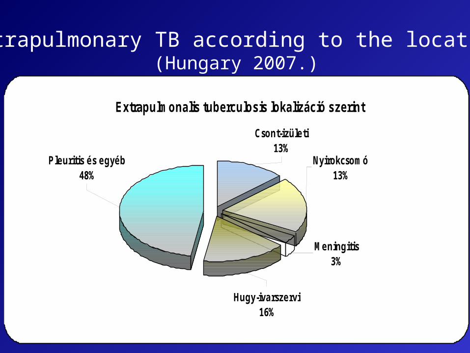
Extrapulmonary TB according to the location(Hungary 2007.)
Extrapulmonalis tuberculosis lokalizáció szerint
Pleuritis és egyéb48%
Nyirokcsomó13%
Hugy-ivarszervi16%
Csont-izületi13%
Meningitis3%
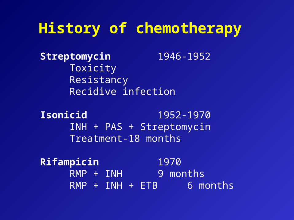
History of chemotherapy
Streptomycin 1946-1952ToxicityResistancyRecidive infection
Isonicid 1952-1970INH + PAS + StreptomycinTreatment-18 months
Rifampicin 1970RMP + INH 9 months RMP + INH + ETB 6 months
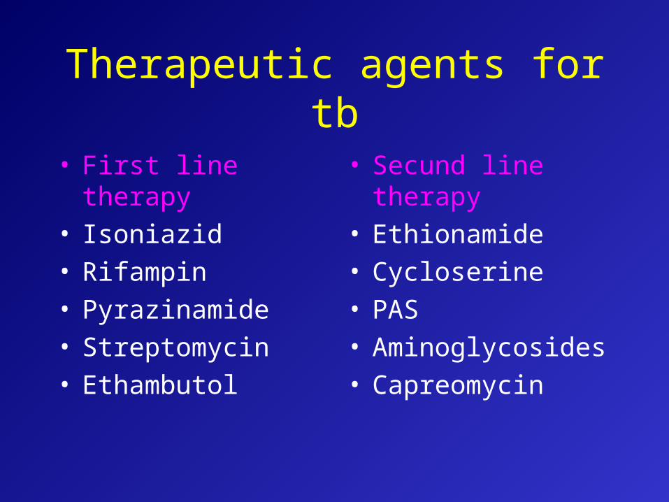
Therapeutic agents for tb
• First line therapy• Isoniazid• Rifampin• Pyrazinamide• Streptomycin• Ethambutol
• Secund line therapy• Ethionamide• Cycloserine• PAS• Aminoglycosides• Capreomycin
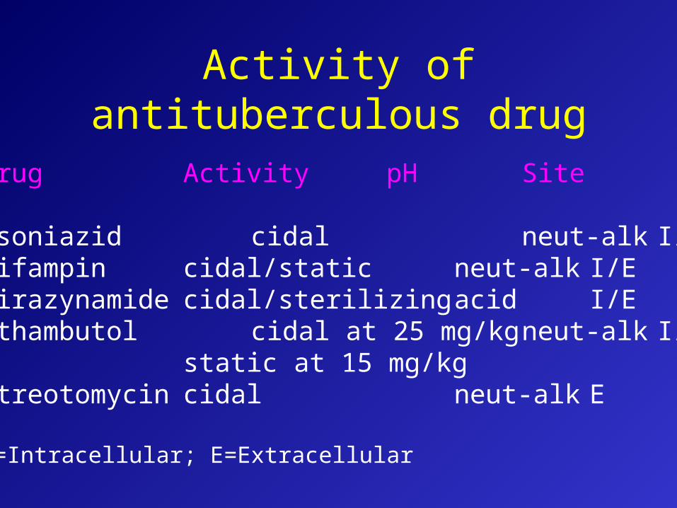
Activity of antituberculous drug
Drug Activity pH Site
Isoniazid cidal neut-alk I/ERifampin cidal/static neut-alk I/EPirazynamide cidal/sterilizing acid I/EEthambutol cidal at 25 mg/kg neut-alk I/E
static at 15 mg/kgStreotomycin cidal neut-alk E
I=Intracellular; E=Extracellular
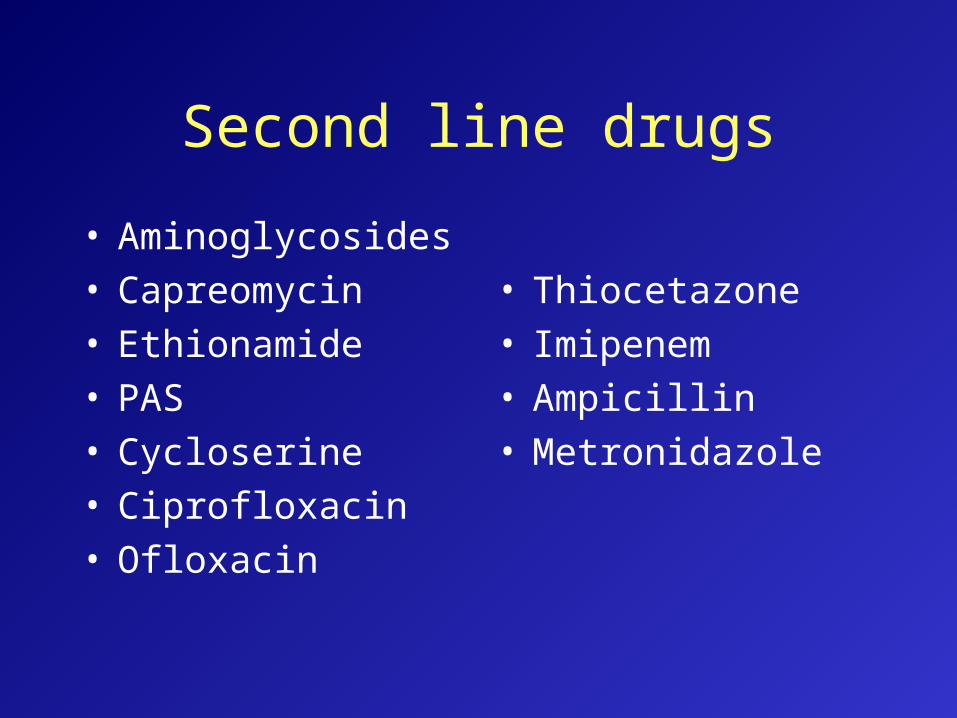
Second line drugs
• Aminoglycosides• Capreomycin• Ethionamide• PAS• Cycloserine• Ciprofloxacin• Ofloxacin
• Thiocetazone• Imipenem• Ampicillin• Metronidazole

Characteristics of 2nd line drugs
• Less effective drugs
• Poor GI tolarence
• Significant side effect profile
• Not well studied
• Some not readily available (PAS)
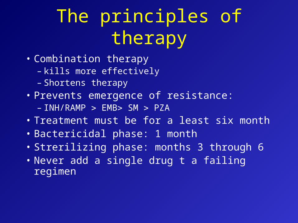
The principles of therapy
• Combination therapy – kills more effectively– Shortens therapy
• Prevents emergence of resistance:– INH/RAMP EMB SM PZA
• Treatment must be for a least six month• Bactericidal phase: 1 month• Strerilizing phase: months 3 through 6• Never add a single drug t a failing regimen

Initial therapy: four drugs
• Isoniazid (INH) 300 mg daily
• Rifampin (RIF) 600 mg daily
• Pyrazinamide (PZA)25-30 mg daily
• Ethambutol (EMB)25 mg initially

Therapeutic Regimens
• Daily therapy• 6 months• Daily treatment• 180 doses• 2-3 % relapse
• Short course• 6 months• Twice or three times
weekly• 52-114 doses• Equivalent relapse
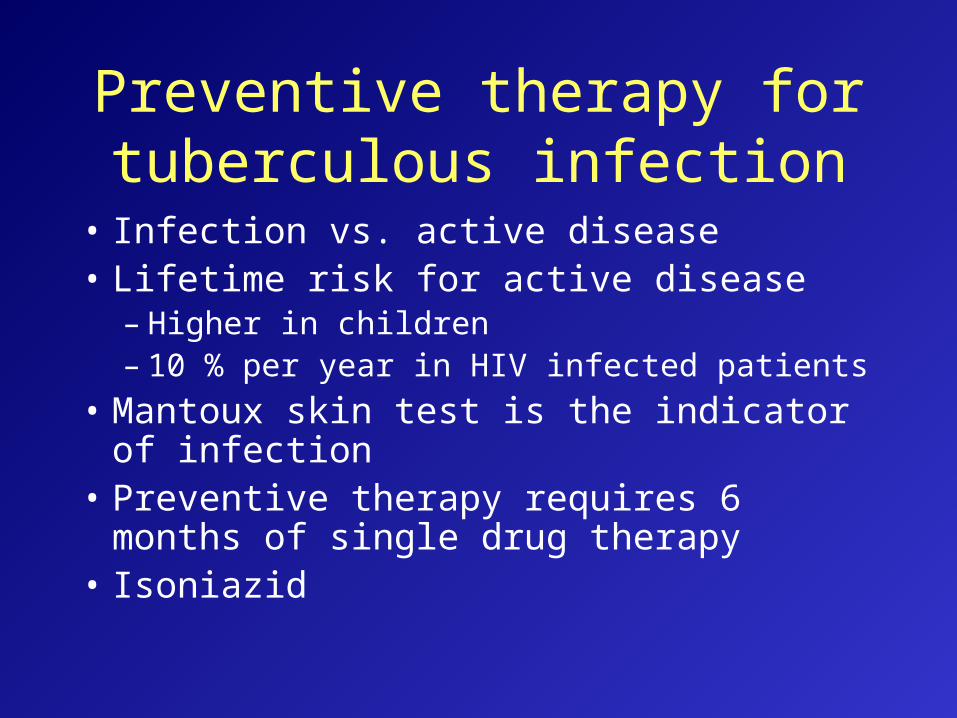
Preventive therapy for tuberculous infection
• Infection vs. active disease• Lifetime risk for active disease
– Higher in children– 10 % per year in HIV infected patients
• Mantoux skin test is the indicator of infection• Preventive therapy requires 6 months of
single drug therapy• Isoniazid

Nontuberculous mycobacteria
• Pumonary disease– M. avium, kansasii, abscessus, xenopi,
malmoense
• Lymphadenitis– M. avium, scrofulaceum, malmoense
• Cutaneous disease– M. marinum, fortuitum, chelonea, ulcerans
• Disseminated disease– M. avium, kansasii,chelonea, haemophilum

Treatment of nontuberculous mycobacteria
• The antituberculotic drugs are usually not effective
• M. kansasii: INH, RIF, EMB
• M. avium: macrolide, Rifamycin, EMB
• Rapid growers: clarithromycin and 2nd agents

History 2.
Outstanding representatives of the arts and political life who suffered from TB
• Balzac• Brontë sisters• Chekov• Chopin• Dostoevsky• Kafka• D.H. Lawrence• Sir Walter Scott
• E. A. Poe• Voltaire• John Keats• Rembrandt’s wife
(Sashka) and his son (Titus)
• Marquise de Pompadur
• Napoleon II
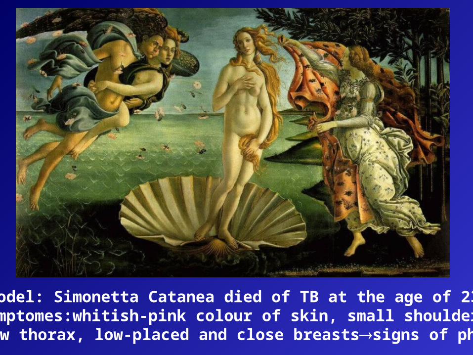
Model: Simonetta Catanea died of TB at the age of 23.Symptomes:whitish-pink colour of skin, small shoulders,
narrow thorax, low-placed and close breastssigns of phtisis

![Beatrix Potter's Gardening Life [Excerpt]](https://static.fdocuments.in/doc/165x107/55cf99b8550346d0339eda31/beatrix-potters-gardening-life-excerpt.jpg)