Triple-helical collagen hydrogels via covalent aromatic ...
34
Triple-helical collagen hydrogels via covalent aromatic functionalization with 1,3-Phenylenediacetic acid Giuseppe Tronci, 1,2 Amanda Doyle, 1,2 Stephen J. Russell, 2 and David J. Wood 1 1 Biomaterials and Tissue Engineering Research Group, Leeds Dental Institute, University of Leeds, Leeds LS2 9LU, United Kingdom 2 Nonwoven Research Group, Centre for Technical Textiles, University of Leeds, Leeds LS2 9JT, United Kingdom Table of contents entry Covalent functionalization of type I collagen with 1,3-Phenylenediacetic acid (Ph) directly leads to triple-helical hydrogels with controlled degree of crosslinking (C), swelling ratio (SR) and denaturation temperature ( T d ), in contrast to state-of-the-art carbodiimide crosslinking. Abstract Chemical crosslinking of collagen is a general strategy to reproduce macroscale tissue properties in physiological environment. However, simultaneous control of protein conformation, material properties and biofunctionality is highly challenging with current synthetic strategies. Consequently, the potentially-diverse clinical applications of collagen-based biomaterials cannot be fully realised. In order to establish defined biomacromolecular systems for mineralised tissue applications, type I collagen was functionalised with 1,3-Phenylenediacetic acid (Ph) and investigated at the molecular, macroscopic and functional levels. Preserved triple
Transcript of Triple-helical collagen hydrogels via covalent aromatic ...

Triple-helical collagen hydrogels via covalent aromatic functionalization with
1,3-Phenylenediacetic acid
Giuseppe Tronci,1,2
Amanda Doyle,1,2
Stephen J. Russell,2
and David J. Wood1
1Biomaterials and Tissue Engineering Research Group, Leeds Dental Institute, University of Leeds, Leeds
LS2 9LU, United Kingdom
2 Nonwoven Research Group, Centre for Technical Textiles, University of Leeds, Leeds LS2 9JT, United
Kingdom
Table of contents entry
Covalent functionalization of type I collagen
with 1,3-Phenylenediacetic acid (Ph) directly
leads to triple-helical hydrogels with controlled
degree of crosslinking (C), swelling ratio (SR) and denaturation temperature (Td), in contrast to
state-of-the-art carbodiimide crosslinking.
Abstract
Chemical crosslinking of collagen is a general strategy to reproduce macroscale tissue
properties in physiological environment. However, simultaneous control of protein
conformation, material properties and biofunctionality is highly challenging with current
synthetic strategies. Consequently, the potentially-diverse clinical applications of collagen-based
biomaterials cannot be fully realised. In order to establish defined biomacromolecular systems
for mineralised tissue applications, type I collagen was functionalised with 1,3-Phenylenediacetic
acid (Ph) and investigated at the molecular, macroscopic and functional levels. Preserved triple

helix conformation was observed in obtained covalent networks via ATR-FTIR (AIII/A1450 ~ 1)
and WAXS, while network crosslinking degree (C: 87-99 mol.-%) could be adjusted based on
specific reaction conditions. Decreased swelling ratio (SR: 823-1285 wt.-%) and increased
thermo-mechanical (Td: 80-88 °C; E: 28-35 kPa; σmax: 6-8 kPa; εb: 53-58 %) properties were
observed compared to state-of-the-art carbodiimide (EDC)-crosslinked collagen controls, likely
related to the intermolecular covalent incorporation of the aromatic segment. Ph-crosslinked
hydrogels displayed nearly intact material integrity and only a slight mass decrease (MR: 5-11
wt. %) following 1-week incubation in either PBS or simulated body fluid (SBF), in contrast to
EDC-crosslinked collagen (MR: 33-58 wt. %). Furthermore, FTIR, SEM and EDS revealed
deposition of a calcium-phosphate phase on SBF-retrieved samples, whereby an increased
calcium phosphate ratio (Ca/P: 0.84-1.41) was observed in hydrogels with higher Ph content. 72-
hour material extracts were well tolerated by L929 mouse fibroblasts, whereby cell confluence
and metabolic activity (MTS assay) were comparable to those of cells cultured in cell culture
medium (positive control). In light of their controlled structure-function properties, these
biocompatible collagen hydrogels represent attractive material systems for potential mineralised
tissue applications.
1. Introduction
Biomaterials play a crucial role in regenerative medicine and pharmaceutics and find huge
applications in wound care, orthopaedics and cardiovascular industries, among others.1
Particularly for mineralised tissue applications, the design of versatile biomimetic systems,
which can be safely implanted in vivo, displaying defined biodegradability, tuneable mechanical
properties, and bioactivity, is a pressing, unmet, clinical need towards next generation
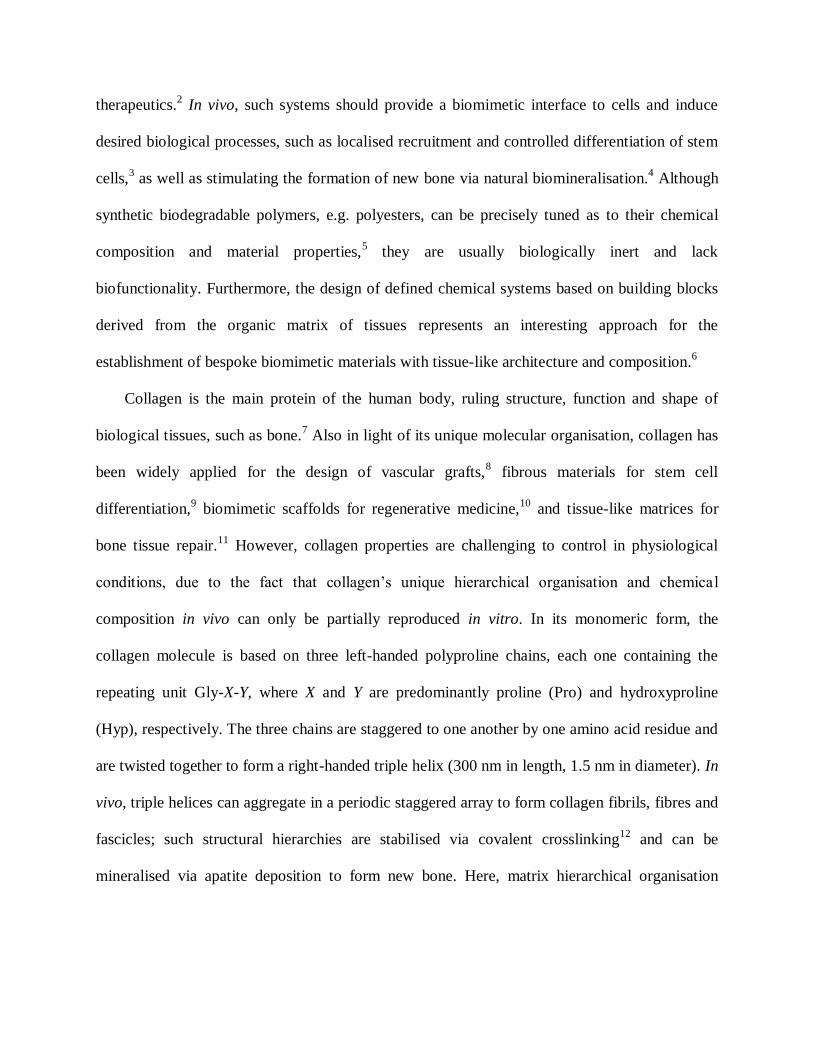
therapeutics.2 In vivo, such systems should provide a biomimetic interface to cells and induce
desired biological processes, such as localised recruitment and controlled differentiation of stem
cells,3 as well as stimulating the formation of new bone via natural biomineralisation.
4 Although
synthetic biodegradable polymers, e.g. polyesters, can be precisely tuned as to their chemical
composition and material properties,5 they are usually biologically inert and lack
biofunctionality. Furthermore, the design of defined chemical systems based on building blocks
derived from the organic matrix of tissues represents an interesting approach for the
establishment of bespoke biomimetic materials with tissue-like architecture and composition.6
Collagen is the main protein of the human body, ruling structure, function and shape of
biological tissues, such as bone.7 Also in light of its unique molecular organisation, collagen has
been widely applied for the design of vascular grafts,8 fibrous materials for stem cell
differentiation,9 biomimetic scaffolds for regenerative medicine,
10 and tissue-like matrices for
bone tissue repair.11
However, collagen properties are challenging to control in physiological
conditions, due to the fact that collagen’s unique hierarchical organisation and chemica l
composition in vivo can only be partially reproduced in vitro. In its monomeric form, the
collagen molecule is based on three left-handed polyproline chains, each one containing the
repeating unit Gly-X-Y, where X and Y are predominantly proline (Pro) and hydroxyproline
(Hyp), respectively. The three chains are staggered to one another by one amino acid residue and
are twisted together to form a right-handed triple helix (300 nm in length, 1.5 nm in diameter). In
vivo, triple helices can aggregate in a periodic staggered array to form collagen fibrils, fibres and
fascicles; such structural hierarchies are stabilised via covalent crosslinking12
and can be
mineralised via apatite deposition to form new bone. Here, matrix hierarchical organisation

directly impacts on the distribution and size of nucleated apatite crystals, so that a clear
relationship between internal collagen architecture and bone physiological state is present.13
Fibrillogenesis can be induced in vitro by exposing triple helical collagen to physiological
conditions. However, hydrogen and covalent bonds stabilising collagen structures in vivo are
partially broken following isolation ex-vivo, so that collagen hierarchical organisation is affected.
As a result, collagen materials display non-controllable swelling and weak mechanical properties
in physiological conditions; reliable synthetic methods must therefore be applied in order to
improve hydrogel thermo-mechanical behaviour, without affecting collagen biofunctionality,14
i.e. biocompatibility and bioactivity. Following a bottom-up synthetic approach, O’Leary et al.
proposed a self-assembling peptide system capable to recapitulate the triple helical and fibrillar
architecture of collagen.15
Based on the peptide concentration and peptide sequence, synthetic
collagen-like hydrogels were successfully developed, although the thermo-mechanical stability
was still not adequate for biomaterial applications. Exploiting the fibrillogenesis process of
native collagen, Nam et al. proposed a heterostructural collagen matrix obtained via layer-by-
layer deposition of collagen solutions at different densities.16
By adjusting the collagen
concentration and the number of layers, mechanical properties and cell proliferation could be
controlled, although the degradation behaviour of these materials in physiological conditions was
not investigated. Collagen fibrillogenesis was also exploited to replicate the hierarchical
organization of mineralized tissues.17
Here, polyacrylic acid was applied to stabilise an
amorphous calcium phosphate phase during the self-assembly of collagen triple helices into
fibrils. In this way, highly-ordered mineralized collagen matrices with improved mechanical
properties were successfully formed, although the material behaviour in physiological conditions

was not fully addressed. Recently, dynamic perfusion-flow mineralization techniques have also
been proposed to recapitulate the natural mineralization process in collagen templates.18
Other than exploiting the inherent triple helix self-assembly, functionalisation and covalent
crosslinking, e.g. via N-(3-Dimethylaminopropyl)-N′-ethylcarbodiimide hydrochloride
(EDC),19,20
glutaraldehyde,21,22
hexamethylene diisocyanate23
and enzymatic treatment24
have
been shown to enhance macroscopic properties and resistance to enzymatic degradation of
collagen in biological environment.25
However, safe and systematic control in network
architecture has proved to be challenging, whereby occurrence of side reactions may lead to the
formation of potentially-toxic materials with only slight variation in macroscopic properties.
Diimidoesters-dimethyl suberimidate (DMS), 3,3’-dithiobispropionimidate (DTBP) and acyl
azide have been employed as alternative crosslinking agents, resulting in stable materials in
physiological conditions, although reduced material elasticity was observed.26
Additionally, both
dehydrothermal and riboflavin-mediated treatments have been applied as physical, benign
stabilisation methods, leading to partial loss of native collagen structure and non-homogeneous
crosslinking.27
In an effort to design water-stable composite materials, collagen fibrils were
functionalized with a silane moiety, in order to promote covalent links between the inorganic
phase and the fibrous matrix.28
The resulting materials displayed preserved collagen fibrous
characteristics and enhanced degradability, although the mechanical properties were not fully
investigated. From all the aforementioned examples it appears rather clear that while the
macroscopic properties may be improved, systematic control of material behaviour at different
length scales is only minimally accomplished in the resulting collagen materials. Thus, the
establishment of novel synthetic methods ensuring tunable collagen functionalisation, preserved
protein conformation together with full biocompatibility and bioactivity is necessary in order to
establish reliable collagen systems with defined structure-function properties for mineralised
tissue repair.
In this work, the design of type I collagen hydrogels via covalent lysine functionalisation
with 1,3-Phenylenediacetic acid (Ph) was investigated. Given that the collagen displays cell-
binding peptides at the molecular level, we investigated whether the incorporation of covalent
crosslinking of collagen triple helices via a stiff, aromatic segment could offer a synthetic
strategy to the formation of defined biomacromolecular materials with relevant mechanical
properties and retained biochemical features. Ph was selected as a novel bifunctional segment, in
order to promote inter-molecular crosslinking of collagen molecules, unlikely to be
accomplished with current synthetic methods.8,19-23
This specific functionalisation was aimed to
achieve controlled swelling and enhanced mechanical properties in resulting hydrogels, due to
the backbone rigidity and hydrophobicity of the Ph aromatic segment. In order to investigate the
effectiveness of this synthetic approach over current strategies, EDC treatment was selected as a
state-of-the-art reference method, since it has been shown to promote the formation of water-
stable collagen materials with no residual toxicity,25
in contrast to aldehyde biomaterial fixation.8
2. Materials and methods
2.1. In-house isolation of type I collagen from rat tail tendons
Type I collagen was isolated in-house via acidic treatment of rat tail tendons.29
Briefly,
frozen rat tails were thawed in ethanol (for about 20 min). Individual tendons were pulled out of
the tendon sheath, minced, and placed in 17.4 mM acetic acid (Sigma Aldrich) solution at 4 °C
in order to extract collagen. After three days extraction, the mixture was centrifuged at 20000
rpm for one hour. The pellet was discarded and the crude collagen solution was neutralised with

0.1 M NaOH (Sigma Aldrich). After stirring (overnight, 4 °C), the neutralised solution was
centrifuged (45 min, 10000 rpm, 4 °C). The supernatant volume was measured and an equal
volume of fresh acetic acid (17.4 mM) solution was used to re-solubilise the collagen pellet. The
mixture was then freeze-dried in order to obtain collagen. The resulting product was analysed via
sodium dodecyl sulphate-polyacrylamide gel electrophoresis (SDS-page) which showed only the
main electrophoretic bands of type I collagen.30
2.2. Synthesis of collagen hydrogels
In-house isolated type I collagen (0.8 wt.-%) was dissolved in 10 mM hydrochloric acid
(Sigma Aldrich) under stirring at room temperature, prior to network formation via either 1,3-
Phenylenediacetic acid (Ph, VWR International) or N-(3-Dimethylaminopropyl)-N′-
ethylcarbodiimide hydrochloride (EDC, Sigma Aldrich). At the same time, Ph was stirred (0°C,
30 min) in sodium phosphate buffer (0.1 M, pH 7.4, 500 µL) at selected molar ratios to target
collagen lysines (0.5-1.5 COOH/Lys molar ratio).31
Here, a three-fold molar content of EDC and
N-Hydroxysuccinimide (NHS) was added. NHS-activated Ph solution was mixed with obtained
collagen solution (1 g), so that the pH of the resulting mixture was 6.5 at 19.2 °C (in the case of
1 COOH/Lys molar ratio). Reacting mixtures were incubated overnight under gentle shaking at
room temperature, in order to allow for the nucleophilic addition reaction of collagen lysines to
Ph carboxylic functions to occur. EDC-crosslinked collagen was synthesised as state-of-the-art
crosslinked collagen control by mixing EDC with obtained collagen solution, as previously
reported.19
Complete gel formation was observed following reaction with either Ph or EDC.
Resulting hydrogels were washed with distilled water and dehydrated in aqueous solutions
of increasing ethanol concentrations.

2.3. Investigation of chemical composition and structural organisation
Attenuated Total Reflectance Fourier-Transform Infrared (ATR FT-IR) was carried out on
dry samples using a Perkin-Elmer Spectrum BX spotlight spectrophotometer with diamond ATR
attachment. Scans were conducted from 4000 to 600 cm-1
with 64 repetitions averaged for each
spectrum. Resolution was 4 cm−1
and interval scanning was 2 cm−1
.
2.4. 2,4,6-Trinitrobenzenesulfonic acid (TNBS) assay
The degree of collagen crosslinking was determined on dry collagen networks via
2,4,6-trinitrobenzenesulfonic acid (TNBS) colorimetric assay.32
11 mg of dry sample were mixed
with 1 mL of 4 wt.-% NaHCO3 (pH 8.5) and 1 mL of 0.5 wt.-% TNBS solution at 40 °C under
mild shaking. After 4 hours reaction, 3 mL of 6 M HCl solution were added and the mixture was
heated to 60 °C to dissolve any sample residuals. Solutions were cooled down and extracted
three times with anhydrous ethyl ether to remove non-reacted TNBS species. All samples were
read against a blank, prepared by the above procedure, except that the HCl solution was added
before the addition of TNBS. The content of free amino groups and degree of crosslinking (C)
were calculated as follows:
xb
nm)Abs(
collageng
Lysmoles
4104.1
02.03462
)(
)( (Equation 1)
100)(
)(1
Collagen
dCrosslinke
Lysmoles
LysmolesC (Equation 2)
where Abs(346 nm) is the absorbance value at 346 nm, 1.4·104 is the molar absorption
coefficient for 2,4,6-trinitrophenyl lysine (in L/mol·cm-1
), b is the cell path length (1 cm), x is the
sample weight, and moles(Lys)Crosslinked and moles(Lys)Collagen represent the lysine molar content

in crosslinked and native collagens. Two replicas were used for each sample composition,
whereby C results were described as average ± standard deviation.
2.5. Wide Angle X-ray Scattering
Wide Angle X-ray Scattering (WAXS) was carried out on dry collagen networks with a Bruker
D8 Discover (40 kV, 30 mA, x-ray wavelength λ = 0.154 nm). The detector was set at a distance
of 150 mm covering 2θ from 5 to 40°. The collimator was 2.0 mm wide and the exposure time
was 10 s per frame. Collected curves were subtracted from the background (no sample loaded)
curve and fitted with polynomial functions.
2.6. Swelling tests
Swelling tests were carried out by incubating dry samples in 5 mL distilled water for 24
hours. Water-equilibrated samples were retrieved, paper-blotted and weighed. The weight-based
swelling ratio (SR) was calculated as follows:
100
d
ds
m
mmSR (Equation 3)
where ms and md are swollen and dry sample weights, respectively. Three replicas were used for
each sample composition, so that SR results were expressed as average ± standard deviation.
2.7. Thermal analysis
Differential Scanning Calorimetry (DSC) temperature scans were conducted on formed
collagen hydrogels in the range of 10-140 °C with either 10 or 1°C·min-1
heating rate (TA
Instruments Thermal Analysis 2000 System and 910 Differential Scanning Calorimeter cell

base). The DSC cell was calibrated using indium with 20 °C·min-1
heating rate under 50
cm3·min
-1 nitrogen atmosphere. 5-10 mg sample was applied in each measurement.
2.8. Compression tests
Water-equilibrated hydrogel discs (ø 0.8 cm) were compressed at room temperature with a
compression rate of 3 mm·min-1
(Instron 5544 UTM). A 500 N load cell was operated up to
sample break. The maximal compressive stress (σmax) and compression at break (εb) were
recorded, so that the compressive modulus (E) was calculated by fitting the linear region of the
stress-strain curve. Four replicas were employed for each composition and results expressed as
average ± standard deviation.
2.9. Hydrolytic degradation
The hydrolytic degradation of crosslinked samples was investigated via 1-week incubation
in PBS (pH 7.4, 25 °C) (Lonza). Retrieved samples were rinsed with distilled water, dried and
weighed. The relative mass change (MR) in retrieved samples was determined as:
100
d
dtR
m
mmM (Equation 4)
whereby mt and md correspond to the dry sample mass after and before PBS incubation,
respectively. Two replicas were used for each composition so that MR was described as average ±
standard deviation. Another sample replica was used for SEM analysis (JEOL SM-35), whereby
any micro-structural alteration was investigated on gold-coated, recovered samples.
2.10. Bioactivity study
A mineralisation experiment was carried out via a 1-week sample incubation at 25 °C in
SBF, with a sample weight to SBF volume ratio of 0.07 g 50 ml-1
.33
Retrieved samples were
rinsed with distilled water, dried, weighed and analysed via ATR-FTIR. The relative mass
change (MR) in retrieved samples was determined via Equation 4. Four replicas were used for
each composition so that MR was described as average ± standard deviation. Following gold-
coating, sample morphological investigations were carried out via SEM and EDS (JEOL SM-
35), in order to identify any structural change in recovered samples and the chemical
composition of potential mineral phase deposited on the material.
An additional incubation test in which samples were incubated in a calcium chloride
solution (2.5 mM CaCl2, pH 7.4, 25 °C) for two days was carried out in order to explore the
mechanism of calcium phosphate deposition on Ph-crosslinked collagen hydrogels, aiming to
elucidate any interaction between solution calcium species and the Ph aromatic ring. Retrieved
samples were rinsed in distilled water and dried before ATR-FTIR analysis.
2.11. Extract cytotoxicity study
A cytotoxicity assay was conducted on freshly synthesised ethanol-treated collagen
hydrogels following European norm standards (EN DIN ISO standard 10993-5). 0.1 mg of
hydrogel sample was incubated in 1 mL cell culture medium (Dulbecco's Modified Eagle
Medium, DMEM) at 37 °C. After 72-hour incubation, sample extract was recovered by
centrifugation and applied to 80% confluent L929 mouse fibroblasts cultured on a polystyrene
96-well plate. Dimethyl sulfoxide (DMSO) was used as negative control, while cell culture
medium was used as positive control. Cell morphology was monitored using a transmitted light

microscope (phase contrast mode, Zeiss, Germany) and coupled with a colorimetric cytotoxicity
assay (CellTiter 96® AQueous Assay, Promega), in order to quantify the number of viable cells in
proliferation. In the presence of phenazine methosulfate (PMS), 3-(4,5-dimethylthiazol-2-yl)-5-
(3-carboxymethoxyphenyl)-2-(4-sulfophenyl)-2H-tetrazolium (MTS) is bioreduced by cells into
a formazan product that is soluble in tissue culture medium. The conversion of MTS into
aqueous, soluble formazan is accomplished by dehydrogenase enzymes found in metabolically
active cells. Thus, the quantity of formazan product as measured by the amount of 490 nm
absorbance is directly proportional to the number of living cells in culture.
3. Results and discussion
Collagen hydrogels were prepared via lysine functionalisation with Ph as stiff, aromatic
segment (Scheme 1), aiming at establishing a reliable, alternative synthetic strategy for the
design of collagen-based, bone tissue-like scaffolds. Following nucleophilic addition of lysine
terminations to NHS-activated carboxylic functions of Ph, a covalent network consisting of
newly-formed, hydrolytically-cleavable, amide bond net-points, was expected. Complete gel
formation was observed following incubation of collagen solution with Ph-containing mixture.
Freshly-synthesised hydrogels were thoroughly washed with distilled water in order to remove
any non-reacted moiety and dehydrated with increasing ethanol solution series for subsequent
characterisation. In the following, molecular architecture, macroscopic properties, and material
functionalities in obtained hydrogels will be described and compared to the state-of-the-art
EDC-crosslinked collagen materials. Sample are coded as Collagen-XXX-YY, where XXX
indicates the type of system (either Ph or EDC-based), while YY identifies the molar ratio of
either Ph carboxylic functions or EDC to collagen lysines.

Scheme 1. Formation of collagen hydrogels via lysine functionalisation with a bifunctional aromatic segment. Ph is
NHS-activated in presence of EDC (1.) prior to reaction with collagen (2.). Lysine functionalisation occurs via
nucleophilic addition to activated carboxylic functions, resulting in the formation of a covalent network.
3.1. Chemical and structural analysis in covalent networks
ATR-FTIR spectral analysis was carried out in order to explore the chemical composition
and molecular conformation in resulting collagen networks. Collagen displays distinct amide
bands via FTIR, which can provide information on its triple helix structure.25,34,35
These are: (i)
amide A and B bands at 3300 and 3087 cm-1
, respectively, which are mainly associated with the
stretching vibrations of N-H groups; (ii) amide I and II bands, at 1650 and 1550 cm−1
, resulting
from the stretching vibrations of peptide C=O groups as well as from N–H bending and C–N
stretching vibrations, respectively; (iii) amide III band centered at 1240 cm−1
, assigned to the C–
N stretching and N–H bending vibrations from amide linkages, as well as wagging vibrations of
CH2 groups in the glycine backbone and proline side chains. Each of the previously-mentioned
amide bands are exhibited in FTIR spectra of both Ph- and EDC-crosslinked samples, whereby
no detectable band shift is displayed compared to the spectrum of native type I collagen (Figure
1). This observation suggests that the collagen triple helical organisation can be preserved in
formed covalent networks. Other than qualitative findings on unchanged band positions, FTIR
absorption ratio of amide III to 1450 cm−1
band (AIII/A1450) was determined in both crosslinked
and native collagens, as well as in gelatin as a denatured collagen control sample, in order to

quantify the degree of triple helix preservation following network formation. As in the case of
native type I collagen, all systems proved to show an amide ratio close to unity
(AIII/A1450 = 1.01-1.14), while a lowered amide ratio (AIII/A1450 = 0.84) was found in the case of
the gelatin sample. These results provide evidence of the preserved triple helix integrity34
following functionalisation of native collagen, which was not observed in the case of gelatin, as a
representative sample of denatured collagen.
Figure 1. Exemplary ATR-FTIR spectra of Ph- (—, left) and EDC- (—, right) crosslinked collagens. Native
collagen (—) and gelatin (—) spectra are displayed as controls in both plots.
Given that the triple helix absorption ratio was determined based on the intensity of amide
bands and given that these bands are present in all protein regardless of their conformation state,
WAXS was applied in order to further confirm the protein conformation in resulting networks.
WAXS has been previously applied in order to elucidate information about the packing features
of collagen in terms of distances between collagen molecules in the lateral plane of the collagen
fibril (intermolecular lateral packing) and distances between amino acids along the polypeptide
chain (helical rise per residue).35-37
Figure 2 displays WAXS spectra of linear intensity vs.
scattering vector resulting from type I collagen and samples Collagen-Ph1 and Collagen-EDC60.
As expected, WAXS spectrum of type I collagen displays three main collagen peaks, identifying

the intermolecular lateral packing of collagen molecules (d ~ 1.1 nm, 2 ~ 8°), the isotropic
amorphous region (d ~ 0.5 nm, 2 ~ 20°) and the axial periodicity (d ~ 0.29 nm, 2 ~ 31°) of
polypeptide subunits (Gly-X-Y) along a single collagen chain. Similarly, the sample Collagen-
Ph1 reveals a similar WAXS spectrum, so that the 1.1 nm peak corresponding to the triple helix
packing is still present following network formation. In contrast to that, the spectrum of EDC-
crosslinked sample shows detectable alterations compared to the one of native collagen. Here,
the peak related to intermolecular lateral packing of collagen molecules is less pronounced.
Figure 2. WAXS spectra of samples Collagen-Ph1 (—), Collagen EDC60 (—) and in-house isolated type I collagen
(—).
These WAXS observations are not in agreement with previous ATR-FTIR results,
suggesting an alteration in the native collagen packing in EDC-crosslinked samples. At the same
time, a preserved collagen conformation is confirmed in Collagen-Ph samples. This is an
interesting result, since aromatic residues are expected to partially destabilise the collagen
trimers due to their inability to form hydrogen bonds, as recently observed in the case of 4-vinyl
benzyl chloride (4VBC)-functionalised type I collagen.30
However, amide bonds are formed
following reaction of collagen lysines with NHS-activated Ph, potentially mediating hydrogen
bonds among collagen molecules. Consequently, despite the presence of destabilising Ph
aromatic moieties, triple helix structure can still be preserved in resulting collagen networks.
3.2. Network architecture in triple-helical collagen hydrogels
Following elucidation of protein conformation, network architecture was investigated in
resulting materials by both quantifying the degree of crosslinking (C) via TNBS assay and by
assessing the swelling ratio (SR). Here, it was of interest to understand whether network
architecture, i.e. crosslinking density, could be adjusted via systematic variation of molecular
parameters, i.e. Ph/Lys molar ratio, so that hydrogel properties could be controlled.
As observed in Table 1, collagen-Ph samples displayed a higher degree of crosslinking (87-
99 mol.-%) compared to state-of-the-art EDC-crosslinked collagen (25-68 mol.-%), despite the
fact that a much lower Ph-Lys, compared to EDC/Lys, molar ratio was applied. Here, a slight
increase of Ph content (0.5 to 1.5 [COOH]/[Lys] ratio) in the crosslinking mixture led to nearly-
complete functionalisation of collagen lysines (> 99 mol.-%). In order to explain the different
yield of crosslinking density in Ph- and EDC-based systems, the network architecture in both
systems must be carefully considered. In Ph-crosslinked collagen, the employment of Ph as
bifunctional segment is likely to promote crosslinking of collagen molecules separated by a
distance.22
On the other hand, EDC-mediated functionalisation results in the formation of zero-
length net-points, whereby only intramolecular crosslinks are likely established.19,20
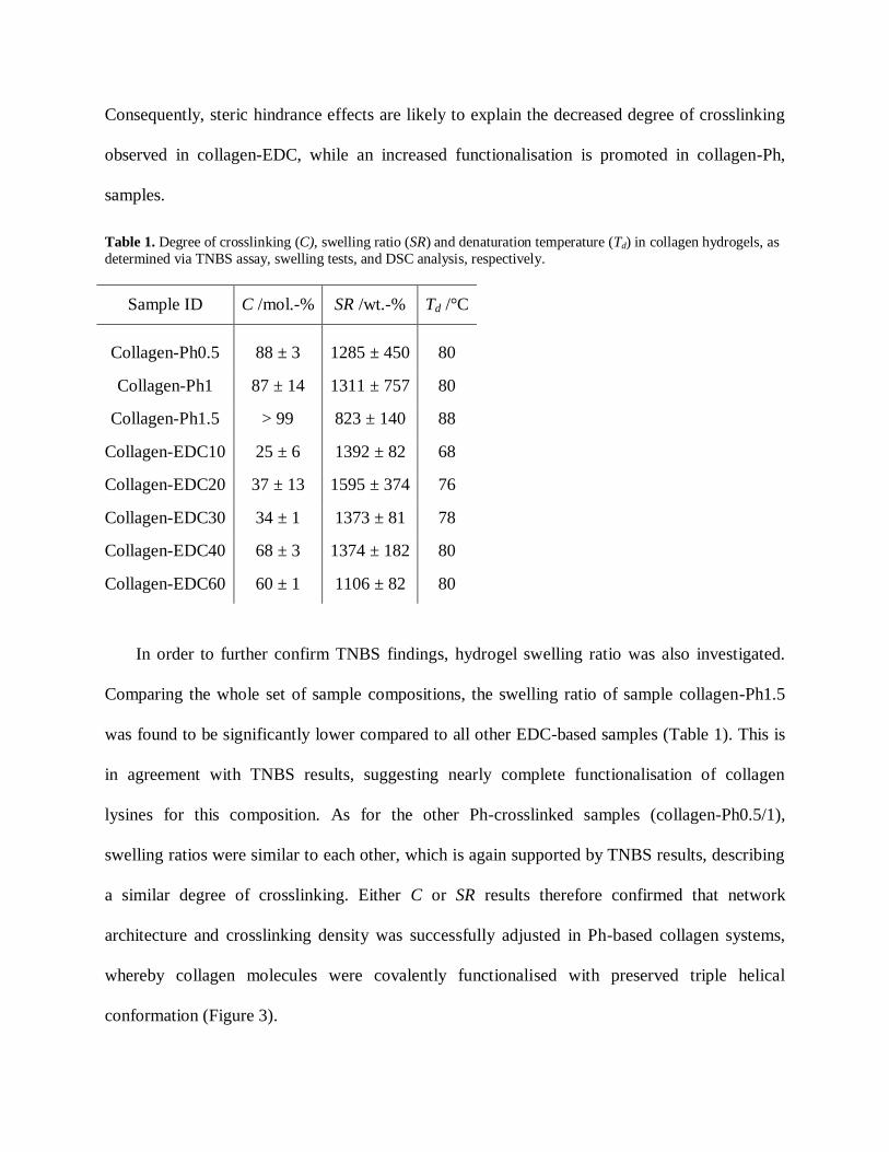
Consequently, steric hindrance effects are likely to explain the decreased degree of crosslinking
observed in collagen-EDC, while an increased functionalisation is promoted in collagen-Ph,
samples.
Table 1. Degree of crosslinking (C), swelling ratio (SR) and denaturation temperature (Td) in collagen hydrogels, as
determined via TNBS assay, swelling tests, and DSC analysis, respectively.
Sample ID
C /mol.-% SR /wt.-%
Td /°C
Collagen-Ph0.5
88 ± 3 1285 ± 450 80
Collagen-Ph1 87 ± 14 1311 ± 757 80
Collagen-Ph1.5 > 99 823 ± 140 88
Collagen-EDC10 25 ± 6 1392 ± 82 68
Collagen-EDC20 37 ± 13 1595 ± 374 76
Collagen-EDC30 34 ± 1 1373 ± 81 78
Collagen-EDC40 68 ± 3 1374 ± 182 80
Collagen-EDC60 60 ± 1 1106 ± 82 80
In order to further confirm TNBS findings, hydrogel swelling ratio was also investigated.
Comparing the whole set of sample compositions, the swelling ratio of sample collagen-Ph1.5
was found to be significantly lower compared to all other EDC-based samples (Table 1). This is
in agreement with TNBS results, suggesting nearly complete functionalisation of collagen
lysines for this composition. As for the other Ph-crosslinked samples (collagen-Ph0.5/1),
swelling ratios were similar to each other, which is again supported by TNBS results, describing
a similar degree of crosslinking. Either C or SR results therefore confirmed that network
architecture and crosslinking density was successfully adjusted in Ph-based collagen systems,
whereby collagen molecules were covalently functionalised with preserved triple helical
conformation (Figure 3).

Figure 3. Network architecture in triple-helical collagen systems. Collagen is Ph-functionalised in diluted acidic
conditions, resulting in intermolecular crosslinking of collagen triple helices. Formed hydrogels highlight preserved
triple helical organisation, while macroscopic properties can be controlled by variation in network architecture.
3.3. Thermo-mechanical properties of collagen hydrogels
Thermo-mechanical analysis was carried out on collagen hydrogels in order to investigate
whether variation of molecular parameters could induce changes in macroscopic properties.
Thus, DSC was performed aiming at investigating the thermal stability of formed hydrogels in
physiological conditions and combined with compression tests in order to study the material
mechanical behaviour in aqueous environment.
Thermal stability of collagen is dictated by its denaturation temperature (Td), which is
related to the unfolding of collagen triple helices into randomly-coiled chains; Td is therefore
expected to be highly affected by the presence of covalent net-points among collagen
molecules.9,22,25
Figure 4 (left) depicts exemplary thermograms of native, Ph- and EDC-
crosslinked type I collagens, all describing an endothermic thermal transition in the range of 67-
88 °C. In the case of native type I collagen, such endothermic transition was recorded at around
67 °C, which is in agreement with previous reports describing a thermal denaturation of collagen
at 55-67 °C (using similar heating rates),25,30
although decreased values (at ca. 34 °C) were also
observed at lowered heating rates (i.e. 0.1-2 °C∙min-1
heating rate).35
Considering the influence

of DSC heating rate on the peak position of collagen denaturation temperature (50100 °C at
0.55 °C∙min-1
),36
the endothermic transitions recorded in this study are likely to identify the
thermal denaturation of collagen. In order to elucidate this point further, ATR-FTIR was
recorded on a DSC-treated sample, aiming to confirm that the observed thermal transition was
not related to a decomposition process. As observed in Figure 4 (right), ATR-FTIR spectrum still
shows the main collagen peaks following DSC analysis, giving supporting evidence that no
decomposition process occurred in the sample. These investigations thus confirmed that the
obtained DSC thermograms effectively displayed an endothermic transition ascribed to collagen
denaturation. These findings provided further evidence of the preserved integrity of triple helices
in formed hydrogels, as already pointed out by ATR-FTIR (Figure 1) and WAXS (Figure 2).
Remarkably, the collagen denaturation temperature is shifted towards higher temperature in
crosslinked compared (68-88 °C) to native collagen (67 °C) samples (Table 1), whereby a slight
influence of DSC heating rate on Td was also observed (Figure 4, left). Considering previous
composition-dependent changes in network architecture, variation of Td seems to be directly
related to changes of crosslinking degree in the hydrogel networks. These results give supporting
evidence that covalent net-points were established during hydrogel formation, so that collagen
triple helices were successfully retained and stabilised. Remarkably, Ph-crosslinked collagen
proved to display higher denaturation temperatures with respect to EDC- (Td: 82 °C, C: 50 mol.-
%),25
glutaraldehyde- (Td: 78 °C, C: 51 mol.-%),21
and hexamethylene diisocyanate- (Td: 74 °C,
C: 48 mol.-%)23
crosslinked collagen materials. This indicates that the incorporation of Ph as a
stiff, aromatic segment superiorly stabilises collagen molecules in comparison with current
crosslinking methods.

Figure 4. Left: DSC thermograms of samples Collagen-Ph1.5 (—), Collagen-EDC60 (—) and type I collagen (—)
obtained with 10 °C·min-1 heating rate. An additional thermogram of sample Collagen-Ph1 was acquired with
1°C·min-1 heating rate (—). All samples describe a triple helix denaturation by an endothermic transition at 67-88
°C. Right: ATR-FTIR spectrum of sample Collagen-Ph1 following DSC analysis; the main peaks of collagen can
still be observed in the spectrum.
Besides thermal analysis, mechanical properties of collagen-Ph hydrogels were measured by
compression tests. Samples described J-shaped stress-compression curves (Figure 5), similar to
the case of native tissues.9 Here, shape recovery was observed following load removal up to
nearly 50% compression, suggesting that the established covalent network successfully resulted
in the formation of an elastic material, as observed in linear biopolymer networks.37
On the other
hand, EDC-crosslinked collagen showed minimal mechanical properties, whereby sample failure
was observed even after sample punching. Consequently, quantitative data of mechanical
properties on collagen-EDC samples could not be acquired. These findings support the idea that
the collagen functionalisation with Ph is effective for the formation of collagen materials with
enhanced mechanical properties.
Compressive modulus (E: 28±1035±9 kPa) and maximal stress of Ph-crosslinked samples
(σmax: 6±28±4 kPa) were measured in the kPa range, while compression at break (εb:
53±558±5 %) did not exceed 60% compression (Figure 6). The resulting compressive modulus

was therefore measured to be almost 20 times higher compared to previously-reported collagen-
based materials.22
Figure 5. Compressive stress-strain plot of hydrogel Collagen-Ph1 compressed up to sample break describing a
J-shaped curve, typical of native tissues. Shape recovery was observed following compression up to nearly 50%.
At the same time, there was little variation in mechanical properties among the different
compositions. Given the unique organisation of collagen, the hierarchical level, e.g. triple helix,
fibrils or fibres, at which covalent crosslinks are introduced, is crucial in order to study the
influence of crosslinking on the mechanical properties of collagen. Olde Damink et al. observed
no variation of mechanical properties in dermal sheep crosslinked collagen.20,21,23
This was
explained based on the fact that crosslinks were mainly introduced within rather than among
collagen molecules. This hypothesis may be supported by above mechanical findings, although it
is not in line with TNBS, swelling and thermal analysis data. Most likely, the variation of Ph
feed ratio among the different compositions (0.51.5 [COOH]/[Lys] ratio) was probably too
low to result in significant changes in mechanical properties. For these reasons, a wider range of
Ph concentrations may be advantageous in order to establish hydrogels with varied mechanical
properties. In that case, investigation via AFM will be instrumental in order to explore the
hierarchical levels (i.e. triple helices, fibrils or fibres) at which covalent crosslinks are

introduced; here, tapping mode12
could be employed in order to scan the material surface and
explore whether any collagen structural assembly is present following covalent crosslinking.
Subsequently, force-volume measurements could give information on the local elasticity of
resulting crosslinked patterns.
Figure 6. Compressive modulus (E), maximal compressive stress (σmax), and compression at break (εb) of
Ph-crosslinked collagen hydrogels.
3.4. Degradability and bioactivity in vitro
SBF incubation is a well-known method to test a material’s ability to form a
hydroxycarbonate apatite (HCA) layer in vitro.33
Collagen is known to trigger bone-like apatite
deposition in vivo during bone formation,11
so it was of interest to investigate collagen hydrogel
behaviour in SBF as an osteogenic-like medium. The mass change was therefore quantified and
the presence of calcium/phosphorous elements in retrieved samples assessed in order to (i)
clarify any occurrence of degradation and mineral deposition and (ii) determine the chemical
composition of any potentially nucleated phases. This investigation was then coupled to a
degradation study in PBS in order to investigate the stability of collagen hydrogels in a calcium-
free medium.

(a) (b)
(c) (d)
Figure 7. SEM on samples Collagen-Ph1 (a), Collagen-Ph1.5 (b), Collagen-EDC10 (c) and Collagen-EDC60 (d), following 1-week incubation in PBS (pH 7.4, 25 °C). Retrieved EDC-crosslinked samples present damaged surface,
providing supporting evidence of hydrolytic degradation taking place in these materials. The extent of surface
damage seems to be directly related to the degree of crosslinking. In contrast to that, Ph-crosslinked samples reveal
no detectable damage on their surfaces.
A slight mass decrease (averaged mass loss ~ 8 wt.-%) was observed in PBS-retrieved
collagen-Ph samples, indicating minimal hydrolytic degradation had occurred (Table 2), in
contrast to EDC-crosslinked samples (MR > 30 wt.-%). The latter high change in mass was also
reflected by SEM of partially-degraded EDC controls (Figure 7), whereby a crack-like formation
was observed following PBS incubation. In contrast to that, the collagen-Ph1.5 sample described
the lowest mass loss and a nearly-intact material microstructure, confirming that increased
Ph/Lys feed molar ratio led to networks with increased crosslinking degree (Table 1).
Interestingly, no specific fibrillar pattern was observed in resulting collagen samples. This likely
suggests that Ph-mediated covalent crosslinking hinders reconstitution of collagen fibres, so that
a triple helical architecture is expected in formed networks (Figure 3), as evidenced by ATR-
FTIR (Figure 1), WAXS (Figure 2) and DSC (Figure 4) analyses.
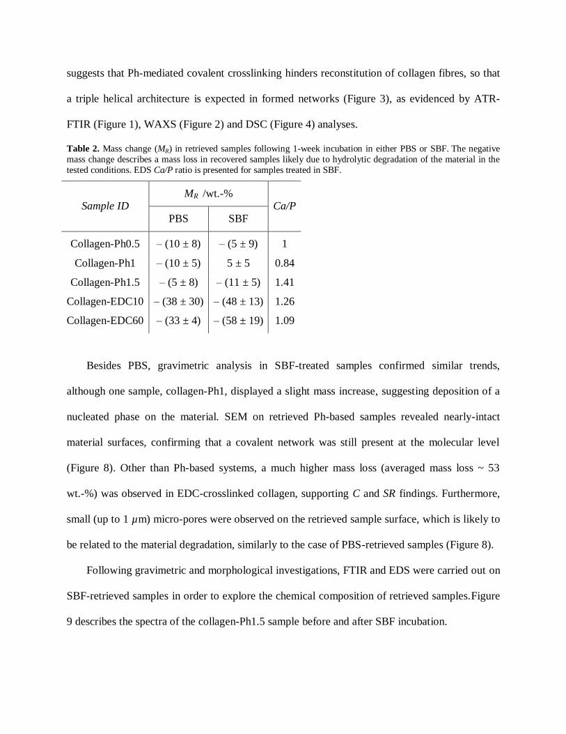
Table 2. Mass change (MR) in retrieved samples following 1-week incubation in either PBS or SBF. The negative
mass change describes a mass loss in recovered samples likely due to hydrolytic degradation of the material in the
tested conditions. EDS Ca/P ratio is presented for samples treated in SBF.
Sample ID MR /wt.-%
Ca/P
PBS SBF
Collagen-Ph0.5 – (10 ± 8) – (5 ± 9) 1
Collagen-Ph1 – (10 ± 5) 5 ± 5 0.84
Collagen-Ph1.5 – (5 ± 8) – (11 ± 5) 1.41
Collagen-EDC10 – (38 ± 30) – (48 ± 13) 1.26
Collagen-EDC60 – (33 ± 4) – (58 ± 19) 1.09
Besides PBS, gravimetric analysis in SBF-treated samples confirmed similar trends,
although one sample, collagen-Ph1, displayed a slight mass increase, suggesting deposition of a
nucleated phase on the material. SEM on retrieved Ph-based samples revealed nearly-intact
material surfaces, confirming that a covalent network was still present at the molecular level
(Figure 8). Other than Ph-based systems, a much higher mass loss (averaged mass loss ~ 53
wt.-%) was observed in EDC-crosslinked collagen, supporting C and SR findings. Furthermore,
small (up to 1 µm) micro-pores were observed on the retrieved sample surface, which is likely to
be related to the material degradation, similarly to the case of PBS-retrieved samples (Figure 8).
Following gravimetric and morphological investigations, FTIR and EDS were carried out on
SBF-retrieved samples in order to explore the chemical composition of retrieved samples.Figure
9 describes the spectra of the collagen-Ph1.5 sample before and after SBF incubation.

(a) (b)
(c) (d)
Figure 8. SEM on samples collagen-Ph1 and collagen-EDC10 prior to (a, b) and following (c, d) 1-week incubation
in 25 °C SBF, respectively. Minimal surface damage is observed in retrieved Ph-crosslinked samples (c), while the formation of pores is displayed in resulting EDC-crosslinked samples (d), likely ascribed to the hydrolytic
degradation of these materials.
Main amide bands are detected in both collagen spectra, proving that collagen composition was
not affected following in vitro bioactivity test. Here, additional bands at 1078, 1030 and 970 cm-1
were observed in spectra of SBF-incubated hydrogels. These bands are characteristic of
phosphate species associated with apatite.38
At the same time, a slight shift is described by the
Amide II band in FTIR spectrum of SBF-incubated sample in comparison to freshly-synthesised
hydrogel. Given that the Amide II band is located in the same region as the C=C band (1540
cm-1
) of Ph aromatic ring, this shift may suggest a specific interaction between the introduced Ph
segment and calcium species of nucleated phase.39
This hypothesis is also suggested by the shape
of this peak, which is broader compared to the same peak in a FTIR spectrum of a
freshly-synthesised sample. In order to further explore the role of the Ph aromatic ring in
templating calcium nucleation, the collagen-Ph1.5 sample was incubated in a simple calcium
containing solution (2.5 mM CaCl2). Interestingly, in the resulting ATR-FTIR spectrum on the
recovered sample (Figure 9), the 1540 cm-1
peak was absent, in contrast to native type I collagen
spectrum. This observation may give further evidence of an interaction between calcium species
present in solution and the aromatic Ph ring present in the collagen network. Although further
studies are needed to confirm this point, these results support the hypothesis that introduced
aromatic moieties may act as a preferential site of calcium phosphate nucleation.
Complementing FTIR, EDS analysis was carried out in order to further characterise the
chemical composition of hydrogel nucleated phase following SBF incubation. In doing this,
complete sample washing with water was crucial in order to remove superficial deposition of
magnesium, sodium and chlorine ions. The presence of calcium and phosphorous elements was
observed in all samples, although with a low Ca/P atomic ratio (Ca/P: 0.84-1.41, Table 2). This
suggests that the mineral phase laid down on the material surface was most likely constituted of
amorphous rather than crystalline calcium phosphate, which may be expected due to the
relatively short incubation (1 week) at 25 °C. The amorphous nature of the hydrogel nucleated
phase is also supported by the broad phosphate peaks in FTIR spectra of resulting samples
(Figure 9).

Figure 9. Exemplary ATR-FTIR spectra of sample Collagen-Ph1.5 directly after synthesis (—), following 2-day
incubation in 2.5 mM CaCl2 solution (—), and after 1-week incubation in SBF medium (—). SBF-retrieved samples
display additional peaks at 970 and 1030 cm-1, distinctive of phosphate phase deposition, while disappearance of the
peak related to aromatic bands (at 1540 cm-1) is observed after incubation in either CaCl2 or SBF solutions.
The collagen-Ph1.5 sample displayed the highest Ca/P atomic ratio (Ca/P ~ 1.41) compared to
all other samples. This finding may provide further evidence of a selective mechanism of apatite
nucleation in hydrogels crosslinked with increased Ph content, as suggested by FTIR (Figure 9).
Here, incorporated aromatic rings are expected to act as selective sites of calcium-phosphate
nucleation following chelation with calcium ions from SBF, similarly to the interaction
mechanism observed in cortical bone between calcium ions and glycosaminoglycan sulfate and
carboxylate groups.40
Although further investigations with longer time at body temperature are
required in order to elucidate the role of Ph in guiding matrix-nucleated phase interaction, this
study provides experimental evidence that these systems could promote calcium-phosphate

nucleation in vitro and therefore may be suitable as implants for the regeneration of mineralised
tissues.
3.5. Cytotoxicity study
Material cyto-compatibility was investigated by extract cytotoxicity assay following
European guidelines for medical device testing. Samples synthesized with the lowest and highest
Ph feed ratio were tested, in order to investigate whether any toxic compound could be released
from formed hydrogels to cell culture medium following sample incubation. Cell morphology
was monitored at 24 and 48 hours following cell culture in both sample compositions as well as
in positive (DMEM) and negative controls (DMSO); these observations were then
complemented with MTS assay quantitative data of cell viability after 48 hours. As pictured in
Figure 10, L929 mouse fibroblasts appeared confluent when cultured in either sample extracts or
cell culture medium, suggesting that sample extracts were well tolerated by cells. On the other
hand, cell death was observed in the case of cell culture in DMSO. These morphology
observations were in agreement with MTS assay results, describing the metabolic activity of vital
cultured cells (Figure 11). Interestingly, measured formazan content was higher in the case of
cells cultured in both samples extracts compared to the case of cell culture in DMEM and
DMSO.

(a) (b)
(c) (d)
(e) (f)
Figure 10. Cell morphology of L929 mouse fibroblasts cultured at different time points during 48-hour culture in
sample extracts, DMEM as positive control and DMSO as negative control. (a, c): morphology of cells cultured in Collagen-Ph0.5 following 24 (a) and 48 (c) hours cell culture; (b, d): morphology of cells cultured in
Collagen-Ph1.5 following 24 (b) and 48 (d) hours cell culture; (e, f): morphology of cells cultured in DMEM (e) and
DMSO (f) following 48 hours cell culture.
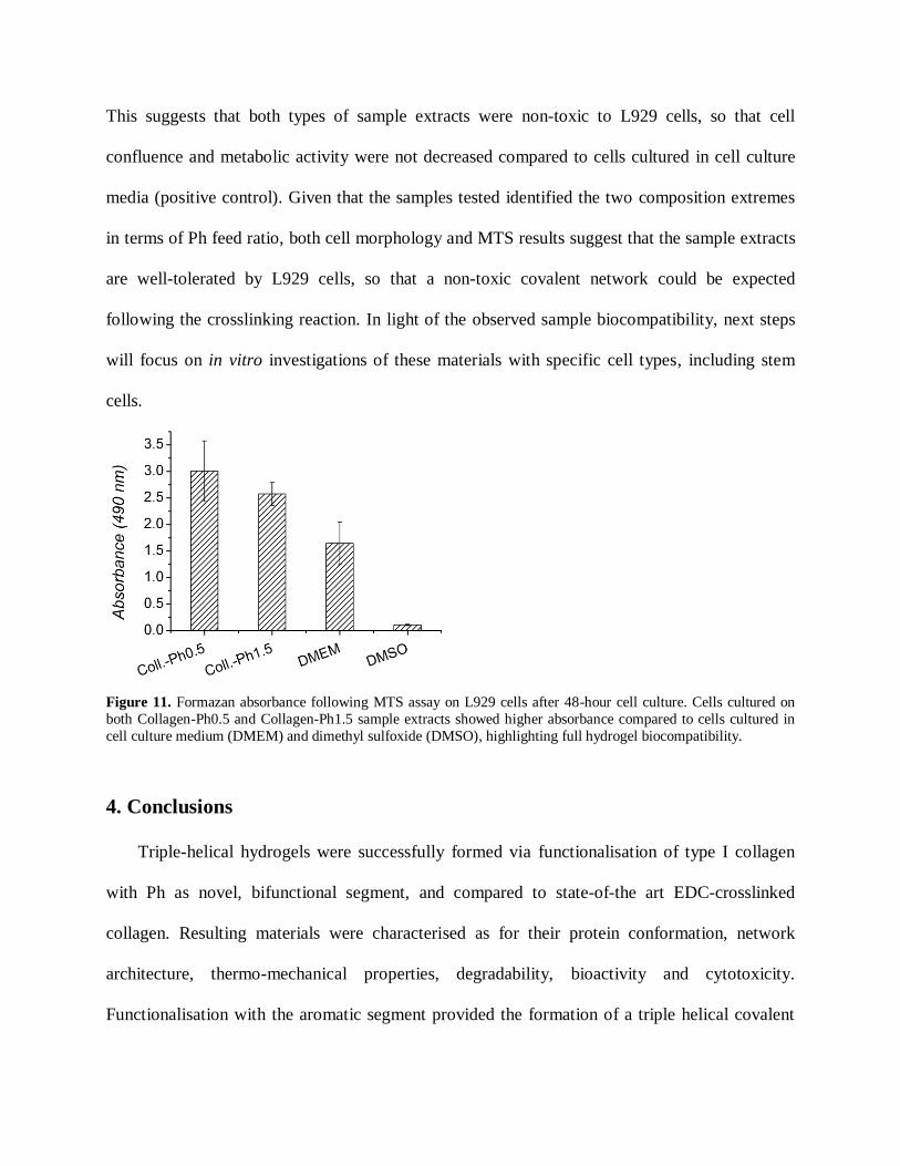
This suggests that both types of sample extracts were non-toxic to L929 cells, so that cell
confluence and metabolic activity were not decreased compared to cells cultured in cell culture
media (positive control). Given that the samples tested identified the two composition extremes
in terms of Ph feed ratio, both cell morphology and MTS results suggest that the sample extracts
are well-tolerated by L929 cells, so that a non-toxic covalent network could be expected
following the crosslinking reaction. In light of the observed sample biocompatibility, next steps
will focus on in vitro investigations of these materials with specific cell types, including stem
cells.
Figure 11. Formazan absorbance following MTS assay on L929 cells after 48-hour cell culture. Cells cultured on
both Collagen-Ph0.5 and Collagen-Ph1.5 sample extracts showed higher absorbance compared to cells cultured in
cell culture medium (DMEM) and dimethyl sulfoxide (DMSO), highlighting full hydrogel biocompatibility.
4. Conclusions
Triple-helical hydrogels were successfully formed via functionalisation of type I collagen
with Ph as novel, bifunctional segment, and compared to state-of-the art EDC-crosslinked
collagen. Resulting materials were characterised as for their protein conformation, network
architecture, thermo-mechanical properties, degradability, bioactivity and cytotoxicity.
Functionalisation with the aromatic segment provided the formation of a triple helical covalent

network with tunable crosslinking density and swelling ratio, whereby enhanced macroscopic
properties were obtained compared to systems with EDC-mediated intramolecular crosslinks.
Consequently, resulting Ph-based systems displayed only minimal mass loss following 1-week
incubation in either PBS or SBF, whilst nucleation of an amorphous calcium phosphate phase
was initiated in retrieved samples. The presence of the Ph aromatic ring in resulting networks
proved is likely to promote an interaction with calcium species present in solution, so that a
specific site for calcium phosphate nucleation may be expected. L929 cell culture revealed no
sign of cytotoxicity following 48-hour incubation in sample extract, whereby cell confluence was
observed as in the case of culture in cell culture medium. Furthermore, the metabolic activity of
cells cultured in the sample extracts was not decreased with respect to the one observed
following cell culture in DMEM. Particularly based on their bioactivity and full biocompatibility,
these materials have significant appeal for applications in mineralised tissue regeneration.
Acknowledgements
This work was funded through WELMEC, a Centre of Excellence in Medical
Engineering funded by the Wellcome Trust and EPSRC, under grant number WT
088908/Z/09/Z. The authors would like to thank Dr. P. Hine, J. Hudson, W. Vickers and S.
Finlay, for kind assistance with WAXS, SEM/EDS, SBF preparation, and compression tests,
respectively.
References
1 T.-P. Nguyen and B.-T. Lee, J. Biomater. Appl. 2011, 27, 311-321.
2 Z. You and Y. A. Wang, Adv. Funct. Mater. 2012, 22, 2812-2820.
3 U. Freudenberg, A. Hermann, P. B. Welzel, K. Stirl, S. C. Schwarz, M. Grimmer, A. Zieris, W.
Panyanuwat, S. Zschoche, D. Meinhold, A. Storch and C. A. Werner, Biomaterials 2009, 30,
5049-5060.
4 A. Bigi, E. Boanini, S. Panzavolta and N. Roveri. Biomacromol. 2000, 1, 752-756.
5 A. T. Neffe, G. Tronci, A. Alteheld and A. Lendlein, Macromol. Chem. Phys. 2010, 211,
182-194.
6 T. G. Kim, H. Shin and D. W. Lim. Adv. Funct. Mater. 2012, 22, 2446-2468.
7 M.M. Giraud-Guille, C. Helary, S. Vigier and N. Nassif, Soft Matter 2010, 6, 4963-4967.
8 T. Huynh, G. Abraham, J. Murray, K. Brockbank, P.-O. Hagen and S. Sullivan, Nat.
Biotechnol. 1999, 17, 1083-1086.
9 L. Meng, O. Arnoult, M. Smith and G. E. Wnek, J. Mater. Chem. 2012, 22, 19412-19417.
10 N. Davidenko, T. Gibb, C. Schuster, S. M. Best, J. J. Campbell, C. J. Watson and R. E.
Cameron, Acta Biomater. 2012, 8, 667-676.
11 S.-J. Hong, H.-S. Yu, K.-T. Noh, S.-A. Oh and H.-W. Kim, J. Biomed. Appl. 2010, 24,
733-750.
12 C. A. Grant, D. J. Brockwell, S. E. Radford, and N. H. Thomson, Biophys. J. 2009, 97,
2985-2992.
13 Y. Wang, T. Azaïs, M. Robin, A. Vallée, C. Catania, P. Legriel, G. Pehau-Arnaudet, F.
Babonneau, M.-M. Giraud-Guille and N. Nassif, Nat. Mater. 2012, 11, 724-733.
14 M. Agostini de Moraes, E. Paternotte, D. Mantovani and M. Masumi Beppu, Macromol.
Biosci. 2012, 12, 1253-1264.
15 L.E.R. O’Leary, J.A. Fallas, E.L. Bakota, M.K. Kang, J.D. Hartgerink, Nature Chemistry
2011, 3, 821-828.

16
K. Nam ,Y. Sakai, Y. Hashimoto, T. Kimura, A. Kishida, Soft Matter 2012, 8, 472-480.
17 Y. Liu, D. Luo, X.-X. Kou, X.-D. Wang, F.R. Tay, Y.-L. Sha, Y.-H. Gan, Y.-H. Zhou, Adv.
Funct. Mater. 2013, 23, 1404-1411.
18 B. Antebi, X. Cheng, J.N. Harris, L.B. Gower, X.-D. Chen, J. Ling, Tissue Eng. Part C 2013,
19, 487-496.
19 S. Yunoki, T. Matsuda, Biomacromol. 2008, 9, 879-885.
20 L. H. H. Olde Damink, P. J. Dijkstra, M. J. A. van Luyn, P. B. van Wachem, P. Nieuwenhuis,
J. Feijen, Biomaterials 1996, 17, 765-773.
21 L. H. H. Olde Damink, P. J. Dijkstra, M. J. A. van Luyn, P. B. van Wachem, P. Nieuwenhuis,
J. Feijen, J. Mater. Sci. Mater. Med. 1995, 6, 460-472.
22 M. J. Haugh, C. M. Murphy, R. C. McKiernan, C. Altenbuchner, F. J. O’Brien, Tissue Eng A
Part A 2011, 17, 1201-1208.
23 L. H. H. Olde Damink, P. J. Dijkstra, M. J. A. Van Luyn, P. B. van Wachem, P. Nieuwenhuis,
J. Feijen, J. Mater. Sci. Mater. Med. 1995, 6, 429-434.
24 J. M. Orban, L. B. Wilson, J. A. Kofroth, M. S. El-Kurdi, T. M. Maul, D. A. Vorp, J Biomed
Mater Res Part A 2004, 68A, 756-762.
25 R. S. Walton, D. D. Brand, J. T. Czernuszka, J. Mater. Sci. Mater. Med. 2010, 21, 451-461.
26 V. Charulatha and A. Rajaram, Biomaterials 2003, 24, 759-767.
27 K. S. Weadock, E. J. Miller, L. D. Bellincampi, J. P. Zawadsky, M. G. Dunn, J. Biomed.
Mater. Res. 1995, 29, 1373-1379.
28 S. Chen, A. Osaka, T. Ikoma, H. Morita, J. Li, M. Takeguchi, N. Hanagata, J. Mater. Chem.
2011, 21, 10942-10948.
29 E. Bell, B. Ivarsson, C. Merrill, Proc. Natl. Acad. Sci. USA 1979, 76, 1274-1278.
30
G. Tronci, S. J. Russell, D. J. Wood, J. Mater. Chem. B 2013, 1, 3705-3715.
31 G. Tronci, A. Doyle, S. J. Russell, D. J. Wood, Mater. Res. Soc. Symp. Proc. 2013,
1498,145-150.
32 W. A. Bubnis, C. M. Ofner, Analyt. Biochem. 1992, 207, 129-133.
33 S. K. Misra, T. Ansari, D. Mohn, S. P. Valappil, T. J. Brunner, W. J. Stark, I. Roy, J. C.
Knowles, P. D. Sibbons, E. V. Jones, A. R. Boccaccini, V. Salih, J. R. Soc. Interf. 2010, 7,
453-465.
34 L. He, C. Mu, J. Shi, Q. Zhang, B. Shi, W. Lin, Int. J. Biol. Macromol. 2011, 48, 354-359.
35 E. Leikina, M. V. Mertts, N. Kuznetsova, S. Leikin, Proc. Natl. Acad. Sci. USA 2002, 99,
1314-1318.
36 S. Skrzyński, A. Sionkowska, A. Marciniak, Journal of Biophysics 2009, 2009,
doi:10.1155/2009/819635
37 G. Tronci, A. T. Neffe, B. F. Pierce, A. Lendlein, J. Mater. Chem. 2010, 20, 8875-8884.
38 B. Marelli, C. E. Ghezzi, A. Alessandrino, J. E. Barralet, G. Freddi, S. N. Nazhat, Biomaterials
2012, 33, 102-108.
39 S. Teng, J. Shi, B. Peng, L. Chen, Comp. Sci. Tech., 2006, 66, 1532-1538.
40 J. Song, E. Saiz, C. R. Bertozzi, J. Am. Chem. Soc., 2003, 125, 1236-1243.