Hybrid hydrogels containing vertically aligned carbon ... hydrogels containing vertically... ·...
11
Hybrid hydrogels containing vertically aligned carbon nanotubes with anisotropic electrical conductivity for muscle myofiber fabrication Samad Ahadian 1 *, Javier Ramo ´n-Azco ´n 1 *, Mehdi Estili 2 , Xiaobin Liang 1 , Serge Ostrovidov 1 , Hitoshi Shiku 3 , Murugan Ramalingam 1,4,5 , Ken Nakajima 1 , Yoshio Sakka 6 , Hojae Bae 7 , Tomokazu Matsue 1,3 & Ali Khademhosseini 1,8,9,10,11 1 WPI-Advanced Institute for Materials Research, Tohoku University, Sendai 980-8577, Japan, 2 International Center for Young Scientists (ICYS), National Institute for Materials Science (NIMS), Tsukuba 305-0047, Japan, 3 Graduate School of Environmental Studies, Tohoku University, Sendai 980-8579, Japan, 4 Centre for Stem Cell Research, A unit of the Institute for Stem Cell Biology and Regenerative Medicine, ChristianMedical College Campus, Vellore 632002, India, 5 Institut National de la Sante ´ Et de la Recherche Me ´dicale U977, Faculte ´ de Chirurgie Dentaire, Universite ´ de Strasbourg, Strasbourg 67085, France, 6 Materials Processing Unit, National Institute for Materials Science (NIMS), Tsukuba 305-0047, Japan, 7 College of Animal Bioscience and Technology, Department of Bioindustrial Technologies, Konkuk University, Hwayang-dong, Kwangjin-gu, Seoul 143-701, Republic of Korea, 8 Department of Medicine, Center for Biomedical Engineering, Brigham and Women’s Hospital, Harvard Medical School, Cambridge, Massachusetts 02139, USA, 9 Harvard-MIT Division of Health Sciences and Technology, Massachusetts Institute of Technology, Cambridge, Massachusetts 02139, USA , 10 Wyss Institute for Biologically Inspired Engineering, Harvard University, Boston, Massachusetts 02115, USA , 11 Department of Maxillofacial Biomedical Engineering and Institute of Oral Biology, School of Dentistry, Kyung Hee University, Seoul 130-701, Republic of Korea. Biological scaffolds with tunable electrical and mechanical properties are of great interest in many different fields, such as regenerative medicine, biorobotics, and biosensing. In this study, dielectrophoresis (DEP) was used to vertically align carbon nanotubes (CNTs) within methacrylated gelatin (GelMA) hydrogels in a robust, simple, and rapid manner. GelMA-aligned CNT hydrogels showed anisotropic electrical conductivity and superior mechanical properties compared with pristine GelMA hydrogels and GelMA hydrogels containing randomly distributed CNTs. Skeletal muscle cells grown on vertically aligned CNTs in GelMA hydrogels yielded a higher number of functional myofibers than cells that were cultured on hydrogels with randomly distributed CNTs and horizontally aligned CNTs, as confirmed by the expression of myogenic genes and proteins. In addition, the myogenic gene and protein expression increased more profoundly after applying electrical stimulation along the direction of the aligned CNTs due to the anisotropic conductivity of the hybrid GelMA-vertically aligned CNT hydrogels. We believe that platform could attract great attention in other biomedical applications, such as biosensing, bioelectronics, and creating functional biomedical devices. H ydrogels are commonly used as biological scaffolds because of their high water content, biocompatibility, and biodegradability 1,2 . However, they are typically mechanically weak and electrically non-conductive, limiting their applications in regulating the behavior of electro-active cells, such as skeletal muscle, cardiac, and neural cells 3 . Therefore, controlling the mechanical and electrical properties of hydrogels is desirable to facilitate the regulation of cell behavior. In particular, anisotropically conductive hydrogels are highly beneficial for the fabrication of functional tissue constructs with the aid of electrical stimulation (ES). Hydrogels with high electrical conductivity and robust mechanical properties have other important applications, for example, in biosensing 4,5 , in the development of hybrid three-dimensional (3D) electronics-tissue materials 6,7 , and as bioactuators 8 . Nanomaterials have been widely used to improve the electrical conductivity and mechanical properties of hydrogels 9 . For example, gold nanostructures have been successfully used to increase the electrical conductivity of alginate hydrogels 3,10 . The resulting hydrogels exhibited higher performance to electrically stimulate cardiomyo- OPEN SUBJECT AREAS: BIOMEDICAL ENGINEERING TISSUE ENGINEERING Received 9 September 2013 Accepted 13 February 2014 Published 19 March 2014 Correspondence and requests for materials should be addressed to T.M. (matsue@bioinfo. che.tohoku.ac.jp) or A.K. ([email protected]. harvard.edu) * These authors contributed equally to this work. SCIENTIFIC REPORTS | 4 : 4271 | DOI: 10.1038/srep04271 1
Transcript of Hybrid hydrogels containing vertically aligned carbon ... hydrogels containing vertically... ·...
Hybrid hydrogels containing verticallyaligned carbon nanotubes withanisotropic electrical conductivity formuscle myofiber fabricationSamad Ahadian1*, Javier Ramon-Azcon1*, Mehdi Estili2, Xiaobin Liang1, Serge Ostrovidov1,Hitoshi Shiku3, Murugan Ramalingam1,4,5, Ken Nakajima1, Yoshio Sakka6, Hojae Bae7,Tomokazu Matsue1,3 & Ali Khademhosseini1,8,9,10,11
1WPI-Advanced Institute for Materials Research, Tohoku University, Sendai 980-8577, Japan, 2International Center for YoungScientists (ICYS), National Institute for Materials Science (NIMS), Tsukuba 305-0047, Japan, 3Graduate School of EnvironmentalStudies, Tohoku University, Sendai 980-8579, Japan, 4Centre for Stem Cell Research, A unit of the Institute for Stem Cell Biology andRegenerative Medicine, ChristianMedical College Campus, Vellore 632002, India, 5Institut National de la Sante Et de la RechercheMedicale U977, Faculte de Chirurgie Dentaire, Universite de Strasbourg, Strasbourg 67085, France, 6Materials Processing Unit,National Institute for Materials Science (NIMS), Tsukuba 305-0047, Japan, 7College of Animal Bioscience and Technology,Department of Bioindustrial Technologies, Konkuk University, Hwayang-dong, Kwangjin-gu, Seoul 143-701, Republic of Korea,8Department of Medicine, Center for Biomedical Engineering, Brigham and Women’s Hospital, Harvard Medical School,Cambridge, Massachusetts 02139, USA, 9Harvard-MIT Division of Health Sciences and Technology, Massachusetts Institute ofTechnology, Cambridge, Massachusetts 02139, USA , 10Wyss Institute for Biologically Inspired Engineering, Harvard University,Boston, Massachusetts 02115, USA , 11Department of Maxillofacial Biomedical Engineering and Institute of Oral Biology, School ofDentistry, Kyung Hee University, Seoul 130-701, Republic of Korea.
Biological scaffolds with tunable electrical and mechanical properties are of great interest in many differentfields, such as regenerative medicine, biorobotics, and biosensing. In this study, dielectrophoresis (DEP) wasused to vertically align carbon nanotubes (CNTs) within methacrylated gelatin (GelMA) hydrogels in arobust, simple, and rapid manner. GelMA-aligned CNT hydrogels showed anisotropic electricalconductivity and superior mechanical properties compared with pristine GelMA hydrogels and GelMAhydrogels containing randomly distributed CNTs. Skeletal muscle cells grown on vertically aligned CNTs inGelMA hydrogels yielded a higher number of functional myofibers than cells that were cultured onhydrogels with randomly distributed CNTs and horizontally aligned CNTs, as confirmed by the expressionof myogenic genes and proteins. In addition, the myogenic gene and protein expression increased moreprofoundly after applying electrical stimulation along the direction of the aligned CNTs due to theanisotropic conductivity of the hybrid GelMA-vertically aligned CNT hydrogels. We believe that platformcould attract great attention in other biomedical applications, such as biosensing, bioelectronics, andcreating functional biomedical devices.
Hydrogels are commonly used as biological scaffolds because of their high water content, biocompatibility,and biodegradability1,2. However, they are typically mechanically weak and electrically non-conductive,limiting their applications in regulating the behavior of electro-active cells, such as skeletal muscle,
cardiac, and neural cells3. Therefore, controlling the mechanical and electrical properties of hydrogels is desirableto facilitate the regulation of cell behavior. In particular, anisotropically conductive hydrogels are highly beneficialfor the fabrication of functional tissue constructs with the aid of electrical stimulation (ES). Hydrogels with highelectrical conductivity and robust mechanical properties have other important applications, for example, inbiosensing4,5, in the development of hybrid three-dimensional (3D) electronics-tissue materials6,7, and asbioactuators8.
Nanomaterials have been widely used to improve the electrical conductivity and mechanical properties ofhydrogels9. For example, gold nanostructures have been successfully used to increase the electrical conductivity ofalginate hydrogels3,10. The resulting hydrogels exhibited higher performance to electrically stimulate cardiomyo-
OPEN
SUBJECT AREAS:BIOMEDICAL
ENGINEERING
TISSUE ENGINEERING
Received9 September 2013
Accepted13 February 2014
Published19 March 2014
Correspondence andrequests for materials
should be addressed toT.M. (matsue@bioinfo.
che.tohoku.ac.jp) orA.K. ([email protected].
harvard.edu)
* These authorscontributed equally to
this work.
SCIENTIFIC REPORTS | 4 : 4271 | DOI: 10.1038/srep04271 1
cytes and neural cells compared with pristine hydrogels. We recentlyreported the use of carbon nanotubes (CNTs) to effectively reinforcemethacrylated gelatin (GelMA) hydrogels and to generate 3D hybridGelMA-CNT hydrogels11. GelMA is a photopolymerizable hydrogelcomposed of natural gelatin functionalized with methacrylic anhyd-ride12. The formation of nanofiber web-like structures of CNTswithin GelMA hydrogels resulted in hybrid GelMA-CNT hydrogelswith enhanced mechanical properties compared with the pureGelMA hydrogels11. Despite the unique electrical properties ofCNTs, few published studies have reported the tuning of electricalproperties of hydrogels using CNTs. Most of the unique properties ofCNTs are exerted in the direction of the tube axis13. Therefore, theanisotropic electrical and mechanical properties of hybrid hydrogel-CNT systems can be controlled by aligning the CNTs within thehydrogels along their tube axes. Based on this, we propose the useof dielectrophoresis (DEP) as an efficient tool to vertically alignCNTs within GelMA hydrogels. DEP is a powerful technique tomanipulate particles in a medium based on their responses to anAC electric field, which induces a charge polarization within theparticles and the surrounding medium14,15. Due to its low ion con-centration and viscosity, GelMA hydrogels provide a suitable milieufor the dielectrophoretic alignment of CNTs. In our previous work,CNTs were horizontally micropatterned inside GelMA hydrogelsusing DEP16. It was shown that horizontally aligned CNTs withinhybrid GelMA hydrogels were aligned through their axial directiondue to DEP forces and formed cylindrically long CNT bundleswithin GelMA hydrogels. Therefore, cells seeded on these hydrogelscould only attach to and sense CNTs in their radial direction16. Inother words, the cells could sense the sides of the CNTs but not thehead or tail end groups, which is somehow limiting as CNTs exhibittheir unique mechanical and electrical properties in their axial dir-ection17. In addition, it was not possible to provide local ES for cellsseeded on GelMA gels containing horizontally aligned CNTs.
Finally, the interconnected network of horizontally aligned CNTsinside GelMA hydrogels generated a situation in which the electricalcurrent of the underlying electrodes could ‘short circuit’ to a neigh-boring electrode without approaching cells that were seeded on topof the hydrogels16. To address these limitations, we changed thedirection of the alignment of CNTs within GelMA hydrogels tovertical.
In this paper, we used DEP to vertically align CNTs within GelMAhydrogels. The anisotropic electrical conductivity and mechanicalfeatures of the hybrid GelMA hydrogels containing vertically alignedCNTs were evaluated and compared with those of pristine GelMA,hybrid GelMA-randomly dispersed CNT, and GelMA-horizontallyaligned CNT hydrogels. To demonstrate the utility of these novelhydrogels, the GelMA-vertically aligned CNT, GelMA-randomCNT, and GelMA-horizontally aligned CNT hydrogels were usedto fabricate muscle myofibers, which were then characterized interms of the expression of myogenic genes and proteins, particularlyafter applying ES along the direction of the aligned CNTs.
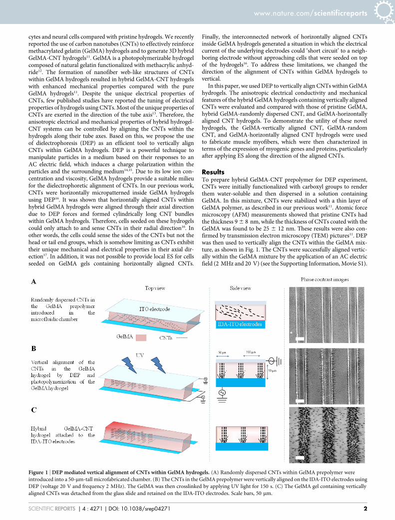
ResultsTo prepare hybrid GelMA-CNT prepolymer for DEP experiment,CNTs were initially functionalized with carboxyl groups to renderthem water-soluble and then dispersed in a solution containingGelMA. In this mixture, CNTs were stabilized with a thin layer ofGelMA polymer, as described in our previous work11. Atomic forcemicroscopy (AFM) measurements showed that pristine CNTs hadthe thickness 9 6 8 nm, while the thickness of CNTs coated with theGelMA was found to be 25 6 12 nm. These results were also con-firmed by transmission electron microscopy (TEM) pictures11. DEPwas then used to vertically align the CNTs within the GelMA mix-ture, as shown in Fig. 1. The CNTs were successfully aligned vertic-ally within the GelMA mixture by the application of an AC electricfield (2 MHz and 20 V) (see the Supporting Information, Movie S1).
Figure 1 | DEP mediated vertical alignment of CNTs within GelMA hydrogels. (A) Randomly dispersed CNTs within GelMA prepolymer were
introduced into a 50-mm-tall microfabricated chamber. (B) The CNTs in the GelMA prepolymer were vertically aligned on the IDA-ITO electrodes using
DEP (voltage 20 V and frequency 2 MHz). The GelMA was then crosslinked by applying UV light for 150 s. (C) The GelMA gel containing vertically
aligned CNTs was detached from the glass slide and retained on the IDA-ITO electrodes. Scale bars, 50 mm.
www.nature.com/scientificreports
SCIENTIFIC REPORTS | 4 : 4271 | DOI: 10.1038/srep04271 2
The alignment of CNTs using the DEP approach was rapid, requiringless than one minute. The AC electric fields appeared to inducedipole moments within the CNTs and forced them to align in thedirection of the electric field.
The proposed approach to align CNTs within hydrogels is versat-ile and can create any desired arrangement of micropatterned CNTswithin hydrogels. Any desired micropatterned CNTs in hydrogelscan be created by using one corresponding micropatterned electrodein the setup of DEP system. In this paper, CNTs within GelMAhydrogels were also vertically aligned between a top ITO electrodeand a bottom interdigitated array of ITO (IDA-ITO) electrodes asshown in Fig. 1 (Supporting Information, Movie S2). Therefore, theCNTs were vertically aligned on the micropatterned ITO electrodes(i.e., IDA-ITO electrodes). The DEP forces disappear when the AC
electric field is switched off. The Supporting Information (Movies S3and S4) demonstrates how dielectrophoretically aligned CNTswithin GelMA hydrogels lose their alignment as the DEP forcesdisappear. Therefore, a stabilization step is required after applyingthe DEP forces to preserve the alignment of the CNTs in the absenceof the AC electric field. Here, the immobilization step was performedin a rapid and non-destructive manner by crosslinking the GelMAhydrogel with UV light (Fig. 1).
We prepared pristine GelMA hydrogels and hybrid GelMAhydrogels containing vertically aligned, random, and horizontallyaligned CNTs with CNT concentrations of 0.5 and 1 mg/mL. Theelectrical conductivities of these hydrogels were then measuredthrough ITO electrodes to obtain accurate and reproducible conduc-tivity values. As shown in Figs. 2 and S1, the DC electrical conduc-
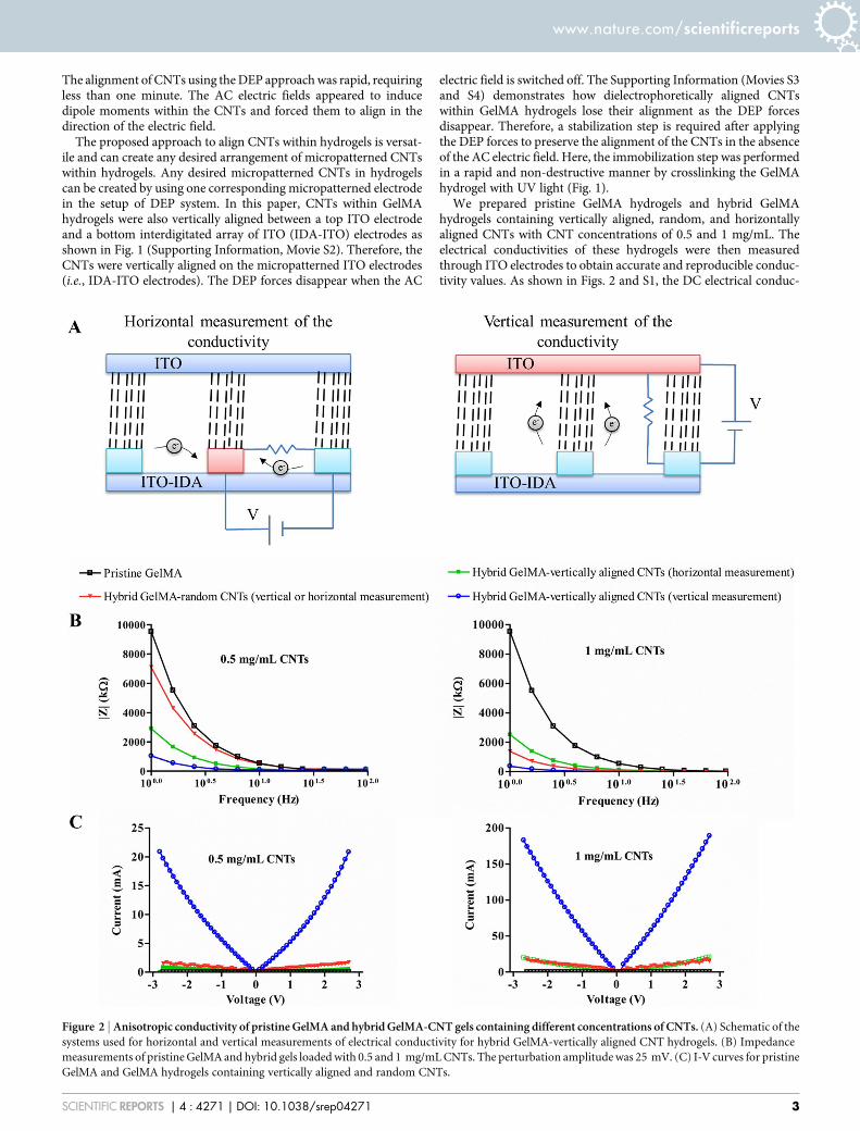
Figure 2 | Anisotropic conductivity of pristine GelMA and hybrid GelMA-CNT gels containing different concentrations of CNTs. (A) Schematic of the
systems used for horizontal and vertical measurements of electrical conductivity for hybrid GelMA-vertically aligned CNT hydrogels. (B) Impedance
measurements of pristine GelMA and hybrid gels loaded with 0.5 and 1 mg/mL CNTs. The perturbation amplitude was 25 mV. (C) I-V curves for pristine
GelMA and GelMA hydrogels containing vertically aligned and random CNTs.
www.nature.com/scientificreports
SCIENTIFIC REPORTS | 4 : 4271 | DOI: 10.1038/srep04271 3
tivity dramatically increased as a function of the CNT concentration.Most importantly, the conductivity of hybrid GelMA-aligned CNThydrogels for both vertical and horizontal CNT alignment along withCNT alignment was higher than that perpendicular to CNT align-ment. For instance, when we applied 2.7 V to the hybrid GelMA-0.5 mg/mL vertically aligned CNT hydrogel, the measured currentswere 20.86 and 0.53 mA along with CNT alignment and perpendic-ular to CNT alignment, respectively. However, the hybrid GelMA-random CNT hydrogels exhibited no anisotropic conductivity. Theconcept of anisotropic conductivity here refers to a significantlyhigher electrical conductivity of underlying hydrogels either in thevertical or horizontal measurement of conductivity as schematized inFigs. 2-A and S1-A. The anisotropic conductivity of the hybridGelMA-aligned CNT hydrogels is primarily due to the generationof an interlinked network of CNTs that enables the propagation ofthe electrical current. We observed a similar trend in the impedanceof the hydrogels (Figs. 2 and S1). For example, the measured impe-dances of the hybrid GelMA-1 mg/mL vertically aligned CNThydrogel at the frequency 6.31 Hz were 2096.84 and 22654.64 kValong with CNT alignment and perpendicular to CNT alignment,respectively. Impedance spectra were acquired for GelMA andhybrid GelMA-CNT hydrogels over a frequency range from 1 to100 Hz with a perturbation amplitude of 25 mV.
We also used a micromechanical mapping approach to measure themechanical properties of GelMA and nanostructured hybrid GelMA-CNT gels. As shown in Fig. 3, the Young’s modulus of the pristineGelMA was 12.5 6 0.1 kPa, consistent with our previously reportedvalue derived from conventional stress-strain measurements12. AsCNTs were added to the GelMA hydrogel, the Young’s modulus, asmeasured by nanoindentation method applied vertically to the hydro-gel, increased (Fig. 3-D). However, this increase was more profoundfor the hybrid GelMA-vertically aligned CNT hydrogels than for thehybrid GelMA-random CNT gels. The Young’s modulus values mea-sured for the hybrid GelMA-0.1 mg/mL aligned CNT gel and theGelMA-0.1 mg/mL random CNT gel were 50.4 6 3.8 and 20.3 6
1.4 kPa, respectively. The increase in the Young’s modulus of thehybrid GelMA-CNT hydrogels compared with the pristine GelMAhydrogel was due to the reinforcing effect of the CNTs within thehydrogels. In this evaluation, the AFM tip vertically tapped the sam-ples along the direction of the aligned CNTs. Therefore, there was anincrease in the Young’s modulus of the hybrid GelMA-aligned CNTgels relative to that of the hybrid GelMA-random CNT gels. Thisresult is consistent with a previous report that CNTs exhibit theirunique mechanical properties in their axial direction17.
The Young’s modulus of GelMA-horizontally aligned CNT wasmeasured to be 23.4 6 1.7 kPa16, which is lower than that of GelMA-vertically aligned CNTs at the same CNT concentration (0.3 mg/mL)(48.5 6 2.3 kPa). This result is in agreement with the previous workthat CNTs show their high and unique mechanical properties in theiraxial direction rather than radial direction17. As a result, there is noneed to increase the mechanical properties of hybrid GelMA-CNThydrogels by increasing the CNT concentration as this can beachieved by vertically aligning CNTs inside GelMA-CNT hydrogelsusing the DEP technique. Minimizing the use of CNTs in hybridhydrogel-CNT materials is always beneficial as to preserve thedesired biological properties of the hydrogel materials for biomedicalapplications.
Interestingly, the Young’s modulus of the hybrid GelMA-alignedCNT gels at a CNT concentration of 1 mg/mL was lower than that ofhybrid GelMA-aligned CNT gels made using lower CNT concentra-tions. This may be due to higher UV absorption at higher concentra-tions, which resulted in a lower degree of crosslinking and as a resultwere softer. In agreement with this result, we recently demonstratedthat the UV absorption of hybrid GelMA-CNT gels is increased uponincreasing the CNT concentration11. The force-deformation curvesare also shown (first images from the right: Fig. 3-A, 3-B, and 3-C),
taking into consideration one representative point of the correspond-ing Young’s modulus maps. Note that the direction of hydrogeldeformation was vertical to the gels. The Young’s modulus mapswere calculated by the Derjaguin, Muller, and Toporov (DMT) fit-ting model18 using the force-deformation plots.
We further evaluated the performance of the hybrid GelMA-ver-tically aligned CNT, GelMA-random CNT, and GelMA-horizontallyaligned CNT hydrogels with a concentration of 0.3 mg/mL in thefabrication of C2C12 muscle myofibers. We previously showed thatCNTs had no detectable adverse effect on the viability of C2C12myoblasts, and more than 95% of myoblasts remained viable on bothpristine GelMA and hybrid GelMA-CNT hydrogels after 3 days ofculture. In addition, the level of muscle cell proliferation was similarin both hydrogel systems16. Here, C2C12 myoblasts were randomlycultured on hybrid GelMA-vertically aligned CNT, GelMA-randomCNT, and GelMA-horizontally aligned CNT hydrogels. The reasonfor this cell culture method was to provide a suitable substrate for thecells to sense the vertically aligned CNTs in their axial direction.Therefore, there was no preferential direction for the cell alignment.After 1 day of culture, myoblast differentiation was induced byswitching to a differentiation medium. During the differentiationperiod, the cells expressed myosin heavy chain protein and fusedtogether to form C2C12 myotubes19. The muscle cells were subjectedto ES (voltage, 8 V; frequency, 1 Hz; duration, 10 ms) for 2 continu-ous days beginning on day 8 of culture. C2C12 myotubes cultured inthe absence of ES were used as control samples. It is well known thatthe calcineurin pathway is responsible for tonic and low frequencynerve stimulation in body leading to fast-to-slow muscle myofibertransformation20. Calcineurin is a Ca21/calmodulin-dependent pro-tein phosphatase that regulates nuclear translocation of the NFAT(nuclear factor of activated T cells) transcription factor and activa-tion of target genes. The employed ES protocol here mimics tonic andlow frequency nerve stimulation for native muscles in body21. As aresult of such stimulation, anabolic processes occur that are requiredfor the expression of proteins and genes related to myofiber forma-tion. Muscle cell proliferation may be also involved during stimu-lation process leading to enlarge muscle myofibers22. Therefore,myotube analysis and gene expression profiles are good measuresto evaluate the effect of ES on muscle myofibers.
The myotube analysis was performed using two groups of elec-trically stimulated myotubes and control samples cultured on hybridGelMA-vertically aligned CNT, GelMA-random CNT, and GelMA-horizontally aligned CNT hydrogels on day 10 of culture (Fig. 4). TheC2C12 myoblasts on the hybrid GelMA-aligned CNT hydrogelsexhibited enhanced differentiation compared with the cells on theGelMA-random CNT and GelMA-horizontally aligned CNT hydro-gels even when no electric field was applied. The average myotubelength for the C2C12 myotubes on the hybrid GelMA-verticallyaligned CNT hydrogels was significantly longer than that on theGelMA-random CNT hydrogels (59.41 6 20.91 mm and 142.03 6
57.07 mm, respectively). The myotube coverage area for the C2C12myotubes on the hybrid GelMA-vertically aligned CNT hydrogelswas significantly higher than that on the GelMA-horizontally alignedCNT hydrogels (12.34 6 2.05 and 16 6 1%, respectively). Thisobserved difference may be due to the differences in the mechanicalproperties of the two substrates. It is expected that the hybridGelMA-vertically aligned CNT hydrogels would provide moreanchoring sites for cellular adhesion and differentiation than theGelMA-random CNT and GelMA-horizontally aligned CNT hydro-gels. In our previous work23, we showed that the C2C12 muscle cellshad more extended pseudopodia and branched filopodia on thehybrid CNT-fibronectin (Fn) surfaces compared with those on thepristine Fn surfaces confirming the significant mechanotaxic effect ofCNTs to create anchoring sites for the cells. Both the average myo-tube length and coverage area values for the C2C12 myotubes thatwere electrically stimulated on the hybrid GelMA-vertically aligned
www.nature.com/scientificreports
SCIENTIFIC REPORTS | 4 : 4271 | DOI: 10.1038/srep04271 4
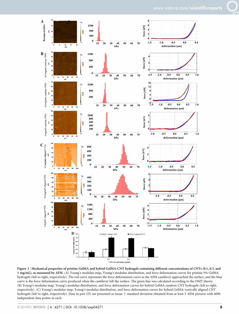
Figure 3 | Mechanical properties of pristine GelMA and hybrid GelMA-CNT hydrogels containing different concentrations of CNTs (0.1, 0.3, and1 mg/mL), as measured by AFM. (A) Young’s modulus map, Young’s modulus distribution, and force deformation curves for pristine 5% GelMA
hydrogels (left to right, respectively). The red curve represents the force deformation curve as the AFM cantilever approached the surface, and the blue
curve is the force deformation curve produced when the cantilever left the surface. The green line was calculated according to the DMT theory.
(B) Young’s modulus map, Young’s modulus distribution, and force deformation curves for hybrid GelMA-random CNT hydrogels (left to right,
respectively). (C) Young’s modulus map, Young’s modulus distribution, and force deformation curves for hybrid GelMA-vertically aligned CNT
hydrogels (left to right, respectively). Data in part (D) are presented as mean 6 standard deviation obtained from at least 5 AFM pictures with 4096
independent data points in each.
www.nature.com/scientificreports
SCIENTIFIC REPORTS | 4 : 4271 | DOI: 10.1038/srep04271 5
Figure 4 | Differentiation of C2C12 myoblasts on GelMA-0.3 mg/mL CNT hydrogels and characterization of the C2C12 myotubes obtained under ES.(A) Schematic representation of the procedure used to produce and electrically stimulate C2C12 myotubes. (B) Immunostaining of the fast skeletal
myosin heavy chain in the C2C12 myotubes fabricated on hybrid GelMA-random CNT, GelMA-vertically aligned CNT, and GelMA-horizontally aligned
CNT hydrogels with and without ES application (indicated as 1ES and 2ES, respectively) on day 10 of culture. Cell nuclei within the C2C12 myotubes
were obvious after the staining procedure. The ES parameters were as follows: a voltage of 8 V, a frequency of 1 Hz, and a duration of 10 ms. The scale bars
represent 50 mm. (C) Quantification of the myotube coverage area and myotube length of the C2C12 myotubes fabricated on hybrid GelMA-random
CNT, GelMA-vertically aligned CNT, and GelMA-horizontally aligned CNT hydrogels with and without ES on day 10 of culture. Data in part (C) are
presented as mean 6 standard deviation obtained from at least 40 myotubes of 2 independent experiments. Asterisks indicate significant differences
between samples (*p , 0.05).
www.nature.com/scientificreports
SCIENTIFIC REPORTS | 4 : 4271 | DOI: 10.1038/srep04271 6
CNT hydrogels (259.89 6 113.05 mm and 33 6 4%, respectively)were significantly greater than those of the unstimulated myotubes(93.83 6 68.84 mm and 16 6 1%, respectively). Although the sametrend was observed when comparing the electrically stimulatedC2C12 myotubes with unstimulated myotubes cultured on theGelMA-random CNT and GelMA-horizontally aligned CNT hydro-gels, the ES had a more profound effect on the muscle myofiberscultured on the hybrid GelMA-aligned CNT hydrogels. In particular,the average myotube length was increased 277% upon applying theES for the C2C12 myotubes cultured on the hybrid GelMA-alignedCNT hydrogels, while this increase was 239% for those cells on thehybrid GelMA-random CNT hydrogels. The average coverage areawas also increased 209% upon applying the ES for the C2C12 myo-tubes cultured on the hybrid GelMA-aligned CNT hydrogels, whilethis parameter was increased 165.65 and 180% for the muscle cellscultured on the hybrid GelMA-random CNT and GelMA-horizont-ally aligned CNT hydrogels, respectively. The enhanced effect islikely due to the vertical alignment of the CNTs in the hybridGelMA-aligned CNT hydrogels, which increased the efficiency ofthe ES along the direction of the CNT alignment for muscle cellson the top of hydrogels in the hybrid GelMA-vertically aligned CNThydrogels compared with that in the GelMA-random CNT andGelMA-horizontally aligned CNT hydrogels. Even though GelMA-horizontally aligned CNT hydrogels also demonstrated the aniso-tropic electrical conductivity, the preferred direction of electric fieldwithin these hydrogels was not favorable for the muscle cells culturedon the top of these hydrogels. In other words, the interconnectednetwork of horizontally aligned CNTs inside GelMA hydrogels pro-vided a short circuit for the electrical current to pass through neigh-boring electrodes without approaching the cells on the top ofhydrogels. Therefore, GelMA-horizontally aligned CNT hydrogelswere less efficient in fabricating functional muscle myofibers com-pared with GelMA-vertically aligned CNT hydrogels.
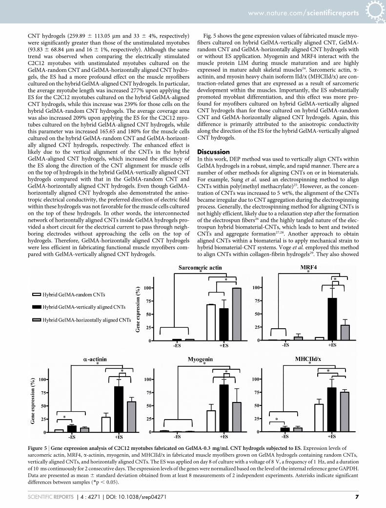
Fig. 5 shows the gene expression values of fabricated muscle myo-fibers cultured on hybrid GelMA-vertically aligned CNT, GelMA-random CNT and GelMA-horizontally aligned CNT hydrogels withor without ES application. Myogenin and MRF4 interact with themuscle protein LIM during muscle maturation and are highlyexpressed in mature adult skeletal muscles24. Sarcomeric actin, a-actinin, and myosin heavy chain isoform IId/x (MHCIId/x) are con-traction-related genes that are expressed as a result of sarcomericdevelopment within the muscles. Importantly, the ES substantiallypromoted myoblast differentiation, and this effect was more pro-found for myofibers cultured on hybrid GelMA-vertically alignedCNT hydrogels than for those cultured on hybrid GelMA-randomCNT and GelMA-horizontally aligned CNT hydrogels. Again, thisdifference is primarily attributed to the anisotropic conductivityalong the direction of the ES for the hybrid GelMA-vertically alignedCNT hydrogels.
DiscussionIn this work, DEP method was used to vertically align CNTs withinGelMA hydrogels in a robust, simple, and rapid manner. There are anumber of other methods for aligning CNTs on or in biomaterials.For example, Sung et al. used an electrospinning method to alignCNTs within poly(methyl methacrylate)25. However, as the concen-tration of CNTs was increased to 5 wt%, the alignment of the CNTsbecame irregular due to CNT aggregation during the electrospinningprocess. Generally, the electrospinning method for aligning CNTs isnot highly efficient, likely due to a relaxation step after the formationof the electrospun fibers26 and the highly tangled nature of the elec-trospun hybrid biomaterial-CNTs, which leads to bent and twistedCNTs and aggregate formation27,28. Another approach to obtainaligned CNTs within a biomaterial is to apply mechanical strain tohybrid biomaterial-CNT systems. Voge et al. employed this methodto align CNTs within collagen-fibrin hydrogels29. They also showed
Figure 5 | Gene expression analysis of C2C12 myotubes fabricated on GelMA-0.3 mg/mL CNT hydrogels subjected to ES. Expression levels of
sarcomeric actin, MRF4, a-actinin, myogenin, and MHCIId/x in fabricated muscle myofibers grown on GelMA hydrogels containing random CNTs,
vertically aligned CNTs, and horizontally aligned CNTs. The ES was applied on day 8 of culture with a voltage of 8 V, a frequency of 1 Hz, and a duration
of 10 ms continuously for 2 consecutive days. The expression levels of the genes were normalized based on the level of the internal reference gene GAPDH.
Data are presented as mean 6 standard deviation obtained from at least 8 measurements of 2 independent experiments. Asterisks indicate significant
differences between samples (*p , 0.05).
www.nature.com/scientificreports
SCIENTIFIC REPORTS | 4 : 4271 | DOI: 10.1038/srep04271 7
that hydrogels with anisotropic CNTs had higher conductivities thanhydrogels containing randomly dispersed CNTs. However, this tech-nique requires several hours and is not suitable for aligning CNTswithin a soft biomaterial. CNTs were also aligned using a simplespinning method30. However, this technique can be used to alignCNTs in the radial direction only. Other vertically aligned nano-structures, such as vertically aligned carbon nanofibers (CNFs), havealso been fabricated for biomedical applications31. CNFs are multi-walled graphene structures that are stacked on top of each other32.These vertically aligned structures have been widely used as neuronalinterfaces, gene delivery arrays, and biosensors33,34. Generally, thesenanostructures are synthesized via plasma-enhanced chemical vapordeposition and are aligned perpendicular to a conductive substrate.This process is long and requires several steps that involve chemicalreactions and must take place under high vacuum. In contrast, ourproposed DEP approach can produce vertically aligned CNT nano-structures in a facile and rapid manner without using toxic chemicalsand complicated experimental setups.
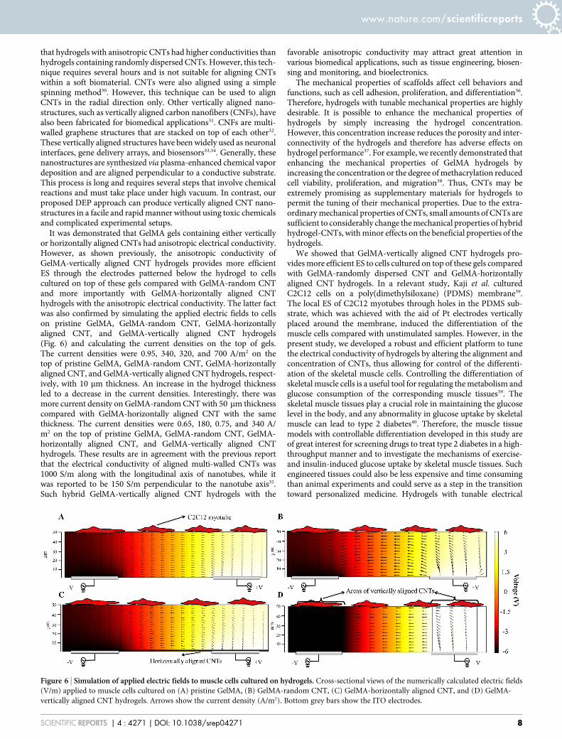
It was demonstrated that GelMA gels containing either verticallyor horizontally aligned CNTs had anisotropic electrical conductivity.However, as shown previously, the anisotropic conductivity ofGelMA-vertically aligned CNT hydrogels provides more efficientES through the electrodes patterned below the hydrogel to cellscultured on top of these gels compared with GelMA-random CNTand more importantly with GelMA-horizontally aligned CNThydrogels with the anisotropic electrical conductivity. The latter factwas also confirmed by simulating the applied electric fields to cellson pristine GelMA, GelMA-random CNT, GelMA-horizontallyaligned CNT, and GelMA-vertically aligned CNT hydrogels(Fig. 6) and calculating the current densities on the top of gels.The current densities were 0.95, 340, 320, and 700 A/m2 on thetop of pristine GelMA, GelMA-random CNT, GelMA-horizontallyaligned CNT, and GelMA-vertically aligned CNT hydrogels, respect-ively, with 10 mm thickness. An increase in the hydrogel thicknessled to a decrease in the current densities. Interestingly, there wasmore current density on GelMA-random CNT with 50 mm thicknesscompared with GelMA-horizontally aligned CNT with the samethickness. The current densities were 0.65, 180, 0.75, and 340 A/m2 on the top of pristine GelMA, GelMA-random CNT, GelMA-horizontally aligned CNT, and GelMA-vertically aligned CNThydrogels. These results are in agreement with the previous reportthat the electrical conductivity of aligned multi-walled CNTs was1000 S/m along with the longitudinal axis of nanotubes, while itwas reported to be 150 S/m perpendicular to the nanotube axis35.Such hybrid GelMA-vertically aligned CNT hydrogels with the
favorable anisotropic conductivity may attract great attention invarious biomedical applications, such as tissue engineering, biosen-sing and monitoring, and bioelectronics.
The mechanical properties of scaffolds affect cell behaviors andfunctions, such as cell adhesion, proliferation, and differentiation36.Therefore, hydrogels with tunable mechanical properties are highlydesirable. It is possible to enhance the mechanical properties ofhydrogels by simply increasing the hydrogel concentration.However, this concentration increase reduces the porosity and inter-connectivity of the hydrogels and therefore has adverse effects onhydrogel performance37. For example, we recently demonstrated thatenhancing the mechanical properties of GelMA hydrogels byincreasing the concentration or the degree of methacrylation reducedcell viability, proliferation, and migration38. Thus, CNTs may beextremely promising as supplementary materials for hydrogels topermit the tuning of their mechanical properties. Due to the extra-ordinary mechanical properties of CNTs, small amounts of CNTs aresufficient to considerably change the mechanical properties of hybridhydrogel-CNTs, with minor effects on the beneficial properties of thehydrogels.
We showed that GelMA-vertically aligned CNT hydrogels pro-vides more efficient ES to cells cultured on top of these gels comparedwith GelMA-randomly dispersed CNT and GelMA-horizontallyaligned CNT hydrogels. In a relevant study, Kaji et al. culturedC2C12 cells on a poly(dimethylsiloxane) (PDMS) membrane39.The local ES of C2C12 myotubes through holes in the PDMS sub-strate, which was achieved with the aid of Pt electrodes verticallyplaced around the membrane, induced the differentiation of themuscle cells compared with unstimulated samples. However, in thepresent study, we developed a robust and efficient platform to tunethe electrical conductivity of hydrogels by altering the alignment andconcentration of CNTs, thus allowing for control of the differenti-ation of the skeletal muscle cells. Controlling the differentiation ofskeletal muscle cells is a useful tool for regulating the metabolism andglucose consumption of the corresponding muscle tissues39. Theskeletal muscle tissues play a crucial role in maintaining the glucoselevel in the body, and any abnormality in glucose uptake by skeletalmuscle can lead to type 2 diabetes40. Therefore, the muscle tissuemodels with controllable differentiation developed in this study areof great interest for screening drugs to treat type 2 diabetes in a high-throughput manner and to investigate the mechanisms of exercise-and insulin-induced glucose uptake by skeletal muscle tissues. Suchengineered tissues could also be less expensive and time consumingthan animal experiments and could serve as a step in the transitiontoward personalized medicine. Hydrogels with tunable electrical
Figure 6 | Simulation of applied electric fields to muscle cells cultured on hydrogels. Cross-sectional views of the numerically calculated electric fields
(V/m) applied to muscle cells cultured on (A) pristine GelMA, (B) GelMA-random CNT, (C) GelMA-horizontally aligned CNT, and (D) GelMA-
vertically aligned CNT hydrogels. Arrows show the current density (A/m2). Bottom grey bars show the ITO electrodes.
www.nature.com/scientificreports
SCIENTIFIC REPORTS | 4 : 4271 | DOI: 10.1038/srep04271 8
conductivity are also promising candidates for the control of thecontraction and therefore the movement of fabricated muscle tissuesin bioactuators or biomedical devices.
In summary, we fabricated GelMA hydrogels with tunable elec-trical conductivity based on CNT alignment and the CNT concen-tration in a robust, simple, and rapid manner. The GelMA-alignedCNT hydrogels exhibited anisotropic electrical conductivity, whichwas not observed in the pristine GelMA hydrogels or the GelMAhydrogels containing randomly distributed CNTs. C2C12 myoblastsgrown on the hybrid GelMA-vertically aligned CNT hydrogelsyielded more functional myofibers, particularly after applying elec-trical stimulation in the direction of the aligned CNTs, than cells thatwere cultured on the GelMA hydrogels with randomly distributedand horizontally aligned CNTs. GelMA-CNT hydrogels with tunableelectrical properties may be useful in drug screening, in the develop-ment of hybrid 3D electronic-tissue materials, and as bioactuators.
MethodsMaterials. The materials and suppliers used were as follows: highly pure multi-walledCNTs (Hodogaya Chemical Co., Ltd.); hexamethyldisilazane (Tokyo Ohka KogyoCo., Ltd., Kanagawa, Japan); developer (MF CD-26) and positive g-line photoresist(S1818) (Shipley Far East Ltd., Tokyo, Japan); methacrylic anhydride, 3-(trimethoxysilyl)propyl methacrylate (TMSPMA), gelatin type A made from porcineskin, and penicillin/streptomycin (P/S) (Sigma-Aldrich Chemical Co., USA); 2-hydroxy-1-[4-(2-hydroxyethoxy)phenyl]-2-methyl-1-propanone (Irgacure 2959)(Ciba Chemicals, Osaka, Japan); fetal bovine serum (FBS) (Bioserum, Japan); andDulbecco’s modified Eagle medium (DMEM), trypsin/EDTA, MEM essential aminoacids, MEM nonessential amino acids, and insulin (Invitrogen, USA).
CNT functionalization. Pristine, highly graphitized CNTs synthesized in a catalyticchemical vapor deposition process are hydrophobic in nature. To make themhydrophilic, their surfaces were functionalized by a controlled acid treatment processas described previously41,42. Briefly, CNTs were refluxed in 120 mL of a 351 v/v ratioof 98% H2SO4 and 68% HNO3 for 20 min at 110uC. The treated CNTs were thenthoroughly washed with ultrapure water on a 1.2 mm membrane and dispersed inultrapure water (,1 mg/mL) by probe sonication at room temperature for severalhours. The resulting CNTs were hydrophilic (zeta potential ,240 mV in an aqueoussolution with a natural pH of ,4.1) and had a high degree of crystallinity, asdemonstrated by Raman spectroscopy, scanning electron microscopy, and TEM inour previous works41,42.
GelMA hydrogel synthesis and preparation of its prepolymer. The GelMAprepolymer was synthesized as described elsewhere43. In brief, 12 mL of methacrylicanhydride was added to 6 g of gelatin in phosphate-buffered saline (PBS) for 1 hr at50uC, resulting in a high degree of methacrylation (,80%) of the gelatin. The mixturewas dialyzed with a 12–14 kDa dialysis membrane against distilled water for one weekat 40uC and then lyophilized for one week. The GelMA hydrogel was stored at 220uCuntil use. The GelMA hydrogel (10% [w/v]) was dissolved in MilliQ water with 1%(w/v) photoinitiator (Irgacure 2959), stored at 60uC to complete the GelMA synthesisprocess, and then used in experiments.
Design and fabrication of the IDA-ITO electrodes. The effective electrodedimensions were 8 3 12 mm2. The band electrode was 50 mm in width, and thedistance between two neighboring band electrodes was 100 mm (Fig. 1). The bandelectrode was fabricated on a glass slide (thickness, 1 mm; Matsunami Co., Japan) byconventional photolithography and chemical etching using an etchant solution(HCl5H2O5HNO3/45251 by volume) for 15 min under ultrasonication.
Fabrication of aligned CNTs within GelMA hydrogels. The IDA-ITO electrodeswere treated with plasma oxygen and methacrylated with TMSPMA under vacuumfor 1 hr. Polyethylene terephthalate film spacers (thickness, 10 mm) were used tocreate a chamber between the ITO electrode and the IDA-ITO electrodes, as shown inFig. 1. GelMA prepolymer (5%) was mixed with different concentrations of CNTs andplaced in an ultrasonic bath for 15 min. The solutions were maintained at 37uC toensure that the liquid state was maintained before and during the dielectrophoreticpatterning of the CNTs within the GelMA hydrogel. To create the gels, 20 mL of thehydrogel-CNT mixture was pipetted between the ITO electrode and the IDA-ITOelectrodes (top and bottom electrodes in Fig. 1, respectively) to fill the chamber (8 3
12 3 0.01 mm3). A sinusoidal AC signal (frequency 2 MHz and voltage 20 V) wasapplied to the IDA-ITO electrodes, and another independent AC signal with anopposite phase was applied to the top ITO electrode to create a nonuniform electricfield and vertically align the CNTs. To obtain horizontally aligned CNTs withinGelMA hydrogels, a sinusoidal AC signal (frequency 2 MHz and voltage 20 V) wasapplied to one electrode band of IDA-ITO electrodes, and another independent ACsignal with an opposite phase was applied to another electrode band of IDA-ITOelectrodes to create a nonuniform electric field. After 1 min, the GelMA prepolymerwas polymerized with 7 mW/cm2 UV light (Hayashi UL-410UV-1; Hayashi
Electronic Shenzen Co., Ltd., Japan) for 150 s. After polymerization, the hybridhydrogels was detached from the DEP device and used in further experiments.
Mechanical properties of GelMA and hybrid GelMA-CNT hydrogels. Themechanical properties of the GelMA and GelMA-CNT hydrogels were investigatedusing a nanomechanical mapping technique using AFM based on force-distancecurve measurements, as described in our previous studies44,45. A MultiMode 8 AFM(Bruker Co., USA) equipped with a liquid chamber was used. The AFM cantilever wasequipped with a colloidal probe with a 1.0 mm radius (PT.GS, NovascanTechnologies, USA) to measure the low elastic modulus values of the underlyinghydrogels. The spring constant of the cantilever was measured using the thermalnoise method46. The force-deformation plots (Fig. 3) were measured by the AFMmethod. They were then used to calculate the Young’s modulus values of hydrogels bythe DMT fitting model. The DMT model18 describes a kind of contact mechanicsbetween an indenting probe and an elastic surface in which adhesion forces are takeninto account and the probe-sample geometry is constrained to be the Hertziancontact. This model has widely been used to analyze AFM-based nano-indentationresults.
Electrical properties of GelMA and hybrid GelMA-CNT hydrogels. The current-voltage (I-V) characterization of the hydrogels at room temperature was performedusing a two-probe station (HiSOL, Inc., model H19S00556) connected to an I-Vanalyzer unit (Agilent, model B1500A). The currents between the probes wereautomatically measured during the automated voltage sweep (23 to 3 V; 0.1 Vsteps). Triaxial cables were used in the measurement setup to reduce noise and thusprecisely measure currents of approximately 10210 A. Note that there was no need touse external electrodes for the measurements because the IDA-ITO and top ITOelectrodes were used as the electrodes.
Electrochemical impedance spectroscopy (EIS) measurements were collected usinga CompactStat potentiostat (CompactStat; Ivium Technologies, Netherlands) con-trolled by a computer equipped with IviumSoft software. EIS spectra were acquiredfor the GelMA and hybrid GelMA-CNT hydrogels over a frequency range from 1 to100 Hz with a perturbation amplitude of 25 mV.
Cell culture. C2C12 myoblasts (American Type Culture Collection, USA) werecultured in DMEM supplemented with 10% FBS and 1% P/S. The C2C12 myoblastswere trypsinized using 0.25% trypsin/0.1% EDTA when they reached 70–80%confluence. The cells were maintained in a cell culture incubator (Sanyo, Japan) with5% CO2 at 37uC.
Culture of C2C12 myoblasts on GelMA and hybrid GelMA-CNT hydrogels. Forcell culture on GelMA and hybrid GelMA-CNT hydrogels, C2C12 myoblasts weretrypsinized, counted, and resuspended in DMEM at a density of 1 3 105 cells/mL.Then, 100 mL of the cell suspension was pipetted onto each hydrogel, and the sampleswere incubated at 37uC for 30 min to allow cell seeding. After seeding, additionalmedium was added for extended cell culture. After 1 day of culture, the culturemedium was replaced with differentiation medium (DMEM with 2% horse serum,1 nM insulin, and 1% P/S). The differentiation medium was replenished every 2 daysduring the culture period.
Immunostaining and characterization of C2C12 myotubes. C2C12 myotubes wereimmunostained as detailed elsewhere47,48. In brief, the C2C12 myotubes were fixedwith 3–4% paraformaldehyde for 12 min, followed by a wash with PBS. Thepermeabilization step was performed with 0.3% Triton X-100 for 5 min at ambienttemperature. Then, the cells were exposed to 5% bovine serum albumin dissolved inPBS for 15 min. A primary mouse monoclonal IgG antibody (ab-7784, AbcamH,Japan) against fast skeletal myosin was added to the samples at a dilution of 151000 inPBS, and the samples were incubated at 4uC for 24 hr. The samples were then washedthree times with PBS, treated with a goat anti-mouse AlexaFluorH 488 antibody(Invitrogen, USA) at a dilution of 151000 in PBS, and incubated at 37uC for 1 hr. TheC2C12 myotubes were imaged with a fluorescence microscope. The average myotubelength was quantified using the AxioVision Rel. 4.8 software package, and themyotube coverage area was computed with the NIH ImageJ software package.
Electrical stimulation of the engineered muscle myofibers. On day 8 of culture,C2C12 myotubes were electrically stimulated through the IDA-ITO electrodes on thebottom of engineered muscle tissues. As reported in our previous studies, this deviceis more efficient for stimulating engineered tissues than conventional electricalstimulators49–51. For the ES of muscle myofibers, the differentiation medium wasreplaced with stimulation medium composed of DMEM with 2% horse serum, 1 nMinsulin, 2% MEM essential amino acids, 1% MEM nonessential amino acids, and 1%P/S. Electrical pulses were applied to the muscle myofibers using a waveformgenerator (WF 1946B Multifunction Synthesizer; NF Co., Japan) with a specifiedregime (voltage, 8 V; frequency, 1 Hz; duration, 10 ms) continuously for 2consecutive days. The generated electric current was confirmed using an oscilloscope(WaveSurfer 424; LeCroy Co., Japan). During ES of the muscle myofibers, thestimulation medium was replenished daily to decrease the negative effects ofaccumulated charge in the medium.
RNA extraction and complementary DNA (cDNA) synthesis. Muscle myofiberswere fixed using liquid nitrogen and thoroughly pulverized with a mortar and pestle.RNA was extracted from the muscle myofibers using b-mercaptoethanol and purified
www.nature.com/scientificreports
SCIENTIFIC REPORTS | 4 : 4271 | DOI: 10.1038/srep04271 9
in accordance with the manufacturer’s protocol (RNeasyE Micro Kit; Qiagen, Venlo,Netherlands). Up to 3 mg of total RNA was reverse transcribed using the QuantiTectReverse Transcription kit as recommended by the manufacturer (Qiagen, Venlo,Netherlands). To synthesize cDNA, 12 mL of sample (3 mg of total RNA) was dilutedwith 14 mL of RNase-free water and 4 mL of gDNA wipeout buffer. The preparedsamples were incubated for 2 min at 42uC and then cooled to 4uC. The QuantiscriptReverse Transcriptase and Reverse Transcriptase primer mix were added, and themixture was incubated for 15 min at 42uC, followed by 3 min at 95uC. The sampleswere kept at 4uC until used for quantitative PCR (qPCR).
Real-time PCR. All primer sets (i.e., primers for GAPDH, myogenin, MRF4,sarcomeric actin, a-actinin, and MHCIId/x) were purchased from OperonBiotechnologies (Tokyo, Japan) and validated for qPCR experiments. The primersequences are 59-TGTCTGTCAGGCTGGGTGTG-39/59-TCGCTGGGCTGGGTGTTAG-39, 59-CGAAAGGAGGAGACTAAAG-39/59-CTGTAGACGCTCAATGTAG-39, 59-ATGGTAGGTATGGGTCAG-39/59-GATCTTCTCCATGTCGTC-39, 59-TCATCCTCCGCTTCGCCATTC-39/59-CTTCAGCATCCAACATCTTAGG-39, and 59-GCGACAGACACCTCCTTC-AAG-39/59-TCCAGCCAGCCAGCGATG-39 for myogenin, MRF4, sarcomericactin, a-actinin, and MHCIId/x, respectively49. Real-time PCR was performed usingthe Roche LightCycler 1.5 (Roche, Mannheim, Germany) with 2 mL of cDNA, 2 mL ofthe appropriate primer set, and 14 mL of LightCycler FastStart DNA Master SYBRGreen 1 reaction mix (Roche, Mannheim, Germany). Following an initialdenaturation step at 95uC for 10 min, real-time PCR was performed over 45 cycles of95uC for 10 s, 62uC for 10 s, and 72uC for 20 s, followed by a melting curve analysis.The expression levels of the target genes were normalized against that of the GAPDHgene52. The gene expression analysis was repeated at least four times for each sample.
Simulation of electric fields. The distribution of electric fields within the gels (i.e.,pristine GelMA, GelMA-random CNT, GelMA-horizontally aligned CNT, andGelMA-vertically aligned CNT hydrogels) was computed using the finite elementanalysis as implemented in the COMSOL Multiphysics 4.2, Stockholm, Sweden(Fig. 6). The size of bottom ITO electrodes was set to 50 mm. The potential differenceand distance between two neighboring ITO electrodes were 6 V and 100 mm,respectively. The height of hydrogels on the top of electrodes was 50 mm. In the case ofthe hybrid GelMA-horizontally aligned CNT, it was assumed that CNT layer betweenthe ITO electrodes has 0.5 mm thickness. For the hybrid GelMA-vertically alignedCNT hydrogel, the CNT alignment was supposed to be on the top of ITO electrodes.The electrical conductivities of all underlying hydrogels were obtained from theexperimental data (see Fig. 2). Viscosity of 5 wt% GelMA hydrogel was set to0.09 cm2/s as reported in our previous work53. The current density (A/m2) was alsocalculated for the hydrogels with 10 and 50 mm thicknesses.
Statistical analysis. Significant differences were identified using the independentStudent’s t-test for two groups of data using the MINITAB 16.0 statistical softwarepackage (Minitab Inc., USA). All data from repeated experiments are reported as theaverage 6 standard deviation, and p-values less than 0.05 were deemed significant.
1. Peppas, N. A., Hilt, J. Z., Khademhosseini, A. & Langer, R. Hydrogels in biologyand medicine: From molecular principles to bionanotechnology. Adv. Mater. 18,1345–60 (2006).
2. Slaughter, B. V., Khurshid, S. S., Fisher, O. Z., Khademhosseini, A. & Peppas, N. A.Hydrogels in regenerative medicine. Adv. Mater. 21, 3307–29 (2009).
3. Dvir, T. et al. Nanowired three-dimensional cardiac patches. Nat. Nano. 6, 720–5(2011).
4. Tian, B. et al. Macroporous nanowire nanoelectronic scaffolds for synthetictissues. Nat. Mater. 11, 986–94 (2012).
5. Xu, L. et al. Design and synthesis of diverse functional kinked nanowire structuresfor nanoelectronic bioprobes. Nano Lett. 13, 746–51 (2013).
6. Schwille, P. Bottom-up synthetic biology: Engineering in a tinkerer’s world.Science 333, 1252–4 (2011).
7. Sapra, K. T. & Bayley, H. Lipid-coated hydrogel shapes as components of electricalcircuits and mechanical devices. Sci. Rep. 2, 848 (2012).
8. Shin, S. R. et al. Carbon-nanotube-embedded hydrogel sheets for engineeringcardiac constructs and bioactuators. ACS Nano 7, 2369–80 (2013).
9. Dvir, T., Timko, B. P., Kohane, D. S. & Langer, R. Nanotechnological strategies forengineering complex tissues. Nat. Nanotech. 6, 13–22 (2011).
10. You, J.-O., Rafat, M., Ye, G. J. C. & Auguste, D. T. Nanoengineering the heart:Conductive scaffolds enhance Connexin 43 expression. Nano Lett. 11, 3643–8(2011).
11. Shin, S. R. et al. Carbon nanotubes reinforced hybrid microgels as scaffoldmaterials for cell encapsulation. ACS Nano 6, 362–72 (2012).
12. Nichol, J. W. et al. Cell-laden microengineered gelatin methacrylate hydrogels.Biomaterials 31, 5536–44 (2010).
13. MacDonald, R. A., Voge, C. M., Kariolis, M. & Stegemann, J. P. Carbon nanotubesincrease the electrical conductivity of fibroblast-seeded collagen hydrogels. ActaBiomater. 4, 1583–92 (2008).
14. Ramon-Azcon, J. et al. Competitive multi-immunosensing of pesticides based onthe particle manipulation with negative dielectrophoresis. Biosens. Bioelectron. 25,1928–33 (2010).
15. Ramon-Azcon, J. et al. Detection of pesticide residues using an immunodevicebased on negative dielectrophoresis. Biosens. Bioelectron. 24, 1592–7 (2009).
16. Ramon-Azcon, J. et al. Dielectrophoretically aligned carbon nanotubes to controlelectrical and mechanical properties of hydrogels to fabricate contractile musclemyofibers. Adv. Mater. 25, 4028–34 (2013).
17. Estili, M., Kawasaki, A. & Sakka, Y. Highly concentrated 3D macrostructure ofindividual carbon nanotubes in a ceramic environment. Adv. Mater. 24, 4322–6(2012).
18. Derjaguin, B. V., Muller, V. M. & Toporov, Y. Effect of contact deformations onthe adhesion of particles. J. Colloid Interf. Sci. 53, 314–26 (1975).
19. Andres, V. & Walsh, K. Myogenin expression, cell cycle withdrawal, andphenotypic differentiation are temporally separable events that precede cell fusionupon myogenesis. J. Cell Biol. 132, 657–66 (1996).
20. McCarthy, J. J. & Esser, K. A. Skeletal Muscle Adaptation to Exercise in Muscle:Fundamental Biology and Mechanisms of Disease. Edited by Hill, J. A., Olson,E. N. & San Diego, C. A: Academic Press, Chapter 64, 911–920.
21. Schiaffino, S. & Serrano, A. Calcineurin signaling and neural control of skeletalmuscle fiber type and size. Trends Pharmacol. Sci. 23, 569–75 (2002).
22. Bickel, C. S. et al. Time course of molecular responses of human skeletal muscle toacute bouts of resistance exercise. J. Appl. Physiol. 98, 482–488 (2005).
23. Fujie, T. et al. Engineered nanomembranes for directing cellular organizationtoward flexible biodevices. Nano Lett. 13, 3185–92 (2013).
24. Arber, S., Halder, G. & Caroni, P. Muscle LIM protein, a novel essential regulatorof myogenesis, promotes myogenic differentiation. Cell 79, 221–31 (1994).
25. Sung, J. H., Kim, H. S., Jin, H.-J., Choi, H. J. & Chin, I.-J. Nanofibrous membranesprepared by multiwalled carbon nanotube/poly(methyl methacrylate)composites. Macromolecules 37, 9899–902 (2004).
26. Ayutsede, J. et al. Carbon nanotube reinforced bombyx mori silk nanofibers by theelectrospinning process. Biomacromolecules 7, 208–14 (2006).
27. Sen, R. et al. Preparation of single-walled carbon nanotube reinforced polystyreneand polyurethane nanofibers and membranes by electrospinning. Nano Lett. 4,459–64 (2004).
28. Ko, F. et al. Electrospinning of continuous carbon nanotube-filled nanofiberyarns. Adv. Mater. 15, 1161–5 (2003).
29. Voge, C. M., Kariolis, M., MacDonald, R. A. & Stegemann, J. P. Directionalconductivity in SWNT-collagen-fibrin composite biomaterials through strain-induced matrix alignment. J. Biomed. Mater. Res. Part A 86A, 269–77 (2008).
30. Namgung, S., Baik, K. Y., Park, J. & Hong, S. Controlling the growth anddifferentiation of human mesenchymal stem cells by the arrangement ofindividual carbon nanotubes. ACS Nano 5, 7383–90 (2011).
31. Melechko, A. V., Desikan, R., McKnight, T. E., Klein, K. L. & Rack, P. D. Synthesisof vertically aligned carbon nanofibres for interfacing with live systems. J. Phys. D:Appl. Phys. 42, 193001 (2009).
32. Nguyen-Vu, T. D. B. et al. Vertically aligned carbon nanofiber arrays: An advancetoward electrical–neural interfaces. Small 2, 89–94 (2006).
33. Yu, Z. et al. Vertically aligned carbon nanofiber arrays record electrophysiologicalsignals from hippocampal slices. Nano Lett. 7, 2188–95 (2007).
34. Tosun, Z. & McFetridge, P. S. A composite SWNT–collagen matrix:Characterization and preliminary assessment as a conductive peripheral nerveregeneration matrix. J. Neural Eng. 7, 066002 (2010).
35. de Heer, W. A., Bacsa, W. S., Chatelain, A., Gerfin, T., Humphrey-Baker, R., Forro,L. & Ugarte, D. Aligned carbon nanotube films: Production and optical andelectronic properties. Science 268, 845–7 (1995).
36. Reilly, G. C. & Engler, A. J. Intrinsic extracellular matrix properties regulate stemcell differentiation. J. Biomech. 43, 55–62 (2010).
37. Lutolf, M. P., Gilbert, P. M. & Blau, H. M. Designing materials to direct stem-cellfate. Nature 462, 433–41 (2009).
38. Aubin, H. et al. Directed 3D cell alignment and elongation in microengineeredhydrogels. Biomaterials 31, 6941–51 (2010).
39. Kaji, H., Ishibashi, T., Nagamine, K., Kanzaki, M. & Nishizawa, M. Electricallyinduced contraction of C2C12 myotubes cultured on a porous membrane-basedsubstrate with muscle tissue-like stiffness. Biomaterials 31, 6981–6 (2010).
40. Nedachi, T., Fujita, H. & Kanzaki, M. Contractile C2C12 myotube model forstudying exercise-inducible responses in skeletal muscle. Am. J. Physiol.Endocrinol. Metab. 295, E1191–E204 (2008).
41. Estili, M. & Kawasaki, A. Engineering strong intergraphene shear resistance inmulti-walled carbon nanotubes and dramatic tensile improvements. Adv. Mater.22, 607–10 (2010).
42. Estili, M. et al. The homogeneous dispersion of surfactantless, slightly disordered,crystalline, multiwalled carbon nanotubes in a-alumina ceramics for structuralreinforcement. Acta Mater. 56, 4070–9 (2008).
43. Ramon-Azcon. et al. Gelatin methacrylate as a promising hydrogel for 3Dmicroscale organization and proliferation of dielectrophoretically patterned cells.Lab Chip 12, 2959–69 (2012).
44. Wang, D., Fujinami, S., Liu, H., Nakajima, K. & Nishi, T. Investigation of truesurface morphology and nanomechanical properties of poly(styrene-b-ethylene-co-butylene-b-styrene) using nanomechanical mapping: Effects of composition.Macromolecules 43, 9049–55 (2010).
45. Liu, H. et al. Quantitative nanomechanical investigation on deformation ofpoly(lactic acid). Macromolecules 45, 8770–9 (2012).
46. Hutter, J. L. & Bechhoefer, J. Calibration of atomic-force microscope tips. Rev. Sci.Instrum. 64, 1868–73 (1993).
www.nature.com/scientificreports
SCIENTIFIC REPORTS | 4 : 4271 | DOI: 10.1038/srep04271 10
47. Hosseini, V. et al. Engineered contractile skeletal muscle tissue on a microgroovedmethacrylated gelatin substrate. Tissue Eng. Part A 18, 2453–65 (2012).
48. Obregon, R. et al. Non-invasive measurement of glucose uptake of skeletal muscletissue models using a glucose nanobiosensor. Biosens. Bioelectron. 50, 194–201(2013).
49. Ahadian, S. et al. Interdigitated array of Pt electrodes for electrical stimulation andengineering of aligned muscle tissue. Lab Chip 12, 3491–503 (2012).
50. Ahadian, S. et al. A contactless electrical stimulator: application to fabricatefunctional skeletal muscle tissue. Biomed. Microdevices 15, 109–15 (2013).
51. Ahadian, S. et al. Electrical stimulation as a biomimicry tool for regulating musclecell behavior. Organogenesis 9, 87–92 (2013).
52. Schmittgen, T. D. & Livak, K. J. Analyzing real-time PCR data by the comparativeCT method. Nat. Protocols 3, 1101–8 (2008).
53. Hancock, M. J. et al. Anisotropic material synthesis by capillary flow in a fluidstripe. Biomaterials 32, 6493–504 (2011).
AcknowledgmentsThe authors thank the MANA Foundry at NIMS for providing the I-V measurementsystem. This work was supported by the World Premier International Research CenterInitiative (WPI), MEXT, Japan.
Author contributionsS.A. and J.R.-A. conceived the idea and designed the research. S.O. synthesized the GelMAprepolymer. M.E. functionalized the CNTs, prepared the aqueous CNT solutions, andperformed the DC conductivity measurements. X.L. helped with the AFM measurementsunder the supervision of K.N. S.A. and J.R.-A. performed all other experiments, analyzedthe results, contributed equally to the work, and wrote the paper. H.S., M.R., H.B., T.M. andA.K. supervised the study. All authors read the manuscript, provided comments, andapproved its content.
Additional informationSupplementary information accompanies this paper at http://www.nature.com/scientificreports
Competing financial interests: The authors declare no competing financial interests.
How to cite this article: Ahadian, S. et al. Hybrid hydrogels containing vertically alignedcarbon nanotubes with anisotropic electrical conductivity for muscle myofiber fabrication.Sci. Rep. 4, 4271; DOI:10.1038/srep04271 (2014).
This work is licensed under a Creative Commons Attribution-NonCommercial-ShareAlike 3.0 Unported license. To view a copy of this license,
visit http://creativecommons.org/licenses/by-nc-sa/3.0
www.nature.com/scientificreports
SCIENTIFIC REPORTS | 4 : 4271 | DOI: 10.1038/srep04271 11