Transport in Animals - WordPress.com
80
Transport in Animals • Explain the need for transport systems in multicellular animals in terms of size, level of activity and surface area: volume ratio. • Explain the meaning of the terms single circulatory system and double circulatory system. • Describe the external and internal structure of the mammalian heart. • Explain the differences in the thickness of the walls of the different chambers of the heart in terms of their functions. • Describe the cardiac cycle, with reference to the action of the valves in the heart. • Describe how heart action is coordinated with reference to the sinoatrial node (SAN), the atrioventricular node (AVN) and the Purkyne tissue. • Interpret and explain electrocardiogram (ECG) traces, with reference to normal and abnormal heart activity.
Transcript of Transport in Animals - WordPress.com

Transport in Animals• Explain the need for transport systems in multicellular
animals in terms of size, level of activity and surface area: volume ratio.
• Explain the meaning of the terms single circulatory systemand double circulatory system.
• Describe the external and internal structure of the mammalian heart.
• Explain the differences in the thickness of the walls of the different chambers of the heart in terms of their functions.
• Describe the cardiac cycle, with reference to the action of the valves in the heart.
• Describe how heart action is coordinated with reference to the sinoatrial node (SAN), the atrioventricular node (AVN) and the Purkyne tissue.
• Interpret and explain electrocardiogram (ECG) traces, with reference to normal and abnormal heart activity.

Transport Systems
• Small organisms may not need a transport
system.
– Why?

A good transport system has…
– A fluid to carry substances around (blood).
– A pump to keep the fluid moving (heart).
– Exchange surfaces to provide nutrients &
remove wastes (lungs, intestines, kidneys).
• Even better if it also has…
– Tubes to carry the fluid (arteries, veins,
capillaries).
– Two circuits (what??????)

Single or
Double
Circulation

Advantages of Double Circulation
• Single:
– BP is reduced as blood passes through gill capillaries.
• Blood flows much slower through tissues.
• Limits the rate of O2/nutrient delivery.
• Double:
– BP is increased again after blood flows through lung capillaries.
• Blood flows much faster through tissues.
• Rate of O2/nutrient delivery is higher.

Double Circulation• Blood pressure is boosted to push it
through both sets of capillaries (Pulmonary & Systemic).

Heart Structure
• 2 separate pumps lying side by side.
– Left side pumps oxygenated blood from the
lungs.
– Right side pumps deoxygenated blood from
the body.
• Each pump has 2 chambers:
– An Atrium.
– A Ventricle.

Basic Structure
Right
Atrium
Left
Atrium
Right
Ventricle
Left
Ventricle

Basic Structure
Right
Atrium
Left
Atrium
Right
Ventricle
Left
Ventricle
Blood
from body
Blood from
lungs

Basic Structure
Right
Ventricle
Left
Ventricle
Blood
from body
Blood from
lungs

Basic Structure
Blood
from body
Blood from
lungs

Basic Structure
Blood
from body
Blood from
lungs
Blood
to lungs
Blood to
body

Basic Structure
Deoxygenated
Blood from
body
Oxygenated
Blood from
lungs
Blood
to lungs
Blood to
body

Basic Structure
Deoxygenated
Blood from
body
Oxygenated
Blood from
lungs
Blood
to lungs
Blood to
body
Tricuspid
valve
Bicuspid
valve
Semilunar
valves
Septum

Basic Structure
Deoxygenated
Blood from
body
Oxygenated
Blood from
lungs
Blood
to lungs
Blood to
body
Tricuspid
valve
Bicuspid
valve
Semilunar
valves
Vena Cava
Pulmonary Artery
Pulmonary Vein
Aorta
Septum

12
3
4
5
6
7
8910
11
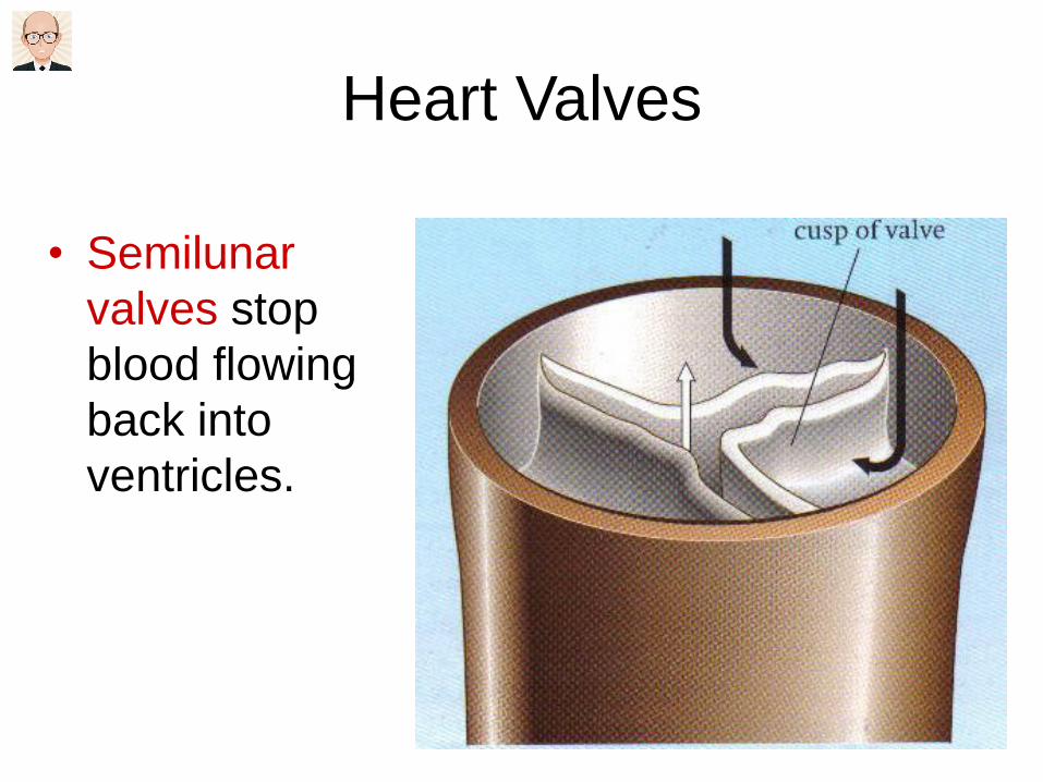
Heart Valves
• Semilunar
valves stop
blood flowing
back into
ventricles.

Heart Valves
• Atrioventricular
valves
(tricuspid &
bicuspid) stop
blood flowing
back into atria.
Tendinous
chords

What is a Coronary?
• The heart muscle uses a lot of oxygen for its own respiration.
• This is supplied by blood vessels called coronary arteries.
• Coronary arteries branch off from the aorta.
• A blockage in these arteries will deprive the heart of oxygen and lead to a heart attack (myocardial infarction).

What Does It Look Like?
• Dissect a heart to find:
– Coronary Arteries.
– Right & Left Atria.
– Right & Left Ventricles.
– Right & Left Atrioventricular valves.
– Tendinous chords
– Semi-lunar valves.
– Difference in ventricle wall thickness.
– Septum

How Does Aerobic Exercise Help?
• Increases the amount of cardiac muscle in
the heart.
– So it can pump more blood with each beat.
– So it can pump blood with greater force.
• This will increase the maximum cardiac
output (heart rate x stroke volume).
• This will decrease the resting heart rate.

Mini Test
The mammalian heart is made up of [1] muscle & is covered by a tough membrane called the [2]. It contains four chambers: a pair of thin walled ones called [3] & a pair of thick muscular ones called [4].Between the chambers on the left side are the [5]valves, those on the right side are called [6] valves. These valves are prevented from turning inside out by [7]. [8] blood from the lungs enters the heart via the [9] & into the chamber called [10]. [11] blood from the body enters the heart via the [12] & into the chamber called [13]. Oxygen is supplied to the heart muscle itself via the [14] that branch off from the [15]. A blockage in these vessels can lead to a [16].

Mini Test Answers
1 Cardiac
2 Pericardium
3 Atria
4 Ventricles
5 Bicuspid
6 Tricuspid
7 Tendinous Chords
8 Oxygenated
9 Pulmonary Vein
10 Left Atrium
11 Deoxygenated
12 Vena Cava
13 Right Atrium
14 Coronary Arteries
15 Aorta
16 Heart Attack/
Cardiac Arrest
The Cardiac Cycle

What is the Cardiac Cycle?
• There are two basic components to the beating heart:
– Contraction (Systole)
– Relaxation (Diastole)
• Each cardiac cycle has three stages:
– Diastole (atria & ventricles relax)
– Atrial Systole (atria contract, pushing blood into ventricles)
– Ventricular Systole (ventricles contract, pushing blood into major arteries).


Valves
• Action of the
atrioventricular
valves.

Interpreting
Graphs

How is this controlled?
• Cardiac muscle is myogenic.
– Contractions are started from the muscle itself rather than from nerve impulses (neurogenic).
• Contractions originate in a region of cells in the right atrium (the sinoatrial node).
• The SA node has a basic rhythm of stimulation that determines the heartbeat.
• The SA node is often called the pacemaker.

Dia
sto
le
A wave of excitation
spreads out from the
SA node across both
atria, causing them
to contract.
All cardiac muscles
relax before the
cycle starts again.
The wave of excitation
is released from the
Purkyne fibres causing
both ventricles to
contract from the apex
upwards.
The bundle branches
into smaller fibres
called Purkyne
fibres. The wave
travels up through
the ventricle walls.
The bundle of His
conducts the wave to
the base of the
ventricles.
The wave is
transmitted down
between the ventricles
along special muscle
fibres called the bundle
of His.
The wave of
excitation is picked
up by a second
group of cells – the
atrioventricular
node.
A layer of non-
conductive tissue
(the atrioventricular
septum) prevents the
wave crossing to the
ventricles

1. SA Node
2. AV Node
3. Bundle of His
4. Left & right
branches of
the bundle
5. Bundle
branches

Electrocardiograms (ECGs)
• Some of the electrical activity in the heart
spreads through other tissues.
• This can be detected by sensors attached
to the skin on the chest.
• A healthy trace has a particular shape.

Interpreting the
ECG
• A healthy trace has shapes labelled as P, Q, R, S & T.
• Wave P shows excitation of atria.
• QRS shows excitation of ventricles.
• T shows diastole.

0 0.2 0.4 0.6 0.8 1.0 1.2 1.4 1.6
Morepositive
Morenegative
Electricalpotential
Time / s
R R
PP T T
Q QSS
Calculate the heart rate
(show working & units)

ECG traces can show heart
abnormalities:• P.194
• Sketch & summarise the abnormal trace
and the disease for:
– Tachycardia
– Bradycardia
– Ectopic heartbeat
– Atrial fibrillation as example of Arrhythmia

Open v Closed Circulation
• Some animals have an open circulation
system.
– Blood is not completely held within vessels.
– Blood circulates through the body cavity.
– Tissue cells are bathed in blood.
• Eg Insects:


Closed Circulation
• Open systems are OK for small insects.
– Blood does not have to travel far.
– Oxygen & Carbon Dioxide do not travel in blood.
• Larger organisms need a closed system;
– Blood speed in a closed system is too slow.
– Blood is used to transport nutrients, O2 & CO2.
– Active muscles would not receive enough blood.
– Tissue fluid bathes the cells instead of blood.

Blood Vessels
• Three types:– Arteries carry blood away from the heart.
• Smaller arteries called arterioles.
– Veins carry blood into the heart.• Smaller veins called venules.
– Capillaries link arteries to veins.

Structure & Function - Arteries
Lumen is small Adds strength to
withstand blood pressure
Thick, muscular wall Prevents rupturing as
artery stretches
Elastic tissue Maintains high BP.
Smooth muscle Allows artery to stretch
as blood flows through.
Endothelium is folded Allows constriction of the
artery to limit blood flow

Structure & Function - Veins
Lumen is large No need to stretch &
recoil. They do not
constrict to reduce flow.
Thin wall Ensure one way flow.
Help blood flow back to
the heart.
Contains valves Eases blood flow

Structure & Function - Capillaries
Lumen is very
narrow
Ensures red blood cells are squeezed as
they pass through – slows them down.
Reduces the diffusion distance for Oxygen,
Carbon dioxide & nutrients.
Very thin walls
(one cell thick)
Allows exchange of materials between
blood & tissue fluid.
Presses red blood cells against walls –
shortens diffusion distance.

How is circulation maintained?
• By the heart.
– A muscular organ that pumps the blood through the vessels.
• By contraction of skeletal muscle.
– Body movements squeeze veins. Valves ensure blood moves towards the heart.
• By enlargement of the thorax during inspiration.
– Reduced pressure in the thorax helps to draw venous blood back to the heart.

Blood
• The medium by which materials are
transported within the body.
• Consists of:
– Plasma (the liquid part)
– Red blood cells
– White blood cells
– Platelets

Functions of the Blood
• Two main functions:
– Transport
• Transports a wide variety of materials around the
body.
– Defence
• White blood cells protect the body by engulfing
foreign material.
• White blood cells produce substances to stimulate
defensive reactions and provide immunity.

Plasma
• Makes up 55% of blood volume.
• It is 90% water and 10% chemicals.
– Chemicals are either dissolved or suspended
in the plasma.

The Chemicals in Plasma Include:
• Nutrients (eg. Glucose, amino acids, vitamins).
• Waste products (eg. Urea).
• Mineral salts (eg. Calcium, iron).
• Hormones (eg. Insulin, adrenaline).
• Plasma proteins (eg. Fibrinogen & prothrombin which are involved in clotting).
• Respiratory gases (eg. Oxygen, carbon dioxide).

Red Blood Cells - Erythrocytes
• Bi-concave discs (8μm diameter).
• Made in bone marrow of certain bones:
– Eg. Cranium, sternum, vertebrae, ribs.
• Contain the red pigment haemoglobin
– Carries oxygen
• Have no nucleus
– Reduces their lifespan (120 days)
– More efficient in transporting oxygen• Larger surface area : volume ratio
• More room for haemoglobin

White Blood Cells - Leucocytes
• Many types of Leucocyte.
– Probably previously known as phagocytes
and lymphocytes.
• Made in the thymus gland and marrow of
limb bones.
• Protect the body against infection.

Platelets - Thrombocytes
• These are small fragments of cells.
• Have a crucial role in blood clotting.
– Prevents blood loss when a vessel is
ruptured.

Tissue Fluid
• Carries oxygen &
nutrients from blood
to cells.
• Carries carbon
dioxide & other
wastes from cells to
blood.

How is tissue fluid formed?
(Oncotic Pressure)

What is Lymph?
• Lymph is a milky liquid made from three
sources:
– Tissue fluid that has not been reabsorbed at
the venous ends of capillaries.
– Fatty acids & glycerol absorbed into the
lacteals in the ileum.
– Lymphocytes produced in the lymph nodes or
that have migrated out of capillaries to fight
infection.

What is the Lymphatic System?
• A network of capillaries that merge into larger vessels which run around the body.
• Lymph vessels drain their contents back into the blood stream at two places:
– The right lymphatic duct• Drains lymph from the right side of the head & right
arm into the right subclavian vein.
– The thoracic duct• Drains lymph from the rest of the body into the left
subclavian vein.

What are Lymph Nodes?
• Points along lymph vessels where lymphocytes are produced & stored.
• These sites filter out any bacteria & other foreign bodies which are engulfed by WBCs.
• This causes the nodes to swell with dead cells.
– This is the cause of tenderness in the groin, armpits & neck during an infection.

How is Lymph Moved Along the
Vessels?
• Lymph is moved along in three ways:
– Hydrostatic pressure of tissue fluid leaving the
capillaries.
– Contraction of skeletal muscles.
• Lymph vessels are squeezed, pushing lymph out
of the way. Valves in the vessels ensure that it
flows in the right direction.
– Enlargement of the thorax.
• During inspiration the pressure inside the thorax is
reduced which draws lymph into this region.

Activity – Copy & Complete
Blood Plasma Tissue Fluid Lymph
Location
How it is
Moved
Direction of
flow
Types of
cells inside

Transport of Oxygen
Even with lungs, blood vessels and a
heart to pump blood around the body,
transport of oxygen would be inadequate if
the gas were simply dissolved in the
plasma.

Introduction
• We have evolved specialised molecules
capable of carrying large quantities of
oxygen.
• These molecules are called respiratory
pigments.
• The most well known is haemoglobin.

Haemoglobin is made of 4 separate polypeptides joined together (2 alpha and 2 beta chains).
The red molecule is a Haem group that contains an iron atom (pink) attached.
Oxygen molecules bind to each of the haem groups, so that each haemoglobin molecule can accommodate four O2 molecules.

Haemoglobin
• A red pigment with a large RMM of 68,000.
• One oxygen molecule can combine with each of its four haem groups to form oxyhaemoglobin.
• Haemoglobin must:
– Readily pick up oxygen at the lungs.
– Readily release oxygen at the respiring tissues.

Carbon Monoxide Poisoning
• Hb has a greater affinity for carbon
monoxide than for oxygen.
• Once attached to Hb, carbon monoxide
stays there permanently.
• This prevents oxygen molecules from
binding.

How do we Measure Oxygen
Concentration?
• The amount of gas present in a mixture of gases
is measured by the pressure it contributes to the
total pressure of the gas mixture.
• This is called partial pressure of the gas, or the
gas tension.
• Measured in kPa.
• For example:
– Normal atmospheric pressure is 100kPa. Oxygen
makes up 21% of air. So the partial pressure of
oxygen (pO2) is normally 21kPa.

Loading Haemoglobin with Oxygen
• At low pO2, the four Hb polypeptides are close together.
– Difficult for the first molecule of O2 to load.
• Once the first O2 has loaded, the Hb polypeptides separate slightly.
– Easier for subsequent O2 molecules to load.
• Once the Hb is almost saturated, the last O2 molecule is difficult to load.

Oxygen Dissociation Curve
Partial Pressure of Oxygen (kPa)0 20
%a
ge
sa
tura
tio
n o
f H
b w
ith
oxyg
en
100
0
Loading with oxygen becomes
easier once the first one has
loaded.
Loading the
final oxygen
molecule is
difficult.

How Much Oxygen is Carried?
Partial Pressure of Oxygen (kPa)
0 20
%a
ge
sa
tura
tio
n o
f H
b w
ith
oxyg
en
95
50
5
15105
If pO2 is high (in lungs)
then more oxygen is
carried by Hb.
In normal atmospheric
oxygen the saturation
will never be more than
95%
If pO2 is low (in
respiring tissue) then
more oxygen is
released by Hb.Loadin
g T
ensio
n
Un
loa
din
g T
en
sio
n

The Effect of Carbon Dioxide
• The increased acidity from the presence of
CO2 reduces the affinity of haemoglobin
for oxygen.
Hb
O O
OO
O
OO
OHb
Low CO2High CO2
O
O
O
O
OO
O
O

What Effect Does Carbon Dioxide
Have?
Partial Pressure of Oxygen (kPa)
0 20
%a
ge
sa
tura
tio
n o
f H
b w
ith
oxyg
en
95
50
5
15105
Low
pCO2
High
pCO2
In the lungs, pO2 is
high and pCO2 is low.
In the respiring
tissues, pO2 is low
and pCO2 is high.
The dissociation
curve is shifted to the
right. This is known
as the Bohr Effect.

The Bohr Effect
• Named after Christian Bohr in 1904.
• It is the result of increased acidity due to
dissolved carbon dioxide.
– Hydrogen ions lower the pH.
– Hydrogen ions reduce the affinity of Hb for
oxygen.
• Other acids (eg. Lactic acid have the same
effect).

How is CO2 Transported?
• Carbon dioxide is carried from the tissues in three ways:
– In solution in the blood plasma.• Only 5% of CO2 is carried this way.
– In combination with haemoglobin.• Only 10% of CO2 is carried this way.
– As hydrogen carbonate ions in the blood plasma.
• Most of the CO2 is carried this way.

In Combination with Haemoglobin
CO2
Carbon
Dioxide in
the plasma
CO2
Carbon
Dioxide in
the RBC
NHb
H
H
Amino group
on
haemoglobin
Carbamino-
haemoglobin+
NHb
COO-
H
H++
Hydrogen
ions
Erythrocyte
Bohr Effect

As Hydrogen Carbonate Ions
CO2
Carbon
Dioxide in
the plasma
CO2
Carbon
Dioxide in
the RBCWater
Carbonic
Acid
+
H++
Hydrogen
Carbonate
Ions
Erythrocyte
H2O
H2CO3
Carbonic
Anhydrase
HCO3-
HCO3-
Hydrogen
Carbonate
Ions in
plasma
Bohr
Effect
Cl-

Hydrogen Ions & the Bohr Effect
• The hydrogen ions produced from the dissociation of Carbaminohaemoglobin & Hydrogen Carbonate ions bind to haemoglobin to form haemoglobinic acid.
• This causes haemoglobin to release its oxygen.
H+ + Hb-O8 H-Hb 4O2+

Hb Changes its affinity for oxygen
under different conditions.
Region of
the body
Oxygen
tension
Carbon
dioxide
tension
Affinity of
Hb for
oxygen
Result
Gas
exchange
surfaceHigh Low High
Oxygen
is
absorbed
Respiring
tissues Low High Low
Oxygen
is
released

What about other respiratory
pigments?
• Haemoglobin exists in different forms.
– Adult haemoglobin
– Foetal haemoglobin
• Myoglobin is found in muscles of
vertebrates.
– Concerned with storage rather than transport
of oxygen.

Foetal Haemoglobin
• If mother & foetus had the same
haemoglobin, there would be no reason
for oxygen to pass from one to the other.
• Foetal RBCs are produced in the liver and
foetal Hb has a higher affinity for oxygen
than adult Hb.
• Once born, RBCs are produced in the
bone marrow with adult Hb.

Foetal v Adult Haemoglobin
Partial Pressure of Oxygen (kPa)
%a
ge
sa
tura
tio
n o
f H
b w
ith
oxyg
en
Foetal Hb has a higher
affinity for oxygen.
Oxygen can be
obtained from mother’s
Hb in the placenta.
Adult Haemoglobin.

Myoglobin
• Provides an emergency supply of oxygen
to muscles when demand exceeds supply.
• Myoglobin has a higher affinity for oxygen
than haemoglobin.
– Ensures that oxygen is taken from
haemoglobin first.
– Ensures that oxygen taken from myoglobin is
rapidly replaced once exercise has stopped.

Myoglobin v Haemoglobin
Partial Pressure of Oxygen (kPa)
%a
ge
sa
tura
tio
n o
f H
b w
ith
oxyg
en
Myoglobin has a
higher affinity for
oxygen.
It acts as an oxygen
store in the muscles.
Haemoglobin.