


Transcriptome Analysis of KSHV During De Novo Primary Infection ...
53
1 Transcriptome Analysis of KSHV During De Novo Primary Infection of 1 Human B and Endothelial Cells 2 3 4 Pravinkumar Purushothaman, Suhani Thakker, and Subhash C. Verma # 5 6 Department of Microbiology & Immunology 7 University of Nevada, Reno, School of Medicine 8 1664 North Virginia Street, MS 320 9 Center for Molecular Medicine 10 Reno, NV 89557 11 Phone: 775-682-6743, Fax: 775-327-2332 12 13 Running Head: KSHV expression profile during primary infection. 14 # Address correspondence to: Subhash C. Verma, [email protected] 15 16 17 18 JVI Accepts, published online ahead of print on 31 December 2014 J. Virol. doi:10.1128/JVI.02507-14 Copyright © 2014, American Society for Microbiology. All Rights Reserved. on March 15, 2018 by guest http://jvi.asm.org/ Downloaded from
Transcript of Transcriptome Analysis of KSHV During De Novo Primary Infection ...

1
Transcriptome Analysis of KSHV During De Novo Primary Infection of 1
Human B and Endothelial Cells 2
3
4
Pravinkumar Purushothaman, Suhani Thakker, and Subhash C. Verma# 5
6
Department of Microbiology & Immunology 7
University of Nevada, Reno, School of Medicine 8
1664 North Virginia Street, MS 320 9
Center for Molecular Medicine 10
Reno, NV 89557 11
Phone: 775-682-6743, Fax: 775-327-2332 12
13
Running Head: KSHV expression profile during primary infection. 14
#Address correspondence to: Subhash C. Verma, [email protected] 15
16
17
18
JVI Accepts, published online ahead of print on 31 December 2014J. Virol. doi:10.1128/JVI.02507-14Copyright © 2014, American Society for Microbiology. All Rights Reserved.
on March 15, 2018 by guest
http://jvi.asm.org/
Dow
nloaded from

2
ABSTRACT: 19
Kaposi’s sarcoma-associated herpesvirus (KSHV) infects many target cells (e.g., endothelial, 20
epithelial, and B cells, keratinocytes, and monocytes) to establish lifelong latent infections. Viral 21
latent-protein expression is critical in inducing and maintaining KSHV latency. Infected cells are 22
programmed to retain the incoming viral genomes during primary infection. KSHV transcribes 23
many lytic genes immediately after infections that modulate various cellular pathways to 24
establish successful infection. Analysis of the virion particle showed that the virions contain viral 25
mRNAs, micro-RNAs, and other noncoding RNAs that are transduced into the target cells during 26
infection, but their biological functions are largely unknown. We performed a comprehensive 27
analysis of the KSHV virions packaged transcripts and the profiles of viral genes transcribed 28
after de novo infections of various cell types (human PBMCs, CD14+ monocytes, and TIVE 29
cells), from viral entry until latency establishment. A next-generation sequence analysis of the 30
total transcriptome showed that several viral RNAs (polyadenylated nuclear RNA, ORF58, 31
ORF59, T0.7, and ORF17) were abundantly present in the KSHV virions and effectively 32
transduced into the target cells. Analysis of the transcription profiles of each viral gene showed 33
specific expression patterns in different cell lines with majority of the genes, other than latent 34
genes, silencing after 24h post-infection. We differentiated the actively transcribing genes from 35
the virions transduced transcripts using a nascent RNA capture approach (Click-It chemistry), 36
which identified transcription of a number of viral genes during primary infection. Treating the 37
infected cells with phosphonoacetic acid (PAA), to block the activity of viral DNA polymerase, 38
confirmed the involvement of lytic DNA replication during primary infection. To further 39
understand the role of DNA replication during primary infection, we performed de novo PBMC 40
infections with a recombinant ORF59 deleted KSHV virus, which showed significantly reduced 41
on March 15, 2018 by guest
http://jvi.asm.org/
Dow
nloaded from
3
viral copies in the latently infected cells. In summary, the transduced KSHV RNAs as well as the 42
actively transcribed genes control critical processes of early infection to establish KSHV latency. 43
IMPORTANCE: 44
Kaposi’s sarcoma-associated herpesvirus (KSHV) is the causative agent of multiple human 45
malignancies in immunocompromised individuals. KSHV establishes a lifelong latency in the 46
infected host, during which only a limited number of viral genes are expressed. However, a 47
fraction of latently infected cells undergo spontaneous reactivation to produce virions that infect 48
the surrounding cells. These newly infected cells are primed early on to retain the incoming viral 49
genome and induce cell growth. KSHV transcribes a variety of lytic proteins during de novo 50
infections that modulate various cellular pathways to establish the latent infection. Interestingly, 51
a large number of viral proteins and RNA are encapsidated in the infectious virions and 52
transduced into the infected cells during a de novo infection. This study determined the kinetics 53
of the viral gene expression during de novo-KSHV infections and the functional role of the 54
incoming viral transcripts in establishing latency. 55
56
on March 15, 2018 by guest
http://jvi.asm.org/
Dow
nloaded from
4
INTRODUCTION: 57
Kaposi’s sarcoma-associated herpesvirus (KSHV), also called human herpesvirus 8 (HHV8), is a 58
double-stranded DNA virus that causes Kaposi’s sarcoma, primary effusion lymphomas, and 59
multicentric Castleman’s disease (1-3). Like other herpesviruses, KSHV exhibits both latent and 60
lytic modes of infection, persisting predominantly in the latent state in which only a subset of the 61
viral proteins are expressed, including the latency-associated nuclear antigen (LANA) protein (4-62
8). Although the expression of latent proteins plays a critical role in inducing and maintaining 63
KSHV latency, the infected cells are primed early on during the primary infection to retain the 64
viral genomes and induce tumors (9). During the primary infection, KSHV undergoes a short 65
lytic-replication cycle that transcribes an array of viral genes, which are shown to modulate 66
various pathways for establishing the latent infection (9). In addition, a small fraction (1–5%) of 67
the infected cells spontaneously undergo lytic reactivation to produce infectious virions, which is 68
likely to be essential for increasing the population of infected cells and inducing viral 69
pathogenesis (10-13). 70
The infection of target cells with KSHV is a complex multistep process involving a variety of 71
host-cell surface receptors and multiple viral glycoproteins. Irrespective of its mechanism of 72
entry, for a successful infection, KSHV must overcome the obstacles it encounters during the 73
transportation of viral capsids from the plasma membrane into the nucleus. The main obstacles 74
include apoptosis triggered by the virus’s binding and entry, autophagy, and the induction of 75
various intrinsic, innate, and adaptive immune responses (14, 15). The mechanisms by which 76
KSHV successfully circumvents these obstacles are beginning to be resolved. During de novo 77
infections, KSHV generally establishes latency by 24 h post-inoculation (hpi) in cell culture 78
systems (14, 16-20). However, very early on, immediately after a de novo infection, KSHV 79
on March 15, 2018 by guest
http://jvi.asm.org/
Dow
nloaded from
5
undergoes a limited initial burst of lytic transcript accumulation (14). At this point, the viral gene 80
expression shows a more complex pattern, wherein the latent and lytic genes are expressed 81
concurrently, with an initial moderate proportion of lytic transcripts followed by the onset of 82
accumulating more latent transcripts (14). 83
It has been shown that another gamma herpesvirus, Epstein-Barr virus (EBV), exhibits a 84
similar pattern during early infection. The primary infection of B cells by EBV shows an 85
expression of lytic genes (21-23). The expression in the lytic genes before the latent genes during 86
early infection suggests that this initial expression of lyitc gene are important for the successful 87
establishment of EBV latency (21, 22). It has also been shown that EBV viral particles contain 88
mRNAs and other nonstructural RNAs that are transduced into the target cells early during 89
infection (22). These mRNAs play crucial roles during infection to establish the EBV latency 90
(22). The detection of lytic transcripts early on during de novo-KSHV infections followed by the 91
latent transcripts suggests that a mechanism similar to that of EBV may be occurring during the 92
primary infection of KSHV. Additionally, the concurrent expression of ORF73 and ORF50 93
transcripts, detected early during KSHV infection at 2 and 4 hpi (9), respectively, indicates that 94
these transcripts might be transported into the newly infected cell during the virus entry and are 95
involved in altering the cellular environment prior to transcription from the incoming viral 96
genome. 97
Indeed, several recent studies have shown that herpesviruses, including KSHV virus particles, 98
contain a variety of viral proteins (24, 25) and diverse RNA species such as viral mRNA, non-99
coding RNA, viral and cellular micro-RNA (miRNA), and unusual small RNA (usRNA) (26, 27). 100
These RNAs are selectively packaged during virion budding and released into the target cells 101
during de novo infection, and they have been shown to be biologically functional (27). The 102
on March 15, 2018 by guest
http://jvi.asm.org/
Dow
nloaded from

6
KSHV ORF59 transcript is also present in the virions and translated very early during primary 103
infection (27). Recent studies have similarly shown that several of the viral and cellular miRNAs 104
are selectively packaged into the virions and released into the target cells during infection (26). 105
However, the exact composition of the virion transcripts in the KSHV particles and their 106
functions remain unclear (27). A recent study reported a higher transient expression of lytic 107
genes at 24 hpi in comparison to the latently infected cells. Moreover, the KSHV genome 108
undergoes rapid chromatinization following infection, indicating that the initial burst of lytic 109
gene transcription likely originates from transcriptionally permissive chromatin rather than from 110
incoming naked DNA (28). 111
This study aimed to analyze the viral genes transcribed early on during the primary infection 112
of PBMCs, CD14+ and TIVE cells. To differentiate the RNAs present in the virion and 113
transcribed early during primary infection, non-internalized virions were removed from the cell 114
surface by treating the cells with trypsin at 2 hpi, followed by capturing the nascently 115
transcribing RNA after 4 and 24hpi. Also, the cells were collected at 0, 4, 24, 48, 72, 96, and 120 116
hpi to extract total RNA for the transcriptomic profiling and real-time quantitative PCR (qPCR) 117
analysis. The encapsulated RNA in the virions was determined by an RNA-sequencing (RNA-118
seq) analysis as well as by reverse transcription quantitative real-time PCR (RT-qPCR) for all of 119
the viral genes. We captured newly synthesized RNA with a Click-iT technology during the de 120
novo-PBMC infections to identify the actively transcribing gens. The treatment of KSHV 121
infected human PBMCs with phosphonoacetic acid (PAA), to block the viral DNA polymerase 122
activity, determined the involvement of lytic DNA replication during primary infection. To 123
understand the functional significance of the lytic genes transcription during KSHV infection and 124
the establishment of latency, we investigated the role of ORF59, an abundantly packaged mRNA 125
on March 15, 2018 by guest
http://jvi.asm.org/
Dow
nloaded from
7
in the virions that is transcribed during primary infection. The infectious recombinant KSHV 126
BAC36WT and its ORF59-deletion mutant (BAC36ΔORF59) were used for de novo-PBMC 127
infections. The infected cells showed that virions from the ΔORF59 cells were not able to attain 128
latent genomic copies as high as the wild-type BAC36 virus, suggesting a defect in the lytic 129
DNA replication. Overall, our data shows that a large number of virion transcripts are transduced 130
into the host cells along with the viral genome during the infection of the target cells, which 131
helps in amplifying the viral genome copies and latency establishment. 132
MATERIALS AND METHODS 133
Cell culture, plasmids, and antibodies. The KSHV-positive cell line TRExBCBL1-RTA, 134
provided by Dr. Jae Jung (University of Southern California), was cultured in RPMI 1640 135
medium supplemented with 10% fetal bovine serum (FBS), 2 mM L-glutamine, 5 U/mL 136
penicillin, 5 μg/mL streptomycin, and 20 μg/mL hygromycin B. Cells (293L) harboring either 137
BAC36WT or BAC36ΔORF59 were cultured in Dulbecco modified Eagle medium 138
supplemented with 10% FBS, 2 mM L-glutamine, 5 U/mL penicillin, 5 μg/mL streptomycin, and 139
50 μg/mL hygromycin B. Telomerase-immortalized vascular endothelial (TIVE) cells, kindly 140
provided by Dr. Erle S. Robertson, were cultured in endothelial basal medium-2 (Lonza) 141
supplemented with endothelial growth factor supplements (Lonza). Human PBMCs (ReachBio, 142
WA) were cultured in RPMI 1640 medium (Hyclone) supplemented with 10% FBS, 2 mM L-143
glutamine, and penicillin/streptomycin (5 U/mL and 5 μg/mL, respectively). Human CD14+ cells 144
were isolated from cord blood received from the Colorado Cord Blood Bank (University of 145
Colorado). Cells were maintained in Iscove Modified Dulbecco Medium (Hyclone), 146
supplemented with 20% heat-inactivated FBS (Hyclone), 50 ng/mL macrophage colony-147
stimulating factor (M-CSF), 50 ng/mL stem cell factor, 50 ng/mL granulocyte CSF (G-CSF), 50 148
on March 15, 2018 by guest
http://jvi.asm.org/
Dow
nloaded from
8
ng/mL GM-CSF, and 50 ng/mL interleukin-3 (R&D Systems), at a density of 1 × 10⁶
cells/mL 149
on a low cell-binding plate (Nunc Hydrocell). Protocols to obtain blood-associated cells were 150
approved by the Institutional Review Board and Office of Human Research Protection at the 151
University of Nevada, Reno. The plasmid pA3F-LANA, carrying a flag-tagged ORF73, has been 152
previously described (29, 30). Commercially available antibodies mouse anti-K8.1 and rat anti-153
LANA (Advanced Biotechnologies, Inc.), were used in this study. 154
KSHV virion purification. KSHV virions were purified as previously described (26, 27). 155
Approximately 100 million TRExBCBL1-RTA cells were induced with doxycycline (1 μg/mL) 156
for 5 days, after which the culture supernatant was collected, centrifuged at 4000 rpm for 10 157
minutes, and filtered through a 0.45 μM filter to remove cellular debris before concentrating the 158
virus by centrifugation at 25,000 rpm for 2 h at 4°C. The concentrated virions were resuspended 159
in 1 mL phosphate-buffered saline (PBS), layered onto a 30–50% sucrose gradient, and 160
centrifuged at 70,000 × g for 2 h. The purified virions formed a white opaque ring, which was 161
collected in a syringe by puncturing the tube with the needle at the band site. The collected band 162
was diluted with PBS before centrifuging for 2 h at 25,000 × g to pellet the virions. 163
Western blot. The purified virus particles were resuspended in 100 μL of lysis buffer [50 mM 164
Tris-HCl (pH 7.5), 150 mM NaCl, 1 mM EDTA, 0.25% sodium deoxycholate, and 1% NP40] 165
supplemented with protease inhibitors (1 mM phenylmethylsulfonyl fluoride, 10 μg/mL 166
pepstatin, 10 μg/mL leupeptin, and 10 μg/mL aprotinin). The lysates were centrifuged at high 167
speed, mixed with protein sample buffer, resolved the protein on a 4-15% SDS-PAGE, and 168
Western blotted using standard protocols (Bio-Rad Laboratories). The KSHV-encoded structural 169
glycoprotein K8.1 was detected using mouse anti-K8.1 antibody followed by incubation with 170
on March 15, 2018 by guest
http://jvi.asm.org/
Dow
nloaded from
9
infrared dye-tagged (IR680, IR800) secondary antibodies and scanning with an Odyssey infrared 171
scanner (LI-COR Biosciences, Lincoln, NE). 172
KSHV de novo infection. Approximately 8 × 10⁷ human PBMCs were infected with KSHV 173
purified from the induced TRExBCBL1-RTA cells. Two hours after infection at 37°C in the 174
presence of 8 μg/mL polybrene, the cells were washed once with 0.005% trypsin in PBS and 175
three times with PBS to remove the loosely bound virions, after which they were resuspended 176
and maintained in RPMI 1680 medium until harvesting. The de novo KSHV-infected PBMCs 177
(~5 × 10⁶) were collected at different time points, washed twice with PBS, and processed 178
separately to isolate the DNA and RNA. 179
Indirect immunofluorescence microscopy. At 120 hpi, KSHV-infected PBMCs were 180
washed with PBS, spread evenly on cover slips, and air-dried. The cells were fixed for 10 min at 181
room temperature with 4% paraformaldehyde and permeabilized with 0.2% Triton X-100 in PBS 182
for 10 min, also at room temperature. The cells were blocked by incubating in PBS containing 183
0.4% fish skin gelatin and 0.05% Triton X-100. The fixed cells were then incubated with primary 184
rat anti-LANA antibody for 1 h at room temperature, washed with PBS, incubated with Alexa 185
Fluor 488 secondary antibody (Molecular Probes) for 45 min at room temperature, and washed 186
with PBS. The nuclear stain TO-PRO-3 (Molecular Probes) was used for counterstaining the 187
nucleus. Images were obtained using a laser scanning confocal microscope (Carl Zeiss, Inc.). 188
Flow cytometry. PBMCs de novo-infected with KSHV were harvested 120 hpi and fixed in 189
Streck tissue fixative (STF, Streck Laboratories) for 30 min. The fixed cells were washed twice 190
in 1× PBS, permeabilized with 0.2% Triton X-100, blocked with 0.4% fish skin gelatin, and 191
incubated with fluorescently tagged mouse anti-CD19 (Alexa Fluor 488), mouse anti-CD3 192
(Alexa Fluor 488; Rockland Immunochemicals, Inc.), and rat anti-LANA (Advanced 193
on March 15, 2018 by guest
http://jvi.asm.org/
Dow
nloaded from

10
Biotechnologies, Inc.), which was detected with a secondary antibody conjugated to Alexa Fluor 194
555 (Rockland Immunochemicals, Inc.). The data were acquired on a FACSCalibur flow 195
cytometer equipped with CellQuest Pro software and analyzed using FlowJo software. 196
Viral genome extraction and quantification. De novo-infected cells were collected by 197
centrifugation (~2 × 10⁶ cells per sample) and washed twice with PBS before extracting the total 198
DNA using a modified Hirt lysis method (31). The PCR primers used for the KSHV genome 199
quantification were selected from the ORF73 gene as previously described (9, 32). Two-fold 200
serial dilutions of the pA3F-LANA plasmid were used as template in qPCR reactions to produce 201
a standard curve for the quantifications. The extracted total DNA was resuspended in 50 μL 202
sterile water, and a 5 μL aliquot of the DNA was used for the qPCR amplification of the KSHV-203
ORF73-specific sequence. The viral DNA copy numbers were calculated with reference to the 204
standard curve 205
RNA preparation and sequencing. The RNA-seq of the KSHV virions and de novo-infected 206
PBMCs was performed on a HiSeq next-generation sequencer (Illumina, Inc.). Total RNA was 207
isolated from the purified KSHV virions after treating them with micrococcal nuclease (NEB) 208
for 30 min at 37°C and terminating the reaction with EGTA. The total RNA was isolated with 209
TRIzol reagent per the manufacturer instructions (Life Technologies), and viral DNA was 210
eliminated by treating with DNase I (GE Health Care Life Sciences), which was subsequently 211
inactivated. The total RNA from the de novo-infected PBMCs was prepared using an Illustra 212
RNAspin Mini kit with an in-column DNase treatment per the manufacturer instructions (GE 213
Health Care Life Sciences). The concentration and purity of the extracted RNA was determined 214
with a NanoDrop 2000c spectrophotometer (NanoDrop Technologies). A TrueSeq RNA sample 215
preparation kit v2 (Illumina, Inc.) was used to prepare cDNA libraries for the RNA-seq 216
on March 15, 2018 by guest
http://jvi.asm.org/
Dow
nloaded from
11
according to the manufacturer instructions. The fragment sizes and purity of the mature libraries 217
were confirmed by analyzing on a Bioanalyzer 2100 (Agilent Technologies). The quantities of 218
the libraries required for RNA-seq were determined by real-time qPCR using a KAPA library 219
quantification kit for the Illumina platform (Kapa Biosystems). The libraries were sequenced 220
using HiSeq (Illumina), and the sequences were mapped to the KSHV reference genome 221
(accession number NC_009333) using the RNA-Seq Analysis tool in the CLC Genomic 222
Workbench 7 (CLC Bio) software. The relative expression of the viral genes at different time 223
points was determined based on RPKM (reads per kilobase of exon per million mapped reads) 224
values, which were also similarly used to generate heat maps showing the relative expression of 225
those genes at different time points utilizing the hierarchical-clustering feature of the CLC 226
Genomic Workbench 7 software. 227
Nascent RNA capture by Click-iT technology. To identify the newly synthesized RNA 228
during the de novo infections, a Click-iT RNA capture technology was used as described by the 229
manufacturer (Life technologies, Inc.). Briefly, 8 × 10⁷ human PBMCs were infected with 230
KSHV purified from the induced TRExBCBL1-RTA cells as described above. Four hours post-231
infection, the cells were incubated for 1 h with ethylene uridine (33) ribonucleotide homologs 232
containing an alkyne-reactive group. Additionally, a de novo infection of KSHV was performed 233
in the presence of phosphonoacetic acid (PAA), an inhibitor of viral DNA polymerase and lytic 234
DNA replication. The PBMCs were pretreated with 0.5mM PAA for 1 h prior to the infection, 235
with the treatment continuing until harvesting at 24 hpi, similarly to a previously described 236
method (34, 35). The de novo-infected PBMCs were collected 4 and 24 hpi and washed twice 237
with PBS before isolating total RNA with an Illustra RNAspin Mini kit per the manufacturer 238
instructions (GE Health Care). Biotin azide was 'clicked on’ to the isolated RNA, and the newly 239
on March 15, 2018 by guest
http://jvi.asm.org/
Dow
nloaded from

12
synthesized RNA was captured using streptavidin magnetic beads. DMSO instead of biotin azide 240
was added as control for the click reaction. The captured RNAs were further utilized for 241
preparing cDNA libraries with a TrueSeq RNA sample preparation kit v2 (Illumina, Inc.) and for 242
the RNA-seq analysis on a Hi-seq Illumina sequencer. Relative copies of the newly synthesized 243
transcripts were determined by the ratios of the biotin enriched (clicked) versus control, DMSO 244
enriched (non-clicked) copies of the transcripts identified by RNA-sequence analysis. 245
Quantitative real-time PCR. The real-time qPCR reactions were performed in a 96-well 246
plate in a total volume of 20 μL that included 10 μL of SYBR green PCR 2× master mix 247
(Applied Biosystems) and 0.5 μM of each KSHV ORF-specific primer. Primers for the human 248
housekeeping genes -actin and GAPDH were included for normalizing the Ct values. Purified 249
genomic DNA samples or the virion cDNA samples and KSHV-infected PBMCs were amplified 250
on an ABI StepOnePlusTM
Real-Time PCR machine (Applied Biosystems). As a standard 251
practice, a no reverse-transcription (no-RT) control reaction was included for all of the prepared 252
RNA samples to ensure their purity. 253
Accession numbers. The RNA-seq data was deposited in the NCBI Gene Expression 254
Omnibus (GEO) database, and the accession number for the complete data set is GSE62344. 255
RESULTS: 256
Purification of the KSHV virions. We isolated the KSHV particles from doxycycline-induced 257
(1μg/mL) TRExBCBL1-RTA cells and purified them on a 20–50% sucrose gradient. The three 258
white bands observed on the sucrose gradient (Fig. 1A) were consistent with previous reports (26, 259
36), representing the A-type (empty virus particles), B-type (intermediate virus particles), and C-260
type KSHV particles (mature virions). The lower band representing the mature C-type KSHV 261
virion particles was collected with a syringe and diluted before pelleting the virions by 262
on March 15, 2018 by guest
http://jvi.asm.org/
Dow
nloaded from

13
ultracentrifugation. These purified virions were tested for the presence of the KSHV envelope 263
glycoprotein K8.1 by immunoblotting with anti-K8.1 antibody (Fig. 1B). The virion DNA was 264
extracted to quantify the number of viral copies in a real-time qPCR assay using ORF73-specific 265
primers (Fig. 1C), which showed approximately 8.0 × 10⁷ virions/ml. This fraction of mature 266
virions, which was confirmed by both analyses, was further used for de novo infections of the 267
indicated target cells. 268
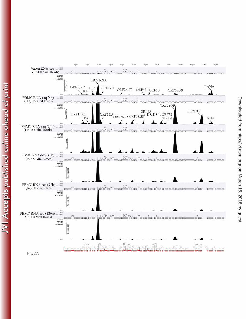
KSHV viral transcripts were abundantly present during the primary infection of human 269
PBMCs, CD14+ and TIVE cells. To determine the profiles of the viral transcripts expressed 270
during the primary infection of B and endothelial cells (natural target cells of KSHV), purified 271
virions were used for the de novo infection of human PBMCs, CD14+ monocytes, and 272
endothelial (TIVE) cells. An RNA-seq analysis was performed on total RNA extracted from 273
these infected cells at different time points (4, 24, 48, 72, and 120 hpi). Furthermore, 10 pM of 274
the quantified libraries were sequenced and mapped to the reference KSHV genome 275
(NC_009333). The transcriptome analysis revealed that several of the KSHV lytic and latent 276
transcripts accumulated as early as 4 hpi (Figs. 2A–C and 3). Apart from the ORF50 replication 277
and trans-activator protein (RTA), a number of other lytic transcripts involved in immune 278
modulation, lytic-DNA replication, and nucleic acid processing/synthesis were detected in 279
significant amounts early during the infection. These included polyadenylated nuclear (PAN) 280
RNA, ORF58/59, kaposin B, K2, K4, K6, ORF11, ORF17, ORF45, ORF27, ORF37, ORF57, 281
ORF64, ORF65, ORF73, and the recently identified T0.7. Among these, PAN RNA was the 282
most abundant as determined by the relative peak height (Figs. 2A–C) and RPKM values (Fig. 3) 283
in all the three tested cell types. Although the overall patterns of the viral gene expressions were 284
similar, i.e. majority of the genes expressing at 24hpi in all the three tested cells, CD14+ 285
on March 15, 2018 by guest
http://jvi.asm.org/
Dow
nloaded from

14
monocytes showed complete silencing of viral gene expressions, other than LANA, after 24hpi 286
(Figs. 2A–C and 3). However, the human PBMCs and TIVE showed detectable levels of PAN 287
RNA and ORF58/59 transcripts along with LANA after 48hpi (Figs. 2A–C and 3). This suggests 288
a cell type specific viral gene expression patterns during primary de novo infection and latency 289
establishment. Relative abundance of many of the viral transcripts including ORF1, K2, K4, T1.5, 290
PAN RNA, ORF17.5, ORF26, ORF27, ORF37 ORF38, ORF45, K8, K8.1, ORF52, vIRF-1, 291
ORF58/59, K12/T0.7 and LANA showed an increase in their abundance in PBMCs at 4hpi and 292
24hpi when compared with their abundance in the virions (Fig. 2A). However, the CD14+ 293
monocytes cells showed increases in the abundance of only fewer genes as compared to the 294
PBMCs (Fig. 2B). Importantly, LANA was the only gene abundantly detected after 72hpi in 295
these CD14+ cells representing a true latency model of KSHV infection. The endothelial cells 296
(TIVE) showed detectable levels of viral transcripts but their relative abundance increased only 297
slightly at 24hpi (Fig. 2C). The heat-maps generated based on the RPKM values showed an 298
increase in their copies from 4hpi to 24hpi, suggesting an active transcription and these included, 299
PAN RNA, ORF73, ORF45, ORF58, T0.7, Kaposin B, ORF59, K4, K6, ORF17, ORF65, 300
ORF27, K8.1, ORF11, ORF37, ORF38, ORF57, ORF69, ORF52, ORF54 and T1.5 (Fig. 3). The 301
viral DNA polymerase, ORF9 along with other lytic DNA replication proteins were detected but 302
their expression level did not increase (Fig. 3). Also, there were several other detectable 303
transcripts (K1, ORF4, ORF10, K6, ORF43, ORF44, ORF67, ORF68, and K14) but their 304
abundance did not increase and the RPKM values remained unchanged during the time course 305
tested, suggested that these genes did not transcribe but were present due to their transduction 306
through the virion particles during infection. 307
on March 15, 2018 by guest
http://jvi.asm.org/
Dow
nloaded from
15
Earlier studies reported that immediately after de novo infection, the herpesviruses express 308
genes involved in lytic DNA replication (28, 37, 38). Our RNA-seq analysis detected the 309
transcripts of proteins involved in viral DNA replication as early as 4 hpi, and these included 310
ORF59 (processivity factor), ORF9 (DNA polymerase), ORF6 (single-stranded DNA binding 311
protein), ORF56 (DNA replication protein), ORF40 (primase-associated factor), ORF54 312
(dUTPase) along with ORFs 60 and 61 (ribonucleoprotein reductase), ORF70 (thymidylate 313
synthase), and ORF 37 (alkaline exonuclease). Apart from these transcripts, several other 314
transcripts encoding the viral structural proteins ORF8 (glycoprotein B), K8.1 (glycoprotein), 315
and ORFs 64 and 75 (tegument protein) were also detected in our transcription profiling data. 316
Moreover, many of the noncoding RNAs, including PAN RNA, T0.7, and T1.5 (OriLyt 317
transcript), were abundantly detected in the RNA-seq analysis. RNA-seq profiling of the virion 318
encapsidated RNA, as relative abundance, showed packaging of a large number of viral 319
transcripts, which are brought into the target cells by the KSHV virion during the de novo 320
infection (Fig. 2 and Table 1). 321
In agreement with the previous report, our results showed the concurrent expression of 322
ORF50 and ORF73 transcripts during early primary infection (Figs. 2A–C and 3). However, the 323
expression of ORF50 decreased after 24 h, whereas the expression of ORF73 increased 324
exponentially (Fig. 3, heat maps). The KSHV latency-associated protein ORF73 is the 325
predominant protein expressed during latency and is responsible for immune evasion, latent 326
DNA replication, and genome maintenance, whereas the ORF50-encoded protein RTA activates 327
lytic cycle-specific KSHV genes in a cascaded manner to facilitate the lytic replication process 328
(39-43). In a cell culture system, the over-expression of RTA is capable of triggering the lytic 329
DNA replication (39, 43). Our data detected the transcripts of ORF50 and other lytic genes along 330
on March 15, 2018 by guest
http://jvi.asm.org/
Dow
nloaded from
16
with ORF73 during early infection suggest that KSHV may enter into a DNA replicative phase 331
before establishing latent infection. 332
Quantitative real-time PCR validation of the RNA sequencing analysis. To further 333
confirm the results of the transcriptome analysis by an independent method, we performed a real-334
time qPCR analysis on de novo-infected PBMCs harvested at different time points (4, 24, 48, 96, 335
and 120 hpi). A custom qPCR array of the KSHV genes in a 96-well format (Bar Harbor 336
Biotechnology) was used for the quantification of the viral mRNA levels. The fold-changes of 337
the individual KSHV genes were calculated for the various times post-infection using the Ct 338
method and plotted separately (Fig. 4). The viral genes showed expression kinetics similar to 339
those seen with the RNA-seq analysis. As first shown by the RNA-seq analysis, even at 4 hpi, 340
several lytic transcripts (ORF58/59, ORF11, ORF8, K8, K8.1, ORF17, ORF22, ORF57, ORF 45, 341
ORF27, ORF 37, and ORF64) were detected at high levels, and the expression of these 342
transcripts was gradually depleted after 24 hpi (Fig. 4). In addition, latent transcripts (ORF73, 343
K12, and ORF72) were consistently detected at significant levels during early infection (Fig. 4). 344
As reported previously (9), the real-time qPCR analysis showed the concurrent expression of 345
ORF50 and ORF73 at 4 hpi, and ORF50 showed a much higher fold-increase during early 346
infection than ORF 73. However, the expression of the ORF73 transcript increased exponentially 347
until 96 hpi. Additionally, the non-coding PAN RNA and recently identified T0.7 transcripts 348
were also detected abundantly at 4 hpi (Fig. 4). The qPCR validation of the RNA-seq analysis 349
suggested that the limited lytic-gene accumulation observed during primary infection might have 350
been influenced by the RTA mediated gene expression as well as by the transduction of viral 351
transcripts with the virions during the de novo infection. 352
on March 15, 2018 by guest
http://jvi.asm.org/
Dow
nloaded from
17
Detection of actively transcribing genes during de novo infection. The transcriptome 353
analysis of de novo-infected cells during early infection revealed a significantly higher 354
expression of several genes, which are expressed during lytic replication cycle including ORF50, 355
ORF6, ORF9, ORF8, ORF7, ORF10, ORF11, ORF22, ORF27, ORF31, ORF37, ORF40/41, 356
ORF45, ORF54, ORF55, ORF56, ORF58, ORF59, and ORF69 (Figs. 2A–C and 3). In addition, 357
many of the immediate-early KSHV unique genes such as K7, K8, K3, K5, K9, and K12, as well 358
as late K8.1, were also detected. Many of these transcripts have crucial regulatory roles in 359
immune evasion and nucleic acid processing and synthesis, which are associated with lytic-DNA 360
replication. The real-time qPCR analysis of the viral transcripts fully corroborated the RNA-seq 361
analysis (Fig. 4). 362
To further identify the actively transcribing genes during de novo infection, we used an 363
approach to capture the nascent transcribed RNA from the KSHV infected PBMCs by labeling 364
them with EdU and isolating them by Click-It approach. The sequencing of newly synthesized 365
RNA captured from the KSHV infected PBMCs showed peaks of many viral genes at 4hpi and 366
these included ORF2, K3, T1.5, PAN RNA, ORF26, ORF29, ORF36, ORF37, ORF40, ORF50, 367
K8.1, ORF55, vIRF-2, ORF58/59, ORF60, ORF63, ORF64, ORF67, T0.7, ORF72, ORF73 and 368
ORF74 (Fig. 5A). Importantly, majority of these were showing increased expression in relative 369
abundance graph as well as in the heat maps (Figs. 2, 3 and 5). Interestingly, majority of the 370
active transcription were limited to the ORF73, detected by the sequence reads, at 24hpi 371
suggested for the beginning of a latency establishment (Fig. 5, 24h panel). We also confirmed the 372
transcription during de novo infection by treating the cells with actinomycin D to block active 373
transcription. Comparison of the transcript levels at 4 hpi in the untreated with actinomycin D 374
treated cells revealed almost similar profiles to the nascent RNA capture data of gene 375
on March 15, 2018 by guest
http://jvi.asm.org/
Dow
nloaded from

18
transcription at 4hpi (data not shown). As expected, the ORF50 gene showed a moderate level of 376
de novo transcription within 4 hpi (Fig. 5). Interestingly, not all the genes required for lytic DNA 377
replication showed an active transcription during de novo infection, but were detected in RNA-378
seq analysis suggesting that those genes were transduced with the virions. Not surprisingly, the 379
ORF50 gene transcripts and those of genes regulated by ORF50 decreased after 24 hpi. Also, a 380
gradual increase in ORF73 transcripts by 24 hpi may aid in suppressing the expression of other 381
viral genes to promote the establishment of latency. 382
KSHV genome copy numbers increased exponentially after de novo infection. To 383
investigate whether the detection of lytic-reactivation genes leads to lytic-DNA replication and 384
genome amplification, we analyzed the KSHV genome copy number during the de novo 385
infection of PBMCs. Total DNA from different time points (0, 4, 24, 48, 72, 96, and 120 hpi) 386
were subjected to a real-time qPCR analysis to analyze the relative viral copies in the infected 387
cells. We used ORF73-specific primers and ORF73-plasmid standards to obtain the copy 388
numbers of the KSHV genome. The relative number of KSHV genome copies was calculated in 389
the real-time qPCR assay by amplifying the ORF73 gene and normalizing it against GAPDH. 390
KSHV viral DNA was detected as early as 4 hpi, which increased exponentially up to 48 hpi, and 391
then slightly decreased over time after establishing latency (Fig. 6A). This exponential increase 392
in the genome copies confirms for a genome replication during early infection till the 393
establishment of latency. The punctate immunolocalization of LANA in the de novo-infected 394
PBMCs (120 hpi) clearly demonstrated the establishment of latency in these cells (Fig. 6B). A 395
flow cytometry analysis of de novo-infected PBMCs using anti-LANA and B- and T-lymphocyte 396
markers gated for LANA expression showed that the populations of both the B- (~55.94%) and 397
T-lymphocytes (~24.75%) were successfully infected with KSHV (Fig. 6C and D). 398
on March 15, 2018 by guest
http://jvi.asm.org/
Dow
nloaded from

19
Since the viral genome copies increased about 6 fold at 48hpi from the 4hpi (the non-399
internalized virions were removed at 2hpi by treating the cells with trypsin), we wanted to 400
determine whether the virus has undergone to latent or lytic modes of DNA synthesis to amplify 401
the genome copies. To this end, we treated the cells with a viral DNA polymerase inhibitor, 402
phosphonoacetic acid (PAA) to block the lytic DNA replication. The cells pretreated with PAA 403
or untreated were infected with KSHV virions for 4h and 24h for the transcriptome analysis. 404
Analysis of the selected genes of latent, immediate early and early genes showed almost no 405
effect of PAA on the transcription of latent (ORF73) and immediate early genes (ORF50) at 4hpi 406
(Fig. 6E). However, the expression of late gene (ORF65) was significantly reduced with PAA at 407
both, 4 and 24hpi (Fig. 6D). We also determined the genome copies during de-novo infection of 408
PBMCs in the presence of PAA. The relative genome copies showed a significant reduction in 409
viral genome amplification in cells treated with PAA (Fig. 6F). Since there was still slight 410
increase in the viral genome copies at 24hpi, we speculate that other mechanism; besides the 411
lytic DNA replication, takes place during de novo infection of PBMCs. 412
KSHV virions packaged the viral transcripts. To analyze the RNA composition in the 413
encapsidated virions, total RNA extracted from the purified C-type KSHV virions was used for 414
the RNA-seq analysis. After confirming the purity of the RNA, the total RNA from two 415
independent virus purifications was used to construct cDNA libraries. After quantifying, 10 pM 416
of the mature library samples were used for the RNA-seq analysis. In comparison with previous 417
reports (26, 27), the transcriptome analysis with the CLC Genomic Workbench 7 software using 418
KSHV as the reference genome detected a larger number of KSHV mRNAs in the virions. A 419
previous DNA microarray study identified 11 KSHV mRNAs in the virions (27), but in this 420
study, we identified more than 60 KSHV-specific transcripts, including mRNAs for latent genes 421
on March 15, 2018 by guest
http://jvi.asm.org/
Dow
nloaded from

20
(Table 1). The mRNAs with 50 or more specific-gene reads and a 10-fold higher abundance in 422
the qPCR assays were considered significant (Fig. 7A and Table 1). The most abundant 423
transcripts detected in the virions included PAN RNA, K7, K14, K12, ORF58/59, and the 424
recently identified T0.7, with PAN RNA being the most abundant (Fig. 7A). In addition, many 425
lytic-specific mRNAs were also present in the virions, such as ORF50, ORF8, ORF9, ORF21, 426
ORF22, ORF31, ORF40/41, ORF45, ORF54, ORF55, ORF69, and ORF75. Moreover, many of 427
the immediate-early and early KSHV-unique K genes (K7, K8, K3, K5, K9) and the late K8.1 428
gene were also detected. Interestingly, in contrast to the previous report (27), a significant 429
amount of latent-specific ORF73 mRNA was detected in the virions (Fig. 7A). Both this and the 430
previous studies suggest that virion transcripts are released into the target cells immediately after 431
de novo infection (26, 27). This may be the reason for the immediate concurrent expression of 432
ORF50 and ORF73 in the target cells (9). It has been shown that proteins encoded by these 433
transcripts have significant regulatory roles in various cellular pathways, including cell signaling, 434
immune modulation, and apoptosis, that could critically affect the phenotype of the newly 435
infected cell and its microenvironment (14, 15, 44, 45) to provide the necessary factors required 436
for a successful infection. 437
Virion-packaged transcript specificity. To further clarify the RNA-Seq results, we 438
performed a real-time qPCR analysis on cDNA prepared from the same source of total RNA 439
(extracted from purified C-type KSHV virions) used in the sequencing analysis. A custom qPCR 440
array representing each of the KSHV-specific ORF primers was used for the analysis. The 441
uninfected PBMCs used as a reference control did not amplify, and a no-RT control was used to 442
ensure the quality of the RNA. The qPCR data showed results similar to those of the RNA-seq 443
analysis, confirming the presence of viral transcripts in the virions. 444
on March 15, 2018 by guest
http://jvi.asm.org/
Dow
nloaded from
21
To determine whether there is a specific mechanism of RNA encapsidation during virion 445
assembly, we compared the abundance of viral transcripts in the virions with the amounts present 446
during the lytic reactivation of the cells. An earlier study indicated that RNA encapsidation by 447
the KSHV virion could be a specific event (27), and a recent report on KSHV virion miRNA also 448
suggested that transcript encapsidation might be a specific process (26). To confirm this, we 449
performed transcriptome (Fig. 7B) and real-time qPCR analyses of the viral genes in the cDNA 450
extracted from induced BCBL1 cells for comparison with the transcripts present in the purified 451
virions (Fig. 7A and B). Many of the transcripts present in the virions were expressed in 452
abundance during lytic reactivation in the TRExBCBL1-RTA cells. To understand the specificity 453
of RNA encapsidation in the virion, we calculated the ratio of virion transcripts to the transcript 454
expression levels present during reactivation in the induced TRExBCBL1-RTA cells. Several of 455
the transcripts, including PAN RNA, T0.7, DR1, ORF58, and ORF59, that were detected in high 456
abundance in the virions were highly expressed during reactivation, and thus the ratios of 457
encapsidated to mRNA transcripts in the induced cells were lower. We therefore concluded that 458
they were packaged simply due to their abundance, not because of any specificity. However, 459
some of the transcripts, including ORF73, ORF31, ORF40, ORF50, ORF56, ORF49, and ORF64, 460
showed significantly higher ratios (≥0.5) of virion transcripts in comparison to their abundance 461
during reactivation, suggesting the involvement of a specific mechanism in their encapsidation. 462
The viral genes with the ratio of virion packaged to the induced cells above 0.5 were considered 463
significant and are marked by asterisks in Fig. 7C. Many of the virion-encapsidated latent- and 464
lytic-specific transcripts that were detected early during de novo infection showed a significantly 465
higher ratio; the ratio for the latent transcript ORF73 was 1.0843, whereas the ratio for the lytic 466
transcript ORF50 was 2.6103. Taken together, this suggests that KSHV may selectively 467
on March 15, 2018 by guest
http://jvi.asm.org/
Dow
nloaded from
22
encapsidates the latent and lytic transcripts required during early infection for priming the cells 468
to successfully establish latency. 469
Early ORF59 expression is required for KSHV de novo infection and genome 470
amplification. 471
Our transcriptome data showed a high expression of ORF59 during the primary early 472
infection of both B and endothelial cells, which led us to believe that ORF59 may be required 473
during the early events of KSHV infection and latency establishment. To this end, we used a 474
recombinant KSHV with a stop codon in the ORF59 gene (BAC36ΔORF59) (46) for de novo 475
infection. Knowing that the ORF59-deleted BAC36 cannot produce virion particles, we 476
generated a 293L-cell line stably expressing the ORF59 gene fused with DsRed to complement 477
the ORF59 in BAC36ΔORF59. These bacterial artificial chromosomes (BACs), WT in 293L and 478
BAC36ΔORF59 in ORF59-DsRed-complemented 293L, were transfected and selected with 479
hygromycin to obtain pure cell populations maintaining the BACs. These stable cells were 480
induced with sodium butyrate (NaB) and tetradecanoyl phorbol acetate to produce virion 481
particles, and the C-type virions were purified by sucrose density-gradient centrifugation for 482
DNA extraction and the infection studies. 483
To determine the composition of the KSHV virion-encapsidated RNA in BAC36WT and 484
BAC36ΔORF59, we performed an RNA-seq analysis on the virions from these two cell lines. 485
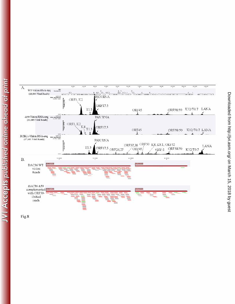
Unsurprisingly, the patterns of the viral mRNA transcripts were similar (Fig. 8A and Table 2). 486
We also compared the virion mRNA patterns from the BACs with those of the TRExBCBL1-487
RTA virions, which showed a slightly different packaging of the viral mRNA, but similar levels 488
of PAN RNA (Fig. 8A). Interestingly, the virions produced in the 293L cells complemented with 489
ORF59-DsRed and containing BAC36ΔORF59 also encapsidated ORF59, which was confirmed 490
on March 15, 2018 by guest
http://jvi.asm.org/
Dow
nloaded from
23
by mapping the sequence reads with ORF59-DsRed (Fig. 8B). The sequence reads from 491
BAC36WT mapped only to the ORF59 regions, confirming that BAC36WT packaged the 492
parental ORF59 gene (Fig. 8B), as expected. Interestingly, the total number of virions produced 493
by BAC36ΔORF59 was three logs lower than that produced by BAC36WT (Fig. 9B), even with 494
the ORF59-DsRed complementation, which is reflected in the C-type band intensity in the 495
BAC36ΔORF59 tube (Fig. 9A). 496
We normalized the number of virions used for the infection of human PBMCs by extracting 497
the virion DNA and quantifying it in a real-time qPCR assay. Cells were collected at 4, 24, 48, 498
72, 96, and 120 hpi and analyzed for viral gene expression and the number of KSHV genome 499
copies during early infection. Our results showed that both BAC36WT and BAC36ΔORF59 500
trans-complemented with ORF59-DsRed produced infectious virions and infected PBMCs (Fig. 501
9C). However, the KSHV genome copy analysis of the PBMCs infected with the trans-502
complemented BAC36ΔORF59 showed a reduced number of KSHV genome copies in 503
comparison to BAC36WT (Fig. 9C). This suggested that the lytic-cycle gene ORF59 is required 504
for amplifying the viral genome during primary infection, probably by allowing DNA replication 505
through lytic origins. 506
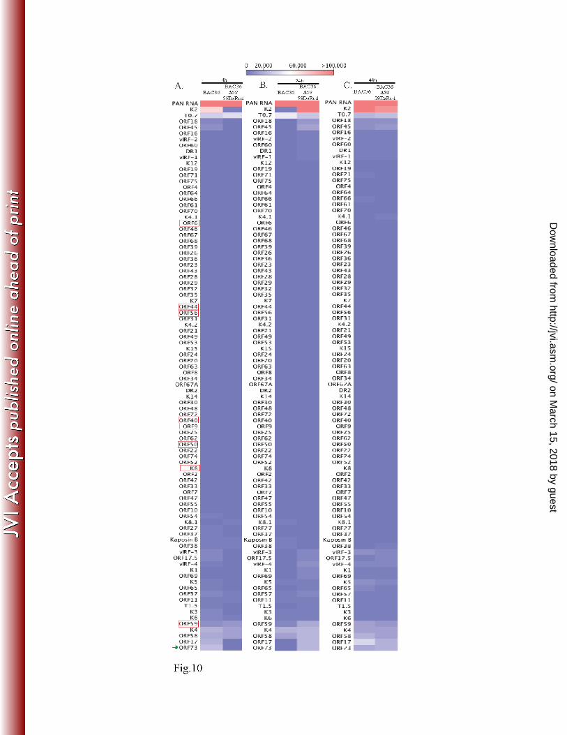
The total RNA extracted from the de novo-infected PBMCs at different time points (4, 24, and 507
48 hpi) were used for a transcriptome analysis to determine whether there were differences in the 508
viral gene expression profiles in the ORF59-deleted virus. The expression profiles of the viral 509
genes were determined based on RPKM values and compared with the wild-type virus at each of 510
the time points. The heat map generated based on the RPKM values showed subtle differences in 511
the viral gene expression profiles (Fig. 10). The genes required for lytic-DNA replication (ORF9, 512
ORF6, ORF40/41, ORF44, ORF56, ORF50, and K8), encircled by red boxes showed almost no 513
on March 15, 2018 by guest
http://jvi.asm.org/
Dow
nloaded from
24
difference in their expression profiles (Fig. 10). ORF59 showed a gradual decrease in virion-514
released ORF59 transcripts for the ORF59-deleted virus. However, BAC36WT showed a slight 515
increase in the number of ORF59 transcripts. The PAN RNA levels were comparable in the wild 516
type and ORF59-deleted viruses at the time points analyzed. The K2 and ORF17 expression was 517
distinctly different in the ORF59-deleted virus, being expressed earlier than in the PBMCs with 518
the BAC36WT virus (Fig. 10). Interestingly, the ORF59-deleted virus showed a higher 519
expression of the ORF73 gene (LANA) at earlier time points than did BAC36WT, suggesting an 520
early onset of latency in the virus lacking ORF59. Overall, these data suggest that ORF59 is 521
required for replicating the DNA and amplifying the viral genome during primary infection of 522
human PBMCs. 523
DISCUSSION: 524
KSHV infection is a complex multi-step process involving the regulation of various cellular 525
pathways. KSHV infects a variety of cellular targets and establishes a latent infection, generally 526
by 24 hpi (14, 16-20). To understand the differential expression of viral transcripts early during 527
infection, PBMCs, CD14+, and TIVE cells were de novo infected with KSHV for different 528
lengths of time (0, 4, 24, 48, 72, and 120 h). The transcriptome and real-time qPCR analyses on 529
the de novo-infected PBMCs revealed that several lytic- and latent-specific mRNAs were 530
concurrently expressed very early on following the de novo infections. Previous reports have 531
shown that immediately after de novo infection with herpesviruses such as EBV and herpes 532
simplex virus, the virus undergoes a short lytic DNA replication phase (37, 38). Our data also 533
showed the presence of the lytic DNA replication-associated transcripts, suggesting the 534
activation of a lytic cycle. Many of the lytic-cycle replication-associated transcripts were 535
detected as early as 4 hpi, including ORF59 (processivity factor), ORF9 (DNA polymerase), 536
on March 15, 2018 by guest
http://jvi.asm.org/
Dow
nloaded from
25
ORF6 (single-stranded DNA binding protein), ORF56 (DNA replication protein), ORF40 537
(primase associated factor), ORF54 (dUTPase), ORFs 60 and 61 (Ribonucleoprotein reductase), 538
ORF70 (thymidylate synthase), ORF37 (alkaline exonuclease). In addition, the ORF50, ORF2, 539
K3, PAN RNA, ORF26, ORF29, ORF37, ORF55, ORF60 ORF63, ORF73 and ORF74 540
transcripts were found to be actively transcribing at 4hpi detected by capturing the nascent RNA 541
and the actinomycin D treatment. 542
Apart from these lytic transcripts, the expression of several other transcripts encoding 543
structural proteins such as K8.1 (glycoprotein), and ORFs 65 (tegument protein) were also 544
detected early during infection. Moreover, many of the noncoding RNAs, including PAN RNA, 545
T0.7, and T1.5 (OriLyt transcript), were also expressed during early infection. PAN RNA has 546
been shown to modulate viral and cellular transcription as well as activate KSHV lytic cycle by 547
interacting with the ORF50 promoter and viral genome (47, 48). Furthermore, there were a large 548
number of viral transcripts, whose expression did not increase from their levels at 4hpi 549
suggesting that those were transduced with the virions. 550
The detection of PAN RNA in the virion and during infection suggests that PAN RNA could 551
be playing major role in triggering lytic cycle cascade. The KSHV ORF50-encoded immediate-552
early protein RTA is known to activate the cascade of lytic genes for facilitating the lytic 553
replication process (39-43). Studies on the primary infection of B cells by EBV showed an 554
accumulation of lytic transcripts during infection, indicating the requirement of a short lytic 555
replication prior to the establishment of latency (21, 23). Earlier studies have shown that the 556
KSHV virion also contains several KSHV lytic proteins, including ORF50, ORF8, ORF25, 557
ORF26, K12, ORF62, ORF63, and ORF75, that are brought into the target cells during de novo 558
infection (24). The ORF50 protein accompanying the virion may also be important for 559
on March 15, 2018 by guest
http://jvi.asm.org/
Dow
nloaded from
26
orchestrating the activation of RTA-responsive genes early on during infection to induce a short 560
lytic cycle. A significant amount of the concurrent expression of ORF50 and the KSHV latent 561
protein, ORF73 was detected as early as 4 hpi. Similar observations were reported in a previous 562
study based on microarray and real-time qPCR analyses, but the possibility of lytic DNA 563
replication was not proposed due to the lack of detecting all of the proteins required for lytic-564
DNA replication (9). 565
A recent study on histone modifications of the KSHV genome during de novo infection 566
demonstrated that there is a distinct pattern of activating the histone H3K4me3 mark across the 567
KSHV genome, which is gradually replaced by the repressive H3K27me3 mark by the LANA-568
mediated displacement of soluble sp100 (49). Additionally, in comparison to the repressive 569
H3K27me3 mark and the rest of the genome, a LANA ChIP-seq analysis of TRExBCBL1-RTA 570
and TIVE-LTC showed a clear association of H3K4me3 mark and RNA pol II activation on the 571
latent gene promoters (50). Our results showed that immediately after the de novo infection of 572
PBMCs, the viral genome increased exponentially up to 48 hpi, followed by a slight decrease at 573
72 hpi. The accumulation of ORF50 and other RTA-regulated lytic-cycle genes during early 574
infection is suggestive of the involvement of lytic-DNA replication for genome amplification. 575
Treating the cells with a late gene expression inhibitor, PAA showed significantly reduced viral 576
genome copies further substantiated the involvement of lytic DNA replication for genome copies 577
amplification in human PBMCs. 578
Discriminating whether the transcripts detected at 4 hpi were due to the virion-transduced 579
mRNA or active transcription, RNA-seq analysis of the newly synthesized transcripts at 4hpi of 580
human PBMCs showed transcription of only a limited number of genes during primary infection. 581
However, a large number of viral transcripts are detected at 4hpi as well as in the virions 582
on March 15, 2018 by guest
http://jvi.asm.org/
Dow
nloaded from
27
particles, which confirms that those transcripts are introduced into the infected cells along with 583
the virions. Detection of actively synthesizing mRNA in KSHV infected PBMCs at 24hpi 584
showed transcription of primarily the ORF73 gene, which confirmed that the viral genome gets 585
chromatinized and epigenetically modified for restricted gene expression by 24hpi. An earlier 586
study reported the viral gene transcription of ORF59 during primary infection (27), and here we 587
provide a comprehensive list of the genes transcribed during a de novo infection. The 588
transcription of the viral genes as early as 4 hpi suggests that the viral DNAs entering the targets 589
cells are capable of transcribing genes before the assembly of the epigenetic histone marks. Not 590
surprisingly, the latent protein ORF73 gene showed active transcription at 4 hpi, suggesting that 591
LANA begins transcription as early as 4 hpi and accumulates over time to help establish latency. 592
The immediate-early protein ORF50 undergoes a moderate level of transcription immediately 593
after infection, which may contribute in triggering the transcription of the lytic cascade to 594
complete the DNA replication. The KSHV virions have also been shown to encapsidate a small 595
quantity of the RTA protein, which may also be important in triggering the lytic-gene expression 596
cascade (24, 27). 597
It has been shown that both KSHV and EBV encapsidate biologically functional virion 598
transcripts that are transported into the target cells during de novo infections (22, 26, 27). A 599
previous study using a DNA microarray and real-time qPCR identified 11 of the virus transcripts 600
packaged into the virion (27). In this study, using RNA-seq and transcriptome analyses, we 601
detected additional KSHV latent- and lytic-cycle transcripts encapsidated in the virions that were 602
also transported into the targets cells. This data was validated by a real-time qPCR analysis using 603
KSHV gene-specific primer arrays, which showed consistent results. The transcriptome analysis 604
identified a variety of virion transcripts in high abundance, including PAN RNA, K7, K14, K12, 605
on March 15, 2018 by guest
http://jvi.asm.org/
Dow
nloaded from

28
ORF 58/59, and the recently identified T1.5 and T0.7. Additionally, many lytic-specific mRNAs 606
(ORF50, ORF8, ORF9, ORF21, ORF22, ORF31, ORF 40/41, ORF45, ORF54, ORF55, ORF 69, 607
and ORF75) were present in the virion in moderate amounts. A number of immediate-early and 608
early KSHV-unique K genes (K7, K8, K3, K5, K9, and late K8.1) were also detected in the 609
virion, in addition to a significant amount of latent-specific mRNAs (ORF73, ORF72, and K2). 610
The mechanism of RNA encapsidation by KSHV is currently unknown. Reports show that, 611
similarly to the herpes simplex virus, the RNA packaging and encapsidation by KSHV could be 612
a specific event (26, 27). In addition, the specific encapsidation of miRNA by the KSHV virion 613
has recently been demonstrated (26). However, both the previous study and this study 614
determined that the majority of mRNAs detected in the KSHV virions were present at very high 615
levels during lytic reactivation (9, 27). This suggests that these transcripts might have been 616
randomly packaged into the virions simply due to their higher abundance. Nonetheless, the 617
qPCR analysis to determine the specificity of the virion versus the reactivated TRExBCBL1-618
RTA showed that the ORF31, ORF40, ORF50, ORF56, ORF49, ORF64, and ORF73 transcripts 619
had significantly higher proportions of encapsidated mRNA than did the PAN RNA, ORF58, 620
ORF59, T0.7, and DR1 transcripts, suggesting a specific packaging mechanism for those 621
transcripts. 622
Several of these highly expressed KSHV lytic genes have been shown to have regulatory roles 623
in immune modulation, anti-apoptosis, and lytic DNA replication, indicating that their expression 624
may be priming the host cell for retaining the incoming viral DNA during the initial infection. 625
This is evident from the de novo infection of PBMCs with virions produced from the 626
BAC36ΔORF59 virus. Although significantly fewer virions were produced from 627
BAC36ΔORF59 than from BAC36WT, a similar number of virions were used to compare the 628
on March 15, 2018 by guest
http://jvi.asm.org/
Dow
nloaded from

29
viral genome copy numbers and expression profiles. The lower number of virion copies in 629
BAC36ΔORF59 complemented with ORF59-DsRed may have been due to a comparably lower 630
expression of ORF59 in those cells. The transcriptome analysis of the de novo-infected PBMCs 631
showed the differential expression of only a few latent- and lytic-specific genes (PAN RNA, 632
T0.7, K2, K4, ORF17, ORF18, ORF58, ORF45, and ORF73), confirming that the incoming 633
DNA is transcription competent. 634
The virions from both the BAC36WT and ORF59-complemented cells showed the packaging 635
of ORF59 transcripts. However, the BAC36ΔORF59 virus genome in de novo-infected PBMCs 636
failed to show an appreciable increase of copy number in comparison to BAC36WT. This 637
indicates that the extended expression of ORF59 during early infection is probably required for 638
synthesizing viral DNA through lytic DNA replication to increase the copy number. The exact 639
mechanism requiring the initial transient accumulation of lytic-specific transcripts for the 640
establishment of latency currently remains elusive. To fully understand the early events of 641
KSHV infection:- latency establishment and genome amplification, further experiments 642
identifying the viral proteins expressed and the mechanism of viral DNA replication during 643
primary infection in various cells are currently ongoing. 644
ACKNOWLEDGMENTS 645
We thank Prof. Erle S. Robertson at the University of Pennsylvania for providing the cell lines 646
and LANA expression plasmids. This work was supported by public health grants from the NIH 647
(CA174459 and AI105000) to SCV. STT was supported by Mick Hitchcock Fellowship at the 648
University of Nevada, Reno. 649
on March 15, 2018 by guest
http://jvi.asm.org/
Dow
nloaded from
30
REFERENCES 650
1. Chang PSMaY. 2010. Why do viruses cause cancer? Highlights of the first century of 651
human tumour virology. Nature Reviews Cancer 10:878-889. 652
2. Moore PS, Chang Y. 2003. Kaposi's sarcoma-associated herpesvirus 653
immunoevasion and tumorigenesis: two sides of the same coin? Annu Rev Microbiol 654
57:609-639. 655
3. Verma SC, Robertson ES. 2003. Molecular biology and pathogenesis of Kaposi 656
sarcoma-associated herpesvirus. FEMS Microbiol Lett 222:155-163. 657
4. Fakhari FD, Dittmer DP. 2002. Charting latency transcripts in Kaposi's sarcoma-658
associated herpesvirus by whole-genome real-time quantitative PCR. J Virol 659
76:6213-6223. 660
5. Jenner RG, Alba MM, Boshoff C, Kellam P. 2001. Kaposi's sarcoma-associated 661
herpesvirus latent and lytic gene expression as revealed by DNA arrays. J Virol 662
75:891-902. 663
6. Verma SC, Lan K, Robertson E. 2007. Structure and function of latency-associated 664
nuclear antigen. Curr Top Microbiol Immunol 312:101-136. 665
7. Zhong W, Wang H, Herndier B, Ganem D. 1996. Restricted expression of Kaposi 666
sarcoma-associated herpesvirus (human herpesvirus 8) genes in Kaposi sarcoma. 667
Proc Natl Acad Sci U S A 93:6641-6646. 668
8. Ballestas ME, Chatis PA, Kaye KM. 1999. Efficient persistence of 669
extrachromosomal KSHV DNA mediated by latency-associated nuclear antigen. 670
Science 284:641-644. 671
9. Krishnan HH, Naranatt PP, Smith MS, Zeng L, Bloomer C, Chandran B. 2004. 672
Concurrent expression of latent and a limited number of lytic genes with immune 673
modulation and antiapoptotic function by Kaposi's sarcoma-associated herpesvirus 674
early during infection of primary endothelial and fibroblast cells and subsequent 675
decline of lytic gene expression. J Virol 78:3601-3620. 676
10. Cannon JS, Ciufo D, Hawkins AL, Griffin CA, Borowitz MJ, Hayward GS, 677
Ambinder RF. 2000. A new primary effusion lymphoma-derived cell line yields a 678
highly infectious Kaposi's sarcoma herpesvirus-containing supernatant. J Virol 679
74:10187-10193. 680
11. Grundhoff A, Ganem D. 2004. Inefficient establishment of KSHV latency suggests 681
an additional role for continued lytic replication in Kaposi sarcoma pathogenesis. J 682
Clin Invest 113:124-136. 683
12. Renne R, Zhong W, Herndier B, McGrath M, Abbey N, Kedes D, Ganem D. 1996. 684
Lytic growth of Kaposi's sarcoma-associated herpesvirus (human herpesvirus 8) in 685
culture. Nat Med 2:342-346. 686
13. Wang CY, Sugden B. 2004. New viruses shake old paradigms. J Clin Invest 113:21-687
23. 688
14. Chandran B. 2010. Early events in Kaposi's sarcoma-associated herpesvirus 689
infection of target cells. J Virol 84:2188-2199. 690
15. Chakraborty S, Veettil MV, Chandran B. 2012. Kaposi's Sarcoma Associated 691
Herpesvirus Entry into Target Cells. Frontiers in microbiology 3:6. 692
on March 15, 2018 by guest
http://jvi.asm.org/
Dow
nloaded from
31
16. Akula SM, Naranatt PP, Walia NS, Wang FZ, Fegley B, Chandran B. 2003. Kaposi's 693
sarcoma-associated herpesvirus (human herpesvirus 8) infection of human 694
fibroblast cells occurs through endocytosis. J Virol 77:7978-7990. 695
17. Akula SM, Pramod NP, Wang FZ, Chandran B. 2002. Integrin alpha3beta1 (CD 696
49c/29) is a cellular receptor for Kaposi's sarcoma-associated herpesvirus 697
(KSHV/HHV-8) entry into the target cells. Cell 108:407-419. 698
18. Bechtel JT, Liang Y, Hvidding J, Ganem D. 2003. Host range of Kaposi's sarcoma-699
associated herpesvirus in cultured cells. J Virol 77:6474-6481. 700
19. Lagunoff M, Bechtel J, Venetsanakos E, Roy AM, Abbey N, Herndier B, McMahon 701
M, Ganem D. 2002. De novo infection and serial transmission of Kaposi's sarcoma-702
associated herpesvirus in cultured endothelial cells. J Virol 76:2440-2448. 703
20. Chakraborty S, Veettil MV, Bottero V, Chandran B. 2012. Kaposi's sarcoma-704
associated herpesvirus interacts with EphrinA2 receptor to amplify signaling 705
essential for productive infection. Proceedings of the National Academy of Sciences 706
of the United States of America 109:E1163-1172. 707
21. Halder S MM, Verma SC, Kumar P, Yi F, Robertson ES. 2009. Early events 708
associated with infection of Epstein-Barr virus infection of primary B-cells. PloS one 709
4. 710
22. Jochum S RR, Moosmann A, Hammerschmidt W, Zeidler R. 2012. RNAs in 711
Epstein-Barr virions control early steps of infection. Proceedings of the National 712
Academy of Sciences of the United States of America 109:1396-1404. 713
23. Wangrong Wen DI, Koji Yamamoto, Seiji Maruo, Teru Kanda, and Kenzo 714
Takada. 2007. Epstein-Barr Virus BZLF1 Gene, a Switch from Latency to Lytic 715
Infection, Is Expressed as an Immediate-Early Gene after Primary Infection of B 716
Lymphocytes. Journal of virology 81. 717
24. Bechtel JT, Winant RC, Ganem D. 2005. Host and viral proteins in the virion of 718
Kaposi's sarcoma-associated herpesvirus. J Virol 79:4952-4964. 719
25. Zhu FX, Chong JM, Wu L, Yuan Y. 2005. Virion proteins of Kaposi's sarcoma-720
associated herpesvirus. J Virol 79:800-811. 721
26. Lin X, Li X, Liang D, Lan K. 2012. MicroRNAs and unusual small RNAs discovered in 722
Kaposi's sarcoma-associated herpesvirus virions. Journal of virology 86:12717-723
12730. 724
27. Bechtel J, Grundhoff A, Ganem D. 2005. RNAs in the virion of Kaposi's sarcoma-725
associated herpesvirus. J Virol 79:10138-10146. 726
28. Toth Z, Brulois K, Lee HR, Izumiya Y, Tepper C, Kung HJ, Jung JU. 2013. Biphasic 727
euchromatin-to-heterochromatin transition on the KSHV genome following de novo 728
infection. PLoS pathogens 9:e1003813. 729
29. Verma SC, Bajaj BG, Cai Q, Si H, Seelhammer T, Robertson ES. 2006. Latency-730
associated nuclear antigen of Kaposi's sarcoma-associated herpesvirus recruits 731
uracil DNA glycosylase 2 at the terminal repeats and is important for latent 732
persistence of the virus. J Virol 80:11178-11190. 733
30. Verma SC, Choudhuri T, Kaul R, Robertson ES. 2006. Latency-associated nuclear 734
antigen (LANA) of Kaposi's sarcoma-associated herpesvirus interacts with origin 735
recognition complexes at the LANA binding sequence within the terminal repeats. J 736
Virol 80:2243-2256. 737
on March 15, 2018 by guest
http://jvi.asm.org/
Dow
nloaded from
32
31. Hirt B. 1967. Selective extraction of polyoma DNA from infected mouse cell 738
cultures. . J. Mol. Biol. 26:365-369. 739
32. Lallemand F, Desire N, Rozenbaum W, Nicolas JC, Marechal V. 2000. Quantitative 740
analysis of human herpesvirus 8 viral load using a real-time PCR assay. J Clin 741
Microbiol 38:1404-1408. 742
33. Bates I, Bedu-Addo G, Jarrett RF, Schulz T, Wallace S, Armstrong A, Sheldon J, 743
Rutherford T. 2001. B-lymphotropic viruses in a novel tropical splenic lymphoma. 744
Br J Haematol 112:161-166. 745
34. Medveczky MM, Horvath E, Lund T, Medveczky PG. 1997. In vitro antiviral drug 746
sensitivity of the Kaposi's sarcoma-associated herpesvirus. AIDS 11:1327-1332. 747
35. Chang PJ, Boonsiri J, Wang SS, Chen LY, Miller G. 2010. Binding of RBP-Jkappa 748
(CSL) protein to the promoter of the Kaposi's sarcoma-associated herpesvirus 749
ORF47 (gL) gene is a critical but not sufficient determinant of transactivation by 750
ORF50 protein. Virology 398:38-48. 751
36. Nealon K, Newcomb WW, Pray TR, Craik CS, Brown JC, Kedes DH. 2001. Lytic 752
replication of Kaposi's sarcoma-associated herpesvirus results in the formation of 753
multiple capsid species: isolation and molecular characterization of A, B, and C 754
capsids from a gammaherpesvirus. J Virol 75:2866-2878. 755
37. Sabyasachi Halder MM, Subhash C. Verma, Pankaj Kumar, Fuming Yi, Erle S. 756
Robertson. 2009. Early Events Associated with Infection of Epstein-Barr 757
Virus Infection of Primary B-Cells. PloS one 4. 758
38. Roizman BaDMK. 2001. Herpes Simplex Viruses and Their Replication. Chapter 72. 759
In: Fields Virology, Fourth Edition, D.M. Knipe, P.M. Howley et al., eds., Lippincott, 760
Williams & Wilkins, Philadelphia, PA. :pp. 2399-2459. 761
39. Guito J, Lukac DM. 2012. KSHV Rta Promoter Specification and Viral Reactivation. 762
Frontiers in microbiology 3:30. 763
40. Dourmishev LA, Dourmishev AL, Palmeri D, Schwartz RA, Lukac DM. 2003. 764
Molecular genetics of Kaposi's sarcoma-associated herpesvirus (human 765
herpesvirus-8) epidemiology and pathogenesis. Microbiol Mol Biol Rev 67:175-212, 766
table of contents. 767
41. Sun R, Lin SF, Staskus K, Gradoville L, Grogan E, Haase A, Miller G. 1999. 768
Kinetics of Kaposi's sarcoma-associated herpesvirus gene expression. J Virol 769
73:2232-2242. 770
42. West JT, Wood C. 2003. The role of Kaposi's sarcoma-associated 771
herpesvirus/human herpesvirus-8 regulator of transcription activation (RTA) in 772
control of gene expression. Oncogene 22:5150-5163. 773
43. Nakamura H, Lu M, Gwack Y, Souvlis J, Zeichner SL, Jung JU. 2003. Global 774
changes in Kaposi's sarcoma-associated virus gene expression patterns following 775
expression of a tetracycline-inducible Rta transactivator. J Virol 77:4205-4220. 776
44. Chan SR, Bloomer C, Chandran B. 1998. Identification and characterization of 777
human herpesvirus-8 lytic cycle-associated ORF 59 protein and the encoding cDNA 778
by monoclonal antibody. Virology 240:118-126. 779
45. Unal A, Pray TR, Lagunoff M, Pennington MW, Ganem D, Craik CS. 1997. The 780
protease and the assembly protein of Kaposi's sarcoma-associated herpesvirus 781
(human herpesvirus 8). J Virol 71:7030-7038. 782
on March 15, 2018 by guest
http://jvi.asm.org/
Dow
nloaded from

33
46. McDowell ME, Purushothaman P, Rossetto CC, Pari GS, Verma SC. 2013. 783
Phosphorylation of Kaposi's sarcoma-associated herpesvirus processivity factor 784
ORF59 by a viral kinase modulates its ability to associate with RTA and oriLyt. J 785
Virol 87:8038-8052. 786
47. Rossetto CC, Pari G. 2012. KSHV PAN RNA associates with demethylases UTX and 787
JMJD3 to activate lytic replication through a physical interaction with the virus 788
genome. PLoS pathogens 8:e1002680. 789
48. Rossetto CC, Pari GS. 2011. Kaposi's sarcoma-associated herpesvirus noncoding 790
polyadenylated nuclear RNA interacts with virus- and host cell-encoded proteins 791
and suppresses expression of genes involved in immune modulation. J Virol 792
85:13290-13297. 793
49. Gunther T, Schreiner S, Dobner T, Tessmer U, Grundhoff A. 2014. Influence of 794
ND10 components on epigenetic determinants of early KSHV latency establishment. 795
PLoS pathogens 10:e1004274. 796
50. Hu J, Yang Y, Turner PC, Jain V, McIntyre LM, Renne R. 2014. LANA binds to 797
multiple active viral and cellular promoters and associates with the 798
H3K4methyltransferase hSET1 complex. PLoS Pathog 10:e1004240. 799
800
Figure Legends: 801
Figure 1: Purification of KSHV Virion. (A) Virions were purified from culture supernatant of 802
approximately 9x10⁸
reactivated TRExBCBL1-RTA cells by a 20-50% sucrose gradient 803
ultracentrifugation. The three white bands as indicated represent A type (empty), B type 804
(intermediate), and C type (mature) KSHV virus particles, respectively. (B) Western blot on 805
purified virions for the presence of KSHV envelope glycoprotein K8.1 by immunoblotting with 806
anti-K8.1 antibody. Total lysate from induced TRExBCBL1-RTA was used as a positive control. 807
(C) Supernatants of uninduced or induced TRExBCBL1-RTA cells were used for real time 808
qPCR quantification of viral genome copy using ORF73 specific primers along with ORF73 809
plasmid standards. Approximately 8.06x10⁷ copies of viral DNA representing C-type virions 810
were detected. 811
Figure 2A: Transcriptome analysis of KSHV during de novo infection of human PBMCs. 812
Total RNA extracted from de novo infected PBMCs harvested at different time points 4h, 24h, 813
48h, 72h, and 120h post-infection were subjected for cDNA library preparation using TrueSeq 814
on March 15, 2018 by guest
http://jvi.asm.org/
Dow
nloaded from
34
RNA-seq library Kit. The libraries were sequenced using HiSeq and the sequences were mapped 815
to the reference KSHV genome to determine the relative abundance of viral transcripts. Purified 816
virions treated with micrococcal nuclease to eliminate the nucleic acid contamination were 817
subjected for total RNA extraction. RNA treated with DNAse was used for sequencing after 818
cDNA library preparation. The number of transcripts mapping to the viral genome are shown in 819
parenthesis on each panel. The peak height represents the number of reads for indicated genes 820
and the predominant peaks are marked in virion RNA-seq and 4h post-infection samples. PAN 821
RNA showed the highest peak in virions as well as KSHV infected PBMCs. Bottom panel shows 822
the location of KSHV genes on the coordinates. 823
Figure 2B: Transcriptome analysis of de novo infected CD14 (+) cells. Total RNA extracted 824
from de novo infected CD14 (+) cells harvested at different time points 4h, 24h, 48h, 72h, and 825
96h post-infection were subjected for cDNA library preparation using TrueSeq RNA-seq library 826
Kit. The libraries were sequenced using HiSeq and the sequences were mapped to the reference 827
KSHV genome to determine the relative abundance of viral transcripts. The number of 828
transcripts mapping to the viral genome are shown in parenthesis on each panel. The peak height 829
representing the number of reads for indicated genes are marked in 4h post-infection and 24h 830
post-infection samples. PAN RNA showed the highest peak until 48h post-infection followed by 831
decline to very low levels. LANA expression progressively increased showing establishment of 832
latency. Bottom panel shows the location of KSHV genes on the coordinates. 833
Figure 2C: Transcriptome analysis of de novo infected TIVE cells. Total RNA extracted from 834
de novo infected TIVE cells harvested at different time points 4h, 24h, 48h, 72h, and 120h post-835
infection were subjected for cDNA library preparation using TrueSeq RNA-seq library Kit. The 836
libraries were sequenced using HiSeq and the sequences were mapped to the reference KSHV 837
on March 15, 2018 by guest
http://jvi.asm.org/
Dow
nloaded from

35
genome to determine the relative abundance of viral mRNA. The number of transcripts mapping 838
to the viral genome are shown in parenthesis on each panel. The peak height representing the 839
number of reads for indicated genes are marked in 4h post-infection and 24h post-infection 840
samples. Sample of 72h post-infection showed lower peaks because of lower number of total 841
reads, which were normalized by calculating the RPKM values in the heat-maps. Bottom panel 842
shows the location of KSHV genes on the coordinates. 843
Figure 3: Transcriptome analysis of KSHV in de novo infected PBMCs, TIVE and CD14 844
(+) cells. RPKM values, calculated based the number of reads for each gene, were used for 845
analyzing relative expression of KSHV genes as heat-maps. Hierarchal clustering of genes was 846
performed using CLC Workbench 7.0. Increasing RPKM values are represented from blue to red. 847
(A) KSHV transcriptome from de novo infected human PBMCs. (B) KSHV transcriptome from 848
de novo infected human TIVEs. (C) KSHV transcriptome from de novo infected human CD14 849
(+) cells. 850
Figure 4: Real time qPCR validation of de novo infected PBMCs. Real time qPCR analysis 851
was performed on the cDNA prepared from total RNA extracted harvested at 4h, 24h, 48h, 96h 852
and 120h post-infection of human PBMCs. A 96 well format qPCR plate representing each of the 853
KSHV specific ORF primer array was used for the qPCR analysis. Fold change of individual 854
KSHV transcripts at various time points are plotted. The lytic genes are shown in red color. 855
Figure 5: Viral genes transcribing at 4hpi and 24hpi identified by nascent RNA capture 856
approach. Human PBMCs infected with KSHV virions were incubated with EdU (alkyne) at 857
4hpi and 24hpi to label the newly transcribing RNA. Total RNA extracted from these cells was 858
subjected for click-reaction with biotin-azide (Invitrogen, Inc.). DMSO instead of biotin-azide 859
was used in control click-reaction. Newly synthesized, biotin labeled RNA was enriched by 860
on March 15, 2018 by guest
http://jvi.asm.org/
Dow
nloaded from
36
streptavidin magnetic beads as per manufacturer’s instruction. These enriched RNA were 861
subjected for RNA-seq analysis by Hi-seq and the sequences were mapped to KSHV reference 862
genome (NC_00039) using CLC Bio Workbench 7.5. Relative copies of the newly synthesized 863
viral RNA were determined by ratios of specific (biotin labeled) and non-specific (DMSO) 864
sequence reads. 865
Figure 6: KSHV genome copies exponentially increases after infection. (A) Approximately 866
8x10⁷ human PBMCs were infected with KSHV isolated from reactivated TRExBCBL1-RTA, 867
with a multiplicity of infection MOI=10. De novo infected PBMCs were harvested at different 868
time points 4h, 24h, 48h, 72h, 96h and 120h post-infection followed by extracting total DNA 869
using modified Hirt’s lysis method. This DNA was used to analyze the genome copy number by 870
real time qPCR using ORF73 specific primers and ORF73 plasmid as standard. (B) Indirect 871
immuno fluorescence assay (IFA) for LANA on de novo infected PBMCs. KSHV infected 872
PBMCs 120h post-infection were subjected to IFA with rat anti-LANA antibody followed by 873
detection with Alexa Fluor 488 secondary antibody. Nuclear stain TO-PRO-3 was used for 874
staining the nuclei. LANA is shown in green and nuclear stain TO-PRO 3 is shown in blue. (C, 875
D) Flow cytometry analysis of de novo infected PBMCs. KSHV infected PBMCs 120h post-876
infection were subjected to flow cytometry analysis to detect the percentage of B and T-877
lymphocytes infected with KSHV. Mouse anti-CD19 was used for the detection of B-878
lymphocytes and mouse anti-CD3 was used for the detection of T-lymphocytes. Rat anti-LANA 879
was used to gate KSHV positive cells. Data were acquired on FACSCalibur equipped with 880
CellQuest Pro software and analyzed using FlowJo software. E) PAA treatment reduced the 881
expression of late genes. Untreated or PAA pre treated human PBMCs were infected with KSHV 882
virions (10 MOI) for 4h or 24h. Total RNA extracted at 4hpi and 24hpi were subjected for the 883
on March 15, 2018 by guest
http://jvi.asm.org/
Dow
nloaded from
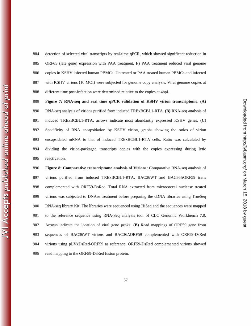
37
detection of selected viral transcripts by real-time qPCR, which showed significant reduction in 884
ORF65 (late gene) expression with PAA treatment. F) PAA treatment reduced viral genome 885
copies in KSHV infected human PBMCs. Untreated or PAA treated human PBMCs and infected 886
with KSHV virions (10 MOI) were subjected for genome copy analysis. Viral genome copies at 887
different time post-infection were determined relative to the copies at 4hpi. 888
Figure 7: RNA-seq and real time qPCR validation of KSHV virion transcriptome. (A) 889
RNA-seq analysis of virions purified from induced TRExBCBL1-RTA. (B) RNA-seq analysis of 890
induced TRExBCBL1-RTA, arrows indicate most abundantly expressed KSHV genes. (C) 891
Specificity of RNA encapsidation by KSHV virion, graphs showing the ratios of virion 892
encapsidated mRNA to that of induced TRExBCBL1-RTA cells. Ratio was calculated by 893
dividing the virion-packaged transcripts copies with the copies expressing during lytic 894
reactivation. 895
Figure 8: Comparative transcriptome analysis of Virions: Comparative RNA-seq analysis of 896
virions purified from induced TRExBCBL1-RTA, BAC36WT and BAC36ΔORF59 trans 897
complemented with ORF59-DsRed. Total RNA extracted from micrococcal nuclease treated 898
virions was subjected to DNAse treatment before preparing the cDNA libraries using TrueSeq 899
RNA-seq library Kit. The libraries were sequenced using HiSeq and the sequences were mapped 900
to the reference sequence using RNA-Seq analysis tool of CLC Genomic Workbench 7.0. 901
Arrows indicate the location of viral gene peaks. (B) Read mappings of ORF59 gene from 902
sequences of BAC36WT virions and BAC36ΔORF59 complemented with ORF59-DsRed 903
virions using pLVxDsRed-ORF59 as reference. ORF59-DsRed complemented virions showed 904
read mapping to the ORF59-DsRed fusion protein. 905
on March 15, 2018 by guest
http://jvi.asm.org/
Dow
nloaded from

38
Figure 9: De novo infection of PBMCs with BAC36WT and BAC36ΔORF59 virions. (A) 906
Virions were purified from culture supernatant of approximately 9x10⁸
reactivated 293L cells 907
harboring either BAC36WT or BAC36ΔORF59 on 20-50% sucrose gradient. The three white 908
bands as indicated represent A type (empty), B type (intermediate), and C type (mature) KSHV 909
virus particles, respectively. (B) Supernatants from 293L cells containing BAC36WT or 910
BAC36ΔORF59 were used for real time qPCR quantification of viral genome copy using ORF73 911
specific primers along with ORF73 plasmid standards. (C) Human PBMCs were infected with 912
KSHV isolated from reactivated 293L cells containing BAC36WT or BAC36ΔORF59, with a 913
multiplicity of infection MOI=10. De novo infected PBMCs were harvested at different time 914
points 4h, 24h, 48h, 72h, 96h and 120h post-infection followed by extracting total DNA using 915
modified Hirt’s lysis method. This DNA was used for analyzing the genome copy number by 916
real time qPCR using ORF73 specific primers and ORF73 plasmid as standard. 917
Figure 10: Expression profiles of viral genes in BAC36 WT and BAC36ΔORF59 at 918
different time post-infection. Normalized expression (RPKM values) of viral genes in BAC36 919
WT and BAC36ΔORF59 at different time post-infection was used for generating hierarchal 920
clustering. (A) Relative expression of KSHV genes at 4h post-infection of PBMCs with BAC36 921
WT and BAC36ΔORF59 virions. (B) Relative expression of KSHV genes at 24h post-infection 922
of PBMCs with BAC36 WT and BAC36ΔORF59 virions. (C) Relative expression of KSHV 923
genes at 48h post-infection of PBMCs with BAC36 WT and BAC36ΔORF59 virions. The red 924
boxes encircle KSHV genes required for lytic DNA replication. Latency Associated gene, LANA 925
is indicated with green arrow, which showed early expression in cells infected with 926
BAC36ΔORF59. 927
on March 15, 2018 by guest
http://jvi.asm.org/
Dow
nloaded from
39
Table 1: List of mRNAs detected in the TRExBCBL1-RTA virions: Compiled list of mRNAs 928
detected in the RNA-seq analysis from purified TRExBCBL1-RTA virions. Total gene reads 929
above 50 and a qPCR relative fold change above 10 were considered significant. The list also 930
shows RPKM values and total gene reads. The list also includes mRNA transcribed during de 931
novo 4h post-infection of human PBMCs. The asterisk represents virion specific mRNAs 932
identified in this study. 933
Table 2: RPKM and gene reads of viral genes packaged in BAC36WT and ORF59-BAC36 934
virions. 935
936
on March 15, 2018 by guest
http://jvi.asm.org/
Dow
nloaded from

Table 1: KSHV transcripts in BCBL1 virions by RNA-seq analysis.
No mRNA RPKM Gene reads
Function 4h PBMC de novo transcribed
Status * This study
1 ORF4 2633.55 101 Complement control protein (KCP) regulates complement activation * 2 ORF6 3804.2 300 ssDNA binding protein essential for lytic DNA replication * 3 ORF7 2332.60 113 Probably DNA packaging * 4 ORF8 4432.43 261 Virion envelope-associated glycoprotein * 5 ORF9 5318.55 375 KSHV DNA polymerase * 6 ORF10 6446.37 188 Specific suppressor of IFN signaling * 7 ORF11 6275.82 191 Viral lytic protein Yes * 8 K2 7218.64 103 K2(vIL-6) regulate cellular proliferation Yes Bechtel et al (4) 9 K3 6004.86 135 E3 ubiquitin ligases that reduce surface expression in infected cells * 10 ORF70 5568.35 131 A homolog of the thymidylate synthase (vTS) * 11 K4.2 4632.04 59 * 12 T1.5 10245.93 358 * 13 K6 10027.11 67 vMIP-IA Bechtel et al (4) 14 PAN RNA 159519.90 3986 Late gene expression, lytic DNA replication, immune modulation Bechtel et al (4) 15 ORF17 13534.71 504 Yes Bechtel et al (4) 16 ORF18 2840.02 51 Late gene regulation * 17 ORF19 3134.66 120 * 18 ORF21 3140.50 127 Thymidine kinase * 19 ORF22 1729.56 88 Glycoprotein H * 20 ORF23 2305.84 65 Predicted glycoprotein * 21 ORF24 2823.83 148 Essential for DNA replication * 22 ORF25 3338.78 320 Major capsid protein * 23 ORF26 3239.66 69 Minor capsid protein * 24 ORF27 6270.22 127 Glycoprotein Yes * 25 ORF29 3163.27 390 Packaging protein * 26 ORF34 2233.92 51 * 27 ORF35 5613.67 59 * 28 ORF36 4907.44 152 Serine protein kinase Yes * 29 ORF37 2625.62 89 Alkaline exonuclease -Sox protein Yes * 30 ORF39 2149.70 60 Glycoprotein M * 31 ORF40 2904.36 144 Helicase/primase * 32 ORF44 2676.778 147 Helicase * 33 ORF45 6972.31 198 Virion-associated tegument protein regulating IFN function Yes * 34 ORF46 7969.30 142 Uracil deglycosylase * 35 ORF48 3101.60 87 Activates JNK/p38 * 36 ORF50 4389.71 309 Replication transcriptional activator protein (RTA) * 37 K8 4072.23 89 bZIP Yes Bechtel et al (4) 38 K8.1 3918.32 71 Glycoprotein Yes * 39 ORF52 6095.17 56 Tegument protein Yes * 40 ORF54 9659.02 199 dUTPase / Immune modulation Bechtel et al (4) 41 ORF55 6364.41 101 Tegument protein * 42 ORF56 3830.11 225 DNA replication * 43 ORF57 3533.39 121 Lytic replication protein homologous to EBV SM protein- mRNA splicing Yes * 44 vIRF-1 7407.08 232 Immune modulation Yes * 45 vIRF-4 2324.47 153 Facilitates lytic replication by targeting cellular IRF4 and Myc gene Yes * 46 vIRF-3 2302.58 96 Immune modulation * 47 vIRF-2 2429.94 122 Type I IFN signaling * 48 ORF58 16775.11 418 Yes Bechtel et al (4) 49 ORF59 26201.13 724 Processivity factor Yes Bechtel et al (4) 50 ORF60 4601.25 98 Ribonucleoprotein reductase * 51 ORF61 3659.74 202 Ribonucleoprotein reductase * 52 ORF63 2891.99 187 NLR homolog * 53 ORF64 3897.02 715 Deubiquitinase Yes * 54 ORF65 5629.25 67 Capsid protein * 55 ORF66 5646.64 169 Capsid protein * 56 ORF67 4753.85 90 Nuclear egress complex * 57 ORF68 5372.34 175 Glycoprotein * 58 ORF69 6353.81 134 BRLF2 nuclear egress Yes * 59 T0.7 9310.93 151 * 60 ORF72 3118.46 56 vCyclin * 61 ORF73 23623.23 1858 Latency associated nuclear antigen (LANA) * 62 K14 3116.41 59 vOX2 * 63 ORF74 4481.89 107 vGPCR * 64 ORF75 3810.57 344 Tegument protein - vFGARAT Yes *
on March 15, 2018 by guest
http://jvi.asm.org/
Dow
nloaded from

Table 2: Viral mRNA in BAC36WT and ΔORF59-BAC36 virions.
No mRNA BAC36WT virion BAC36Δ59 virion Function
RPKM Gene reads RPKM Gene reads 1 K1 4106.397 65 3545.13 36 Glycoprotein
2 ORF11 9104.682 210 5136.19 76 Viral lytic protein
3 K2 264559.9 3066 1800100.6 1339 K2 (vIL-6) regulate cellular proliferation
4 K3 13800.78 252 1621.957 19 E3 ubiquitin ligases that reduce surface expression in infected cells
5 K4 14151.28 76 17124.46 59 vMIP-II
6 T1.5 2572.319 73 3735.02 68 7 K5 9016.62 131 4291.56 40 E3 Ubiquitin ligase
8 K6 10134.38 55 2010.55 7 vMIP-IA
9 PAN RNA 456221 9259 554384.1 7218 Late gene expression, lytic DNA replication, immune modulation
10 ORF16 5125.818 51 2506.66 16 Bcl2 homolog
11 ORF17 61895.31 1872 40406.45 784 Protease
12 ORF18 12752.6 186 13786.64 129 Late gene regulation
13 ORF45 5332.74 123 4798.29 71 Virion-associated tegument protein regulating IFN function
14 ORF54 4601.55 77 94.15 1 dUTPase / Immune modulation
15 ORF57 16862.17 469 4203.24 75 Lytic replication protein homologous to EBV SM protein- mRNA splicing
16 vIRF-4 6565.60 351 10321.76 354 Facilitates lytic replication by targeting cellular IRF4 and Myc gene
17 vIRF-3 8888.84 301 11738.21 255 Immune modulation
18 vIRF-2 2305.14 94 2493.35 77 Type I IFN signaling
19 ORF58 167550.29 339 17252.55 224
20 ORF59 9980.75 224 14863.18 214 Processivity factor
21 ORF60 3005.99 52 2793.37 31 Ribonucleoprotein reductase
22 ORF61 1672.91 75 1599.45 46 Ribonucleoprotein reductase
23 ORF64 899.21 134 1234.312 118 Deubiquitinase
24 ORF65 8585.93 83 8384.85 52 Capsid protein
25 ORF66 3085.30 75 3077.94 48 Capsid protein
26 T0.7 10097.21 125 18224.4 154
27 ORF73 2081.99 133 1445.63 68 Latency Associated Nuclear Antigen (LANA)
28 ORF50 192.39 11 545.28 20 Replication and Transcription Activator (RTA)
on March 15, 2018 by guest
http://jvi.asm.org/
Dow
nloaded from