Transaxillary Subpectoral Placement of Cardiac riginal ... · Symptom (s) Indication Device Model,...
8
34 Copyright © 2017 The Korean Society of Plastic and Reconstructive Surgeons This is an Open Access article distributed under the terms of the Creative Commons Attribution Non-Commercial License (http://creativecommons.org/ licenses/by-nc/4.0/) which permits unrestricted non-commercial use, distribution, and reproduction in any medium, provided the original work is properly cited. www.e-aps.org Original Article INTRODUCTION Cardiac implantable electronic devices (CIEDs) are an essential part of cardiologic treatment in high-risk patients with a wide range of structural or functional heart diseases [1,2]. Over the last few decades, numerous procedural changes have been made Transaxillary Subpectoral Placement of Cardiac Implantable Electronic Devices in Young Female Patients Joo Hyun Oh 1 , Chae Min Kim 1 , Seung Yong Song 1 , Jae Sun Uhm 2 , Dae Hyun Lew 1 , Dong Won Lee 1 Departments of 1 Plastic and Reconstructive Surgery and 2 Cardiology, Severance Hospital, Yonsei University College of Medicine, Seoul, Korea Background The current indications of cardiac implantable electronic devices (CIEDs) have expanded to include young patients with serious cardiac risk factors, but CIED placement has the disadvantage of involving unsightly scarring and bulging of the chest wall. A collaborative team of cardiologists and plastic surgeons developed a technique for the subpectoral placement of CIEDs in young female patients via a transaxillary approach. Methods From July 2012 to December 2015, subpectoral CIED placement via an axillary incision was performed in 10 young female patients, with a mean age of 25.9 years and mean body mass index of 20.1 kg/m 2 . In the supine position, with the patient’s shoulder abducted, an approximately 5-cm linear incision was made along one of the deepest axillary creases. The submuscular plane was identified at the lateral border of the pectoralis major, and the dissection continued over the clavipectoral fascia until the subpectoral pocket could securely receive a pulse generator. Slight upward dissection also exposed an entrance to the subclavian vein, allowing the cardiology team to gain access to the vein. One patient with dilated cardiomyopathy underwent augmentation mammoplasty and CIED insertion simultaneously. Results One case of late-onset device infection occurred. All patients were highly satisfied with the results and reported that they would recommend the procedure to others. Conclusions With superior aesthetic outcomes compared to conventional methods, the subpectoral placement of CIEDs via a transaxillary approach is an effective, single-incision method to hide operative scarring and minimize bulging of the device, and is particularly beneficial for young female or lean patients. Keywords Defibrillators, implantable / Pacemaker, artificial / Cardiac resynchronization therapy Correspondence: Dong Won Lee Institute for Human Tissue Restoration, Department of Plastic and Reconstructive Surgery, Severance Hospital, Yonsei University College of Medicine, 50-1 Yonsei-ro, Seodaemun-gu, Seoul 03722, Korea Tel: +82-2-2228-2215 Fax: +82-2-393-6947 E-mail: [email protected] This study was presented at the Sixth Research and Reconstructive Forum of the Korean Society of Plastic Surgeons on April 7th, 2016 in Daegu, Korea. No potential conflict of interest relevant to this article was reported. Received: 22 Apr 2016 • Revised: 5 Oct 2016 • Accepted: 19 Oct 2016 pISSN: 2234-6163 • eISSN: 2234-6171 • https://doi.org/10.5999/aps.2017.44.1.34 • Arch Plast Surg 2017;44:34-41
-
Upload
phungkhuong -
Category
Documents
-
view
219 -
download
1
Transcript of Transaxillary Subpectoral Placement of Cardiac riginal ... · Symptom (s) Indication Device Model,...
34
Copyright © 2017 The Korean Society of Plastic and Reconstructive SurgeonsThis is an Open Access article distributed under the terms of the Creative Commons Attribution Non-Commercial License (http://creativecommons.org/ licenses/by-nc/4.0/) which permits unrestricted non-commercial use, distribution, and reproduction in any medium, provided the original work is properly cited. www.e-aps.org
Orig
inal
Art
icle
INTRODUCTION
Cardiac implantable electronic devices (CIEDs) are an essential
part of cardiologic treatment in high-risk patients with a wide range of structural or functional heart diseases [1,2]. Over the last few decades, numerous procedural changes have been made
Transaxillary Subpectoral Placement of Cardiac Implantable Electronic Devices in Young Female PatientsJoo Hyun Oh1, Chae Min Kim1, Seung Yong Song1, Jae Sun Uhm2, Dae Hyun Lew1, Dong Won Lee1
Departments of 1Plastic and Reconstructive Surgery and 2Cardiology, Severance Hospital, Yonsei University College of Medicine, Seoul, Korea
Background The current indications of cardiac implantable electronic devices (CIEDs) have expanded to include young patients with serious cardiac risk factors, but CIED placement has the disadvantage of involving unsightly scarring and bulging of the chest wall. A collaborative team of cardiologists and plastic surgeons developed a technique for the subpectoral placement of CIEDs in young female patients via a transaxillary approach.Methods From July 2012 to December 2015, subpectoral CIED placement via an axillary incision was performed in 10 young female patients, with a mean age of 25.9 years and mean body mass index of 20.1 kg/m2. In the supine position, with the patient’s shoulder abducted, an approximately 5-cm linear incision was made along one of the deepest axillary creases. The submuscular plane was identified at the lateral border of the pectoralis major, and the dissection continued over the clavipectoral fascia until the subpectoral pocket could securely receive a pulse generator. Slight upward dissection also exposed an entrance to the subclavian vein, allowing the cardiology team to gain access to the vein. One patient with dilated cardiomyopathy underwent augmentation mammoplasty and CIED insertion simultaneously.Results One case of late-onset device infection occurred. All patients were highly satisfied with the results and reported that they would recommend the procedure to others.Conclusions With superior aesthetic outcomes compared to conventional methods, the subpectoral placement of CIEDs via a transaxillary approach is an effective, single-incision method to hide operative scarring and minimize bulging of the device, and is particularly beneficial for young female or lean patients.
Keywords Defibrillators, implantable / Pacemaker, artificial / Cardiac resynchronization therapy
Correspondence: Dong Won LeeInstitute for Human Tissue Restoration, Department of Plastic and Reconstructive Surgery, Severance Hospital, Yonsei University College of Medicine, 50-1 Yonsei-ro, Seodaemun-gu, Seoul 03722, KoreaTel: +82-2-2228-2215Fax: +82-2-393-6947E-mail: [email protected]
This study was presented at the Sixth Research and Reconstructive Forum of the Korean Society of Plastic Surgeons on April 7th, 2016 in Daegu, Korea.
No potential conflict of interest relevant to this article was reported.
Received: 22 Apr 2016 • Revised: 5 Oct 2016 • Accepted: 19 Oct 2016pISSN: 2234-6163 • eISSN: 2234-6171 • https://doi.org/10.5999/aps.2017.44.1.34 • Arch Plast Surg 2017;44:34-41

Vol. 44 / No. 1 / January 2017
35
in pacemaker implantation, mostly attributable to technological advancements throughout multiple generations of the devices [3]. The size of the pulse generators has also decreased over time, making it possible, most notably in pediatric patients, to replace abdominal implantation of an epicardial pacemaker with an endocardial device in the prepectoral or subpectoral space.
The clinical use of CIEDs has expanded to include not only middle-age patients with chronic conditions, but also younger patients predisposed to sudden cardiac death. However, several problems have arisen with conventional subcutaneous device insertion in the subclavian area, most notably the conspicuous contour of the pacemaker in the subcutaneous pocket of the an-terior chest and/or an often unsightly surgical scar (Fig. 1) [4].
With this problem in mind, a collaborative team of cardiolo-gists and plastic surgeons in our institution developed a tech-nique for the subpectoral placement of CIEDs in young female patients via a transaxillary approach. This transaxillary approach has been frequently utilized in augmentation mammoplasty to hide visible operative scars in the axillary fold, and therefore our procedure can be deemed an extension of a familiar surgical ap-proach for a different purpose. We present our experience with this procedure in 10 young female patients, including 1 patient who received augmentation mammoplasty together with CIED placement.
METHODS
PatientsFrom July 2012 to December 2015, subpectoral CIED place-ment via the transaxillary approach was performed in 10 young
Patient no.
Age (yr)
Height (cm)
Weight (kg)
BMI (kg/m2)
Symptom (s) Indication DeviceModel, company, and
countryFollow-up
(wk)
1 38 151.6 53.5 23.28 Palpitation, chest discomfort Sick sinus syndrome Pacemaker Evia, Biotronik, Germany 214.4
2 29 166 55 19.96 Sudden cardiac arrest Ventricular fibrillation ICD Incepta ICD, Boston Scientific, United States
130.4
3 34 155 51 21.09 Syncope Dilated cardiomyopathy ICD Ellipse DR, St. Jude Medical, United States
126.0
4 16 162 49 18.67 Sudden cardiac arrest Dilated cardiomyopathy ICD Ellipse DR, St. Jude Medical, United States
76.0
5 22 151 51.9 22.49 Dizziness Chemotherapy-induced dilated cardiomyopathy
CRT-D VivaQuad XT CRT-D, Medtronic, Ireland
65.4
6 37 154 48 20.24 Syncope High-degree atrioventricular block
Pacemaker Accolade EL, Boston Scientific, United States
59.1
7 18 160 48.3 18.75 Syncope Sick sinus syndrome Pacemaker Accolade EL, Boston Scientific, United States
57.4
8 25 153 43 18.37 Syncope Hypertrophic cardiomyopathy ICD Evera MRI XT DR, Medtronic, Ireland
32.4
9 20 162.1 53 20.17 Dyspnea, dizziness High-degree atrioventricular block
Pacemaker Advisa DR MRI, Medtronic, Ireland
36.3
10a) 20 161 45.2 17.44 Dyspnea Dilated cardiomyopathy ICD Ellipse DR, St. Jude Medical, United States
84.1
Mean 25.9 157.6 49.8 20.05 - - - 88.2
CIED, cardiac implantable electronic device; BMI, body mass index; ICD, implantable cardioverter-defibrillator; CRT-D, cardiac resynchronization therapy defibrillator. a)A patient who received concomitant augmentation mammoplasty together with CIED placement.
Table 1. Patient demographics, cardiologic indications for CIED implantation with device information, and postoperative follow-up periods
Conspicuous scarring and disfiguring bulging often result from the conventional subcutaneous insertion of cardiac implantable elec-tronic devices in the subclavian area.
Fig. 1. Cardiac device in a conventional subclavian pocket

Oh JH et al. Transaxillary subpectoral CIED placement
36
female patients who were referred from a group of 3 cardiolo-gists at our institution (Table 1).
All subjects were young and lean female patients with a mean age of 25.9 years (range, 16–38 years) and a mean body mass in-dex (BMI) of 20.1 kg/m2 (range, 17.44–23.28 kg/m2). The mean postoperative follow-up period was 88.2 weeks.
The patients had been diagnosed with a range of anatomical and functional cardiac abnormalities. Five patients had been di-agnosed with various types of cardiomyopathy, of whom 3 had dilated cardiomyopathy, 1 had chemotherapy-induced cardio-myopathy, and 1 patient had hypertrophic cardiomyopathy. Three patients had symptomatic sick sinus syndrome. Two pa-tients had high-degree atrioventricular block, and 1 patient had ventricular fibrillation.
All patients were highly concerned with any noticeable physi-cal changes on their chest following the CIED insertion, and they wanted any surgical scar to be concealed as much as possi-ble. One patient with dilated cardiomyopathy inquired about the possibility of combining augmentation mammoplasty using the conventional endoscopic approach and CIED insertion un-der the same subpectoral plane, and we carried out a combina-tion of these 2 procedures in that patient.
Each patient was informed that the inserted device would re-quire a battery change every 10 years at the time of battery de-pletion, and that the same subpectoral plane would be used again during each of these future procedures.
We also reviewed all surgical complications that could lead to device removal, such as postoperative pain, device migration, lead dislodgement, and infection.
Surgical techniqueAll patients were under general anesthesia throughout the entire surgical procedure. In the supine position, the patient’s shoulder was abducted to expose the axillary area and the lateral border of the pectoralis major muscle. On the axillary fossa, a linear in-cision approximately 5–7 cm in length was made along one of the deepest axillary creases, mostly perpendicular to the lateral border of the pectoralis major. Care was taken not to extend this incision beyond the anterior axillary fold. The subcutaneous dissection proceeded, with undermining remaining close to the lateral border of the pectoralis major muscle, since major neuro-vascular structures are located posteriorly under the axillary fat pad. The scrub assistants, if any, should be informed not to re-tract the lateral tissue too strongly, because this can stretch the intercostobrachial nerve and the patient may complain of numbness of the upper medial aspect of the affected arm after surgery.
When the fascia of the pectoralis major was encountered at its
lateral border, the submuscular plane was identified while main-taining intact attachment of the pectoralis minor muscle to the chest wall. The clavipectoral fascia enclosing the pectoralis mi-nor muscle can be readily differentiated from the pectoral fascia of the pectoralis major. The dissection continued over the clavi-pectoral fascia; otherwise, further dissection under the pectora-lis minor may damage vital structures, such as the thoracoacro-mial artery, cephalic vein, or lateral pectoral nerve, which may appear beyond the muscle. The medial pectoral nerve mainly supplies the pectoralis minor and partly the pectoralis major, and it may also be exposed and inadvertently damaged during undermining. This alone, however, would not result in signifi-cant functional sequelae. Under the submuscular plane of the pectoralis major, dissection is relatively easy without being dis-turbed by arterial perforators, and the surgeon continued until a pocket became large enough to securely receive a cardiac pulse generator. Slight upward dissection also exposed an entrance window to the subclavian vein, allowing the cardiology team to easily gain access to the vein (Fig. 2).
Using the Seldinger technique, the subclavian vein was punc-tured, and a cardiologist introduced the ventricular lead and, if necessary, an atrial lead via a 9-Fr guiding sheath while stably anchoring them on the right ventricular apex and right atrial ap-pendage, respectively. The ventricular and atrial leads were then connected to a pulse generator, and device function was evalu-ated using wireless telemetry with the device in place. The de-vice was then stably fixed onto either the periosteum of the rib bone or the outermost fascia of the intercostalis muscle with ab-sorbable sutures, and 1 or 2 other additional sutures were made into the surrounding tissues (Fig. 3). A negative-pressure drain
The device is placed inside the subpectoral pocket along the mid-clavicular level, and is fixated with absorbable sutures onto the chest wall. A slight upward dissection provides a window to the subclavian vein through which the atrial and ventricular leads are introduced.
Fig. 2. Device positioning using the transaxillary approach

Vol. 44 / No. 1 / January 2017
37
was inserted, and the subcutaneous tissue was repaired in layers, first with absorbable sutures and then with nonabsorbable su-tures for skin closure (Fig. 4).
Blood pressure and other signs of hemodynamics should be carefully monitored during the entire procedure, particularly while the patient is sitting up for an evaluation of the shape and position of the implants.
Postoperative pain was usually not remarkable, and analgesia use was similar to or less than that observed in patients who un-dergo augmentation mammoplasty. We maintained negative-pressure drains until the daily amount of drainage became less than 20 mL.
RESULTS
Cardiac implantable electronic devices All patients were treated with subpectoral CIED insertion at a single institution by 2 plastic surgeons in collaboration with 3 cardiologists (Table 1). Four patients received pacemakers, 5 re-ceived implantable cardioverter defibrillators (ICDs), and 1 pa-tient was treated with a cardiac resynchronization therapy defi-brillator.
Patient satisfactionIn general, all patients were highly satisfied with the outcomes. They responded with an average of 8.5 points on a scale of 1 to 10 when asked about how much they were pleased with the benefits of the transaxillary procedure. Some patients com-plained of minor discomfort, such as temporary hypoesthesia on and around the incision area. Some did not like the fact that they were required to limit their arm movements for the first
few postoperative weeks. However, all patients said that they would recommend this procedure to others of the same age with similar cardiac conditions.
ComplicationsOne case of device pocket infection occurred. A bacterial swab culture and subsequent blood culture revealed methicillin-resis-tant Staphylococcus aureus. The device was ultimately surgically removed. One month after device removal, when the inflamma-tion had subsided completely, a new device was inserted into the right anterior chest wall using the conventional subcutane-ous plane.
Another patient experienced mild tingling on the medial as-pect of the right upper arm, which resolved spontaneously with-in several weeks. No other significant complications were found in the other patients.
DISCUSSION
As implantable cardioverter defibrillators have become a main-stay of antiarrhythmic treatments for the primary and secondary prevention of sudden cardiac death, providing substantial bene-fits in terms of utility, efficacy, and safety [1,2,5], several re-searchers have investigated the quality of life among recipients of these devices [6,7]. Moreover, the indications for CIEDs are expanding to include younger patients, with the early recogni-tion of inherited cardiac syndromes, such as long QT syndrome, Brugada syndrome, and hypertrophic cardiomyopathy.
Once implanted, CIEDs should be maintained for the rest of the patient’s life, and the physical and psychological impact can therefore be much greater among young patients. In addition,
Intraoperative view of an implantable cardioverter-defibrillator pulse generator inside the subpectoral pocket placed via the trans-axillary approach.
Fig. 3. Pulse generator in the subpectoral pocket
Immediate postoperative view after negative-pressure drainage in-sertion and wound closure. Note that the axillary incision did not extend beyond the anterior axillary fold.
Fig. 4. Immediate postoperative view of the axillary area

Oh JH et al. Transaxillary subpectoral CIED placement
38
these patients will experience psychological discomfort if visible scars and bulging of the device are not addressed properly, lead-ing to a more intense need to alleviate the stigma experienced by patients.
The psychosocial impact of CIEDs can be of particular con-cern, making young female patients most vulnerable. Vazquez re-ported that younger female patients showed worse perceptions of shock anxiety, death anxiety, and body image distress than old-er female patients [4]. In 2012, Marshall et al. [8] reported that female patients were more distressed than male patients about the impact of CIEDs on their appearance, especially women younger than 39 years of age. Earlier research also revealed that younger CIED patients had concerns about various issues, such as the fit of clothing, socialization, and sexual activity.
The conventional technique, which involves a subclavian skin incision with subcutaneous pocket formation, fails to address these quality of life issues. CIED recipients have often been stig-matized due to the operative scar in the infraclavicular area, which is highly susceptible to hypertrophic scarring, because unusual tension is applied due to the convexity of the chest wall and weight of the breast. The superficial placement of the device also involves a greater risk of skin ulceration or device exposure.
Several alternative surgical approaches, therefore, have been suggested in response to these problems. A surgical approach through the inframammary fold was the first alternative route described in the literature in an effort to place the pacemaker or CIED behind the breast tissue or pectoralis major muscle. In 1983, Belott and Bucko [9] first presented his experience with 2 female patients utilizing an inframammary approach for pace-maker placement. Allan [10], in 1985, published a case report describing his use of the familiar inframammary approach em-ployed in augmentation mammoplasty to implant a pacemaker. In 1993, Kolettis et al.[11] reported successful retromammary CIED device placement with an endocardial lead system in a 62-year-old woman. These early attempts were done exclusively using a subglandular pocket, followed by Ozin et al. [12] and Schaverien et al. [13].
Giudici [14] and Collegues [15], in 2001 and 2010, published his comprehensive experience with the subpectoral placement of pacemakers and CIEDs in 51 female patients over a 9-year period. He used the subpectoral plane to place the devices with the skin incision on the inframammary fold for device insertion and a separate 15–20-mm incision on the anterior axillary line for lead insertion. Patient satisfaction was high, and 3 cases of lead dislodgement and 1 case of pneumothorax were reported as complications. Persichetti also presented, in 2014, 30 consec-utive female patients who underwent submuscular implantation or substitution of CIED devices, which was the largest clinical
series for submuscular ICD implantation [16]. The inframam-mary approach leads to excellent cosmetic results, since the op-erative scar is seen only when the patient raises her arm. Howev-er, as a 2-incision method, it still requires another skin incision in the infraclavicular area for lead insertion and narrow subcuta-neous tunneling to the device pocket behind the breast.
Other alternative routes include subpectoral placement via a conventional infraclavicular incision. In 1992, Hammel et al. [17] inserted a CIED under the pectoralis major with a cephalic venotomy incision in an obese woman. In 2014, Asamura et al. [18] presented results from 21 male patients for the secondary replacement of CIED devices; by using the previous operative scar in the infraclavicular area, he inserted the devices under the pectoralis major muscle by splitting the muscle fibers between the clavicular and sternocostal attachment points. Half of the patients were lean, with a BMI under 18.5 kg/m2, and the bulge from the device became much smoother after the procedure than is the case for conventional subcutaneous pockets, al-though an infraclavicular scar was still obvious.
A skin incision via the lateral border of the pectoralis muscle was also utilized by several researchers, particularly among pedi-atric patients. Molina [19], in 1991, published his long-term ex-perience with 83 adult female patients and 21 pediatric patients. Two skin incisions, a short subclavian puncture and a longer lat-eral breast incision, were used to insert the device in the sub-glandular pocket in female patients. Several other reports of subpectoral pacemaker insertion for pediatric patients were published by Shefer et al. [20] in 1996 and Rosenthal [21] in 2000, using a direct skin incision along the lateral border of the pectoralis major. However, this operative scar on anterior axil-lary fold was still readily visible upon even slight arm abduction.
Despite the variety of these previous approaches, we believe that the transaxillary approach is the most suitable method for young female patients. It can effectively conceal the operative scar in the axillary fossa, as it can be disguised between natural axillary creases, as long as the operator does not extend the inci-sion anteriorly beyond the anterior axillary fold. The bulge from the device is hardly noticeable, allowing the patients to continue their normal lives without continuously paying attention to the existence of the cardiac device (Fig. 5). This approach may eliminate even the small inconveniences in daily life caused by the bulge of the device when wearing a seatbelt or carrying a bag with shoulder straps.
The young female patients in our study also showed a high level of satisfaction regarding the benefits and outcomes of the procedure, and unanimously said that they would recommend this transaxillary subpectoral approach to those in similar cir-cumstances.
Vol. 44 / No. 1 / January 2017
39
This approach is already time-tested in cosmetic plastic sur-gery with regard to operative scar management. Moreover, it does not necessitate a separate skin incision in the infraclavicular area, since the subclavian vein can also be approached via a sin-gle axillary incision. An additional skin stab incision, no matter how short it may be, can be a psychological burden for the pa-tient if it is located in a potentially visible area.
The transaxillary approach is not regarded as a technically de-manding task for a plastic surgeon. When performing cosmetic augmentation mammoplasty, bleeding from perforators can be troublesome given the limited surgical field, but the subpectoral pocket for CIED placement is laterally positioned and small, such that medial perforators are rarely encountered. Plastic sur-geons should not find it difficult to implement this transaxillary CIED placement procedure, since it is a simpler form of an al-ready familiar approach, only for a different purpose.
Our first patient experienced bacterial infection inside the de-vice pocket, and the CIED eventually had to be removed. How-ever, we encountered no other complications, such as device dislocation, lead dislodgement, or pneumothorax. Device dislo-cation is a very unlikely occurrence, since the device is fixated with absorbable sutures tightly onto the chest wall fascia or peri-
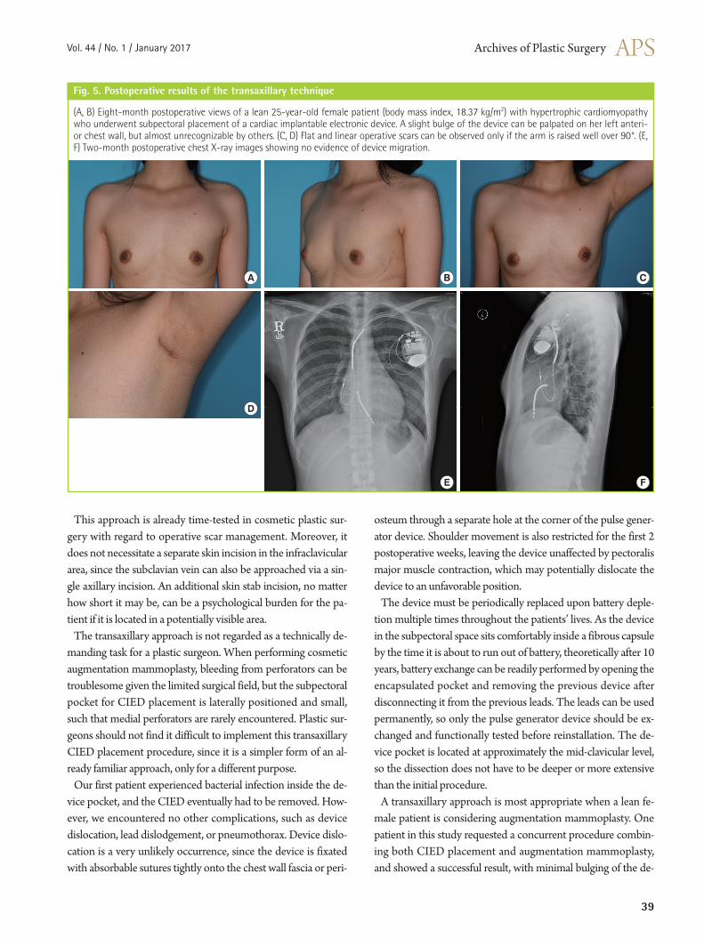
Fig. 5. Postoperative results of the transaxillary technique
(A, B) Eight-month postoperative views of a lean 25-year-old female patient (body mass index, 18.37 kg/m2) with hypertrophic cardiomyopathy who underwent subpectoral placement of a cardiac implantable electronic device. A slight bulge of the device can be palpated on her left anteri-or chest wall, but almost unrecognizable by others. (C, D) Flat and linear operative scars can be observed only if the arm is raised well over 90°. (E, F) Two-month postoperative chest X-ray images showing no evidence of device migration.
A
D
B
E
C
F
osteum through a separate hole at the corner of the pulse gener-ator device. Shoulder movement is also restricted for the first 2 postoperative weeks, leaving the device unaffected by pectoralis major muscle contraction, which may potentially dislocate the device to an unfavorable position.
The device must be periodically replaced upon battery deple-tion multiple times throughout the patients’ lives. As the device in the subpectoral space sits comfortably inside a fibrous capsule by the time it is about to run out of battery, theoretically after 10 years, battery exchange can be readily performed by opening the encapsulated pocket and removing the previous device after disconnecting it from the previous leads. The leads can be used permanently, so only the pulse generator device should be ex-changed and functionally tested before reinstallation. The de-vice pocket is located at approximately the mid-clavicular level, so the dissection does not have to be deeper or more extensive than the initial procedure.
A transaxillary approach is most appropriate when a lean fe-male patient is considering augmentation mammoplasty. One patient in this study requested a concurrent procedure combin-ing both CIED placement and augmentation mammoplasty, and showed a successful result, with minimal bulging of the de-

Oh JH et al. Transaxillary subpectoral CIED placement
40
vice and no additional cardiologic risk. When the procedure is combined with augmentation mammoplasty, the surgical ap-proach is largely identical, as the mammary implants are insert-ed under the same submuscular plane, but the pocket of the breast implant can be distinguished from that of the more supe-riorly positioned CIED. The pocket dissection should not be too extensive compared with the size and shape of the cardiac device and breast implant. Otherwise, the breast implant be-comes susceptible to gradual migration.
Despite the benefits of the transaxillary approach for subpec-toral placement, not all medical centers may be able to perform this procedure, since it requires a certain degree of dedication from cardiologists and plastic surgeons alike. For cardiologists, the use of a transaxillary approach in the subpectoral plane uti-lizes a completely different set of procedures, so they should not be expected to learn this approach in a center without plastic surgeons. This technique can be made possible by the collabora-tive efforts of dedicated cardiologists and plastic surgeons.
To date, this is the largest clinical series to be published on the subpectoral placement of CIEDs via a transaxillary approach, which results in superior aesthetic outcomes compared to con-ventional methods, because this approach is an effective, single-incision method to obscure operative scars and minimize bulg-ing of the device. Patients do not have to be aware of the device at all times, and they can remain as socially active as before the procedure. Young female or lean patients are specific subgroups that would benefit the most from this approach.
REFERENCES
1. Connolly SJ, Hallstrom AP, Cappato R, et al. Meta-analysis of the implantable cardioverter defibrillator secondary pre-vention trials: AVID, CASH and CIDS studies. Antiarrhyth-mics vs Implantable Defibrillator study. Cardiac Arrest Study Hamburg. Canadian Implantable Defibrillator Study. Eur Heart J 2000;21:2071-8.
2. Goldberger Z, Lampert R. Implantable cardioverter-defi-brillators: expanding indications and technologies. JAMA 2006;295:809-18.
3. Hammill SC, Kremers MS, Stevenson LW, et al. Review of the registry’s fourth year, incorporating lead data and pediat-ric ICD procedures, and use as a national performance mea-sure. Heart Rhythm 2010;7:1340-5.
4. Vazquez LD, Kuhl EA, Shea JB, et al. Age-specific differenc-es in women with implantable cardioverter defibrillators: an international multi center study. Pacing Clin Electrophysiol 2008;31:1528-34.
5. A comparison of antiarrhythmic-drug therapy with implant-
able defibrillators in patients resuscitated from near-fatal ventricular arrhythmias: the Antiarrhythmics versus Im-plantable Defibrillators (AVID) Investigators. N Engl J Med 1997;337:1576-83.
6. Sears SF Jr, Todaro JF, Lewis TS, et al. Examining the psy-chosocial impact of implantable cardioverter defibrillators: a literature review. Clin Cardiol 1999;22:481-9.
7. Thomas SA, Friedmann E, Kao CW, et al. Quality of life and psychological status of patients with implantable cardio-verter defibrillators. Am J Crit Care 2006;15:389-98.
8. Marshall P, Ketchell A, Maclean J. Comparison of male and female psychological outcomes related to implantable car-dioverter defibrillators (COMFORTID). Eur J Cardiovasc Nurs 2012;11:313-21.
9. Belott PH, Bucko D. Inframammary pulse generator place-ment for maximizing cosmetic effect. Pacing Clin Electro-physiol 1983;6:1241-4.
10. Allan D. Augmentation mammaplasty approach to pace-maker implantation. Ann Plast Surg 1985;15:242-3.
11. Kolettis TM, Saxena A, Krol RB, et al. Submammary im-plantation of a cardioverter-defibrillator with nonthoracoto-my lead system. Am Heart J 1993;126:1222-3.
12. Ozin B, Borman H, Bozbas H, et al. Implantation of sub-mammary implantable cardioverter defibrillators. Pacing Clin Electrophysiol 2004;27:779-82.
13. Schaverien MV, Elder D, Munnoch DA. Inframammary car-diac pacemaker insertion in women: an aesthetic alternative. Plast Reconstr Surg 2013;131:464e-465e.
14. Giudici MC. Experience with a cosmetic approach to device implantation. Pacing Clin Electrophysiol 2001;24:1679-80.
15. Giudici MC, Carlson JI, Krupa RK, et al. Submammary pacemakers and ICDs in women: long-term follow-up and patient satisfaction. Pacing Clin Electrophysiol 2010;33: 1373-5.
16. Persichetti P, Brunetti B, Cagli B, et al. Aesthetic subpectoral placement of implantable cardioverter defibrillators. Ann Plast Surg 2014;72:188-92.
17. Hammel D, Block M, Borggrefe M, et al. Implantation of a cardioverter/defibrillator in the subpectoral region com-bined with a nonthoracotomy lead system. Pacing Clin Electrophysiol 1992;15:367-8.
18. Asamura S, Kurita T, Motoki K, et al. Efficacy and feasibility of the submuscular implantation technique for an implant-able cardiac electrical device. Eplasty 2014;14:e40.
19. Molina JE. New technique for pacemaker implantation in the upper chest of children and women. Ann Thorac Surg 1991;51:992-5.
20. Shefer A, Lewis BS, Gang ES. The retropectoral transaxil-

Vol. 44 / No. 1 / January 2017
41
lary permanent pacemaker: description of a technique for percutaneous implantation of an “invisible” device. Pacing Clin Electrophysiol 1996;19:1646-51.
21. Rosenthal E. A cosmetic approach for pectoral pacemaker implantation in young girls. Pacing Clin Electrophysiol 2000;23:1397-400.