Subpectoral Bicipital Tendon Tenodesis with All Suture · PDF fileSubpectoral Bicipital Tendon...
11
Transcript of Subpectoral Bicipital Tendon Tenodesis with All Suture · PDF fileSubpectoral Bicipital Tendon...


Subpectoral Bicipital Tendon Tenodesis with All Suture AnchorJohn G. Coury, DO; Eric G. Huish, DO; Robert Cash, MD
IntroductionTendinitis involving the long head of the biceps occurs as an inflammatory tenosynovitis along the course of the tendon within the bicipital groove of the humerus.[1] Surgical management is indicated for partial-thickness tear of the long head of the biceps tendon from 25-50%, medial subluxation of the tendon, and tendon subluxation in addition to a tear of the subscapularis tendon or biceps pulley.[2-4] When comparing tenotomy and tenodesis, the literature suggest that tenodesis provides beneficial closer restoration of normal anatomy, prevents loss of strength and cramping, and has less risk for cosmetic, popeye, deformity. The subpectoral tenodesis location has proven to be a reasonable method as it allows the surgeon to assess and treat any intra-articular pathology, prevents cosmetic deformity, and provides technical simplicity.[5]
Materials & MethodsOur study included 31 patients who underwent subpectoral tenodesis of the long head of the biceps tendon utilizing all suture anchors from December, 2013 to November, 2015. Of the 31 total patients, 28 had additional procedures performed along with the tenodesis including rotator cuff repair (24), subacromial decompression (17), labraldebridement (3), Mumford (1), and lateral epicondylar release (1). These patients were contacted via telephone and verbally completed the American Shoulder and Elbow Surgeons Shoulder (ASES) questionnaire as well as asked for their pain score, subjective shoulder value (SSV), need for repeat or revision surgery, and questioned regarding the presence or absence of a popeye deformity or any spasm.
DiscussionPostoperatively, average values for the cohort for pain score was 3.04 (SD of ±2.37), ASES score was 74.1 (SD of ±20.12), SSV was 81.84 (SD of ±17.9). Five of the patients reported a popeye deformity and 9 reported spasm. There was one failure in the cohort who required revision of both the tenodesis of the long head of the biceps tendon as well as the rotator cuff repair. Seven patients, including the aforementioned failure, required repeat or revision surgeries including manipulation under anesthesia (3), rotator cuff repair (3), tenodesis revision (1), and hematoma evacuation (1).
References1. Nho SJ, Strauss EJ, Lenart BA, et al. Long head of the biceps
tendinopathy: diagnosis and management. J Am Acad Orthop Surg. 2010;18(11):645-656.
2. Ahrens PM, Boileau P. The long head of biceps and associated tendinopathy.J Bone Joint Surg Br 2007;89(8):1001-1009.
3. Sethi N, Wright R, Yamaguchi K: Disorders of the long head of the biceps tendon. J Shoulder Elbow Surg 1999; 8(6):644-654.
4. Franceschi F, Longo UG, Ruzzini L, Rizzello G, Maffulli N, Denaro V: No advantages in repairing a type II superior labrum anterior and posterior (SLAP) lesion when associated with rotator cuff repair in patients over age 50: A randomized controlled trial. Am J Sports Med 2008;36(2):247-253.
5. Frost A, Zafar MS, Maffulli N. Tenotomy versus tenodesis in the management of pathologic lesions of the tendon of the long head of the biceps brachii. Am J Sports Med. 2009;37(4):828-833.
Figure 1: Animation demonstrating the location of the incision to perform subpectoral tenodesis.
Figure 2: Animation demonstrating the final appearance of the subpectoral tenodesis of the tendon of the long head of the biceps.

Radiographic Outcomes of Dorsal Spanning Plate for Complex Distal Radius Fractures
Eric Huish, DO*; John Coury, DO*; Mohamed Ibrahim, MD*; Marc Trzeciak, DO**Valley Orthopedic Surgery Residency, Modesto, CA; +Western University of Health Sciences
IntroductionDorsal plating of distal radius fractures has mostly given way to locked volar plating. However, when the dorsal rim of the distal radius is fractured and depressed the carpus is often unstable and the fragments may be too small for adequate fixation with volar plating. This pattern is termed dorsal marginal impaction. Historically these have been treated with external fixation which leaves the patient with bulky instrumentation and a risk for pin site infections while poorly managing volar tilt. An alternative to this is dorsal plating using a spanning or distraction plate. Other indications for this technique include polytrauma, complex articular fractures, severe osteoporosis, and volar soft tissue compromise. The technique relies on ligamentotaxis for reduction but may be augmented by k-wires, bone graft, or additional hardware. The volar approach also allows for direct visualization of the joint surface. With this technique good radiographic outcomes are achievable..
Methods• 19 patients (11 male, 8 female) average age 47.8 (22-82) with distal radius
injuries determined best treated with dorsal distraction plating by senior authors (MT, MI)
• 7 Dominant extremity injuries, 12 non-dominant• Procedures includes open reduction with joint visualization• Distraction across radiocarpal joint protects articular repair and allows for
ligamentotaxis• Repair is augmented with k wires, additional plates, suture anchors, or bone
graft as needed• Plate removed after 80.5 days (54-123 days) when union is achieved• Aggressive therapy is done after initial fixation to maintain finger motion and
then after hardware removal to restore wrist ROM
Results• Union rate: 100%• Radial inclination: 20.5 degrees (13.2-25.5 degrees)• Volar tilt: 7.9 degrees (-3 to 15 degrees)• Radial height: 10.7 mm (7.5-14 mm)• Infections/wound complications: none• Tendon ruptures/irritation: none
DiscussionThe dorsal marginal impaction fracture of the distal radius can be problematic. If traditional volar plating is used and the dorsal cortex cannot be engaged this may leave the joint malreduced or the carpus unstable. There are many benefits of using the dorsal approach including the ability to directly visualize the joint and creation of a dorsal buttress. In addition this construct has been shown to be more stable than external fixation (Wolf 2006) which can be attributed to the shorter working distance. In addition to providing better fixation this allows for immediate weight bearing with crutches for polytrauma patients, while avoiding the reported 62% complication rate of external fixation (Weber 1986). With the entire construct buried beneath the skin there is also not the risk of pin site infection as seen with external fixators. Even if the fixation is augmented by k wires, they can be removed if there is concern for the pin site with the majority of the fixation remaining in place. Maintenance of volar tilt has also been difficult with external fixators (Brogan 2015), but in our series the volar tilt was maintained. Other authors have similarly reported maintenance of volar tilt with this technique and decreased but functional ROM (Richard 2012, Ruch 2005). Comparing ROM to the contralateral wrist there is significant loss of flexion, extension, and ulnar deviation (Lauder 2015). Lauder showed return of grip strength was improved in dominant extremity injuries while Ruch showed an inverse relationship between grip strength and both duration of plate fixation and proximal extent of comminution. Our study had a plate duration of 80.5 days compared to theirs of 124 days, a difference of greater than 6 weeks. We anticipate that our shorter duration of plate use will allow for greater ROM and grip strength at long term clinical follow up.
ReferencesBurke EF, Singer RM. Treatment of comminuted distal radius with the use of an internal distraction plate. Tech Hand Up Extrem Surg. 1998;2:248–252. Brogan DM, Richard MJ, Ruch D, Kakar S. Management of Severely Comminuted Distal Radius Fractures. J Hand Surg Am. 2015 Sep;40(9):1905-14.Lauder A, Agnew S, Bakri K, Allan CH, Hanel DP, et al. Functional Outcomes Following Bridge Plate Fixation for Distal Radius Fractures. J Hand Surg Am. 2015 Aug;40(8):1554-62.Richard MJ, Katolik LI, Hanel DP, et al. Distraction plating for the treatment of highly comminuted distal radius fractures in elderly patients. J Hand Surg. 2012;37:948–956. Ruch DS, Ginn TA, Yang CC, et al. Use of a distraction plate for distal radial fractures with metaphyseal and diaphyseal comminution. J Bone Joint Surg Am. 2005;87:945–954. Weber SC, Szabo RM. Severely comminuted distal radial fracture as an unsolved problem: complications associated with external fixation and pins and plaster techniques. J Hand Surg Am. 1986 Mar;11(2):157-65. Wolf JC, Wayne MW, Hanel DP, et al. A biomechanical comparison of an internal radiocarpal-spanning 2.4-mm locking plate and external fixation in a model of distal radius fractures. J Hand Surg. 2006;31A:1578–1586. Dodds SD, Save AV, Yacob A. Dorsal Spanning Plate Fixation for Distal Radius Fractures. Tech Hand Up Extrem Surg. 2013;17:192-198.
Case Report: Combined Minimally Invasive Release and Digit Wigit Applicationfor the Treatment of Dupytren’s Contracture
Jesua Law DO, John G Coury DO, Marc A Trzeciak DO
Introduction:Dupytren’s Disease is a debilitating disease that is
difficult frought with complications and a high rate of reoccurances. In this study we report a case
study of combined collagenase and needle aponeurotomy along with the application of a
dynamic external fixator (“Digit Wigit”) in efforts to maximize digital release and decrease
reoccurances. Case Report
HPI: 49 y.o. M with right small finger contracture for greater than 2 years. He states he has not had
any treatments prior and denies any other associated contractures.
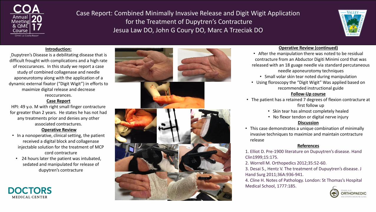
Operative Review• In a nonoperative, clinical setting, the patient
received a digital block and collagenase injectable solution for the treatment of MCP
cord contracture• 24 hours later the patient was intubated,
sedated and manipulated for release of dupytren’s contracture
Operative Review (continued)• After the manipulation there was noted to be residual contracture from an Abductor Digiti Minimi cord that was
released with an 18 guage needle via standard percutaneous needle aponeurotomy techniques
• Small volar skin tear noted during manipulation• Using floroscopy the “Digit Wigit” Was applied based on
recommended instructional guideFollow-Up course
• The patient has a retained 7 degrees of flexion contracture at first follow up
• Skin tear has almost completely healed• No flexor tendon or digital nerve injury
Discussion• This case demonstrates a unique combination of minimally
invasive techniques to maximize and maintain contracture release
References1. Elliot D. Pre-1900 literature on Dupuytren’s disease. Hand Clin1999;15:175.2. Worrell M. Orthopedics 2012;35:52-60.3. Desai S., Hentz V. The treatment of Dupuytren’s disease. J Hand Surg 2011;36A:936-941.4. Cline H. Notes of Pathology. London: St Thomas’s Hospital Medical School, 1777:185.

ODI Talon DistalFix Nail for Intertrochanteric Femur Fracture: A Report of Two CasesJohn Coury, DO; Jacob Duncan, DO; Robert Cash, MD
IntroductionThe Talon Distalfix Femoral Nail System utilizes deployable talons for proximal fixation within the femoral head as well as distal fixation in the femoral shaft. These talons are deployed after nail insertion with the application of a remote screw driver without the need for an additional incision. As opposed to traditional nails, this method of proximal and distal fixation provides theoretical advantages of reduced blood loss, operative time, fluoroscopic time, wound complications, and interlocking screw hardware failure.
Case ReportA 56-year-old man involved in a MVC sustained a comminuted left subtrochanteric fracture. The patient underwent ORIF utilizing a long ODI Talon nail. Intraoperative imaging demonstrates the ability of the nail to auto-center within the medullary canal during deployment of the distal locking talons (Figure 1).A 96-year-old woman who presented to the emergency department after a ground level fall. Radiographs taken at that time demonstrated an intertrochanteric fracture of the left femur. After nail placement during distal talon deployment, rather than auto-centering the nail as demonstrated in the previous case, the talons penetrated the cortex and the nail failed to center in the medullary canal, therefore maintaining the valgus distal positioning of the device. There were no immediate complications related to the cortical perforation, and the patient otherwise had an uneventful immediate post operative course and was discharged from the hospital on the fourth day.
DiscussionThese cases serve to illustrate a potential reference for the use of the ODI Talon nail. The potential benefits to its use including ability of the nail to center itself within the medullary canal and eliminating the need to perform “perfect circles” for insertion of distal interlocking screws. The indications for use of the talon nail are not impacted by bone quality; however, our experience with its use in an older patient with softer bone demonstrated that bone quality should probably play a role in selecting this method of fixation. When considering older, female patients who are more likely have softer, osteopenic bone, the nail may be incapable of self-centering within the medullary canal and can result in eccentric intramedullary placement of the device and a final distal talon position outside of the cortex of the bone.
References1. Tanner DA, Kloseck M, Crilly RG, et al. Hip Fracture Types in Men and Women Change Differently with Age. BMC Geriatr., 2010;10:12.2. Zehir S, Sahin E, Zehir R. Comparison of Clinical Outcomes with Three Different Intramedullary Nailing Devices in the Treatment of Unstable Trochanteric Fractures. Ulus Travma Acil Cerrahi Derg., 2015;21(6):469-476.3. Bellabarba C, Hersovici D Jr., Ricci WM. Percutaneous Treatment of Peritrochanteric Fractures Using the Gamma Nail. Clin. Orthop., 2000;375:30-42.4. Brumback RJ, Uwagie-Ero S, Lakatos RP, et al. Intramedullary Nailing of Femoral Shaft Fractures. Part II: Fracture-healing with Static Interlocking Fixation. J Bone Joint Surg AM, 1988;70(10):1453-1462.
Figure 1
Figure 2

Radiographic Comparison of Adolescent with Elbow Osteochondritis Dessicans Ulnar Collateral Ligament Tears and Controls
Brian C. Lau1, Nirav K. Pandya1
1Department of Orthopaedic Surgery, University of California, San Francisco
INTRODUCTION METHODS
• Single Sport Specialization and y ear-round athletic activity has led to a rapid increase in the number of youth sporting injuries (1,2).
• This is particular prevalent in upper extremity athletes such as baseball pitchers and gymnast who are prone to Osteochondritis dissecans (OCD) lesions of the capitellum and ulnar collateral ligament (UCL) tears.
• Multiple extrinsic factors for the development of these injuries include work-load (pitch count months/year of pitching) (3).
• There has been minimal attention paid to alignment and radiographic indices. This is particularly important as lower extremity alignment has been shown to be a risk factor for the development of OCD lesions of the knee and anterior cruciate ligament tears (4,5).
• A recent article described normal radiographic anatomy of the elbow in 178 pediatric and adolescent patients with objective measurements (6).
• To our knowledge, no studies have evaluated these radiographic measurements in adolescent patients with elbow OCD lesions or ulnar collateral ligament tears.
The purpose of the present study was to investigate objective radiographic measurements in an adolescent population with confirmed OCD lesions of the capitellum or UCL tears in comparison to normal cohort.
We hypothesize that patients with OCD/UCL injuries will vary from a normal population in regards to overall joint alignment.
METHODS
We received institutional review board approval for this clinical research study. Patients:- Retrospective Review of adolescent patients at a single institution from 2011-
2016 who had an elbow MRI were evaluated. - Inclusion criteria: Between ages 10-18, had both Antero-Posterior and Lateral
Radiographs of the affected elbow and had an elbow MRI. - Exclusion Criteria- History of neuromuscular disease, prior elbow surgery, or
connective tissue disorders.- 3 groups identified:
1) Isolated OCD of the capitellum (n=19)2) Isolated complete UCL tear (n=8)3) Normal Elbows (n=16)
Radiographic Evaluation• Radiographic measurements were based on descriptions by Goldfarb et al and
comments by Lawrence et al (6,7) on Phillips iSite picture archiving communication systems (PACS).
• Carrying angle, radial-neck shaft ankle, distal humeral articular angle, anterior angulation of distal humeral articular surface were evaluated due to their relationship with alignment of the elbow joint.
On Anterior-Posterior RadiographCarrying angle: The angle between a longitudinal line down the shaft of humeral shaft and a line down the ulna. An higher number indicates more valgus (Figure 1).
Distal humeral articular angle: Angle between a longitudinal line drawn down the humeralshaft & a transverse line drawn along the most distal aspect of the bony trochlea and thecapitellum. An increasing angle represents increasing valgus alignment of the distal humerus(Figure 2).
On Anterior-Posterior RadiographRadial neck-shaft angle: An angle between a longitudinal line perpendicular to the articular surface of the radial neck and a longitudinal line along the radial shaft (Figure 3).
Figure 3 Radial Neck-Shaft Angle
Table 2: Radiographic Means and Normative Data
DISCUSSION AND CONCLUSIONS
ACKNOWLEDGMENTS
1) Adolescents with OCD lesions and UCL tears of the elbow have greater varus carrying angle and greater valgus distal humeral articular surface compared to normal controls.
Leads to greater distance between the radial head and capitellumMay lead to greater valgus laxity of the elbow joint that leads to greater valgus force exacerbating medial stretch and
lateral compressionMay represent anatomic variant from genetic predisposition or may also be th consequence of repetitive valgus loading
2) UCL tear patients were older than OCD lesions (16.89 vs 13.73 years; p<0.0001)Supports that OCD and UCL injuries are part of spectrum that may be related to skeletal maturity
3) Findings lend support to use of previous definitions of elbow radiographic measurements made by Goldfarb et al. (6, 7)Significance: Preliminary study that demonstrates objective radiographic measurements of the adolescent elbow may be a usefulclinical test. These elbow parameters, similar to lower extremity alignment for development of OCD lesions of knee (5) may be risk factors for the development of OCD or UCL injuries. Future studies are needed to correlate these findings with biomechanical tests and track changes in these measurements through bony development.
1)Bell DR, et al. Prevalence of Sport Specialization in High School Athletics: A 1-Year Observational Study. Am J Sports Med. 2016 Feb 26. [Epub ahead of print] 2) Myer GD, et al. Sports Specialization, Part II: Alternative Solutions to Early Sport Specialization in Youth Athletes. Sports Health. 2016 Jan;8(1):65-73. 3) Erickson BJ, et al. Predicting and Preventing Injury in Major League Baseball. Am J Orthop (Belle Mead NJ). 2016 Mar-Apr;45(3):152-6. 4) Amaraee D, et al. Predictor factors for lower extremity malalignment and non-contact anterior cruciate ligament injuries in male athletes. Knee Surg Sports Traumatol Arthrosc. 2015 Dec 24 [Epub ahead of print] 5) Jacobi M, et al. Association between mechanical axis of the leg and osteochondritis dissecans of the knee: radiographic study on 103 knees. J Sports Med. 2010 Jul;38(7):1425-8. 6) Goldfarb CA, et al. Elbow
REFERENCES
1a.
1Aa.
RESULTS
Figure 2: Distal Humeral Articular Angle
Figure 1: Carrying Angle
On Lateral Radiograph:Anterior angulation of articular surface of distal humerus: The angle between line along humeral shaft and line bisecting the capitellum (Figure 4).
Statistical Analysis:• The patients were grouped into normal controls, osteochondritis
dissecans lesions, and ulnar collateral ligament injuries based on elbow MRI findings.
• Descriptive data are presented as means and standard deviations.
Figure 4: Anterior Angulation of Articular Surface of Distal Humerus
Table 1: Patient Demographics
Table 3: Comparision Between Groups (P-Values)
*= Data published by Goldfarb et al (6)*=P-value with significance p<0.05

A Pilot Study: Pocket-sized Ultrasound to Assess Distal-Radius Fracture and Quality of Closed Reduction
Brian C. Lau1, Aaron Robertson1, Daria Motamedi3, Nicolas Lee1
1Department of Orthopaedic Surgery, University of California, San Francisco2Department of Radiology and Biomedical Imaging, University of California, San Francisco
INTRODUCTION METHODS
• Distal Radius Fractures are one of the most common bony injuries (1)• Treatment in emergency department consists of closed reduction• Closed reduction may be performed as image-unassisted or with fluoroscopy• Image-unassisted closed reductions are checked with radiographs and if reduction is
unsatisfactory may require repeat reduction and radiographs• Fluoroscopy requires radiation and is not regularly available• Preliminary studies demonstrate that hospital-grade ultrasounds may guide reductions of
distal radius fractures (3,4,5,6)• Handheld ultrasounds are mobile, low-cost, and may be used in developing world, or
nontraditional medical setting such as disaster areas or combat zones- No studies have yet investigated the efficacy of hand-held ultrasounds in fracture care.
Purpose: To study was to evaluate if hand-held ultrasound was sensitive and specific indiagnosing a distal radius fracture and determining a satisfactory reduction.
Hypothesis: Hand-held ultrasound will) sensitivity and specificity in diagnosing distalradius fractures and determining a satisfactory reduction
METHODS
The study was approved by the Committee for Human Research at our institution; informed consent was obtained from all subjects.
Subjects• Forty- five total subjects were enrolled. 25 patients with acute distal radius
fractures (average age 49.6, 13 female) and 20 healthy controls (average age53.3, 10 female).
• Inclusion Criteria:1) Age ≥ 18 with acute (<48 hr) isolated, closed distal radius fracture that require
reduction• Exclusion Criteria:
1) Open Fractures2) Polytrauma3) Prior hardware within distal radius from previous surgery4) History of infection or inflammatory arthritis
Ultrasound Images obtained with 12.5 MHz ultrasoundprobe from hand-heldMobisante Inc. (Redmond, WA)
Clinical Procedures• All distal radius fracture patients had 3-view wrist (AP/Lateral/Oblique)
radiographs performed before fracture reduction• Ultrasound 3-views of the distal radius ( Dorsal, Volar, and Radial
(“snuffbox”) (Figure 1) were obtained
• Standard closed reduction was performed1) Oral analgesic provided2) Hematoma block with 1% Lidocaine injected into
fracture site3) Application of traction4) Closed manipulation and reduction
• Following reduction, the arm was re-imaged with hand-held ultrasound.• A standard plaster sugar-tong splint applied• Post-reduction Xrays were obtained per standard treatment protocol
Image Review• All ultrasound exams performed by single investigator (B.C.L)• A hand fellowship trained orthopaedic surgeon and a musculoskeletal fellowship trained radiologist served as evaluators and were
blinded to which images were obtained from healthy controls, before or after fracture reduction.• Evaluators independently reviewed radiographs and ultrasound images and determined:
1) No Fracture 2) Reduced Fracture
3) Non-reduced FractureStatistical Analysis• Radiographs were used as gold standards• Descriptive statistics and standard 2x2 tables to measure sensitivity, specificity, positive predictive value, and negative predictive
value of ultrasound images were performed.• For diagnosing a distal radius fracture -- Healthy Controls vs pre-reduction fracture ultrasound images were compared.• For determining a satisfactory fracture reduction Pre- and Post- fracture reduction ultrasound images were compared.
DISCUSSION AND CONCLUSIONS
ACKNOWLEDGMENTS
1) Pocket-sized portable handheld ultrasounds demonstrate excellent accuracy in diagnosing distal radius fractures and assessingsatisfactory reduction.
2) Pocket-sized portable handheld ultrasound demonstrated good to excellent ability to determine satisfactory reduction• This is the first study to evaluate portable handheld ultrasounds in fracture care.• Prior studies using hospital-grade ultrasound demonstrated 88-94% sensitivity in determining satisfactory reduction (4-6).• Dorsal and Radial views were most diagnostic. Future study should investigate accuracy with volar view removed.• Handheld ultrasounds may reduce radiation exposure to practitioners compared to using fluoroscopy and may reduce need for
repeat reductions and improve ED efficiency and wait times• Handheld may also be useful in the pediatric population for forearm (wrist fractures and both bone fractures) and elbow fracture
diagnosis and reduction to decrease radiation exposure.Significance: Portable handheld ultrasound may be cost-effective alternative for fracture diagnosis and assessing quality ofclosed reduction to reduce radiation to vulnerable populations (i.e pediatrics) and improve care in nontraditional settingswith limited access to fluoroscopy or radiographs (i.e. developing world, disaster zones).
1. 2. Court-Brown CM, Caesar B. Epidemiology of adult fractures: A review. Injury2006;37:691–697.2. Ang SH, Lee SW, Lam KY. “Ultrasound-guided reduction of distal radius fractures.” Am J of Emer Med 2010(28):1002-1008. 3. Chern TC, Jou IM, Lau KA, Yang CY, Yeh SH, Cheng SC. “Sonography for monitoring closed reductions of displaced extra-articular distal radius fractures.” J Bone Joint Surg Am. 2002 Feb;84-A(2):194-203.
REFERENCES
1a.
1Aa.
RESULTS
METHODS

Reliability:
Glenoid bone less: Intra-observer reliability Pearson correlation coefficient 0.91 (p=0.02); Inter-observer reliability: 0.86 (p=0.03)Hill-Sachs size: Intra-observer reliability: Pearson correlation coefficient 0.87 (p=0.03)Inter-observer reliability: Pearson correlation coefficient 0.83 (p=0.04)Kappa statistic for glenoid tracking classification: intra-observer 0.86; inter-observer 0.81.
Bipolar Bone Loss with Anterior Shoulder Dislocation: A Comparison of Adolescent Versus Adult Patients
Brian C. Lau1, Devin Conway1, Patrick F. Curran1, Brian T. Feeley1, Nirav K. Pandya1
1Department of Orthopaedic Surgery, University of California, San Francisco
INTRODUCTION
• Adolescence has been identified as the most significant risk factor for recurrent anterior shoulder dislocations(1,2).
• Adolescents have a 75-80% rate of recurrent dislocations with non-operative treatment (3) and a higher rate of failure, up to 49% at 5 years, following arthroscopic treatment (4) compared to adult rate of 8-11% after 11 years (5).
• Recent studies have described glenoid track concept to simultaneously characterize the relationship between the glenoid bone loss and Hill-Sachs lesion: OFF-track and ON-track
The purpose of this study was to compare bipolar bone loss by evaluating the degree of glenoid bone loss, Hill-Sachs lesion size, and glenoid track lesions in adolescents and adults with anterior shoulder dislocations.
We hypothesize that adolescent patients will have more glenoid OFF-track lesions compared to adults.
METHODS
We received institutional review board approval for this clinical research study. Patients:- Retrospective Review of adolescent patients of surgical and non-surgical
patients with a history of primary and recurrent anterior shoulder dislocations over a 4-year period (2012-2016) that had an magnetic resonance (MR) of their affected shoulder.
- Exclusion Criteria- Prior surgery to the affected shoulder, multidirectional instability, and posterior dislocation. Demographic data, sports played, and number of dislocations were collected.
- 2 groups identified: 1) Adolsecents (10-19 years old)2) Adults (>20 years old)
Glenoid Track Definition:• As described by Yamamoto, the size of the glenoid track was determined by the
amount of glenoid bone loss6. • When there is no glenoid defect, the width of the glenoid track is 84% of the
glenoid width6.• When there is a bony defect at the anterior rim of the glenoid, the defect width
should be subtracted from the 84% length to obtain a true width of the glenoid track14.
• If the medial margin of a Hill-Sachs lesion is more medial than the glenoid track, it was defined as OFF-track.
• If the medial margin of the Hill-Sachs lesions is within the glenoid track it was defined as ON-track and less likely to engage6 (Figure 1).
or angulation of distal humeral articular surface were evaluated due to their relationship with alignment of the elbow joint.
MRI Evaluation:• Glenoid Bone Loss: Based on method by Metzger et al (7) and Huysmans (8).• Bare spot identified on sagittal oblique image and glenoid width measured.• To determine expected glenoid width, a best-fit circle was placed on the
inferior third of the glenoid centered on the bare spot and the diameter of the circle was measured to calculate the expected width prior to bone loss.
• The glenoid bone loss percentage and glenoid track were both determined on these measurements. (Figure 2A)
• Glenoid track=84% of actual glenoid width• In setting of bone loss:
Glenoid Track= [84% of Actual Glenoid Width] -- [Amount of bone loss]Figure 2: Glenoid Track Calculation
• Hill-Sachs Lesion: Based on method by Saito et al (9).• Distance from medial margin of rotator cuff footprint to medial margin of Hill-
Sachs lesion using largest distance on a coronal-oblique image. (Figure 2B).
• OFF-TRACK: If Hill-Sachs measured greater than glenoid track then humeral lesion
• ON-TRACK: If Hill-Sachs measured less than glenoid track then humeral lesion
Statistics:Student’s T-tests for parametric data were performed to compare the two groups: adolescent (10-19 years old) and adults (≥20 years old). Adjusted odds ratios were utilized to determine the risk for adolescents to have off-track lesions.
A subgroup analysis comparing all patients (adolescent and adult) with 1 dislocation (SINGLE) compared to those with 2 or more dislocations (MULTIPLE)
Table 2: Adolescent vs Adult Comparison of Glenoid Bone Loss, Hill-Sachs Size, and Glenoid Tracking
ACKNOWLEDGMENTS
1) Adolescents patients with anterior shoulder dislocation have a 9.4x increased risk of having an OFF-track lesion.• Adolescents is a known risk factor for recurrent dislocation during non-operative management and following operative
treatment and these findings suggest that OFF-track lesions may contribute to this risk.2) Glenoid bone loss and Hill-Sachs lesion size similar between adolescent and adults
• This highlights that although size of a Hill-Sachs lesion is important, its location is equally important.3) A history of multiple dislocations (2 or more) regardless of age had a 4.2x increased risk of having an OFF-track lesion
• A history of multiple dislocations did not affect amount of glenoid bone loss between groups but was associated with larger Hill-Sachs size.
• The findings from this study suggest that subsequent dislocations effect the humeral head and glenoid tracking.Significance: This study demonstrates that adolescence and a history of multiple dislocations are independent risk factors for greater likelihood of glenoid OFF-track lesions. The findings from this study support use of bipolar assessment of shoulder dislocators, especially in adolescents and multiple dislocators.
1) Flinkkilä T, Hyvönen P, Ohtonen P, Leppilahti J. Arthroscopic Bankart repair: results and risk factors of recurrence of instability. Knee Surg Sports Traumatol Arthrosc 2010; 18:1752-1758. 2) Te Slaa RL, Wijffels MP, Brand R, Marti RK. The prognosis following acute primary glenohumeral dislocation. J Bone Joint Surg Br 2004; 86:58-64. 3) Arciero RA, Wheeler JH, Ryan JB, McBride JT. Arthroscopic Bankart repair versus nonoperative treatment for acute, initial anterior shoulder dislocations. Am J Sports Med. 1994 sep-Oct;22(5):589-94. 4) Shymon SJ, Roocroft J, Edmonds EW. Traumatic Anterior Instability of the Pediatric Shoulder:A comparison of arthroscopic and open bankart repairs. J Pediatric Orthopaedics. 2015 January; 35(1):1-6. 5) Harris JD, Gupta AK, Mall NA, Abrams GD, McCormick FM, Cole BJ, Bach BR Jr, Romeo AA, Verma NN. Long-term outcomes after bankart shoulder stabilization. Arthroscopy. 2013 May; 29(5):920-33. 6) Yamamota N, Itoi E, Abe H, et al. Contact between glenoid and the humeral head in abduction, external rotation, and horizontal extension: a new concept of glenoid track. J Shoulder and Elbow Surg. 2007;16:649-656. 7) Metzger PD, Barlow B, Leonardelli D, Peace W, Solomon DJ, Provencher MT. Clinical application of the “glenoid track” concept for defining humeral head engagement in anterior shoulder instability. Orthop J Sports Med. 2013 July 1(2):
REFERENCES
1a.
1Aa.
Table 1: Patient Demographics
Table 3: Single vs Multiple Dislocation Comparison of Glenoid Bone Loss, Hill-Sachs Size, and Glenoid Tracking
Single group had history of one dislocation prior to magnetic resonance imaging. Multiple group had history of 2 or more dislocations prior to magnetic resonance imaging.
Figure 1: Diagram of Glenoid Tracking
The distances from the medial margin of the contact area
to the edge of the articular surface of the humeral head
(MA) and to the medial margin of the cuff attachment
site on the greater tuberosity (MF) are measured. C-
articular center of the humeral head; M-most medial
point; A-lateral margin of articular surface, F-footprint. Figure courtesy of Sonali Feeley.
Adolescent Adult P-value
N 45 30
Mean Age 16.1 (range 13-19) 28.9 (range 20-39) <0.001
Gender (M/F) 33/12 26/4
BMI 25.7 (range 20.5-33.2) 23.45 (range 19.9-30.1) 0.31
Adolescent Adult P-Value
N 45 30
Glenoid Bone
Loss (mean %)
8.4% 9.9% 0.67
Expected
Glenoid
Diameter (mean)
25.4mm 26.0mm 0.32
Resultant
Glenoid
Diameter (mean)
23.3mm 23.5mm 0.75
Hill-Sachs Size
(mean mm)
12.77mm 10.0mm 0.12
Glenoid Tracking
Off-Track 11 1
On-Track 34 29
Single
Dislocation
Multiple
Dislocation
P-value
N 34 41
Adolescent/Adu
lt
21/13 24/17
Glenoid Bone
Loss (mean %)
8.4% 9.5% 0.47
Hill-Sachs Size
(mean mm)
8.1mm 14.3mm 0.0003
Glenoid
Tracking
OFF-Track 2 10
ON-Track 32 31
Odds Ratio 95% Confidence
Interval
P-Value
Adolescence
(13-19 years
old)
9.38 1.14-77.1 P<0.01
History of
Multiple
Dislocations
4.15 0.85-20.23 P<0.01
Table 4: Odds Ratio of Adolescence and History of Multiple Dislocations
METHODS RESULTS
DISCUSSION AND CONCLUSIONS

Secondary Ossification Center Appearance and Closure in the Pelvis and Proximal FemurKevin C. Parvaresh, MD,1 Vidyadhar V. Upasani, MD,1,2 James D. Bomar, MPH,2 Andrew T. Pennock, MD1,2
1 Department of Orthopaedic Surgery, University of California, San Diego2 Department of Orthopaedic Surgery, Rady Children’s Hospital
Introduction:
The known secondary ossification centers of the pelvis include the iliac crest (IC), anterior superior iliac spine (ASIS), anterior inferior iliac spine (AIIS), posterior superior iliac spine (PSIS), symphysis pubis (SP), and ischial tuberosity (IT). Proximal femoral secondary ossification centers include the femoral head (FH), greater trochanter (GT), and lesser trochanter (LT). Inconsistencies in ossification timing amongst prior studies and lack of gender comparisons underscore the need for a more comprehensive characterization of the secondary ossification centers. Our primary purpose was to characterize ages of appearance and closure for all pelvic and proximal femur secondary ossification centers. Our secondary goal was to evaluate gender differences in timing of appearance and closure.
Methods:
Inclusion: Patients 2-32 years old with CT imaging of the abdomen or pelvisRady Children’s Hospital: 11/2011 – 1/2012UCSD: 1/2009 – 12/2014
Exclusion: Orthopaedic trauma or pathology
Outcomes: Appearance, closure, and fusion of all secondary ossification centersAppearance: visible ossification at the secondary ossification siteClosure: cortical congruity on one axisFusion: cortical congruity on two or all three axes
Discussion:
The appearance and closure of the pelvis and proximal femur secondary ossification centers follow a predictable pattern of development, occurring slightly earlier in females than males. Knowledge of more precise ages of development and gender differences better characterize this complex skeletal development. Future studies may utilize secondary ossification centers to further evaluate skeletal maturity, assess pediatric pathology, and aid surgical management.

Does Pre-Culture Antibiotic Administration Lead to Non-Diagnostic Bone and Joint Culture Results in Pediatric Osteomyelitis and Septic Arthritis: A 12-Year Retrospective ReviewMichael Basso-Williams, DO, MPH 1,2, John A. Schlechter, DO 1,2, Bishal Bhandari, BA1. CHOC Children’s Hospital, 2. Riverside University Health System
Introduction:Pediatric musculoskeletal infections can be devastating, with lasting morbidity if
left untreated • Children often receive antibiotics in various clinical settings prior to having cultures obtained.
• A commonly held notion is that administration of antibiotics prior to obtaining a sample from
the involved musculoskeletal site will result in negative cultures.
PURPOSE: To evaluate whether culture results for children receiving antibiotics
prior to obtaining cultures for osteomyelitis and/or septic arthritis is affected.
HYPOTHESIS: Antibiotic administration prior to bone and/or joint culture in
children with osteomyelitis and/or septic arthritis will result in a greater number of
negative cultures.
Materials / Methods:A retrospective chart review of 107 patients was performed.• Children age < 18 years with osteomyelitis and/or septic arthritis that received antibiotics
(oral or IV) within seven days of obtaining a formal culture were included.
• Comparison based on pre-sample antibiotic administration and final culture results were
made as well as the recording of type of antibiotic, organisms isolated, blood cultures,
diagnoses, location of infection, age, sex, fever, WBC, ESR, and CRP.
• Results:• One-hundred seven children, average age of 8 years and 2 months old (range: 6
weeks – 16 years), 76 males and 31 females were included
• 59/107 (55%) children received antibiotics prior to culture.
• In those who had bone and/or joint culture obtained prior to receiving antibiotics (Group 1), 47 had
positive cultures and 12 had negative cultures.
• Of those who received antibiotics after culture (Group 2), 30 had positive cultures and 18 had
negative cultures.
• Children who received antibiotics prior to obtaining a sample were more likely to have positive results
(p = 0.049).
• There was a clinical trend for increased WBC and CRP in children with positive cultures
• Discussion:• Receiving antibiotics prior to obtaining a culture in children with osteomyelitis and/or
septic arthritis did not lead to a statistically significant number of negative cultures.
• Delaying antibiotic treatment until formal samples were obtained demonstrates
no benefit for diagnostic purposes in our cohort.
•
Group 1 Group 2
Positive Culture Negative Culture Positive Culture Negative Culture P-value
WBC (K/UL) 11.8 (2.9-26.6) 11 (3.5-23.7) 11.4 (5-20.9) 10.8 (6.3-18.8) p=0.805
ESR (mm/hr) 60.3 (5-140) 55.5 (1-140) 55.2 (12-114) 55.6 (8-140) p=0.71
CRP (mg/L) 28.3 (1.5-175.1) 24.5 (0.02-126.5) 37.8 (0.55-416.4) 21 (0.3-70.6) p=0.87
T-max (Celsius) 39.4 (38.1-40.5) 39.5 (38.3-40.5) 39.3 (38.2-40.5) 39.1 (38.2-40) p=0.10
Bulk allograft is more susceptible to infection than stainless steel in a novel mouse modelStephen D. Zoller M.D, Vishal Hegde M.D., Howard Y. Park M.D., William Sheppard B.S., Christopher Hamad B.S., Amanda H. Loftin B.S., Daniel Johansen
B.S., Ryan Smith B.S., Marina M. Sprague M.S., John Huang B.A., Scott Nelson M.D., Nicholas M. Bernthal M.D.Department of Orthopaedic Surgery, David Geffen School of Medicine at UCLA, Los Angeles, CA
Introduction:Bulk allograft remains a viable reconstruction option for large bone defects, withinfection being a devastating complication.
Currently, infection treatment principles follow those of metal implant infections.
We hypothesized:
i) to generate a novel mouse model of bulk allograft infection that will allow theinvestigation of infection and host response longitudinally and non-invasively.
ii) allograft will behave similarly to metal implants in terms of susceptibility toinfection.
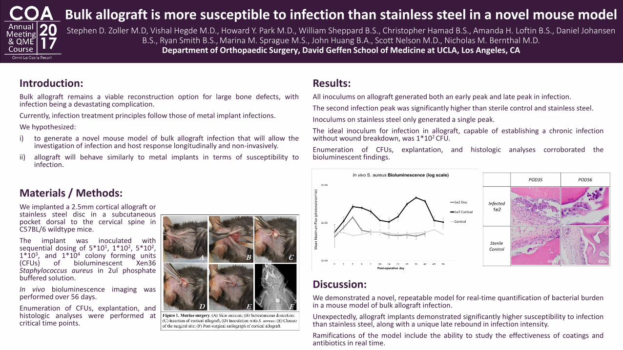
Results:All inoculums on allograft generated both an early peak and late peak in infection.
The second infection peak was significantly higher than sterile control and stainless steel.
Inoculums on stainless steel only generated a single peak.
The ideal inoculum for infection in allograft, capable of establishing a chronic infectionwithout wound breakdown, was 1*102 CFU.
Enumeration of CFUs, explantation, and histologic analyses corroborated thebioluminescent findings.
Discussion:We demonstrated a novel, repeatable model for real-time quantification of bacterial burdenin a mouse model of bulk allograft infection.
Unexpectedly, allograft implants demonstrated significantly higher susceptibility to infectionthan stainless steel, along with a unique late rebound in infection intensity.
Ramifications of the model include the ability to study the effectiveness of coatings andantibiotics in real time.
Materials / Methods:We implanted a 2.5mm cortical allograft orstainless steel disc in a subcutaneouspocket dorsal to the cervical spine inC57BL/6 wildtype mice.
The implant was inoculated withsequential dosing of 5*101, 1*102, 5*102,1*103, and 1*104 colony forming units(CFUs) of bioluminescent Xen36Staphylococcus aureus in 2ul phosphatebuffered solution.
In vivo bioluminescence imaging wasperformed over 56 days.
Enumeration of CFUs, explantation, andhistologic analyses were performed atcritical time points.