TRACING THE CRIMINAL Part thirteen: Cooperation at investigation or Clinical Microbiology IV...
55
TRACING THE CRIMINAL Part thirteen: Cooperation at investigation or Clinical Microbiology IV Institute for microbiology shows
-
Upload
miranda-robbins -
Category
Documents
-
view
216 -
download
1
Transcript of TRACING THE CRIMINAL Part thirteen: Cooperation at investigation or Clinical Microbiology IV...
TRACING THE CRIMINAL
Part thirteen:Cooperation at investigation or
Clinical Microbiology IV
Institute for microbiology shows
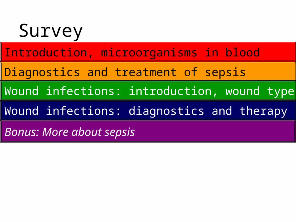
SurveyIntroduction, microorganisms in blood
Diagnostics and treatment of sepsis
Wound infections: introduction, wound types
Wound infections: diagnostics and therapy
Bonus: More about sepsis

Introduction, microbes in
blood
Presence of microbes in blood• At normal situation microbes are present in blood at most
as transitory (e. g. during teeth cleaning). In heart tissue and vessel endothelium, of course, bacteria should not be at all.
• The term „bloodstream infection“ (BSI) is almost used for bacterial, eventually mycotic (yeast) infections
• Viraemia (presence of viruses in blood) is a part of various viral diseases, almost hepatitis and HIV infection (it is mentioned in the corresponding virology topic)
• Among blood parasites we have malaria plasmodia, trypanosomes and filariae (see parasitological topic)
Bacterial (eventually mycotic) bloodstream infections (BSI)
• Sepsis are infections of proper bloodstream, in the same time they are systemic infections of the complete organism. They are primary (in typhus) or secondary (catheter sepsis, urosepsis, abdominal sepsis). They are caused by bacteria or yeasts.
• Endocarditis are connected to previous ones, but besides presence of microbe in blood there is also a more narrow relationship to the endocardium, especially in case that it is damaged by a previous disease or treatment (rheumatoid fever, implant)

Important terms• Sepsis/septicaemia is a complex situation, presence of
bacteria in blood PLUS clinical symptoms (there exist clinical criteria that should be fulfilled)
• Bacter(i)aemia (eventually fungaemia, i. e. presence of yeasts) is just evidence of presence of bacteria (fungi) in blood without evaluation of their clinical importance. Transient bacteraemia may be a part of spreading of bacteria in the organism, although it is not BSI (especially in pneumonia or pyelonephritis – possible use in diagnostics).
• Pseudobacter(i)aemia is a situation, when bacteria only seem to be present in blood (badly performed blood examination, usually skin contamination). More later

Diagnostics and treatment of
sepsis

Diagnostics of sepsis• blood cultures (see further) and more microbiology
examinations (changed catheter, sputum, urine according to likely original focus, lumbal punction in suspicion for meningitis)
• biochemical laboratory – inflammatory markers (CRP, procalcitonin, differential blood picture)
• Laboratory markers of disseminated intravascular coagulation (DIC): thrombocytopenia, lower AT III etc.
• Search for infection focuses: RTG of heart and lung, otorhinolaryngology, ultrasound (oesophagus – focuses on heart), CT…
• Neurologic examination
Blood cultures – sampling• It means not clotted blood, principally very different from
serological examinations (it is neither antibody nor antigen detection, microbe should remain alive and is detected by cultivation)
• Today we usually sample into special vessels with transport-cultivation medium for automatic culture (sooner just not-clotted blood without any medium was sent)
• We have to ensure minimalisation of risk of pseudobacteriaemia (see more)
• In adults we take 20 to 30 ml of blood, in children usually 1–5 ml according to age (sampling is more difficult in adults, and also in children also less bacteria are important)

Types of blood culture vials• There are various types regarding to microbes that
are to be detected (aerobes, anaerobes, yeasts)• Some vials include charcoal. They are designed for
culturing blood of patients already treated by antibiotics (classical vessel could give a false negative result – the antibiotic would suppress the growth)
• The most frequent types are: aerobic standard, aerobic with charcoal and anaerobic with charcoal.

Examples of vials for blood culture – system BacT/ALERT
Photo: archive of Institute
Standard
aerobic
Charcoal
anaerobic
Charcoal
aerobic
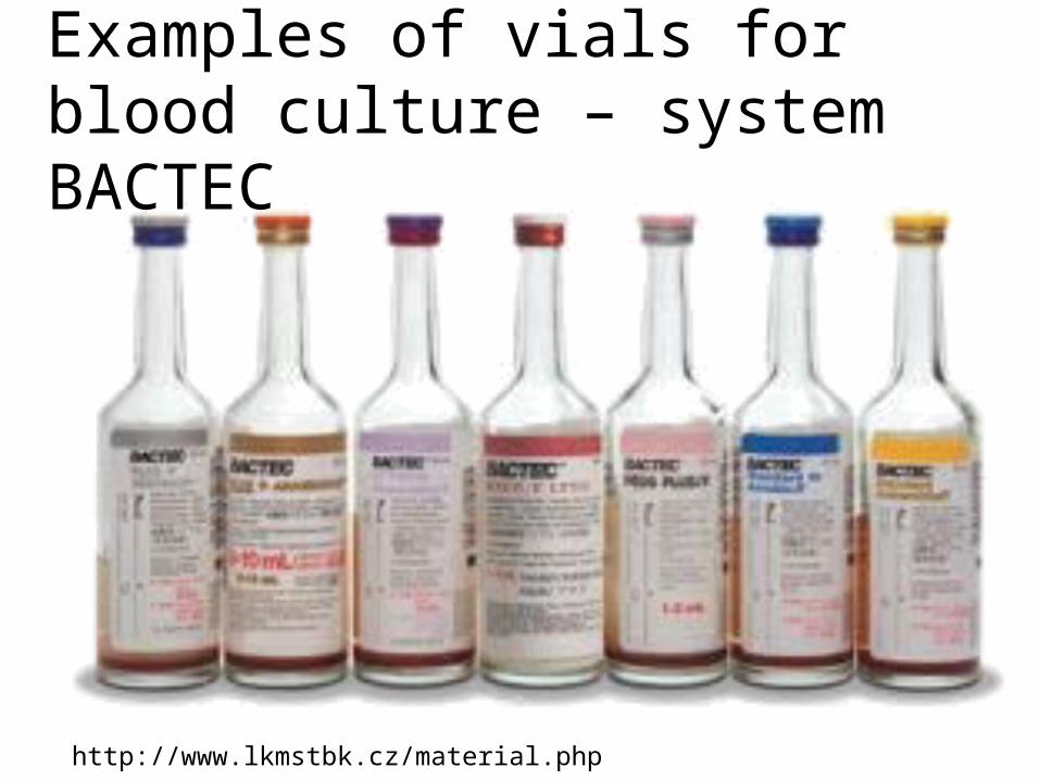
Examples of vials for blood culture – system BACTEC
http://www.lkmstbk.cz/material.php
Pseudobacteraemia – reasons• Not properly done sampling, not sufficient asepsis
during taking blood• Sampling only from artificial catheters (we only
catch bacteria colonizing the catheter, but it is not always real cause of bacteraemia, not speaking about sepsis)
Why bacteriemia is bad? That means that the patient is treated uselessly for not-existing infection. It is also possible that the infection exists, but instead of the pathogen another microbe was found – this finding would stop the search for the real pathogen
How to avoid pseudobacteraemia – I• To take blood targetedly, when presence of
bacteria in blood is likely; not take it „just for sure“ when another examination is indicated
• To take sufficient amount of specimens: one is useless, even two are too little, three is optimum
• To take blood cultures from suitable sites: at least one from a new venepunction, ideally three venepunctions plus one from venous catheter
• To take blood cultures in suitable moment, in sceptic status typically at temperature increase
How to avoid pseudobacteriaemia – II• To take blood cultures properly, very important and
frequently neglected is keeping aseptic sampling (to disinfect, not only clean skin, and to let the disinfection really dry). To send blood cultures together with well filled in request form; necessary is not only date, but also time and place of sampling – for result interpretation
• Surprisingly for someone less important than the previous policy is removed, blood cultures to the correct kit: there is usually no reason to even send the anaerobic, if there is not actual suspicion for anaerobic infection (the estimated origin of sepsis is in the abdominal cavity). Use of a vial with active carbon is required at the very least, where the patient is already treated by an antibiotic.

How to avoid pseudobacteraemia – III• In suspicion for contamination of vascular catheter
the catheter is changed. The old catheter is not discarded, but sent for bacteriology examination. Today there are already methods capable to estimate whether it is a real colonisation of the catheter or a random find (see below)
• The same of course applies to any of the implants, which are removed from the body – the microbiological testing can bring substantial information for further treatment

How to find pseudobacteraemia when it occurred already
• Typical for pseudobacteraemia (false positivity of a blood culture) is, that– just one of three blood cultures is positive– or even more blood cultures are positive, but from each
one another strain would be cultured (different susceptibility, different colony appearance) and would have different TTD (time to detection) – this corresponds to different quantity
– clinical patient problems are not corresponding to the findings
– Eventually the same strain would be also found in patient‘s skin

Evaluation of TTD (time to detection)• Time between sampling and the report of positivity from
the automat (e. g. the automat beeps and signals positivity by a red rectangle) is shorter in case of massive presence of bacteria in blood and longer in case of few bacteria
• In real bacteraemias the time uses to be shorter (less than 48 h) and the same for all taken blood cultures (plus minus two hours)
• Eventually the time might be shorter for the blood culture from place that is source of infection (e. g. blood culture from central venous catheter, that represents the source of a catheter sepsis)
Do you understand already why it is so important to write time and site of sampling to the request form?
Importance of TTD for finding interpretation
• Example 1: Three blood cultures were taken, all of them were positive, but one after 12 hours, the second after 36 hours, and the third after 3 days. The strains are phenotypically different it is very likely that it is a skin contamination
• Example 2: Three blood cultures were taken, all of them were positive, all about the same time after the collection, the strains look similar it is likely that the finding is the actual pathogen
Function of cultivators• Cultivator, connected to a computer, keeps
automatically optimal conditions of cultivation, and also evaluates status of the vessel and indicates eventual growth (e. g. change of reflectance, i. e. optical characteristics of the vial)
• The growth is signalized optically and by a sound. When nothing is growing even after a week, the apparatus signalizes it too (it is time to give out a negative result)
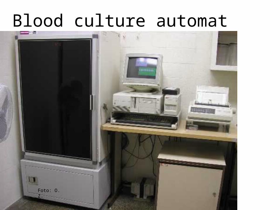
Blood culture automat
Foto: O. Z.

The same automat – open
Foto: O. Z.

When a blood culture is positive…• The vial is taken out from the machine• It is necessary to mark the time, or TTD –
period from admission to positivity. The longer this time is, the more the contamination is likely
• We perform inoculation to solid media, Gram stained smear and according to its result „directly“ orientation disc test of susceptibility; instead of standard suspension just fluid from vessel is used the result is unsure

What to do after that• We have to count that direct tests are only
orientation tests, for not standard content of bacteria in individual blood samples (non-standard inoculum). Usually in another step we perform proper susceptibility testing (often using quantitative tests)
• Exceptions are likely contaminations (only one of three is positive, or all three, but different strains, positivity after longer time, coagulase negative staphylococci), here we usually only perform a new qualitative test from a standard inoculum

Cooperation laboratory – ward• Laboratory tries to cooperate with clinicians
already during blood culture, mostly in form of telephonic report, sending preliminary results (even in negative blood cultures) etc.
• Also long term evidence of positive findings is useful in frame of a systematic surveillance of hospital infections
• Details of cooperation should be mediated individually

Microbiology examination of blood catheters
• Catheters are today usually sent in a sterile test tube, without being put in a liquid. In the laboratory– either biofilm is broken by ultrasound and released into
the solution (so called sonication)– or the catheter is rolled on the surface of agar medium
• Both methods are semiquantitative, i. e. the result decides whether it is rather significant finding or contamination
• Traditional method of a catheter just placed into the broth and here let to multiply is now considered obsolete
More options in examination of BSIs• Examination of urine, sputum, cerebrospinal fluid,
etc. is performed with regard to the suspected source of sepsis
• In some microbes direct antigen detection is possible in the blood without cultivation, i.e. with the possibility of almost immediate obtaining a result: mannan antigens in yeast, or antigens, the cause of typhoid fever, meningitis pathogens and the like

Also in typhoid fever there exist a possibility of direct antigen detection in blood www.drdo.org/labs/dls/drde/products/typhigen.htm.
Therapy of sepsis• symptomatic therapy – emergency or intermediary units
• Monitoring, addition of circulating liquids, oxygen, support for circulation (noradrenalin), use of peripheral and/or central venous catheters, artificial lung ventilation etc.
• antibiotics (initial „blind“ therapy, later targeted) • In case of presence of abscesses their surgical removal• corticosteroids – in initial phasis of sepsis approx. 300 mg
of hydrocortisone (< 3 days) • anticoagulation therapy – in case of signs of disseminated
intravascular coagulation only• change of glycaemia, level of calcium etc.

Complications and prognosis of bacterial sepsis
• acute respiratory failure syndrome: 40 % of patients with sepsis
• acute renal failure (elevated urea and creatinin) • circulation failure – BP decrease (systolic BP < 90 mmHg) • disseminated intravascular coagulation – Gram-negative
sepsis• digestive tract failure – vomiting, diarrhoea, bleeding
(stress ulcus) • hepatic failure – elevated bilirubin, ALT, AST etc.• CNS damage – alteration of consciousness• total lethality of sepsis is approx. 40 %• septic shock lethality is 70–90 %
Wound infections – introduction, wound types

Wound infections Infection of wounds are fairly disparate group (different
origins, different location). In any case they are serious, because microbes penetrated body surface to sites that are normally sterile.
Specific situation is a pyogene inflammation of surgical wounds. Its prevention and treatment is one of the important topics for the surgeons. (Today we almost use abbreviation SSI – surgical site infection.)
Purulent wound infections only occur, when the bacterial infection of wound is accompanied by infiltration of polymorphonuclear granulocytes (due to the immune response of the host organism).

Wound infection
www.ageless.co.za/case-infection.htm www.ehagroup.com/nosocomial/

Wound classification• Wound classification with regard to depth:
– surface wound infection (skin, hypodermis)– deep wound infection– infection of organs and body spaces
• Wound classification of with regard to the risk:– 1/clean– 2/clean-contaminated (surgery of sites with normal
microflora)– 3/contaminated (trauma, bacteria from outside)– 4/dirty-infected (inflammation in a wound)

Areal wounds (diabetic ulcers, ulcus cruris, bedsore)
Often mixture of various bacteria, often participation of bacterial biofilm treatment should be almost local (biofilm destroying) and only sometimes also supportive oral/venous antibiotic use
The most serious causative agents are Streptococcus pyogenes and Staphylococcus aureus
Bacteria that rather colonize the wound: Escherichia coli, Proteus mirabilis and other enterobacteria, Pseudomonas aeruginosa and yeasts
Infection × colonization of a wound
• Sometimes it is difficult to say which microbe is responsible for an invasive wound infection and which one only colonized it (and formed a biofilm in it)
• A typical sign of true invasive infection is presence of causative bacteria deeper in the body (e. g. in blood culture) and also inflammatory markers
• Pure colonization should not be treated with oral/venous antibiotics; topic treatment and wound debridement etc. should be performed

Wound infections: diagnostics
including interpretation
and treatment

Sampling in deep focal infections (1)
• In case of sufficient amount of pus or another liquid (exsudate, cyst contain etc.) that liquid in a test tube should be sent and not just swab (if possible)
• In suspicion for anaerobic infections (especially pus from abdominal region) it is recommended to send it directly in a syringe. To cap a syringe (just syringe, no needle) use combi cap (see picture)
• Sending of a syringe with a needle placed to a sterile rubber cap, that was recommended sooner, is in the matter of fact forbidden for safety reasons, (needle manipulation is risky for sampling person)
http://www.mediform.cz/default.asp?nDepartmentID=63&nLanguageID=1
Sampling in deep focal infections (2)
• In case of impossible liquid sending (not enough liquid) we need to send swab with transport medium. Recently also E-swabs are sent (see further)
• In some cases also smear or imprint on slide may be effective (catching of pathogens that could not be cultured)
• In special cases a microbiologist may be even called to an operation hall

E-swab (1)Transport system ESwab is sterile and consists of two
parts• Polypropylene screw cap test tube with liquid
Amies transport medium• The proper sampling swab, ended by soft nylon
fibres. It is made by an advanced technology of parallel nylon fibres in electrostatic field.
• Microorganisms are actively captured by electrostatic force of the fibbers (in classical swab they just remain passively in the swab).
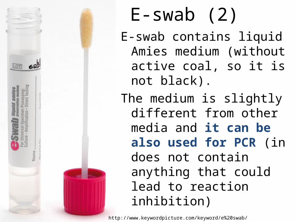
E-swab (2)E-swab contains liquid Amies
medium (without active coal, so it is not black).
The medium is slightly different from other media and it can be also used for PCR (in does not contain anything that could lead to reaction inhibition)
http://www.keywordpicture.com/keyword/e%20swab/
Sampling in superficial wounds• Classical method is a swab with transport medium• The swab should catch the pathogen (we have to
reach the infection focus) and in the same time not contaminated, especially from skin
• It is also possible to use imprint method: a square peace of a sterile square of filtration paper is placed on an areal wound (e. g. diabetic ulcus) and then moved to the surface of culture medium (blood agar). In the laboratory it is also placed to other media; this enables better semiquantitative evaluation of the result

Swab, or imprint?
• At swab we use sterile tool with Amies transport medium. Result is qualitative.
• For imprint, we use 5x5 cm sterile square of filtration paper. The result is semiquantitative.
ww.fnkv.cz/kliniky/ustav_lekarske_mikrobiologie/download/200801111-prirucka-pro-odber-materialu.pdf
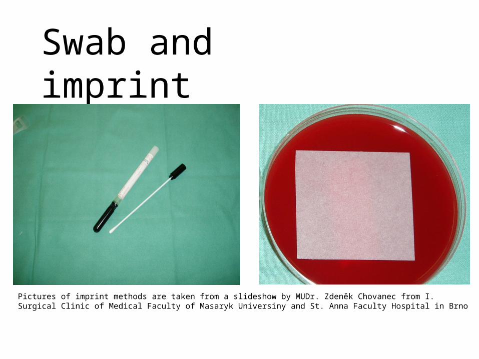
Swab and imprint
Pictures of imprint methods are taken from a slideshow by MUDr. Zdeněk Chovanec from I. Surgical Clinic of Medical Faculty of Masaryk Universiny and St. Anna Faculty Hospital in Brno
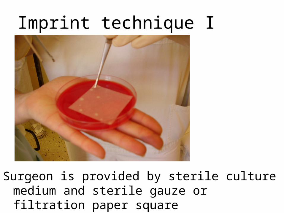
Imprint technique I
Surgeon is provided by sterile culture medium and sterile gauze or filtration paper square
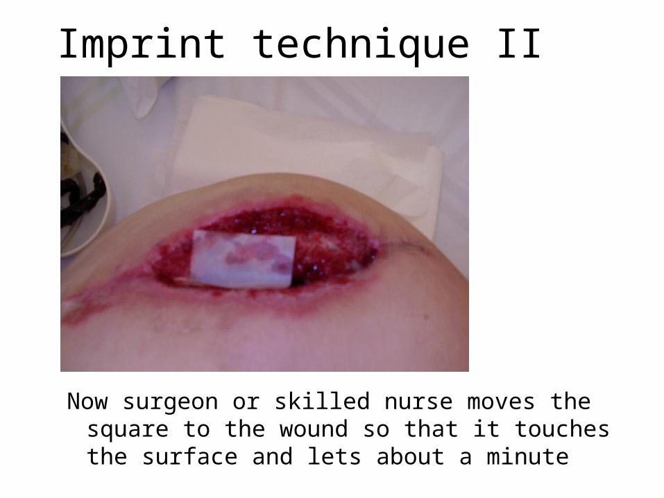
Imprint technique II
Now surgeon or skilled nurse moves the square to the wound so that it touches the surface and lets about a minute
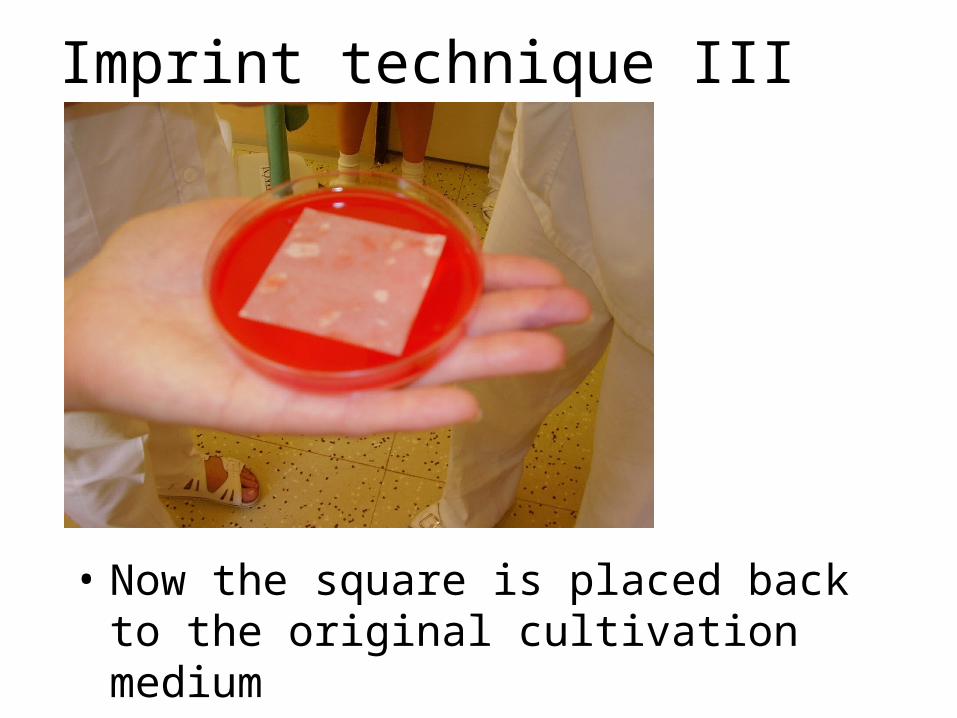
Imprint technique III
• Now the square is placed back to the original cultivation medium
Request form filling in in wounds• The person taking the specimens should fill in the
request carefully, „wound swab“ not sufficient, it is necessary to add– Wound type (origin) – surgical wound, bite, punctual
wound etc.– Localization of wound on body– Eventually special examinations (although e. g.
anaerobic culture for swabs from abdominal cavity is automatic)
• Also important anamnestic data (coming from abroad, work in agriculture) may be useful
Wound infection diagnostics• Laboratory performs microscopy (Gram staining) in
liquid specimens only (not swabs); always culture, precise determination of pathogens is done and examination of antibiotic susceptibility
• In microscopy, not only microbes, but also WBCs etc. are important
• At culture we use liquid multiplying media (for situations with few microbes) and also selective media (with NaCl for staphylococci, with amikacin or streptococci), especially in bedsores etc.

Wound swabs (without anaerobes):Possible diagnostic scheme
• (Different in different types of wound etc.)• Day 0: start of culture only• Day 1: result of primary culture of specimen on
blood agar (BA), Endo agar, NaCl and BA + amikacin. If all solid media are negative, broth is observed; if turbid, a subcultivation to solid media is performed
• Day 2: expedition of negative and some positive results; too resistant bacteria more tests
• Days 3, 4: expedition of remaining results
Wound swab – result interpretation• Common flora: none, all findings are looked as
pathogen (even including microbes suspicious of being contamination) – so also antibiotic tests are performed
• Pathogens: any bacteria or Candida are considered possible pathogen, perhaps with the exception of coagulase-negative staphs and corynebacteria on the surface of staphylococcal skin wounds.
• Colonisation: in surface wounds it is questionable, whether the situation corresponds to an infection or rather to a colonisation (especially in Pseudomonas and Proteus). Antibiotic susceptibility testing is nevertheless performed even here.

Pyogene infections treatment
• Local wound care is important (local preparations, cleaning, debridement, eventually also larvotherapy)
• If we do not suppose finding of anaerobic, oxacilin (as an anti-staphylococcal drug) would be a drug of choice
• For rather streptococcal origin, high doses of G-penicillin should be used.
• In hospital wound infections targeted treatment is necessary

EndE. coli in blood culture, phase contrast
http://www.visualsunlimited.com/browse/vu198/vu19873.html
Bonus: Types of septicaemia• Primary sepsis – some bacteria do sepsis
„normally“, e. g. typhoid fever salmonellae or partially also meningococci
• Secondary sepsis – sepsis coming after failure of an organ
• Special types of sepsis– urosepsis – sepsis in kidney failure– sepsis related with pneumoniae– sepsis of abdominal origin– catheter sepsis as hospital disease

Sepsis – clinical picture• instable body temperature• decreased muscle tonus• intolerance of food, diarrhoea• respiratory problems – frequent, irregular
breathing, breath pause, failure• blood circulation problems – pulse more or
less frequent, blood pressure decrease• common icterus, hyper/hypoglycaemia,
metabolic failure, bleeding, neural symptoms etc.