TRACING THE PATHOGEN Part eleven: Cooperation at investigation or Clinical Microbiology II Institute...
60
TRACING THE PATHOGEN Part eleven: Cooperation at investigation or Clinical Microbiology II Institute for Microbiology shows
-
Upload
geoffrey-wood -
Category
Documents
-
view
217 -
download
0
Transcript of TRACING THE PATHOGEN Part eleven: Cooperation at investigation or Clinical Microbiology II Institute...

TRACING THE PATHOGEN
Part eleven:
Cooperation at investigation orClinical Microbiology II
Institute for Microbiology shows

Survey of topicsRespiratory infections – introduction
Indications for examination in respiratory infections
Sampling and examination in respiratory infections
Processing and interpretation of respiratory specimens
Importance and classification of digestive tract infections
Sampling and examination in intestinal infections

Respiratory infections -
introduction

Importance of respiratory infections
The most common infection in general practitioner's (microbes multiply well in respiratory ways)
Big economy impact (inability to work, necessity for parents to stay at home with ill children)
Often seen in collectives and sometimes causing outbreaks
¾ of respiratory infections (even more in children) caused by viruses

Localization of infection in respiratory infections
It is not the same, which part of respiratory ways is affected by the infection (examination, treatment and seriousness is different).– Symptoms of infections of different parts of respiratory
tract are different (sneezing in rhinitis, cough in lower respiratory ways infections)
– Causative agents are different, tooIt is necessary do differentiate infections of:
– Upper respiratory ways (+ anatomically also middle ear)– Lower respiratory ways including lungs (lungs are often
put aside as it is not a „way“)On the other hand the infection often affects more parts
of respiratory ways simultaneously

Classification of respiratory infectionsUpper respiratory ways and
connected organsInfections of nose and
nasopharynxInfections of oropharynx
and tonsilsInfections of paranasal
cavitiesAnatomically usually also
middle ear infections
Lower respiratory ways and lungs
Infections of epiglottisInfections of larynx and
tracheaInfections of bronchiInfections of bronchiolesLung infections
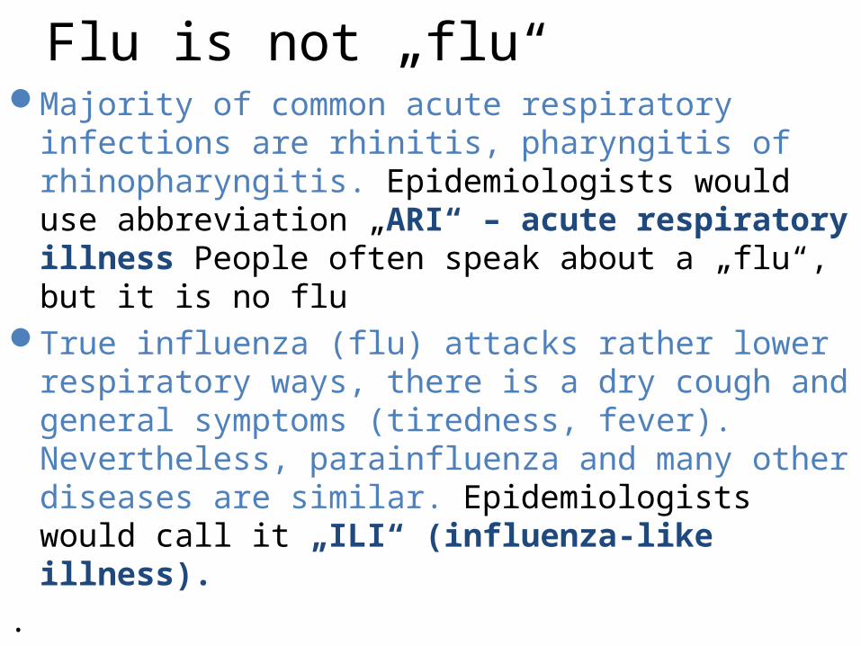
Flu is not „flu“Majority of common acute respiratory infections are
rhinitis, pharyngitis of rhinopharyngitis. Epidemiologists would use abbreviation „ARI“ – acute respiratory illness People often speak about a „flu“, but it is no flu
True influenza (flu) attacks rather lower respiratory ways, there is a dry cough and general symptoms (tiredness, fever). Nevertheless, parainfluenza and many other diseases are similar. Epidemiologists would call it „ILI“ (influenza-like illness).
.

Normal respiratory microfloraNasal cavity has no specific microflora, there is skin
microflora (frontal part) and pharyngeal microflora (back part)
In pharynx (and also oral cavity) we can find oral streptococci, Neisserias, non-virulent strains of haemophili etc. Many other strains are also present, but we cannot culture them.
Lungs and lower respiratory ways use to be nearly microbes-free in a healthy person
Other sites (larynx) have transient microbes (larynx – like pharynx, but less microbes)

Indications for examination in
respiratory ways

Examination and treatment in infections of nose and nasopharynxExamination is useless. Even mucopurulent secretion
is not a reason for bacteriology examination, if it does not persist too long.
Therapy is symptomatic (drops in congested nose; otherwise liquids, e. g. tea; antipyretics are not too useful, as elevated temperature helps against viruses). Antibiotic treatment is not indicated. Sometimes topic treatment by framycoin may be used.
Only in case of infection continuing more than 10–14 days it is useful to examine nasal swab (to avoid skin contamination!) and to use targeted antibiotic treatment according to susceptibility

What do the experts say„More than 80 % of rhinitis is accompanied by changes on
paranasal cavity mucosa, therefore the disease is sometimes also called rhinosinusitis. The cough is present in 60–80 % rhinosinusitis cases. Mucous secretion is in three days after infection oncome changed into a mucopurulent one, containing desquamated epithelial cells and colonizing bacteria commonly found in nasal cavity. This qualitative change of secretion, in practice commonly misinterpreted for „bacterial superinfection“, especially in case of cultivation examination of mucus of nasal swab; but it is a part of a normal rhinitis course.“
(Respiratory infection – recommendation guidelines by Czech Medical Association of John Evangelist Purkyně)
Examination and treatment of sinusitis
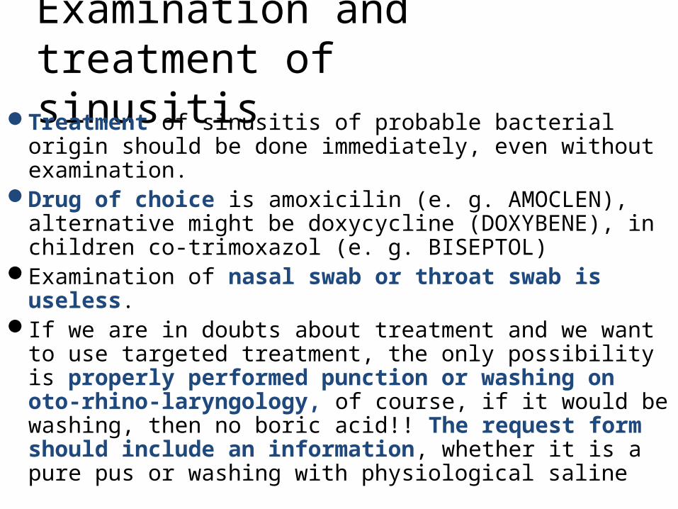
Treatment of sinusitis of probable bacterial origin should be done immediately, even without examination.
Drug of choice is amoxicilin (e. g. AMOCLEN), alternative might be doxycycline (DOXYBENE), in children co-trimoxazol (e. g. BISEPTOL)
Examination of nasal swab or throat swab is useless.If we are in doubts about treatment and we want to use
targeted treatment, the only possibility is properly performed punction or washing on oto-rhino-laryngology, of course, if it would be washing, then no boric acid!! The request form should include an information, whether it is a pure pus or washing with physiological saline

Examination and treatment of otitis media
Treatment is only meaningful if it is a real inflammation (pain, redness, fever) and it does not react to anti-inflammatory treatment
Drug of choice is amoxicillin (e. g. AMOCLEN), as alternative, co-trimoxazole can be used
It has only sense to examine external ear examination after paracenthesis
Otherwise it is useful to send pus containing fluid, sampled during paracenthesis

Tonsilopharyngitis
http://medicine.ucsd.edu/Clinicalimg/Head-Pharyngitis.htm
http://www.newagebd.com/2005/sep/12/img2.html
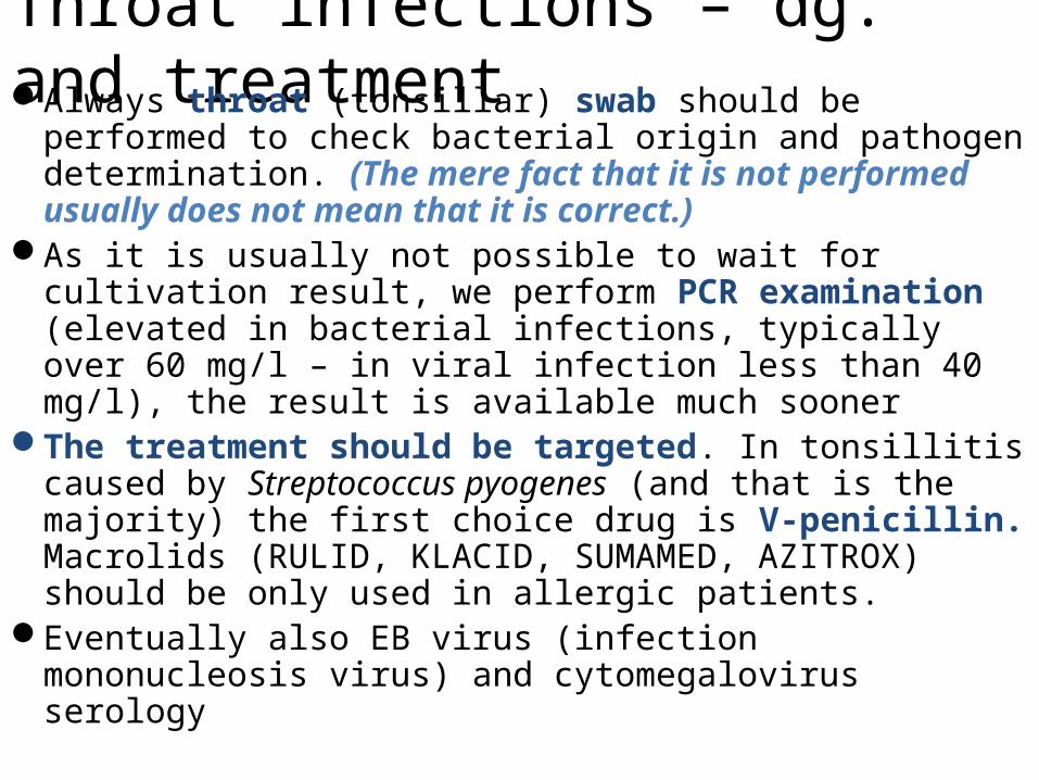
Throat infections – dg. and treatmentAlways throat (tonsillar) swab should be performed to
check bacterial origin and pathogen determination. (The mere fact that it is not performed usually does not mean that it is correct.)
As it is usually not possible to wait for cultivation result, we perform PCR examination (elevated in bacterial infections, typically over 60 mg/l – in viral infection less than 40 mg/l), the result is available much sooner
The treatment should be targeted. In tonsillitis caused by Streptococcus pyogenes (and that is the majority) the first choice drug is V-penicillin. Macrolids (RULID, KLACID, SUMAMED, AZITROX) should be only used in allergic patients.
Eventually also EB virus (infection mononucleosis virus) and cytomegalovirus serology

Examination and treatment of laryngeal (and tracheal) inflammations
It is nothing to examine. It is useless to perform throat swab, as there are completely different bacteria in throat. So, microbiological examination is not performed, except specific situations (chronic disease)
Treatment is symptomatic. Antibiotics are not indicated, not regarding the circumstances.

Examination and treatment of inflammations of bronchi and bronchiolesBasic is clinical examination that shows development of
cough with expectoration, without findings on the lung tissue (according to X-rays and clinical examination)
Microbiology examination is almost useless. In case of pus expectoration sputum may be sent because of the risk of secondary infection. In this case, also CRP could be measured. It is also possible to sent blood for serology of respiratory pathogens
Antibiotic treatment is almost useless, in macrolids and tetracyclins might be used

Special situation: acute deterioration of chronic bronchitis
Characterized by– Cough deterioration– Elevated expectoration, change of character and colour of sputum– Often deterioration of breathlessness
Causative agents: less than 40 % virusesAmong bacteria, the most common agents are
Haemophilus influenzae, Streptococcus pneumoniae or Moraxella catarrhalis.
Routine antibiotic treatment of patients is not recommended
Antibiotics have only effect in cases with presence of all three disease symptoms
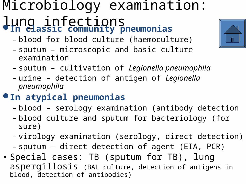
Microbiology examination: lung infectionsIn classic community pneumonias
– blood for blood culture (haemoculture)– sputum – microscopic and basic culture examination– sputum – cultivation of Legionella pneumophila– urine – detection of antigen of Legionella pneumophila
In atypical pneumonias– blood – serology examination (antibody detection– blood culture and sputum for bacteriology (for sure)– virology examination (serology, direct detection)– sputum – direct detection of agent (EIA, PCR)
• Special cases: TB (sputum for TB), lung aspergillosis (BAL culture, detection of antigens in blood, detection of antibodies)

Specimens and examination in
respiratory infections

Specimens for respiratory infection diagnostics – globally (1)
For bacteriology we send– swabs – (throat, tonsillar, nasal etc.), always with
transport medium (e. g. Amies medium), describe the localisation
– sputum, tracheal aspirate or bronchoalveolar lavage, eventually also endotracheal canulas and similar specimens – for bronchitis and pneumonia (eventual request for TB should be written on the form!)
– blood culture in pneumonias– urine for Legionella antigen
For mycology examination swab in FungiQuick (but also common Amies) is sent

Specimens for respiratory infection diagnostics – globally (2)
Viral agents are usually not examinedIn rare need for viral agent determination we use
nasopharyngeal or bronchoalveolar lavages with special medium, or blood for serology of respiratory viruses (i. e. for antibodies; we have to count that antibodies are only formed one or two weeks after start of the disease)
For influenzavirus we use swab from rear face of pharynx using special transport medium

Throat swab – techniqueSampling material: Swab with plastic stick in Amies
transport medium.Way of sampling:
– The swab is placed behind the palatal arcs with help of a spatula without contacting the oral mucosa.
– By rolling movement the surface of both tonsils and palatal arcs is swabbed so that sufficient amount of mucosal secretion would be sucked in the swab.
– Simultaneously back side of pharynx is swabbed.– The swab is pulled out carefully to avoid its contamination, and
placed to a special test tube with transport mediumStorage: Maximum 24 h at room temperature (for
gonorrhoea do not store it and send it immediately)Transport: Maximum 2 h at room temperature

Nasopharyngeal swab („pertussoid“ syndrome, suspicion for pertussis)Specimen: A wired swab; for Bordetella, inoculate
immediately to a special culture medium, for Haemophilus it is sufficient to send in a transport medium
Sampling: The end part (approx. 3 to 4 cm) of the swab on wire is flexed using the edge of test tube to the 90°, lead through oral cavity behind palatal arcs to the back side of the nasopharynx without touching the mucosa of oral cavity or tonsils. By circulating, punka-like movement the swab from pharyngeal mucosa is done (cotton-up)
Storage: Immediate transport to the laboratoryTransport: Maximum two hours at the room
temperature.

Sputum samplingSpecimen: Sterile transparent plastic container with a
screw cap.Sampling:
– Sampling is always performed at supervision of a nurse or a doctor.
– Patient washes the oral cavity and gurgles with water (decrease of oral bacteria contamination)
– After that, the patien should deeply cough so that to press out the secretion from lower respiratory ways, not saliva or nasopharynx secretion.
– So gained sputum is kept in a sterile container in volume of minimally 1ml.
Storage: Maximum 24 h at room temperatureTransport: Maximum 2 h at room temperature

Possible examinations in lung infectionsThe basis is clinical examination and X-rays, important
differentiation classic × atypical pneumonia (different spectrum of causing agents)
In classic pneumonias properly taken sputum has sense, eventually (especially in septic course) also blood for blood culture
In atypical pneumonias serology of Mycoplasma and Chlamydia (eventually in frame of „serology of respiratory viruses“
In hospital pneumonias it also might be useful to perform examination for Legionella. Besides culture examination it is also possible to examine urine for Legionella antigen, eventually serology

What to write on a request formIn addition to filling in the usual fields (name,
number of the patient...) is an important field of the request, what is to be examined.
Examples of formulation on the request form:– throat swab for bacteriology– punctate of frontal sinuses on bacteriology + yeast– blood for serology of agents of atypical pneumonias– sputum for bacteriology– sputum for TB (culture + PCR)– blood culture No. II from a venepunction– bronchoalveolar for Pneumocystis jirovecii

What to knowThe request form should contain an information,
what type of specimen is it, what testing is required, and, where appropriate, other relevant information
Microbiologist has the right to reject the wrong sample of sputum (non-pyogene, does not contain leucocytes, only epithelia it is saliva!!!)
TB culture takes several weeks, similarly also culture of some fungi
For virology and detection of various antigens the speed of examination depends mainly on the organization of work

Processing and evaluation of respiratory specimens

What happens with the samples in the laboratory
Most swabs are cultured on blood agar. On the agar we place disks, whose aim is to suppress the normal flora and to allow detection of pathogens. Because of Haemophilus, which would be only able to grow there in presence of S. aureus, we inoculate a S. aureus line on the agar
In sputum and similar samples, microscopy is usedBesides blood agar, more media (Endo) are usedVirology samples are isolated on the eggs or tissue
cultures, antigen detection is performedIn serology specimens we search for antibodies

How to find a pathogen among common oropharyngeal flora
• Normal flora consists of greyish, viridating colonies (oral streptococci) and yellowish, usually a-haemolytic colonies (oral Neisserias). They use to make a dense „carpet“ on the surface of agar medium and they make search for pathogens quite difficult, nevertheless possible:– Haemolytic streptococci (and also Staphylococcus aureus) are
visible by a strong haemolysis on blood agar– For Haemophilus detection we use antibiotic disc with bacitracin –
higher concentrations than in bacitracin test (to decline the normal microflora)
– For meningococcal detection we use another disk, with mixture of vancomycin and colistine

Detection of pathogen in throat/sputum1 swab inoculation
2 loop inoculation
3 staphylococcus line
4 bacitracin disc (for hemophili)
5 V + K disc (colistine and vancomycine) for meningococci
In all parts of inoculated area we search for colonies with haemolysis. They could be streptococci (rather colourless) or goldish)

Cultivation result of throat swab with common flora
In these sites we search for haemophili
www.medmicro.info
The bacitracin disk may be placed either on the Staphylococcus line, or approx. 1 cm far from it, both ways are used.

Explanations to following screens
• BA – blood agar• EA – Endo agar; usually, McConkey agar may be used as
an alternative• BA+AMIK – blood agar with amikacin, selective for
streptococci a enterococci• NaCl – BA with 10 % NaCl, selective for stafylococci• B – broth

Sputum examination
www.lumen.luc.edu

Sputum examinationDiagnostic schedule (1)
• Day 0: microscopy (Gram staining)• Day 1: result of primary culture on BA and EA. If
only common flora is present, EA is discarded and BA is prolonged to another day. An eventual pathogen is identified and its antimicrobial susceptibility assessed. If there is a small amount of a pathogen, isolation is performed (colony is carefully picked by a loop and reinoculated to a new agar plate to obtain a pure culture)
• NaCl is not used in sputum specimens, but is used in some other specimens (tracheal aspirations, bronchoalveolar lavage) it is used.
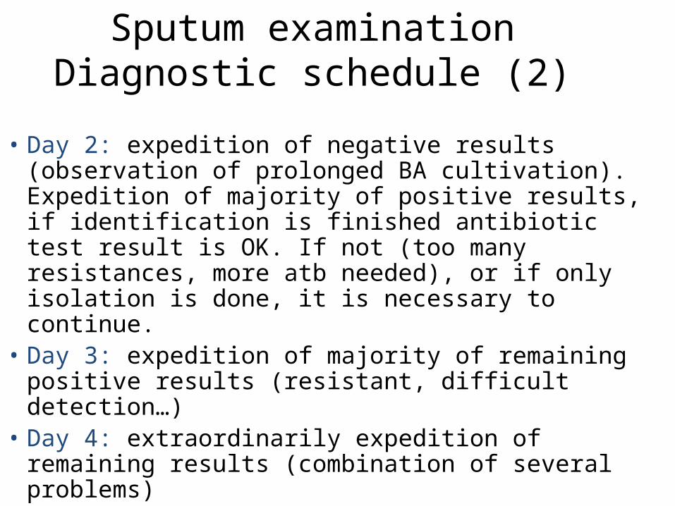
Sputum examinationDiagnostic schedule (2)
• Day 2: expedition of negative results (observation of prolonged BA cultivation). Expedition of majority of positive results, if identification is finished antibiotic test result is OK. If not (too many resistances, more atb needed), or if only isolation is done, it is necessary to continue.
• Day 3: expedition of majority of remaining positive results (resistant, difficult detection…)
• Day 4: extraordinarily expedition of remaining results (combination of several problems)

Sputum – possible findings• Common flora: There is no flora in LRT, but always a
contamination from URT is present: oral streptococci and Neisserias
• Pathogens: pneumococci, pyogenous streptococci, haemophili (typical pneumoniae). Causative agents of atypical pneumoniae are mostly non-culturable.
• One of typical findings is Staphylococcus aureus, you can use treatment using oxacilin, eventually, if oral oxacillin would not be available, to use Ist generation cephalosporins.
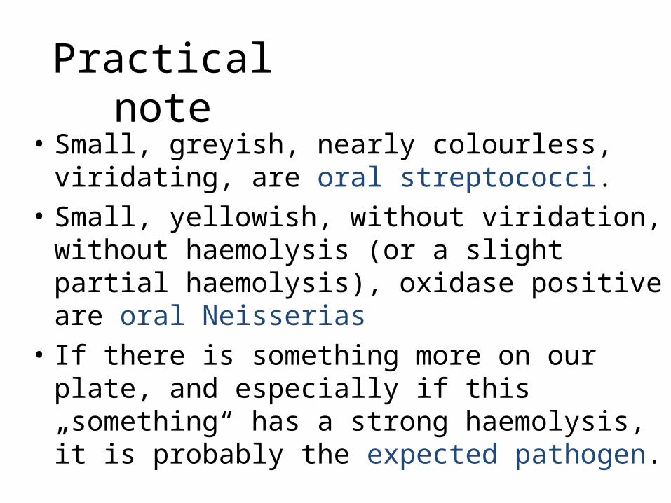
Practical note• Small, greyish, nearly colourless, viridating, are
oral streptococci.• Small, yellowish, without viridation, without
haemolysis (or a slight partial haemolysis), oxidase positive are oral Neisserias
• If there is something more on our plate, and especially if this „something“ has a strong haemolysis, it is probably the expected pathogen.

„Reading“ of bacteriology
www.medmicro.info

Throat swabbiology.clc.uc.edu

Throat swabDiagnostic schedule
• Day 0: only start of the cultures• Day 1: result of primary culture of specimen on BA
and EA. NaCl is not used here. Here, too, BA cultures with common flora are prolonged
• Day 2: expedition of all negative and majority positive results
• Day 3: expedition of mostly all remaining results

Thro
at s
wab
– c
omm
on
flora
and
pyo
geno
us
stre
ptoc
occu
s
www.medmicro.info

Pharynx – possible findings• Common flora: Oral streptococci a Neisserias;
haemophili (mostly H. parainfluenzae), but normal are also small amounts of S. aureus, pneumococci, meningococci, Moraxellas etc. More components of common flora (anaerobes, spirochetes) are not found in normal culture
• Pathogens: pyogene streptococci, arkanobacteria; often nothing is found and it is viral origin (EB viruses and others)
• Treatment: In case of Streptococcus pyogenes found to be a pathogen, V-penicillin is used.
Importance and classification of digestive tract
infections
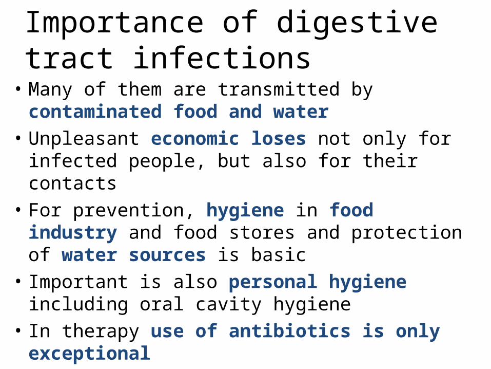
Importance of digestive tract infections
• Many of them are transmitted by contaminated food and water
• Unpleasant economic loses not only for infected people, but also for their contacts
• For prevention, hygiene in food industry and food stores and protection of water sources is basic
• Important is also personal hygiene including oral cavity hygiene
• In therapy use of antibiotics is only exceptional

Classification of digestive infections• We speak about
– Infections in the oral cavity– Infections of pharynx – see respiratory infections– Infections of oesophagus – very rare, usually secondary
after originally non-infectious disease– Infections of stomach (or rather cooperation of gastric
microbes in some diseases)– Infections of small intestine (enteritis)– Infections of large intestine (colitis)– Often infections of both parts (enterocolitis)

Normal microflora of GIT• Lips are transition between skin and oral flora• Oral cavity (as in pharynx) we find oral
streptococci, Neisseria, avirulent Haemophilus strains etc. Many others are present, but cannot be cultured.
• Oesophagus and stomach are normally microbe-free
• In small and large intestine we usually find approx. 1 kg anaerobes, also Enterobacteriaceae, enterococci, yeasts, sometimes even non-pathogenic amoeba
• Anus is again transition intestine-skin

Sampling and examination in
intestinal infections

Sampling and stool transport for individual examinations
• Bacteria – in Amies transport medium• Yeasts – better in FungiQuick medium, but substantially
Amies medium is also sufficient• Viruses – hazelnut-sized specimen; for isolation of a virus
cooling is necessary• Parasites – hazelnut-sized again, not necessarily sterile.
Traveller anamnesis necessary. Usually three specimens (one negative does not mean complete positivity)
• Toxin of Clostridium difficile – hazelnut-sized specimen• Pinworms – Graham method – perianal moulage on a
special tape, for microscopy• Intoxications by bacterial toxins – vomit, food remainders
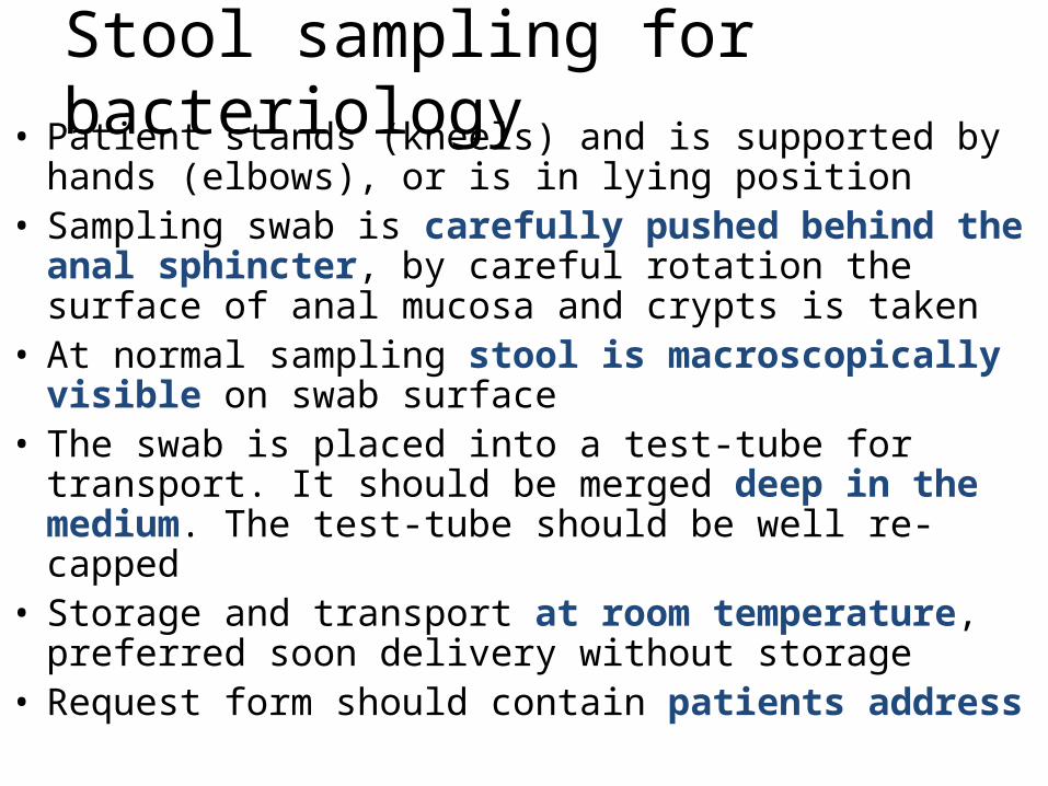
Stool sampling for bacteriology• Patient stands (kneels) and is supported by hands (elbows),
or is in lying position• Sampling swab is carefully pushed behind the anal
sphincter, by careful rotation the surface of anal mucosa and crypts is taken
• At normal sampling stool is macroscopically visible on swab surface
• The swab is placed into a test-tube for transport. It should be merged deep in the medium. The test-tube should be well re-capped
• Storage and transport at room temperature, preferred soon delivery without storage
• Request form should contain patients address

Why address?• In case of obligatory pathogen (Salmonella, Shigella,
Campylobacter, Yersinia) finding, the laboratory (in Czechia) is obliged to send a report to regional public health office that contacts the ill person for depistage (to find the source of infection, and also to know possible risks for other people)
• In case of missing address a telephonic question is addressed to the doctor that has sent the specimen
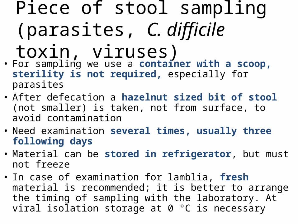
Piece of stool sampling (parasites, C. difficile toxin, viruses)
• For sampling we use a container with a scoop, sterility is not required, especially for parasites
• After defecation a hazelnut sized bit of stool (not smaller) is taken, not from surface, to avoid contamination
• Need examination several times, usually three following days
• Material can be stored in refrigerator, but must not freeze
• In case of examination for lamblia, fresh material is recommended; it is better to arrange the timing of sampling with the laboratory. At viral isolation storage at 0 °C is necessary

More about stool for parasites• Traveller anamnesis is necessary, not only „was
abroad“, but also what countries he/she visited• In case of macroscopic finding of a whole parasite (e.
g. roundworm), it is possible to send the complete organism in a test-tube
• Be careful – patients often use to insist on saying that a worm was present in their stool, but in fact the organism (e. g. earthworm) just fell from the window parapet
• Sometimes the „being sure“ about presence of a parasite is a part of psychiatric diagnosis of the patient

Sampling for pinworms(Graham method)
• The sampling is done in the morning without washing (the female pinworms lay eggs to perianal region during the night)
• Prior to sampling, the transparent (!) tape is carefully removed from the slide, placed to perianal region, the thighs are pressed one against the other, released and the tape is placed back to the slide
• In adults (it would be painful because of hairs) we rather use stool sampling (with lower effectiveness), of we use Schüffner stick

Diagnostics of bacterial pathogens•Microscopy has low practical importance only•Cultivation is performed on various media (choice depends on
the patient‘s age and diagnosis, in travellers eventually more rare media are added), found pathogens are identified – see further
•Direct detection of A and B toxin (Clostridium difficile) as antigen. Toxin detection is more important than mere finding of clostridium or its structural antigen – the antigen may be even present in healthy persons, but positive toxin means serious problem
www.oxoid.cz
Diagnostics of viral agents: usually antigen detection, eventually nucleid acid detection
Diagnostics of parasital and fungal agents: see more in mycology and parasitology lessons
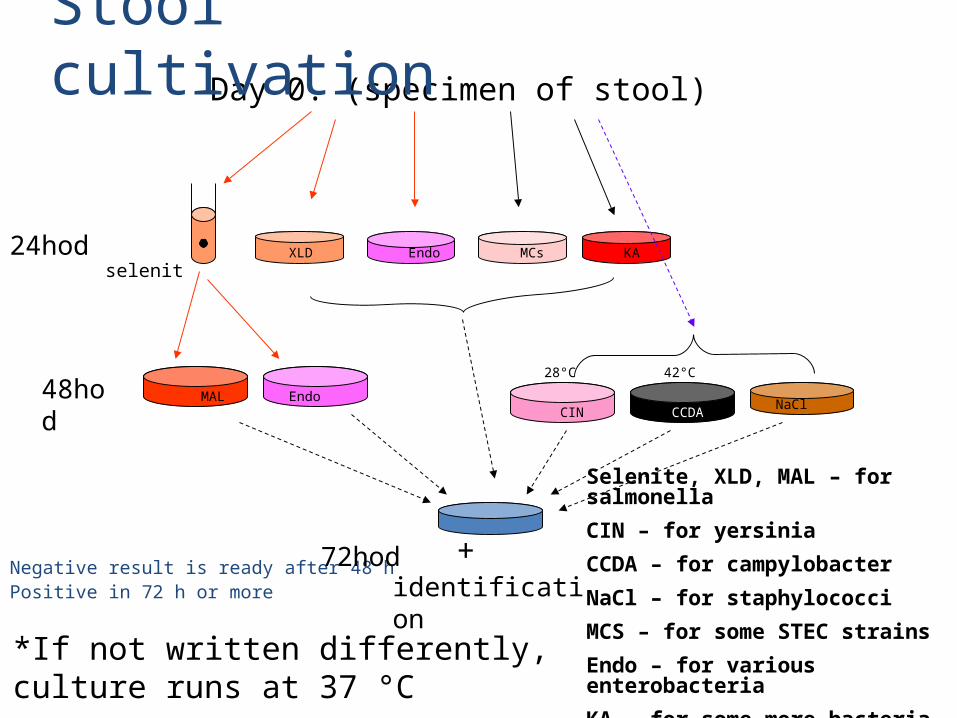
Day 0. (specimen of stool)
Negative result is ready after 48 hPositive in 72 h or more
selenit24hod
48hod
identification
Endo
EndoXLD
MAL
MCs KA
NaCl CCDA CIN
+ 72hod
42°C28°C
*If not written differently, culture runs at 37 °C
Stool cultivation
Selenite, XLD, MAL – for salmonella
CIN – for yersinia
CCDA – for campylobacter
NaCl – for staphylococci
MCS – for some STEC strains
Endo – for various enterobacteria
KA – for some more bacteria

Identification of a bacterium
• Bacteria are cultured on various media, they have specific appearance on them
• Bacteria are further diagnosed by biochemical tests
• In some cases (Salmonella, Escherichia) it is necessary to perform antigen analysis of the strain

Interpretation of stool examination• In results of stool examination it is necessary to
differentiate whether they are primary pathogens (Salmonella, Shigella, Yersinia, Campylobacter) or secondary pathogens; in some secondary pathogens (especially Escherichia coli) further determination is needed (EPEC, STEC, EAggEC etc.)
• Interpretation should be done in context of clinical signs (in high quantity of „non-pathogenic amoeba“ and serious symptoms the treatment might be useful)
• In case of Clostridium difficile infection it is important to know whether clostridium toxin is positive. Mere finding of clostridium antigen or cultivation finding of Clostridium does not mean too much.

The
end
Archiv MiÚ LF MU


![BMC Microbiology BioMed Central · 2017. 8. 27. · Moraxella catarrhalis is an exclusively human, mucosal res-piratory tract commensal and pathogen causing between 5% [1] and 20%](https://static.fdocuments.in/doc/165x107/60b9d1b25ab06638794a37be/bmc-microbiology-biomed-central-2017-8-27-moraxella-catarrhalis-is-an-exclusively.jpg)