Tracheomalacia - mc.vanderbilt.edu · 4‐year‐old girl with recurrent cough. Paired...
22
Transcript of Tracheomalacia - mc.vanderbilt.edu · 4‐year‐old girl with recurrent cough. Paired...


Tracheomalacia
Paul Moore, M.D.Eddie Penn, M.D.David Bichell, M.D.Otis Rickman, M.D.

Normal Trachea
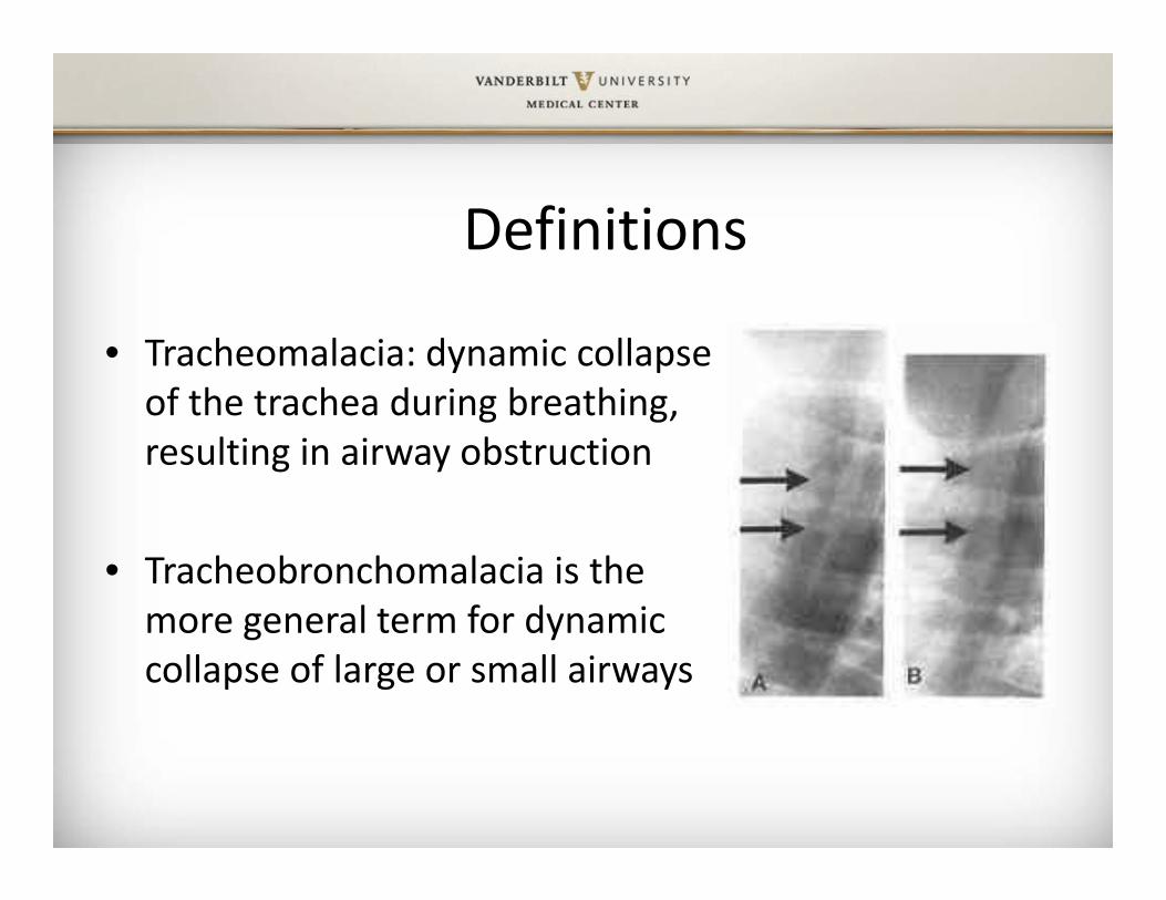
Definitions
• Tracheomalacia: dynamic collapse of the trachea during breathing, resulting in airway obstruction
• Tracheobronchomalacia is the more general term for dynamic collapse of large or small airways

Etiology• Reduction and/or atrophy of the longitudinal elastic fibers of the pars membranacea
• Impairment of cartilage integrity• Faulty division of the foregut into the trachea and esophagus during embryonic development results in the trachea receiving too much tissue in TEF/EA and possibly idiopathic primary TM

Histology• Reduced cartilage to muscle ratio, resulting in easer collapsibility of the trachea (normal ratio 4.5:1)
• Variability in the longitudinal muscle fibers deep to the transverse fibers in the lower trachea

Physiology• By virtue of its compliance, the normal intrathoracic trachea
dilates somewhat with inspiration and narrows with expiration, as a reflection of the difference between intrathoracic and intraluminal pressures.
• The majority of cases of TM are intrathoracic in nature such that excessive narrowing is most prominent when intrathoracicpressure is substantially greater than intraluminal pressure, as it is during forced expiration, cough, or the Valsalva maneuver.
• With extrathoracic TM, negative intrapleural pressures are transmitted to the extrathoracic trachea due to pleural reflections such that the upper airway collapses during inspiration.

Classification: Congenital or Primary Forms
• Isolated finding in healthy infants• Prematurity: inadequate maturity of tracheobronchial
cartilage• Pulsatile collapse with normal inominate artery• Association with TEF (tracheosophageal fistula)• Disorders with abnormal cartilaginous matrix of the
trachea (polychondritis, chondromalacia, mucopolysaccharidoses)
• Other genetic syndromes that may alter collagen maturation or airway tone

Classification: Acquired or Secondary Forms
• Protracted endotracheal intubation (with more significant effects in premature infants)
• Tracheotomy• Severe tracheobronchitis• External tracheal compression
– Vascular– Cardiac– Skeletal– Tumors and cysts

Associated conditions
• Cardiovascular abnormalities (20‐58%)• Bronchopulmonary dysplasia (up to 52%)• Gastroesophageal reflux disease (~50%)• Subglottic stenosis and layrngomalacia• Neurologic impairment (8‐48%)• Severe developmental delay (26%)

MAC (Major Airway Collapse) Classification System
• Type I: intrinsic defect in the cartilaginous portion of the trachea leading to an increased proportion of membranous trachea
• Type II: extrinsic tracheal compression by cardiovascular structures, tumors, lymph nodes, or other masses
• Type III: prolonged positive pressure ventilation or an infectious/inflammatory process that undermines the intrinsic cartilaginous support of the trachea

Incidence
• Congenital form: est. 1 per 1,445 infants• Incidence of tracheomalacia in congenital diseases parallels the incidence of those diseases
• Incidence on increase due to greater survival from NICU and increase # of bronchoscopies
• Mortality of severe TM can approach 80%

Clinical features• Expiratory stridor and cough, often described as barking or brassy
• Cough due to the juxtaposition of the anterior and posterior walls of the trachea, resulting in recurrent vibrations and irritation of the airway
• ±whether symptoms present at birth or after a few weeks
• Inspiratory stridor suggestive of extrathoraciccompression

Additional clinical features• Noisy, medium‐pitched to high‐pitched breathing• Recurrent respiratory distress• Wheezing• Cyanosis• Spontaneous hyperextension of the neck• Breath‐holding spells• Bagpipe sign, an expiratory sibilant note that persists after the
end of visible expiration• Feeding difficulties• Reflex apnea

Clinical Severity Rating
• Mild: Respiratory difficulties associated with infectious processes such as croup or bronchiolitis
• Moderate: classic findings, including stridor, wheezing, recurrent respiratory infections, and even cyanosis associated with exacerbations
• Severe: stridor during tidal breathing, marked sputum retention, upper airway obstruction, reflex apnea, and even cardiac arrest

Diagnosis: Imaging
• Chest radiograph: 62% sensitivity• Standard CT and MRI: not well suited because images are obtained end‐expiration
• Multidetector CT: allows end‐expiratory and end‐inspiratory imaging
• Free breathing cine CT: does not require patient cooperation with expiratory maneuvers
• CT‐Angiogram or Angio‐MRI can be used to diagnose vascular anomalies
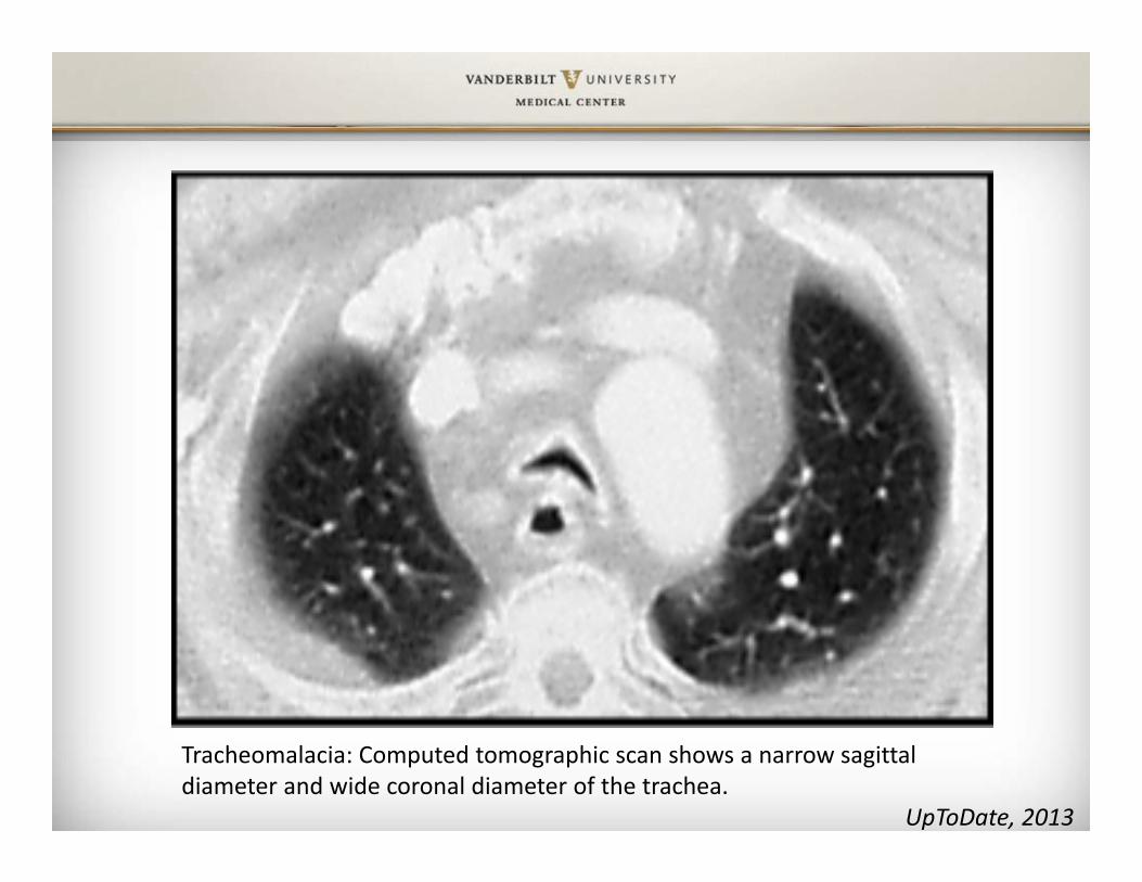
Tracheomalacia: Computed tomographic scan shows a narrow sagittal diameter and wide coronal diameter of the trachea.
UpToDate, 2013

4‐year‐old girl with recurrent cough. Paired inspiratory–expiratory volumetric MDCT was performed for evaluation for possible underlying tracheomalacia.A, Axial end‐inspiratory CT image obtained with lung window shows normal appearance of trachea and lung parenchyma.B, Axial end‐expiratory CT image obtained with lung window at same level as A shows excessive collapse of trachea (curved arrow) consistent with tracheomalacia. Areas of geographically marginated radiolucency (straight arrows) in both lungs are consistent with air trapping.
Lee, AJR, 2009
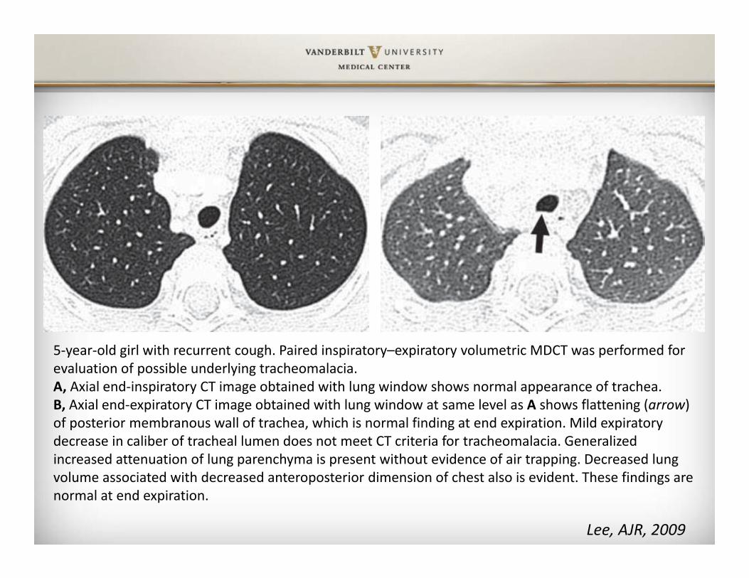
5‐year‐old girl with recurrent cough. Paired inspiratory–expiratory volumetric MDCT was performed for evaluation of possible underlying tracheomalacia.A, Axial end‐inspiratory CT image obtained with lung window shows normal appearance of trachea.B, Axial end‐expiratory CT image obtained with lung window at same level as A shows flattening (arrow) of posterior membranous wall of trachea, which is normal finding at end expiration. Mild expiratory decrease in caliber of tracheal lumen does not meet CT criteria for tracheomalacia. Generalized increased attenuation of lung parenchyma is present without evidence of air trapping. Decreased lung volume associated with decreased anteroposterior dimension of chest also is evident. These findings are normal at end expiration.
Lee, AJR, 2009

Tracheal shapes on freebreathing cine CT according to the ratio of the transverse diameter to the anteroposterior diameter of the trachea. a Round shape shows a ratio of between 0.8 and 1.2.b Lunate shape shows a ratio of ≥1.2. c Elongated shape shows a ratio of <0.8. d Crescentic shape shows a ratio ≥2.0 with posterior concavity Goo Ped Rad. 2013

Management• Most infants improve by 6 to 12 months of age as airway caliber increases and cartilage develops.
• Indications for intervention: life‐threatening episodes of airway obstruction, recurrent infection, respiratory failure, or failure to thrive.

Possible Interventions• Splinting• Tracheotomy • Positive pressure ventilatory support• Aortopexy• Stent placement• Posterior tracheal wall surgery