USEFULNESS OF MDCT IN THE EARLY ABDOMINAL … · USEFULNESS OF MDCT IN THE EARLY ABDOMINAL...
35
USEFULNESS OF MDCT IN THE EARLY ABDOMINAL COMPLICATIONS AFTER ORTHOTOPIC LIVER TRANSPLANTATION Tesi Scuola Specializzazione Radiodiagnostica Pisa Massimiliano Rossi
Transcript of USEFULNESS OF MDCT IN THE EARLY ABDOMINAL … · USEFULNESS OF MDCT IN THE EARLY ABDOMINAL...
USEFULNESS OF MDCT IN THE
EARLY ABDOMINAL COMPLICATIONS
AFTER ORTHOTOPIC LIVER
TRANSPLANTATION
Tesi Scuola Specializzazione
Radiodiagnostica
Pisa
Massimiliano Rossi

Abstract
Purpose
To evaluate the usefulness of MDCT in the detection of early abdominal complications after orthotopic liver transplantation (OLT).
Material and Methods
From September 2009 to October 2013 we retrospectively enrolled 170 subjects who underwent MDCT within the first 90 days after OLT at the Pisa University Hospital. Inclusion criteria were represented by clinical-laboratory and/or echo-color Doppler abnormalities. The examinations were performed at 64-slice MDCT scanner (Somaris\5 Syngo CT 2009\E; Siemens) and the protocol study always included pre- and post-contrast multi-phasic acquisitions. All images were reviewed by two radiologists in conference and imaging results were correlated with DSA, trans-Kehr cholangiography/ERCP, surgery, clinical-laboratory and imaging follow-up.
Results
No significant complication was found in 142 patients, while in the remaining 28 patients vascular complications (hepatic artery thrombosis HAT, n=5 and stenosis HAS, n=5; portal vein thrombosis, n=4; cava vein thrombosis, n=1; arterial bleeding, n=1), biliary complications (anastomotic leak with biloma, n=6), 4 graft complications (abscess, n=1; extended areas of impaired perfusion, n=3), adrenal hemorrhage (n=1) and bowel perforation (n=1) were identified. Four false positive cases (3 hepatic artery stenoses and 1 hepatic artery rupture) and one false negative (biloma) were diagnosed on MDCT. The sensitivity, specificity, PPV, NPV and diagnostic accuracy of MDCT in the identification of various complications were 96%, 97%, 87%, 99% and 97%, respectively.
Conclusion
MDCT is extremely reliable in the identification of early abdominal complications of liver transplant patients.
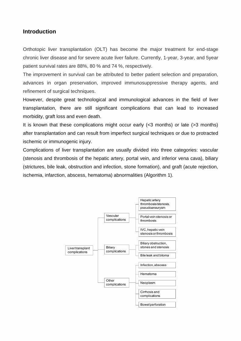
Introduction
Orthotopic liver transplantation (OLT) has become the major treatment for end-stage
chronic liver disease and for severe acute liver failure. Currently, 1-year, 3-year, and 5year
patient survival rates are 88%, 80 % and 74 %, respectively.
The improvement in survival can be attributed to better patient selection and preparation,
advances in organ preservation, improved immunosuppressive therapy agents, and
refinement of surgical techniques.
However, despite great technological and immunological advances in the field of liver
transplantation, there are still significant complications that can lead to increased
morbidity, graft loss and even death.
It is known that these complications might occur early (<3 months) or late (>3 months)
after transplantation and can result from imperfect surgical techniques or due to protracted
ischemic or immunogenic injury.
Complications of liver transplantation are usually divided into three categories: vascular
(stenosis and thrombosis of the hepatic artery, portal vein, and inferior vena cava), biliary
(strictures, bile leak, obstruction and infection, stone formation), and graft (acute rejection,
ischemia, infarction, abscess, hematoma) abnormalities (Algorithm 1).
Diagnosis of acute rejection, one of the most serious complications after liver
transplantation, is established with graft biopsy and histologic study .
The role of imaging methods consists of excluding the other complications, which can
have clinical signs and symptoms similar to acute rejection.
Ultrasound (US) is routinely performed in OLT for assessment of the hepatic parenchyma,
biliary tree, and vasculature.US is sensitive but not specific in the evaluation of
peritransplant fluid collections. Bile, lymph, blood and pus can all present on imaging as a
simple fluid collection.
US with Echo-Color-Doppler (ECD) is the primary modality used to evaluate the vascular
anastomoses in the post-transplantation period.
Multidetector row CT (MDCT) scanner is accepted as a practical noninvasive diagnostic
method in various complications after OLT. MDCT imaging is usually performed for the
evaluation of postoperative collections, vascular complications, or if the ultrasound
examination is technically unsatisfactory.
The aim of this study was to evaluate the usefulness of MDCT in the detection of early
abdominal complications after orthotopic liver transplantation.
Material and methods
Patients
Out of 420 patients undergone OLT from September 2009 to October 2013 we included
retrospectively 170 subjects (113 M, and 57 F; age range, 16– 69 years) who were
studied with MDCT within the first 90 days after OLT at the Pisa University Hospital. In our
transplantation center, CT is not routinely performed in the early period in a regular post-
transplantation course. Inclusion criteria were represented by clinical (fever, right flank
pain), laboratory (leukocytosis) and ECD abnormalities.
Imaging technique
All examinations were performed by using a 64 slice MDCT scanner (Somatom\5 Syngo
CT 2009\E Siemens with spiral technique).
Our imaging protocol included abdomen examination performed before and after the
intravenous injection of non-ionic contrast material (100 -130 ml, 320 to 400 mg of iodine
per milliliter) via a power injector (injection flow-rate, 4 to 5 mL\sec). We used the bolus
trigger technique (positioning of the respective region of interest in the abdominal aorta,
threshold 100 HU).
Post-contrast study included a quadriphasic hepatic scanning (double arterial phase with a
delay of 20-40 sec, portal-venous phase with a delay of 60-70 sec and the last phase at
180 sec after reaching the threshold). The powers of the tube were 100 or 120 KVp and
automatic adjustment for mAs.
Images were reconstructed with a 1 mm thickness.
Imaging analysis
CT examinations were analyzed by two experienced radiologists in conference.
All images were evaluated using a picture archiving and communication system (Esaote-
Fuji) with post-processing of the imaging data using a variety of three-dimensional
reformatting techniques such as maximum intensity projection (MIP) and volume rendering
(VR).
The post processing of the CT images particularly allowed to the reviewers to depict both
HA anatomy and pathology efficiently and accurately.
The reviewers considered as normal findings: right pleural effusion, small amount of free
intra-abdominal fluid or hematomas in the perihepatic region (especially in the hepatic
hilum or adjacent to the anastomosis), the periportal edema and small bowel edema.
Periportal low-attenuation zones were very common in this setting and probably reflect
lymphedema and lymphangiectasia in most cases.
Besides, they evaluated as pathological features arterial stenosis and thrombosis, portal
thrombosis, bleeding, partial caval thrombosis, biloma, abscess, impaired perfusion,
adrenal hemorrage and bowel perforation.
We finally compared our MDCT findings with the gold standard (DSA, trans-Kehr
cholangiography\ERCP, surgery, clinical laboratory and imaging follow-up.)
Statistical analysis
We omitted normal findings in the statistical analysis.
The distribution of the qualitative variables was expressed as the relative frequency of the
various modalities under observation. The distribution of the quantitative variables was
expressed as the mean, standard deviation, minimum, maximum and number of
observations.
MDCT findings were compared with DSA, ERCP, PTC, surgery or clinical and imaging
follow-up results and defined as true positives when they correctly detected complications
confirmed by the final diagnosis reference standards; false positives when they were not
confirmed by DSA, ERCP, PTC, surgery or clinical and imaging follow-up; false negatives
when complications detected by DSA, ERCP, PTC, surgery or clinical and imaging follow-
up were not detected by MDCT; true negatives when the absence of complications was
confirmed by DSA, ERCP, PTC, surgery or clinical and imaging follow-up. To evaluate the
diagnostic yield of MDCT, we determined the sensitivity, specificity, diagnostic accuracy,
positive predictive value (PPV), and negative predictive value (NPV) of the reviewers for
the detection of all complications.
Results
On the basis of our gold standard, no significant complication was found in 142 patients.
The remaining 28 patients presented 16 vascular complications (hepatic artery thrombosis
[Figure 1], n=5; hepatic artery stenosis [Figure 2], n=5; partial portal vein thrombosis, n=4;
partial cava vein thrombosis, n=1; arterial bleeding, n=1); 6 biliary complications
(anastomotic leak with biloma) [Figure 3]; 4 graft complications (abscess [Figure 4], n=1;
extended areas of impaired perfusion, n=3); one adrenal hemorrhage and one bowel
perforation (Table 1).

TAB 1: GOLD STANDARD
Type of complications
Number of
cases
Vascular 16 (57 %) HAT 5
HAS 5
Partial portal
thrombosis (PVT)
4
Bleeding 1
Partial caval
thrombosis
1
Biliary 6 (21 %) Biloma 6
Graft complications
4(13 %)
Abscess 1
Impaired perfusion 3
Adrenal hemorrage
1(3%)
Adrenal
hemorrage
1
Perforation 1(3 %) Bowel perforation 1
28
Vascular complications were treated as follows: all 5 HAT with Re-OLT; all 5 HAS by
stenting; all 4 partial portal thrombosis and 1 partial caval thrombosis by medical therapy;
and 1 bleeding with surgery.
Four bilomas were subjected to drainage and 2 small bilomas resolved without
intervention. The patient with liver abscess underwent medical therapy. The patient with
intestinal perforation was treated with surgery.
All MDCT findings were reported in Table 2.

TAB 2 : CT FINDINGS
No complications (139)
Overall complications (31)
Vascular complications (20) HAT (5)
HAS (8)
Hepatic artery rupture (1)
Partial portal thrombosis (4)
Bleeding (1)
Partial caval thrombosis (1)
Biliary complications (5) Biloma (5)
Graft complications (4) Abscess (1)
Impaired perfusion (3)
Adrenal hemorrage (1)
Bowel perforation (1)
Four false positive cases (3 hepatic artery stenosis and 1 hepatic artery rupture) and one
false negative case (biloma) were diagnosed on MDCT (Table 3).

TAB 3: CT FINDINGS
Positive Negative Total
TP27 FN1 28 true complications
FP4 TN138 142 negative complications
31 139 170
The sensitivity, specificity, positive predictive value (PPV), negative predictive value (NPV)
and diagnostic accuracy of MDCT in the identification of various complications were 96%,
97%, 87%, 99% and 97%, respectively (Table 4).
TAB 4: CT FINDINGS
Sensitivity
96 %
Specificity
97 %
PPV
87 %
NPV
99 %
Diagnostic Accuracy
97 %
There have been number 170 CT examinations of 420 OLT over the last four years, about
40%. (Table 5).

TAB 5: NUMBER OF CT EXAMINATIONS FOR YEAR
YEAR
OLT CT EXAMS %
2009 (September-
December)
36 11 30
2010
95 37 37
2011
115 48 41
2012
85 37 43
2013 (january-october) 89 37 40
420
170 40
We have seen a trend of gradual increase in the demand for CT exams, with a peak in
2012 of 43% and subsequent stabilization at around of 40%.
Discussion
Patients who are candidates for liver transplantation are those with acute liver failure
(fulminant hepatic failure) or chronic liver disease for which conventional treatment is
unavailable or has ceased to be effective. Fulminant hepatic failure, a disorder that
ordinarily affects young people, causes a large portion of liver tissue to be destroyed in a
short time, leading to failure of liver function, changes in mental status (hepatic
encephalopathy), infections, and kidney failure. Chronic liver diseases that may be treated
by transplantation include chronic hepatitis B and C, autoimmune chronic hepatitis,
primary biliary cirrhosis, primary sclerosing cholangitis, alcoholic liver disease, and drug
induced liver injury. Hereditary disorders of metal metabolism including hemochromatosis

and Wilson disease (involving disordered iron and copper metabolism) also may require
transplantation.
After surgery, the patient ordinarily spends 1 to 3 days in the intensive care unit and then
moves to a regular hospital room. The average hospital time after the procedure is about
10 days to 2 weeks.
Potential complications include bleeding in the first 2 to 3 days and bacterial infections up
to 2 to 3 weeks after the surgery. Despite great technological and immunological
advances in the field of liver transplantation, there are still significant complications that
can lead to increased morbidity, graft loss and even death.
Complications of liver transplantation are usually divided into three categories: vascular
(hepatic artery thrombosis is the most common and most feared vascular complication of
liver transplantation), biliary and others.
Diagnosis of acute rejection, one of the most serious complications after liver
transplantation, is established with graft biopsy and histologic study. The role of imaging
methods consists of excluding the other complications, which can have clinical signs and
symptoms similar to acute rejection.
In our study we evaluated patients with complications arising within the first 90 days after
OLT. During this period, the most frequent are vascular complications, such as portal vein
thrombosis, hepatic artery stenosis and thrombosis or ischemia, so that a close monitoring
of the anastomotic vessels, the tissue perfusion and the microcirculation is necessary.
Clinical presentations of vascular complications vary from mild elevation of hepatic
function tests to fulminant hepatic failure. Because the clinical presentation is so varied,
their early detection and adequate management are important elements for ensuring graft
and patient survival after OLT. For this reason, imaging studies can be particularly helpful
in finding or eliminating vascular complications from the differential diagnosis.
Sonography with ECD is the primary modality used to search possible perihepatic fluid
collections and to evaluate the hepatic artery in the post-transplantation period.
Besides, even if ECD depends on a technically experienced operator and the condition of
the patient, it is the routine method in the clinical algorithm for the diagnosis of
complications. According some authors the reported sensitivities of ECD for the detection
of hepatic artery stenosis and thrombosis are, respectively, 80–85% and 60–80% for the
detection of hepatic artery thrombosis. Opinion of other authors the accuracy of ECD for
the diagnosis of HAT is reported to be 92%.

However, ECD can show false positive finding of HAT, probably from hepatic edema,
systemic hypotension or arterial spasm. False-negative results may be due to the
development of subacute extensive arterial collaterals that produce an arterial signal on
the Doppler sonogram, leading to the diagnosis of a suffering artery.
Some authors use microbubble sonography contrast agents to improve the sensitivity of
sonography to detect HAT.
In cases where US-ECD results are inconclusive, confirmation is required, or clinical
suspicion of a complication persists despite normal sonographic results, CT should be
performed. Unlike US, CT is not as dependent on the operator’s skill performing the study
or on the patient’s body habitus.
In particular, MDCT offers excellent spatial and temporal resolution, that combined with
post-processing of the imaging data using a variety of three-dimensional reformatting
techniques such as maximum intensity projection (MIP) and volume rendering (VR) allows
to depict both HA anatomy and pathology efficiently and accurately.
Moreover, MDCT has several advantages over other imaging modalities. Compared with
catheter angiography, MDCT angiography is non-invasive and cost effective. Digital
subtraction angiography (DSA) is the gold standard for diagnosing hepatic artery
complications and allows a concomitant angioplasty-stenting procedure under some
circumstances; however, it is not an ideal screening test because of the associated high
cost and invasive nature of the procedure with the associated risks and potential
complications.
In addition, despite the enormous improvement in post-OLT survival over the years, biliary
tract complications lead to a risk of significant morbidity and mortality (2–7%).
Complications such as leaks, obstructions, strictures, and stones have been observed in
9% to 35% of large clinical series of liver transplant recipients. In our hospital, in the first
period after OLT we don’t use MR study, however, after T-tube removal (after three
months), MR studies allow complete visualization of the extra-hepatic bile ducts and biliary
anastomosis (choledocho-choledocho-stomy) in nearly all the investigated subjects.
In our study we presented our experience with the MDCT evaluation of liver transplant
complications.
There was a low overall incidence of complications (28\420, gold standard), under the
findings reported in literature. The most frequent complications were vascular
complications (16\420).

Vascular complications were found in 16 cases, all correctly identified with MDCT. There
were verified 5 cases of hepatic artery thrombosis, followed with Re-OLT; 5 cases of
hepatic artery stenosis, treated by stenting; 4 partial portal thrombosis; 1 case of partial
caval thrombosis, followed by medical therapy and 1 bleeding treated with re-surgery.
In the literature, HAT in OLT is the most common vascular complication and the second
one is HAS. Risk factors predisposing to HAT include prolonged cold ischemia time of the
donor liver, significant difference in caliber between the donor and recipient hepatic
arteries, small caliber of the anastomosed vessels, ABO blood type incompatibility,
technical errors, CMV infection and acute rejection. Risk factors for HAS are similar to
those for HAS and additional cause causes include clamp injury, intimal trauma from a
perfusion catheter and disruption of vasa vasorum. This complication occurs classically at
the site of anastomosis within 3 months of OLT; if left untreated, HAS can lead to HAT,
and can progress to liver ischemia with hepatic insufficiency, biliary strictures, sepsis and
ultimately graft loss.
In our series MDCT correctly identified 27\28 positive cases, confirmed during surgery,
DSA or follow-up, and 138 \142 negative cases; there were also 4 false positive vascular
cases.
Out of 4 FP, 2 patients with an overestimation of stenosis were considered not significant
at clinical and imaging follow-up; a patient with suspected wrong artery rupture underwent
a revision surgery seven days after; there was coleperitoneo caused by biliary fistula
instead imaging correctly identified. Finally, there was a case of a steal syndrome,
diagnosed at DSA, caused by decreased perfusion of one arterial branch because of
diversion of blood flow into a different arterial branch originating from the same trunk.
About biliary complications there were 6 cases of biloma, 5 correctly identified with MDCT;
we identified also 1 false negative case of biloma, misdiagnosed as a fluid collection.
If a T-tube was inserted, like our patients, the biliary leak most commonly occurs at the site
of the entry of the tube into the duct. Non-anastomotic leaks are associated with hepatic
artery thrombosis in 89 % of the cases. Bile may run off freely into the peritoneal cavity or
may form a peritransplant fluid collection. Clinical presentation is variable and ranges from
mild nonspecific abdominal symptoms to septicemic shock. Most bile leaks are sterile and
their treatment can include biliary stent placement and percutaneous drainage, or
sometimes small leaks may close spontaneously.
About the case of intestinal perforation, it may occur due to deserosalization of the bowel
during a difficult hepatectomy, or an intestinal leak at the jejunostomy site.

Despite it was not the aim of the study, we observed a trend of gradual increase in the
demand for CT exams (170\420 patients were subjected to at least one CT examination in
the first three months after OLT) with a peak of 43% in 2012 and subsequent stabilization
at around 40%.
The explanation of this increase is complex: medico-legal issue, modern equipment 64
slice CT scanner provides thin slices images, very important informations for surgeons,
impossible with the previous equipment.
Due to the limited availability of livers, there was recruitment of older liver donors in the
last years; these patients need increased surveillance and are subject to greater risk of
vascular complications. In fact, there was larger number of vascular complications in 2013
(Table 6), compared to previous years.
TAB 6:HAT and HAS
HAT HAS
2009 1 0
2010 0 0
2011 1 1
2012 0 0
2013 3 4
TOTALE 5 5

Conclusions
Because the clinical presentation of post-OLT complications is frequently nonspecific and
varies widely, imaging studies are critical for early diagnosis.
Our results demonstrated the important role of MDCT in the diagnosis of complications
after OLT. In particular, MDCT can provide imaging of vascular abnormalities and
synchronous evaluation of liver parenchyma, biliary tract, and the other abdominal
structure by a non-invasive means.
In conclusion the early diagnosis and correct management of biliary complications are
important elements for ensuring graft and patient survival after OLT, and the diagnostic
workup has been repeatedly reviewed in an attempt to reach the most accurate strategy .
.
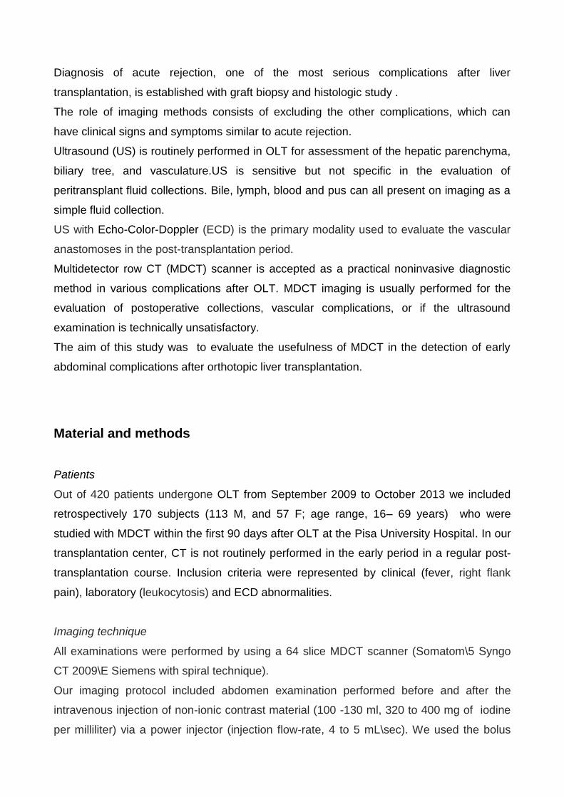
FIG 1a
FIG 1b

FIG 1c
Figure 1 (a-c). Hepatic artery thrombosis (HAT). Coronal oblique MIP (a) and
VR (b) 3D MDCT angiographic images showed occluded HA proximal to the
anastomotic site. DSA (c) confirmed HAT, with absent intrahepatic flow.
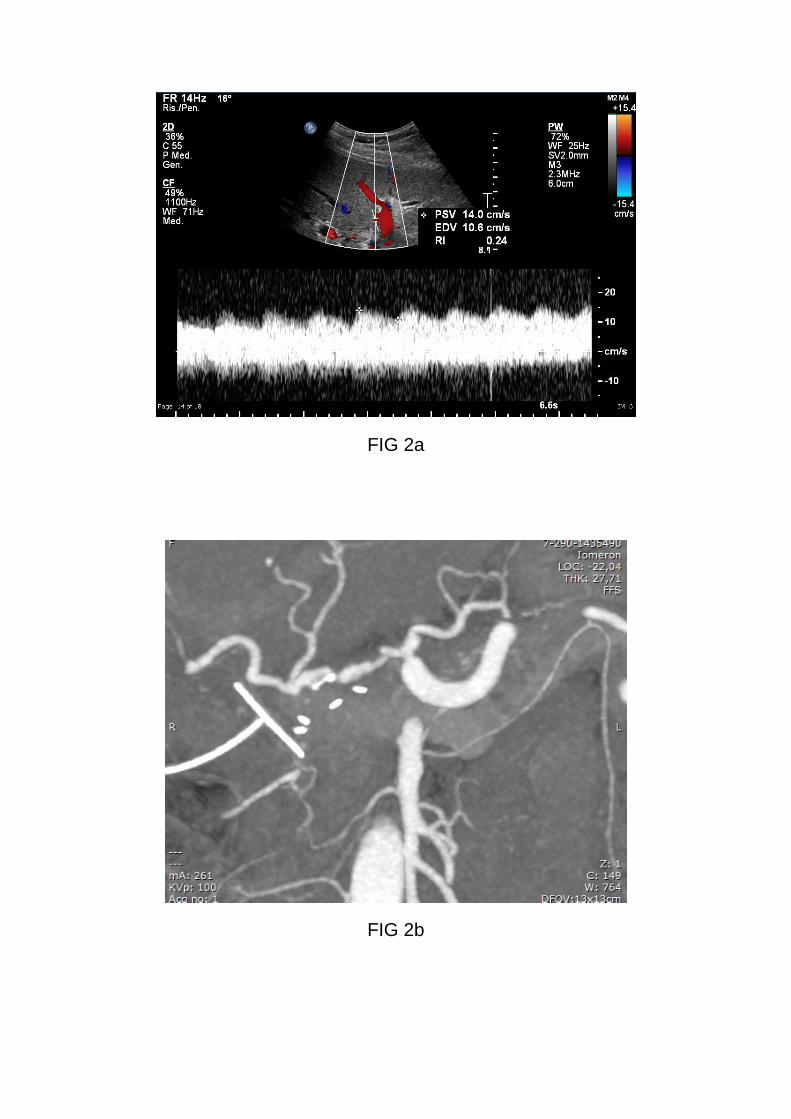
FIG 2a
FIG 2b

FIG 2c
FIG 2d

FIG 2e
FIG 2f

Figure 2 (a-f). Hepatic artery stenosis (HAS). ECD (a) showed tardus and
parvus waveform, low resistive indices and delayed systolic upstroke.
Coronal oblique MIP 3D MDCT angiographic image (b) demonstrated
anastomotic narrowing. DSA (c- e) confirmed a severe stenosis, treated by
stenting. ECD (f) post stenting: normal waveform.
FIG 3a
FIG 3b

FIG 3c
FIG 3d

Figure 3 (a-d). Biloma. Axial (a) and coronal (b) contrast-enhanced CT
images showed a rounded fluid collection at the level of hepatic hilum,
suspicious for biloma. Trans-Kehr cholangiography (c) demonstrated bile
leakage from the biliary anastomosis. The patient underwent no treatment. A
new trans Kehr cholangiography (d) performed 10 days after, didn't show any
extravasation of contrast.
FIG 4a

FIG 4b
FIG 4c

Figure 4 (a-c). Intrahepatic abscesses. Contrast-enhanced MDCT (a)
identified multiple rim-enhancing fluid collections in the right hepatic lobe
suspicious for liver abscesses. At MR study, SS-FSE T2-weighted image (b)
demonstrated multiple hyperintense lesions and 3D Gd-enhanced T1-
weighted LAVA image (c) confirmed the presence of hypovascular lesions
with a thick enhancing rim, like CT findings. This patient underwent medical-
therapy.
FIG 5a
FIG 5b
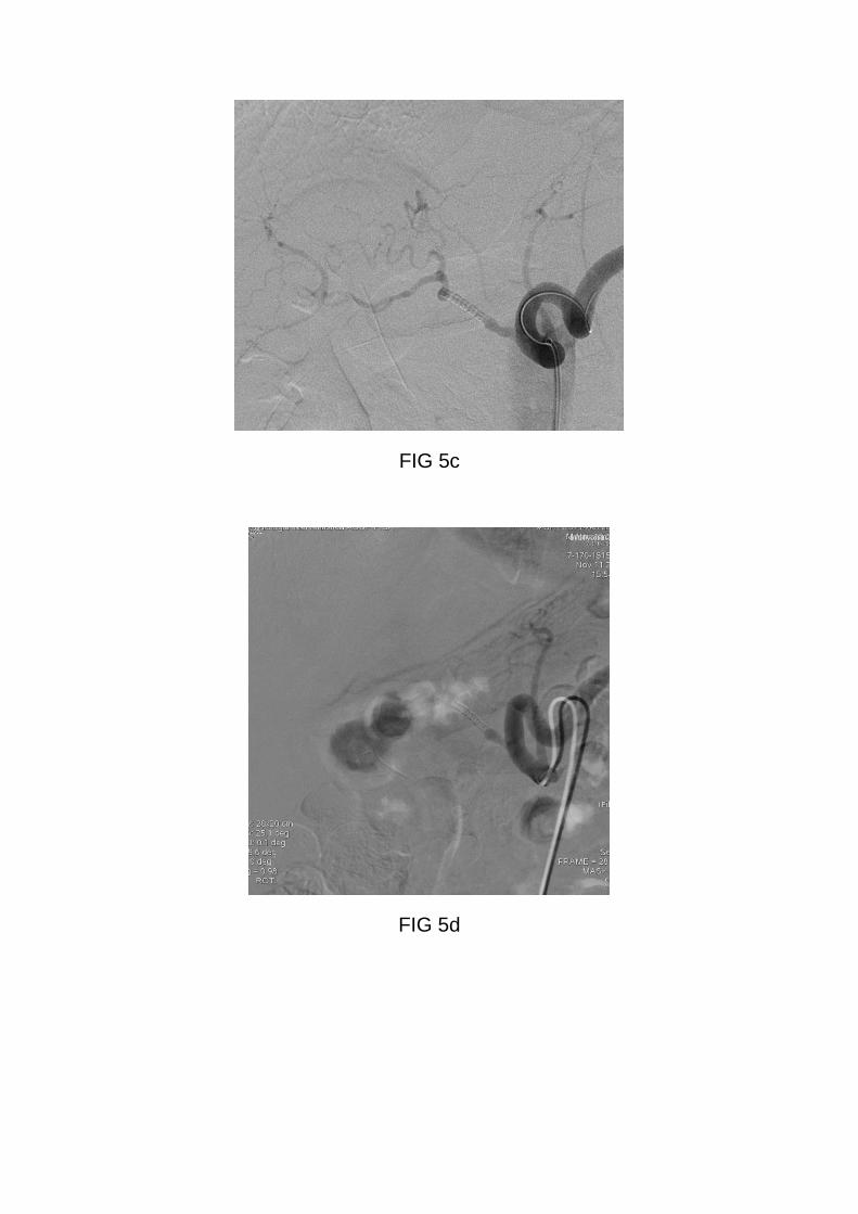
FIG 5c
FIG 5d

FIG 5e
Figure 5 (a-e). Hepatic artery stenosis (HAS). ECD (a) showed tardus and
parvus waveform. Coronal oblique MIP 3D MDCT angiographic image (b)
demonstrates anastomotic narrowing. DSA (c, d, e) confirms a severe
stenosis, treated by stenting. Stent was blocked shortly after placement and
was immediately treated with angioplasty.
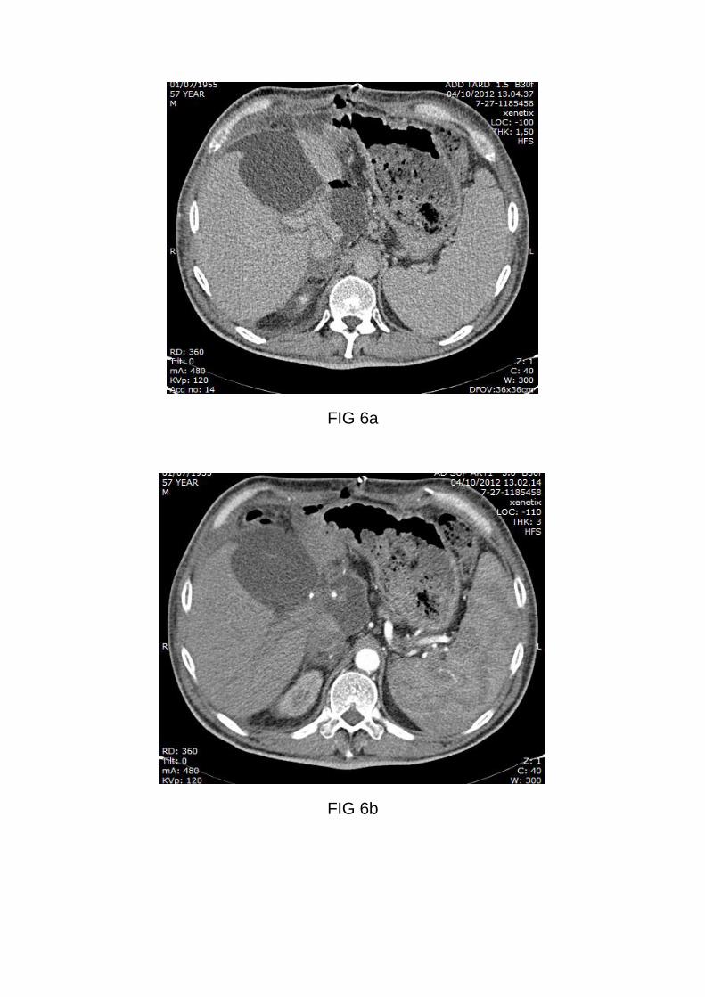
FIG 6a
FIG 6b

FIG 6c
FIG 6d

FIG 6e
FIG 6f

Figure 6 (a-f). False positive case of hepatic artery rupture not confirmed.
Axial unenhanced CT (a) showed fluid collection in the porta hepatis. Post-
contrast CT images showed focal enhancement in the arterial phase (b), with
a subsequent increase in the portal-venous phase (c), suspicion for active
bleeding. Trans-Kehr cholangiography (d) showed anastomotic leak and
communication of an intrahepatic collection with the biliary tree, with
presence of large contrast- filled biloma at CT-colangiography (e). The
patient underwent prosthesis placement with good resolution of the leak (f).
FIG 7 a
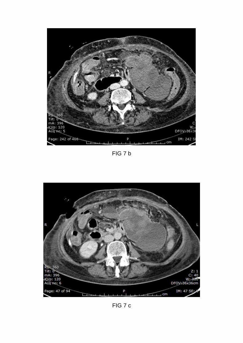
FIG 7 b
FIG 7 c

FIG 7 d
FIG 7 e
Figure 7 (a-e): Bleeding in a 68- year old female with ipotension and
decreased hemoglobin\hematocrit 7 days after OLT. Axial unenhanced CT
and post-contrast CT images (a-c) demonstrated a large fluid collection with
a layering hematocrit level. CT–DSA (d-e): Occurred onset of HAT post-
surgery.

References Bismpa K, Zlika S, Fouzas I, Imvrios G, Papanikolaou V, Petridis A. Imaging of
complications of liver transplantation: multidetector computed tomography
findings.Transplant Proc (2012) 44:2751-2753
Boraschi P, Donati F. Complications of orthotopic liver transplantation: imaging findings.
Abdom Imaging (2004) 2:189-202
Boraschi P, Donati F, Cossu MC, Gigoni R, Vignali C, Filipponi F, Bartolozzi C, Falaschi F.
Multi-detector computed tomography angiography of the hepatic artery in liver transplant
recipients. Acta Radiol (2005) 46:455-61
Busuttil RW,Farmer DG,Yersiz H,Hiatt JR,McDiarmid SV,Goldstein LI,Saab S,Han
S,Durazo F,Weaver M,Cao C,Chen T,Lipshutz GS,Holt C,Gordon S,Gombein J,Amersi
F,Ghobrial RM. Analysis of long-term outcomes of 3200 liver transplantations over two
decades:a single-center experience. Ann Surg (2005) 241:905-916
Kim SY, Kim KW, Kim MJ, Shin YM, Lee MG, Lee SG. Multidetector row CT of various
hepatic artery complications after living donor liver transplantation Abdom Imaging (2007)
32:635–643
Itri JN, Heller MT, Tublin ME. Hepatic transplantation: postoperative complications. Abdom
Imaging (2013) 38:1300-33
Pareja E, Cortes M, Navarro R, Sanjuan N, López R, Mir J. Vascular complications after
orthotopic liver transplantation: hepatic artery thrombosis. Transplant Proc (2010) 42:2970-
2972
Quiroga S, Sebastià MC, Margarit C, Castells L, Boyé R, Alvarez-Castells A.
Complications of orthotopic liver transplantation: spectrum of findings with helical CT.
Radiographics (2001) 21:1085-102
Puneet Bhargava, Sandeep Vaidya, Andrè A.S.Dick, Manjiri Dighe- Imaging of orthotopic
Liver Transplantation: Review. AJR Am J Roentgenol (March 2011) 196: WS15-25
Roberts JH, Mazzariol FS, Frank SJ, Oh SK, Koenigsberg M, Stein MW.
Multimodality imaging of normal hepatic transplant vasculature and graft vascular
complications. J Clin Imaging Sci (2011) 1: 50
Singh AK, Nachiappan AC, Verma HA, Uppot RN, Blake MA, Saini S, Boland GW.
Postoperative imaging in liver transplantation: what radiologists should know.
Radiographics (2010) 30:339-51

Uzochukwu LN, Bluth EI, Smetherman DH, Troxclair LA, Loss GE Jr, Cohen A, Eason JD.
Early postoperative hepatic sonography as a predictor of vascular and biliary
complications in adult orthotopic liver transplant patients. AJR Am J Roentgenol (2005)
185:1558-70.