Topic 6.4 – Gas Exchange. The human respiratory system works in collaboration with the transport...
18
Topic 6.4 – Gas Exchange
-
Upload
daniela-bell -
Category
Documents
-
view
218 -
download
0
Transcript of Topic 6.4 – Gas Exchange. The human respiratory system works in collaboration with the transport...
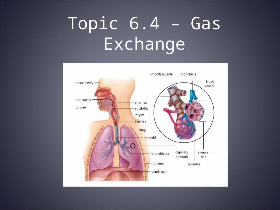
Topic 6.4 – Gas Exchange
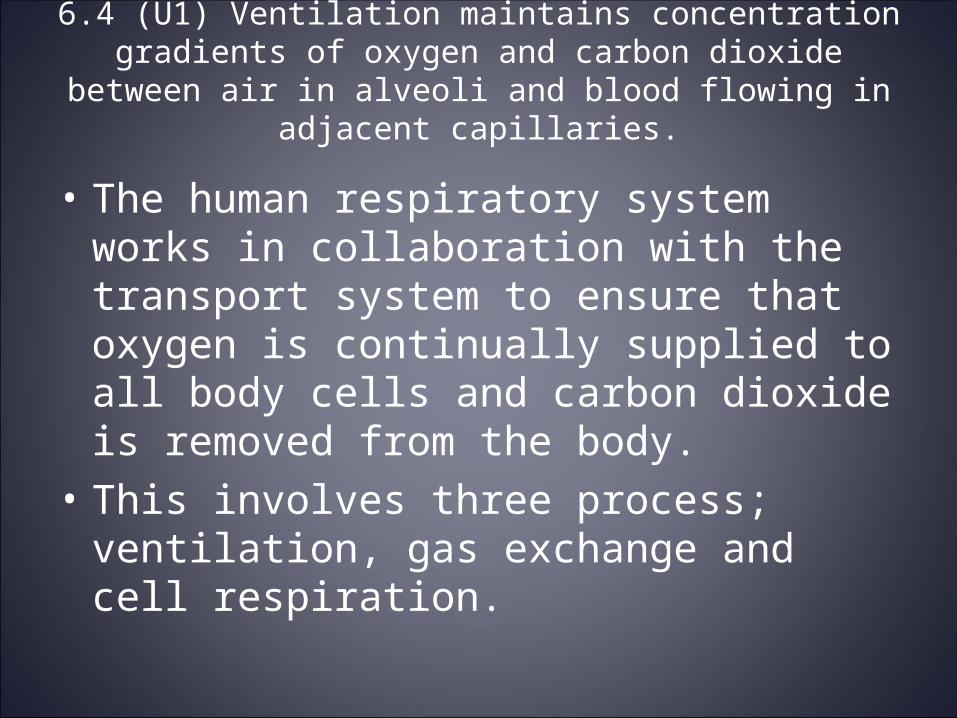
• The human respiratory system works in collaboration with the transport system to ensure that oxygen is continually supplied to all body cells and carbon dioxide is removed from the body.
• This involves three process; ventilation, gas exchange and cell respiration.
6.4 (U1) Ventilation maintains concentration gradients of oxygen and carbon dioxide between air in alveoli and blood flowing in
adjacent capillaries.

Ventilation
• In humans, ventilation involves bringing fresh air to the alveoli and removing stale air.
• In other words, the process of ventilation involves the diffusion of CO2 out of the alveolus and the diffusion of O2 into the alveolus.
• These gases diffuse due to concentration gradients that exist between the alveoli and the blood in the capillaries.

Gas Exchange• Gas exchange can be defined as the uptake of
oxygen molecules from the environment and the discharge of carbon dioxide into the environment.
• This happens in the alveoli of human lungs and the exchange occurs with the blood in the capillaries.
• In other words, CO2 is diffusing into body capillaries and then out of the lung capillary into the alveolus. Oxygen is diffusing into the lung capillary and then out of the capillary and into the body cells.

6.4 (U2) Type I pneumocytes are extremely thin alveolar cells that are adapted to carry out gas exchange.
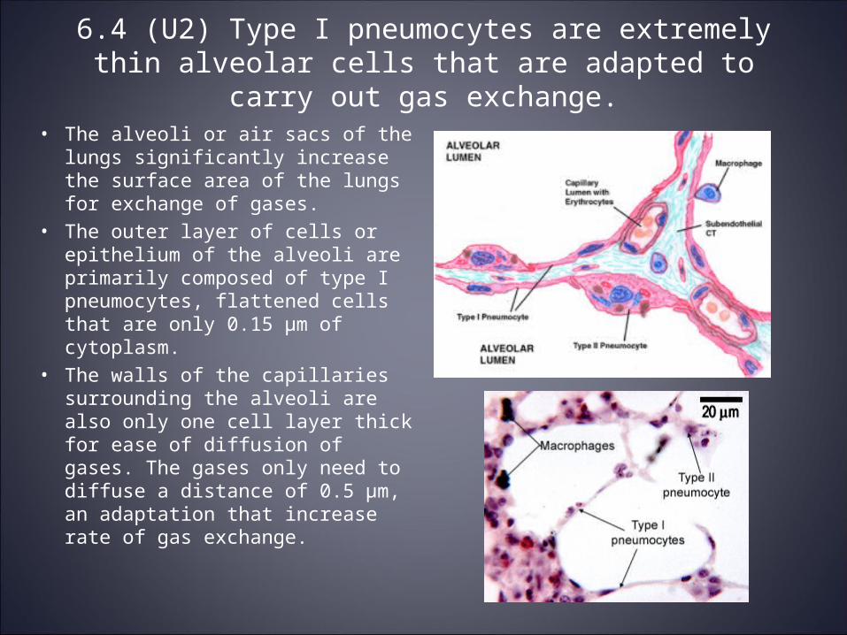
• The alveoli or air sacs of the lungs significantly increase the surface area of the lungs for exchange of gases.
• The outer layer of cells or epithelium of the alveoli are primarily composed of type I pneumocytes, flattened cells that are only 0.15 μm of cytoplasm.
• The walls of the capillaries surrounding the alveoli are also only one cell layer thick for ease of diffusion of gases. The gases only need to diffuse a distance of 0.5 μm, an adaptation that increase rate of gas exchange.

6.4 (U3) Type II pneumocytes secrete a solution containing surfactant that creates a moist surface inside the alveolus
adhering to each other by reducing surface tension.
• Type II pneumocytes make up 5% of the surface area of the aveoli and they secrete a fluid that coats the inner surface of the aveoli creating a film of moisture that the oxygen and carbon dioxide can dissolve into.
• The fluid contains a pulmonary surfactant that prevents the adhering of the walls of the alveoli to one another (prevents collapse of lung). The molecules of the surfactant are similar to phospholipids (hydrophillic heads and hydrophobic tails).
• Human babies born prematurely may suffer from infant respiratory distress syndrome due to the fact that they may be born without enough pulmonary surfactant. They will need oxygen and doses of surfactant.


6.4 (U4) Air is carried to the lungs in the trachea and bronchi and then to the alveoli in bronchioles.
• Air is inhaled in the human body through the trachea and it travels down the bronchi to the bronchioles and into the alveoli.
• The trachea is held open by rings of catrillage and branches off into two primary bronchi (one per lung).
• Each bronchi branch off into smaller bronchioles which have the alveoli attached at the end. The bronchioles have walls of smooth muscle which means the width of the airways may change.

6.4 (U5) Muscle contractions cause the pressure changes inside the thorax that force air in and out of the lungs to ventilate
them.
• Air moves in and out of the lungs as changes in pressure inside the chest cavity occurs. Air will flow from regions of high pressure to low pressure.
• Muscle contractions cause the pressure inside the chest (thorax) to decrease lower than atmospheric pressure and air rushes into the lungs. Another set of muscle contractions cause the air pressure in the chest cavity to increase above atmospheric pressure and air rushes out of the lungs.
• The movement of air into and out of the lungs is known as inhalation and exhalation.

.4 (U6) Different muscles are required for inspiration and expiration and because muscles only do work they contract.
• Muscles are always in one of two states, contracted or relaxed. Muscles can only do work when they are contracted and are often forced into relaxation by the contraction of another muscle that works in opposition.
• Muscles will often work in pairs to achieve movement. The muscles involved in inspiration and expiration work as antagonist pairs.

6.4 (A1) External and internal intercostal muscles, and diaphragm and abdominal are examples of antagonistic muscle
action.
• Ventilation relies on the work of antagonistic pairs of muscles in the thoracic cavity.
• The muscles change the volume of the chest cavity which changes the pressure inside the cavity which results in the movement of air into and out of the lungs.

Inhalation• During inhalation the external intercostals
contract and this moves the rib cage up and out. At the same time the diaphragm is contracting which causes it to move down and flatten out.
• Both of these muscle contractions increase the volume of the thorax (chest) which in turn results in a decrease in pressure inside the chest.
• The decrease in pressure in the chest cavity causes air to rush in from outside until the pressure inside the lungs rises to match the atmospheric pressure outside.

Exhalation• During exhalation the internal intercostal
muscles contract which move the rib cage down and in. At the same time the abdominal muscles contract and the diaphragm is pushed upward into a dome shape.
• The result of these muscles contractions is a decreased volume in the thorax and pressure rises above atmospheric pressure.
• The increased pressure in the chest cavity causes the air to flow out of the lungs until pressure inside the lungs falls back to atmospheric pressure.


6.4 (A2) Cause and consequences of lung cancer
• See handout for assignment completed in class.

6.4 (A3) Causes and consequences of emphysema
• See handout for assignment completed in class.