Title Small-Angle X-ray Scattered from Oligomeric Enzymes ...
13
Title Small-Angle X-ray Scattered from Oligomeric Enzymes in Aqueous Solution Author(s) Wang, Manlin; Takahashi, Nobuya; Hiragi, Yuzuru; Ooi, Tatsuo; Urakawa, Hiroshi; Kajiwara, Kanji Citation Bulletin of the Institute for Chemical Research, Kyoto University (1986), 64(2): 35-46 Issue Date 1986-07-25 URL http://hdl.handle.net/2433/77141 Right Type Departmental Bulletin Paper Textversion publisher Kyoto University
Transcript of Title Small-Angle X-ray Scattered from Oligomeric Enzymes ...
Title Small-Angle X-ray Scattered from Oligomeric Enzymes inAqueous Solution
Author(s) Wang, Manlin; Takahashi, Nobuya; Hiragi, Yuzuru; Ooi,Tatsuo; Urakawa, Hiroshi; Kajiwara, Kanji
Citation Bulletin of the Institute for Chemical Research, KyotoUniversity (1986), 64(2): 35-46
Issue Date 1986-07-25
URL http://hdl.handle.net/2433/77141
Right
Type Departmental Bulletin Paper
Textversion publisher
Kyoto University

Bull. Inst. Chem. Res., Kyoto Univ., Vol. 64, No. 2, 1986
Small-Angle X-ray Scattered from Oligomeric
Enzymes in Aqueous Solution
Manlin WANG*Il, Nobuya TAKAHASHI*, Yuzuru HIRAGI*, Tatsuo OmI*,
Hiroshi URAKAWA**21, and Kanji KAJIWARA***
Received May 2, 1986
The size and shape of two oligomeric enzymes, creatine kinase native dimer and tryptophanase holo tetramer, were evaluated by means of small-angle X-ray scattering. The statistical analysis with least-squares fitting was performed in terms of four triaxial body models; (a) an isosceles triangular prism, (b) a rectangular prism, (c) an elliptic cylinder and (d) an ellipsoid. Most probable shape models; a hollow ellipsoid and an isosceles triangular prism, were proposed for creatine kinase native dimer and trypto-
phanase holo tetramer, respectively. An adequacy/inadequacy of the present analysis was also discussed.
KEY WORDS: Oligomeric enzyme/ Small-angle X-ray scattering/ Crea- tine kinase/ Tryptophanase/ Triaxial body model/
INTRODUCTION
Oligomeric enzymes consist of several constituent units to form a quarternary
structure as categorically defined. The enzymes undergo a quarternary-structural
change which regulates the enzymatic activities through inter-subunit interactions))
Thus the primary importance in the study of the enzymatic activities is placed to the
understanding of the quarternary structure and its change of oligomeric proteins in
solution, which are best analysed with the use of the small-angle X-ray scattering (SAXS) technique.2-4) A present report demonstrates the application of the small-angle X-ray scattering analysis to two systems of oligomeric enzymes; a dimeric enzyme creatine kinase and a tetrameric enzyme tryptophanase. These enzymes are known to be enzymatically active in the form of oligomers.
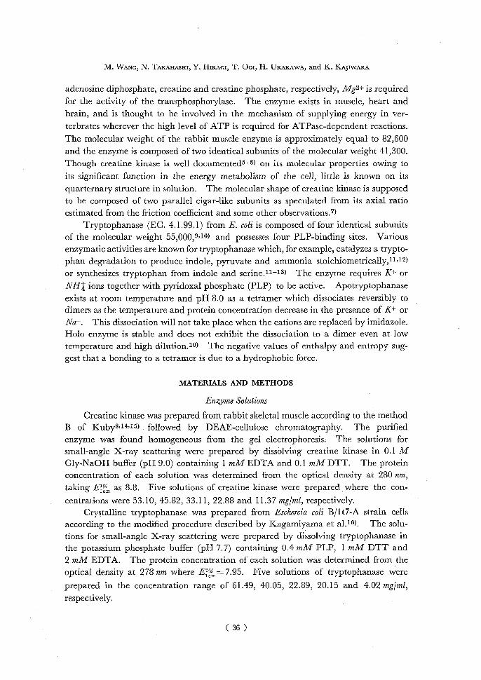
Creatine kinase (EC. 2.7.3.2.) is an enzyme participating in cell energy metabolism and found generally in all verterbrates. It catalyses the reaction :
ATP Cr ATP—Cr --DA transphosphorylase
where ATP, ADP, Cr and Cr—'P denote the abbreviations of adenosine triphosphate,
* R , A E-(_h, 14,S ft ~ : Laboratory of Physical Chemistry of Enzyme, Institute for Chemical Research, Kyoto University, Uji, Kyoto-fu, 611.
** (JI~ : Laboratory of Fiber Chemistry, Institute for Chemical Research, Kyoto University, Uji, Kyoto-fu, 611.
*** NJ,grnj : Laboratory of Molecular Design for Physiological Functions, Institute for Chemical Research, Kyoto University, Uji, Kyoto-fu, 611.
1) Present address; Institute of Biophysics, Academia Sinica, Beijin, China. 2) Present address; Faculty of Polytechnic Sciences, Kyoto Institute of Technology, Kyoto, Japan.
( 35 )
M. WANG, N. TAKAHASHI, Y. HIRAGI, T. Om, H. URAKAWA, and K. KAJIWARA
adenosine diphosphate, creatine and creatine phosphate, respectively, Mg2+ is required
for the activity of the transphosphorylase. The enzyme exists in muscle, heart and brain, and is thought to be involved in the mechanism of supplying energy in ver-
terbrates wherever the high level of ATP is required for ATPase-dependent reactions. The molecular weight of the rabbit muscle enzyme is approximately equal to 82,600 and the enzyme is composed of two identical subunits of the molecular weight 41,300.
Though creatine kinase is well documented5-8) on its molecular properties owing to its significant function in the energy metabolism of the cell, little is known on its
quarternary structure in solution. The molecular shape of creatine kinase is supposed to be composed of two parallel cigar-like subunits as speculated from its axial ratio estimated from the friction coefficient and some other observations.7)
Tryptophanase (EC. 4.1.99.1) from E. coli is composed of four identical subunits of the molecular weight 55,000,9,10) and possesses four PLP-binding sites. Various enzymatic activities are known for tryptophanase which, for example, catalyzes a trypto-
phan degradation to produce indole, pyruvate and ammonia stoichiometrically,11,12) or synthesizes tryptophan from indole and serine.11-13) The enzyme requires K+ or NHt ions together with pyridoxal phosphate (PLP) to be active. Apotryptophanase
exists at room temperature and pH 8.0 as a tetramer which dissociates reversibly to dimers as the temperature and protein concentration decrease in the presence of K+ or Nat. This dissociation will not take place when the cations are replaced by imidazole. Holo enzyme is stable and does not exhibit the dissociation to a dimer even at low temperature and high dilution.10) The negative values of enthalpy and entropy sug-
gest that a bonding to a tetramer is due to a hydrophobic force.
MATERIALS AND METHODS
Enzyme Solutions
Creatine kinase was prepared from rabbit skeletal muscle according to the method B of Kuby8"14,15) followed by DEAE-cellulose chromatography. The purified enzyme was found homogeneous from the gel electrophoresis. The solutions for small-angle X-ray scattering were prepared by dissolving creatine kinase in 0.1 M Gly-NaOH buffer (pH 9.0) containing 1 mM EDTA and 0.1 mM DTT. The protein concentration of each solution was determined from the optical density at, 280 nrn, taking Em Ercx,m as 8.8. Five solutions of creatine kinase were prepared where the con-centrations were 53.10, 45.82, 33.11, 22.88 and 11.37 mg/ml, respectively.
Crystalline tryptophanase was prepared from Eschercia coli B/ 1 t7-A strain cells according to the modified procedure described by Kagamiyama et al) 6). The solu-tions for small-angle X-ray scattering were prepared by dissolving tryptophanase in the potassium phosphate buffer (pH 7.7) containing 0.4 mM PLP, 1 mM DTT and 2 mM EDTA. The protein concentration of each solution was determined from the optical density at 278 nm where E/ „,-- 7.95. Five solutions of tryptophanase were
prepared in the concentration range of 61.49, 40.05, 22.89, 20.15 and 4.02 mg/ml, respectively.
( 36 )
SAXS from Oligomeric Enzymes
Small-Angle X-ray Scattering
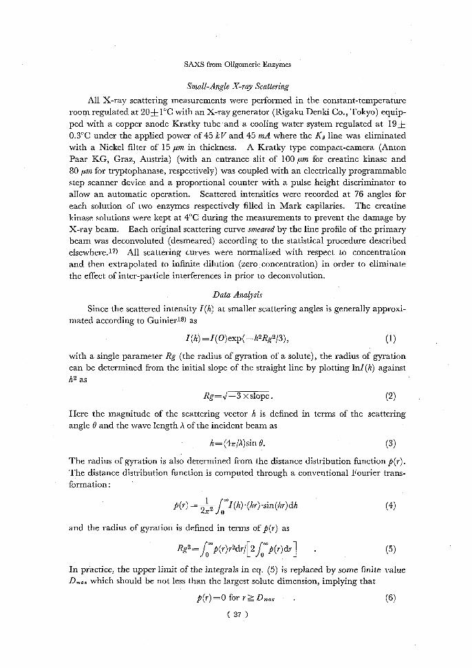
All X-ray scattering measurements were performed in the constant-temperature room regulated at 20±1°C with an X-ray generator (Rigaku Denki Co., Tokyo) equip-
ped with a copper anode Kratky tube and a cooling water system regulated at 19+ 0.3°C under the applied power of 45 kV and 45 mA where the Kp line was eliminated with a Nickel filter of 15 ,um in thickness. A Kratky type compact-camera (Anton Paar KG, Graz, Austria) (with an entrance slit of 100 ,um for creatine kinase and 80 ,um for tryptophanase, respectively) was coupled with an electrically programmable step scanner device and a proportional counter with a pulse height discriminator to allow an automatic operation. Scattered intensities were recorded at 76 angles for each solution of two enzymes respectively filled in Mark capilaries. The creatine kinase solutions were kept at 4°C during the measurements to prevent the damage by X-ray beam. Each original scattering curve smeared by the line profile of the primary beam was deconvoluted (desmeared) according to the statistical procedure described elsewhere.17) All scattering curves were normalized with respect to concentration and then extrapolated to infinite dilution (zero concentration) in order to eliminate the effect of inter-particle interferences in prior to deconvolution.
Data Analysis
Since the scattered intensity 1(h) at smaller scattering angles is generally approxi-mated according to Guinier18) as
I (h) =I (0)exp(—h2Rg2/3),(1)
with a single parameter Rg (the radius of gyration of a solute), the radius of gyration can be determined from the initial slope of the straight line by plotting lnl (h) against h2 as
Rg=,/-3 x slope.(2)
Here the magnitude of the scattering vector h is defined in terms of the scattering angle O and the wave length ? of the incident beam as
h= (4rr/A)sin O.(3)
The radius of gyration is also determined from the distance distribution function p(r). The distance distribution function is computed through a conventional Fourier trans-formation:
p(r) = 212r°I(h) • (hr) •sin (hr) dh (4) and the radius of gyration is defined in terms of p(r) as
Rg2=fp(r)r2dr/[2 f p(r)dr](5) In practice, the upper limit of the integrals in eq. (5) is replaced by some finite value Dmaz which should be not less than the largest solute dimension, implying that
p(r) =0 for r> Amax .(6)
( 37 )

M. WANG, N. TAKAHASHI, Y. HIRAGI, T. OGI, H. URAKAWA, and K. KAJIWARA
The Guinier approximation eq. (1) can be used to extrapolate the scattered intensities to the zero angle, and Porod's fourth-power law19)
I (h)IV~)h----•S(7) may be applied for the extrapolation for larger angles to eliminate the termination
effect in the Fourier transformation. Here S and V denote the total interface and the solute volume, respectively. Eq. (7) omits the term due to the background scattering which is taken to be a constant in the measured angular range. Eq. (5) is expected to
yield a more accurate estimation of Rg than the Guinier approximation, since Rg is calculated from the whole available smeared data points.
Though the radius of gyration Rg would indicate a size of a solute molecule, its exact shape could not be known from this single parameter. The shape of a solute molecule may be speculated by comparing the observed scattering curve with that calculated for various shape models of simple triaxial bodies. The particle scattering factors were analytically obtained for simple triaxial bodies such as isosceles triangular
prism,20) regular polygonal prism of n sides,21,22) elliptic cylinder,23) and ellipsoid,24) respectively, with or without hollow. The geometry of most of oligomeric enzymes can be expressed in terms of the ensemble of these simple triaxial bodies. In this report,
we analyze the scattering data from two oligomeric enzymes (creatine kinase and tryptophanase) with a non-linear least-squares fitting program SALS (provided by Data Processing Center, University of Tokyo) 25) in terms of four triaxial body models mentioned above. The present data treatment is tentative to exhibit an application of a non-linear least-squares fitting to a model analysis. A full analysis will be dealt in a succeeding paper with more elaborated models which directly correlate with enzymatic functions of repective oligomers.
RESULTS AND DISCUSSION
Creatine Kinase Native Dimer: The original (smeared) scattering curves are shown in Fig. 1(a) for five solutions of creatine kinase where the scattering curve extrapolated to the zero concentration is also displayed. The scattering curve at the zero con-centration was desmeared (deconvoluted) as shown in Fig. 2 according to the statistical
procedure.17) 11 knots were found optimum in the cubic spline approximation to determine smoothness of the deconvoluted SAXS curve where AIC (Akaike's In-formation Criterion) takes a minimum value. Here AIC is defined as
AIC= —21oge (Maximized likelihood of a model with respect to given data)
+ (Degree of freedom of the model),(8)
Fig. 1. Original small-angle X-ray scattering (SAXS) curves and those extrapolated to the zero concentration, from creatine kinase and tryptophanase. Solid lines indicate
the SAXS curves at the zero concentration. Symbols denote the concentrations as 53.10 mg/ml (^), 45.82 mg/ml (s), 33.11 mg/ml (A), 22.88 mg/ml (A), 11.37 mg/ml
(0) and 0 mg/ml (e) for creatine kinase, and 61.49 mg/ml (0), 40.05 mg/ml (s), 22.89 mg/ml (0) and 0 mg/ml (0) for tryptophanase.
( 38 )

SAXS from Oligomeric Enzymes
茎
hX102Fig.1.(a) 4
蔓 ㊨2 -
・0 2hx1。24 ヤ°1Q
Fig.1.(b)(39)

M. WANG, N. TAKAHASHI, Y. HIRAM, T. Ooi, H. URAKAWA, and K. KAJIWARA
SMEARED CURVE APPROXIMATED CURVE
° J
- tlo
N n1
y N
13.00 1.60 3.20 4.00 6.40 8.00 Sz°
DESMEAREO CURVE -
m 80.00 1.80 3.20 4.80 6.40 8.00
S
{CREATINE KINASE NATIVE DIMER ENZYME KNOT = 11
ffdITERATION = 7 (OPTIMIZED) AIC =-4.137120+02 RES = 1.03130D-02
91.O0 1.60 3.204.80 0.40 8.00 S Fig. 2. Deconvolution procedure of original SAXS data for creatine kinase. Vertical
broken lines indicate the knot positions in deconvolution process.
so that AIC represents the amount of the deviation of a model from the true curve. The minimum AIC serves as a measure to select a best fitted model to the statistical
data among various sets of parameters specifying the model. This minimum AIC estimation (MAICE) procedure is also employed to judge a best model for trypto-
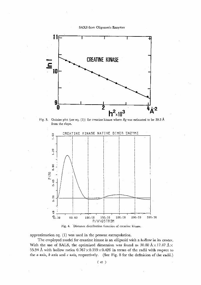
phanase among four available models in the succeeding section. The Guinier plot according to eq. (1) yields 30.3 A for the radius of gyration Rg
of creatine kinase dimer (Fig. 3), while Rg was estimated to be 30.1 A from the distance distribution function (Fig. 4) according to eq. (5). The agreement is satisfactory in
these two values estimated independently for Rg, though the estimation of Rg from the distance distribution function depends on the accuracy in the extrapolation of the scattering data to the zero angle as discussed later. The maximum distance D of
creatine kinase is determined from the distance distribution function (Fig. 3) approxi- mately as 91.2 A which is slightly shorter than 112 A, a longest axis length of the hollow
ellipsoid adapted for a creatine kinase model as estimated from the model fitting. This discrepancy may be mainly due to the error in the extrapolation of the scattering data
to the zero angle, which also causes an unusual behaviour of the distance distribution function at larger distances (see Fig. 4). Since the distance distribution function at
larger distances plays a dominant role in the estimation of Rg, the extrapolation of the scattering data to the zero angle should be performed with much care. The Guinier
(40)
SAXS from Oligomeric Enzymes
11I I I +
CREATINE KINASE
10—
9I i e 24A-22
3 hX10 Fig. 3. Guinier plot (see eq. (1)) for creatine kinase where Rg was estimated to be 30.3 A
from the slope.
o CREATINE KINASE NATIVE DIMER ENZYME
0 - •
0 m _
0
_ a 0
_
0 0
0
1----------------------------------------------------------------------------------------------------------------1 I 1 I I 1 1 1 I 1 (1:1
.0050.00 100.00150.00200.00250.00 300.00
A/ANGSTROM
Fig. 4. Distance distribution function of creatine kinase.
approximation eq. (1) was used in the present extrapolation. The employed model for creatine kinase is an ellipsoid with a hollow in its center.
With the use of SALS, the optimized dimension was found as 30.00 Ax 17.47 A x 55.94 A with hollow ratios 0.767 x 0.359 x 0.426 in terms of the radii with respect to the a axis, b axis and c axis, respectively. (See Fig. 8 for the definition of the radii.)
( 41 )

M. WANG, N. TAKAHASHI, Y. HIRAGr, T. Oor, H. URAKAWA, and K. KAJrwAxA
CREATINE KINASE.NATIVE DINER ENZYME
o O _ a
m _
I -
C
- 0 J o
N I
_
_
O _
4
I _
O I I 1I1 I I 1 I i I------------------------------------------------------------------------------------------------------------------------------------------------------------I
t. 20 -0.80 -0.400.000.400.801.201.80 LOGSxRG
Fig. 5. Experimental scattering curve (x x x) for creatine kinase with that calculated from a model ellipsoid with a hollow in its center (solid line).
Fig. 5 displays the SAXS curve from the model ellipsoid (solid line) with the experi-mental scattering curve where the experimental curve includes the extrapolated data
according to eq. (7) in larger h (scattering vector) region. Though a good agreement was observed in most of the h region between two scattering curves, the experimental curve deviates significantly in high h region from the calculated scattering curve for the model ellipsoid. The deviation might be caused by the inadequate extrapolation to larger h region according to Porod's fourth-power law (see eq. (7)), and/or by the inadequate choice of the shape model. The hydrodynamic observation of creatine
kinase aqueous solution suggests the shape of two cigars arranged parallel.7) The
present analysis employed a simple model which might not be able to represent the shape of creatine kinase native dimer. Considering an excellent agreement in small and intermediate h regions, a hollow ellipsoid, however, could not be discarded totally as a shape model of creatine kinase. The validity of Porod's fourth-power law should be examined with more data in larger h region.
Tryptophanase Holo Tetramer: The original scattering curves are shown in Fig. 1(b) for three solutions of tryptophanase where the scattering curve extrapolated to the zero concentration is also displayed. The scattering curve at the zero concentration was deconvoluted with the same procedure as shown in the previous section (see also Fig. 2),
( 42 )

SAXS from Oligomeric Enzymes
11_-----------------------------------------------------------------------~ I , +
IRMOPHANASE
10-
g------------------------------------------- 0122it
hx10 Fig. 6. Guinier plot for tryptophanase where Rg was estimated to be 40.5 A from the slope.
o TRYPTOPHANASE
0 0
0 In 0
0.. 0 m 0
0 0
o I-------------------------------------------------------------------------------------------------------------I I I I I
b. 00 50.00 100.00 150.00 200.00 250.00 000.00 R/ANGSTROM Fig. 7. Distance distribution function of tryptophanase.
where 11 knots were found optimum in the cubic spline approximation with AIC= —357.538. The radius of gyration Rg was estimated to be 40.5 A from the Guinier
plot in Fig. 6. The scattering data were extrapolated to the larger h region according to eq. (7), and then the distance distribution function was computed through eq. (4) as shown in Fig. 7. Rg was estimated to be 40.6 A by eq. (5) from the distance distri-bution function (Fig. 7). The good agreement was observed between two inde-
pendently estimated values of Rg. The distance distribution function of tryptophanase
( 43 )
M. WANG, N. TAKAHASHI, Y. HIRAGI, T. Oo2, H. URAKAWA, and K. KAJIWARA
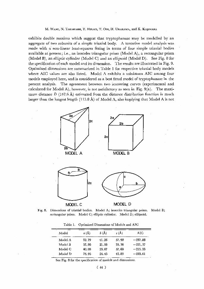
exhibits double maxima which suggest that tryptophanase may be modelled by an aggregate of two subunits of a simple triaxial body. A tentative model analysis was made with a non-linear least-squares fitting in terms of four simple triaxial bodies available at present; i.e., an isosceles triangular prism (Model A), a rectangular prism
(Model B), an elliptic cylinder (Model C) and an ellipsoid (Model D). See Fig. 8 for the specification of each model and its dimension. The results are illustrated in Fig. 9. Optimized dimensions are summarized in Table 1 for respective triaxial body models where AIC values are also listed. Model A exhibits a minimum AIC among four models employed here, and is considered as a best fitted model of tryptophanase in the
present analysis. The agreement between two scattering curves (experimental and calculated for Model A), however, is not satisfactory as seen in Fig. 9(a). The maxi-mum distance D (147.6 A) estimated from the distance distribution function is much larger than the longest length (115.8 A) of Model A, also implying that Model A is not
2a 2b2c1Ø2c MODEL AMODEL B
2cb
411110, MODEL CMODEL D
Fig. 8. Dimensions of triaxial bodies. Model A; isosceles triangular prism. Model B; rectangular prism. Model C; elliptic cylinder. Model D; ellipsoid.
Table 1. Optimized Dimensions of Models and AIC
Model a (A) b (A) c (A)AIC
Model A 32.79 41.26 57.90—237.88
Model B 57.95 21.18 33.38—221.37 Model C 40.08 23.07 57.60—215.33
Model D 75.95 24.45 42.83—233.61
See Fig. 8 for the specification of models and dimensions.
(44)

SAXS from Oligomeric Enzymes
THTPTOPHANASE
• i°
° MODEL A g MODEL B
AIC = -237.88AIC = -221.37
-0.00 -0.40 0.00 0.40 0.00 I.20 -0.00 -0.40 0.00 0.40 0.00 1.20 1.00
LOG SMRG -LOGS.RG
MODEL C _MODEL D _ AIC = -215.33 AIC = -233.61
ii. 20 -0. 00 -0.40 0.00 0.40 0.00 I.20 I. 20 -0.00 -0.40 0.00 0.40 0.00 1.20 I.00 LGG SoRGLOG SNAG Fig. 9. Experimental scattering curve (X X X) for tryptophanase with those calculated for
various shape models, i.e., Model A; isosceles triangular prism, Model B; regular rectangular prism, Model C; elliptic cylinder and Model D; ellipsoid.
adequate to represent tryptophanase. A more elaborated model is required to approximate the shape of tryptophanase tetramer.
CONCLUDING REMARKS
The model analysis is conveniently performed with a non-linear least-squares fitting for the scattering data deconvoluted in advance. Since the characteristics of these models appear in rather large h (scattering vector) regions, the scattering data should be collected up to sufficiently large scattering angles. An inadequate extrapolation to the zero angle causes a large error in computing the distance distribution function at large r values and consequently in evaluating the radius of gyration from the distance distribution function.
(45)
M. WANG, N. TAKAHASHI, Y. HIRAGI, T. Ooi, H. URAKAWA, and K. KAJIWARA
ACKNOWLEDGEMENT
We are grateful to Drs. Lixiang Hou and Haimeng Zhou (Institute of Biophysics, Academia Sinica, Beijin, China), and Professor M. Tokushige (Faculty of Sciences, Kyoto University, Kyoto, Japan) for supplying creatine kinase and tryptophanase, respectively.
REFERENCES
(1) J. Monod, J.-P. Changeux and F. Jacob, J. Mol. Biol., 6, 306 (1963). (2 ) I. Pilz, O. Glatter, and O. Kratky, Methods in Enzymol., 61, 148 (1979). (3) V. Luzatti and A. Tardieu, Ann. Rev. Biophys. Bioeng., 9, 1 (1980). (4) I. Pilz, in Small-Angle X-ray Scattering, (ed. by O. Glatter and O. Kratky), p.239, Academic Press,
London, 1982. (5) G. L. Kenyon and G. H. Reed, Advances in Enzymology and Related Area in Molecular Biology, 54,
367 (1983). (6) G. Bicerstaff and N. C. Price, Int. J. Biochem., 19, 1 (1978). (7) D. C. Watts, in The Enzymes Vol. VIII, (ed. by P. D. Boyer), P383, Academic Press, New York,
1973. (8) Q. Yao, L. Hou, H. Zhou and C. Zou, Sciencia Sinica (Series B), 25, 1186 (1982). (9) W. A. Newton, Y. Morino and E. E. Snell, J. Biol. Chem., 240, 1211 (1965). (10) Y. Morino and E. E. Snell, J. Biol. Chem., 242, 5591 (1967). (11) W. A. Newton and E. E. Snell, Proc. Natl. Acad. Sci., U.S.A., 51, 382 (1964). (12) E. E. Snell, Adv. Enzymol., 42, 287 (1975). (13) M. D. Yudkin, J. Gen. Microbiol., 92, 125 (1976). (14) S. A. Kuby, L. Noda and H. A. Lardy, J. Biol. Chem., 210, 65 (1954). (15) A. Fattoum, R. Kasssab and L. A. Pardel, Biochim. Biophys. Acta, 405, 324 (1975). (16) H. Kagamiyama, H. Wada, H. Matsubara and E. E. Snell, J. Biol. Chem., 247, 1571 (1972). (17) Y. Hiragi, H. Urakawa and K. Tanabe, J. Appi. Phys., 58, 5 (1985). (18) A. Guinier, Ann. Phys., 12, 161 (1939). (19) G. Porod, Kolloid-Z., 124, 83 (1951). (20) W. Bode, J. Engel and R. Winklmair, Eur. J. Biochem., 26, 313 (1972). (21) P. Mittelbach and G. Porod, Acta Phys. Austriaca, 14, 185 (1961). (22) Y. Hiragi and S. Ihara, Acta Crst., A37, 378 (1981). (23) P. Mittelbach and G. Porod, Acta Phys. Austriaca, 14, 405 (1961). (24) P. Mittelbach and G. Porod, Acta Phys. Austriaca, 15, 122 (1962). (25) T. Nakagawa and Y. Koyanagi, Statistical Analysis with Least-Squares Fitting (in Japanese), Tokyo
U.P., Tokyo, 1982. (26) See for example, H. Akaike, in Application of Statistics, (ed. by P. K. Krishniah), p27, North Holland,
Amsterdam, 1977.
( 46 )