This Paper Will Change Your Practice – It Changed Mine ...
27
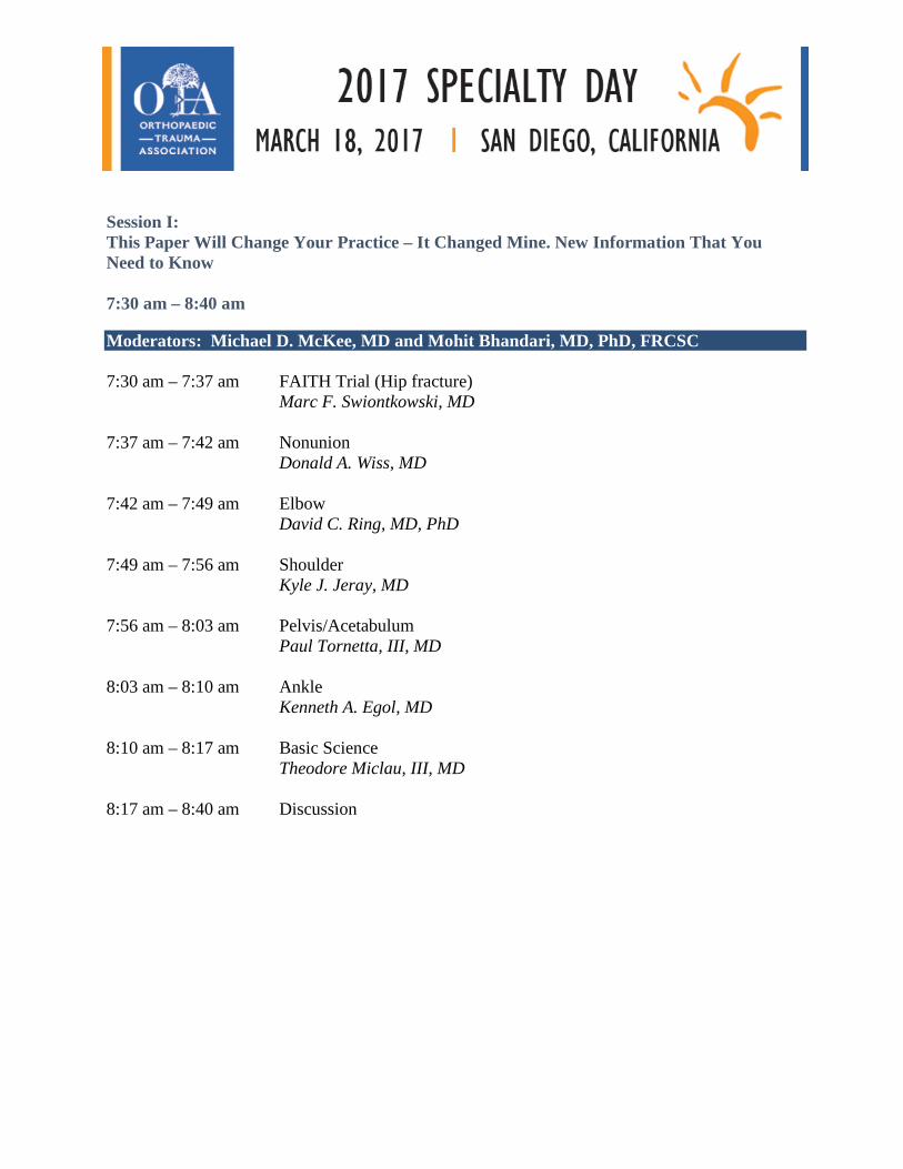
Session I: This Paper Will Change Your Practice – It Changed Mine. New Information That You Need to Know 7:30 am – 8:40 am Moderators: Michael D. McKee, MD and Mohit Bhandari, MD, PhD, FRCSC 7:30 am – 7:37 am FAITH Trial (Hip fracture) Marc F. Swiontkowski, MD 7:37 am – 7:42 am Nonunion Donald A. Wiss, MD 7:42 am – 7:49 am Elbow David C. Ring, MD, PhD 7:49 am – 7:56 am Shoulder Kyle J. Jeray, MD 7:56 am – 8:03 am Pelvis/Acetabulum Paul Tornetta, III, MD 8:03 am – 8:10 am Ankle Kenneth A. Egol, MD 8:10 am – 8:17 am Basic Science Theodore Miclau, III, MD 8:17 am – 8:40 am Discussion
Transcript of This Paper Will Change Your Practice – It Changed Mine ...
Session I: This Paper Will Change Your Practice – It Changed Mine. New Information That You Need to Know 7:30 am – 8:40 am Moderators: Michael D. McKee, MD and Mohit Bhandari, MD, PhD, FRCSC 7:30 am – 7:37 am FAITH Trial (Hip fracture) Marc F. Swiontkowski, MD 7:37 am – 7:42 am Nonunion Donald A. Wiss, MD 7:42 am – 7:49 am Elbow David C. Ring, MD, PhD 7:49 am – 7:56 am Shoulder Kyle J. Jeray, MD 7:56 am – 8:03 am Pelvis/Acetabulum Paul Tornetta, III, MD 8:03 am – 8:10 am Ankle Kenneth A. Egol, MD 8:10 am – 8:17 am Basic Science Theodore Miclau, III, MD 8:17 am – 8:40 am Discussion
Page 1 of 3
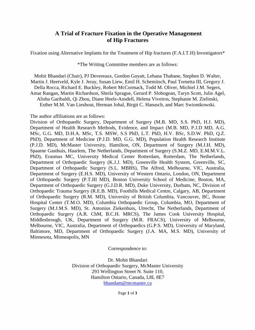
A Trial of Fracture Fixation in the Operative Management of Hip Fractures
Fixation using Alternative Implants for the Treatment of Hip fractures (F.A.I.T.H) Investigators*
*The Writing Committee members are as follows:
Mohit Bhandari (Chair), PJ Devereaux, Gordon Guyatt, Lehana Thabane, Stephen D. Walter, Martin J. Heetveld, Kyle J. Jeray, Susan Liew, Emil H. Schemitsch, Paul Tornetta III, Gregory J.
Della Rocca, Richard E. Buckley, Robert McCormack, Todd M. Oliver, Michiel J.M. Segers, Amar Rangan, Martin Richardson, Sheila Sprague, Gerard P. Slobogean, Taryn Scott, Julie Agel,
Alisha Garibaldi, Qi Zhou, Diane Heels-Ansdell, Helena Viveiros, Stephanie M. Zielinski, Esther M.M. Van Lieshout, Herman Johal, Birgit C. Hanusch, and Marc Swiontkowski.
The author affiliations are as follows: Division of Orthopaedic Surgery, Department of Surgery (M.B. MD, S.S. PhD, H.J. MD), Department of Health Research Methods, Evidence, and Impact (M.B. MD, P.J.D MD, A.G. MSc, G.G. MD, D.H.A. MSc, T.S. MSW, S.S PhD, L.T. PhD, H.V. BSc, S.D.W. PhD, Q.Z. PhD), Department of Medicine (P.J.D. MD, G.G. MD), Population Health Research Institute (P.J.D. MD), McMaster University, Hamilton, ON, Department of Surgery (M.J.H. MD), Spaarne Gasthuis, Haarlem, The Netherlands, Department of Surgery (S.M.Z. MD, E.M.M.V.L. PhD), Erasmus MC, University Medical Center Rotterdam, Rotterdam, The Netherlands, Department of Orthopaedic Surgery (K.J.J. MD), Greenville Health System, Greenville, SC, Department of Orthopaedic Surgery (S.L. MBBS), The Alfred, Melbourne, VIC, Australia, Department of Surgery (E.H.S. MD), University of Western Ontario, London, ON, Department of Orthopaedic Surgery (P.T.III MD), Boston University School of Medicine, Boston, MA, Department of Orthopaedic Surgery (G.J.D.R. MD), Duke University, Durham, NC, Division of Orthopaedic Trauma Surgery (R.E.B. MD), Foothills Medical Centre, Calgary, AB, Department of Orthopaedic Surgery (R.M. MD), University of British Columbia, Vancouver, BC, Boone Hospital Center (T.M.O. MD), Columbia Orthopaedic Group, Columbia, MO, Department of Surgery (M.J.M.S. MD), St. Antonius Ziekenhuis, Utrecht, The Netherlands, Department of Orthopaedic Surgery (A.R. ChM, B.C.H. MRCS), The James Cook University Hospital, Middlesbrough, UK, Department of Surgery (M.R. FRACS), University of Melbourne, Melbourne, VIC, Australia, Department of Orthopaedics (G.P.S. MD), University of Maryland, Baltimore, MD, Department of Orthopaedic Surgery (J.A. MA, M.S. MD), University of Minnesota, Minneapolis, MN
Correspondence to:
Dr. Mohit Bhandari Division of Orthopaedic Surgery, McMaster University
293 Wellington Street N. Suite 110, Hamilton Ontario, Canada, L8L 8E7
Page 2 of 3
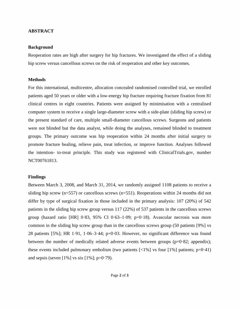
ABSTRACT
Background
Reoperation rates are high after surgery for hip fractures. We investigated the effect of a sliding
hip screw versus cancellous screws on the risk of reoperation and other key outcomes.
Methods
For this international, multicentre, allocation concealed randomised controlled trial, we enrolled
patients aged 50 years or older with a low-energy hip fracture requiring fracture fixation from 81
clinical centres in eight countries. Patients were assigned by minimisation with a centralised
computer system to receive a single large-diameter screw with a side-plate (sliding hip screw) or
the present standard of care, multiple small-diameter cancellous screws. Surgeons and patients
were not blinded but the data analyst, while doing the analyses, remained blinded to treatment
groups. The primary outcome was hip reoperation within 24 months after initial surgery to
promote fracture healing, relieve pain, treat infection, or improve function. Analyses followed
the intention- to-treat principle. This study was registered with ClinicalTrials.gov, number
NCT00761813.
Findings
Between March 3, 2008, and March 31, 2014, we randomly assigned 1108 patients to receive a
sliding hip screw (n=557) or cancellous screws (n=551). Reoperations within 24 months did not
differ by type of surgical fixation in those included in the primary analysis: 107 (20%) of 542
patients in the sliding hip screw group versus 117 (22%) of 537 patients in the cancellous screws
group (hazard ratio [HR] 0·83, 95% CI 0·63–1·09; p=0·18). Avascular necrosis was more
common in the sliding hip screw group than in the cancellous screws group (50 patients [9%] vs
28 patients [5%]; HR 1·91, 1·06–3·44; p=0·03. However, no significant difference was found
between the number of medically related adverse events between groups (p=0·82; appendix);
these events included pulmonary embolism (two patients [<1%] vs four [1%] patients; p=0·41)
and sepsis (seven [1%] vs six [1%]; p=0·79).
Page 3 of 3
Interpretation
In terms of reoperation rates the sliding hip screw shows no advantage, but some groups of
patients (smokers and those with displaced or base of neck fractures) might do better with a
sliding hip screw than with cancellous screws.
Funding
National Institutes of Health, Canadian Institutes of Health Research, Stichting NutsOhra,
Netherlands Organisation for Health Research and Development, Physicians’ Services
Incorporated.
2/15/17
1
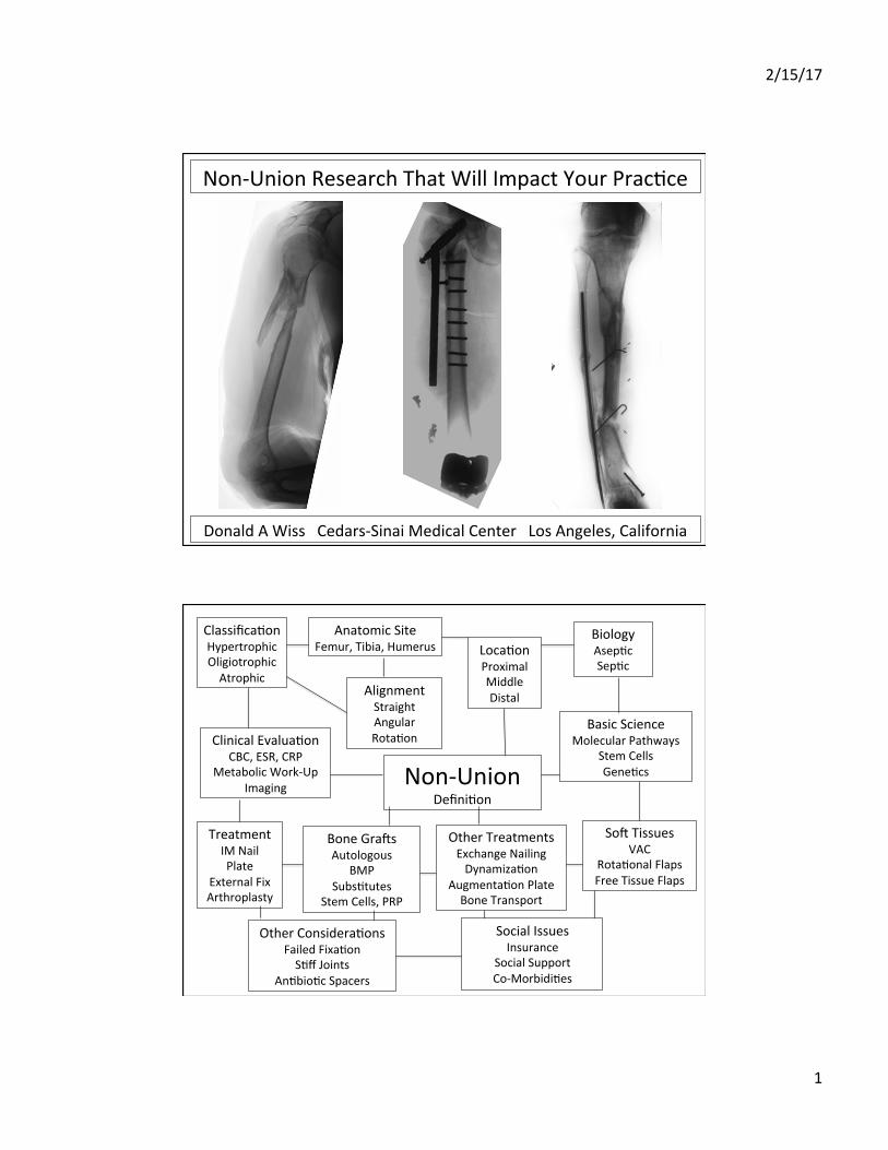
Non-UnionResearchThatWillImpactYourPrac>ce
DonaldAWissCedars-SinaiMedicalCenterLosAngeles,California
Non-UnionDefini>on
Classifica>onHypertrophicOligiotrophicAtrophic
AnatomicSiteFemur,Tibia,Humerus Loca>on
ProximalMiddleDistal
BiologyAsep>cSep>c
AlignmentStraightAngularRota>on
BasicScienceMolecularPathways
StemCellsGene>cs
ClinicalEvalua>onCBC,ESR,CRP
MetabolicWork-UpImaging
TreatmentIMNailPlate
ExternalFixArthroplasty
BoneGraVsAutologous
BMPSubs>tutes
StemCells,PRP
OtherTreatmentsExchangeNailingDynamiza>on
Augmenta>onPlateBoneTransport
SoVTissuesVAC
Rota>onalFlapsFreeTissueFlaps
OtherConsidera>onsFailedFixa>onS>ffJoints
An>bio>cSpacers
SocialIssuesInsurance
SocialSupportCo-Morbidi>es
This Paper Will Change Your Practice: Elbow David Ring MD PhD Lindenhovius AL, Linzel DS, Doornberg JN, Ring DC, Jupiter JB. Comparison of elbow contracture release in elbows with and without heterotopic ossification restricting motion. J Shoulder Elbow Surg. 2007 Sep-Oct;16(5):621-5. PubMed PMID:17644008.
• Surgery for stiffness is MORE effective when there is heterotopic ossification restricting motion
Lindenhovius AL, Doornberg JN, Brouwer KM, Jupiter JB, Mudgal CS, Ring D. A prospective randomized controlled trial of dynamic versus static progressive elbow splinting for posttraumatic elbow stiffness. J Bone Joint Surg Am. 2012 Apr 18;94(8):694-700. doi: 10.2106/JBJS.J.01761. PubMed PMID: 22517385.
• No difference in splints • Not clear that a splint is better than exercises on your own • People improved for more than 12 months • In the absence of HO, ulnar neuropathy, errant implants, malunion, etc. the
capsule can be stretched Teunis T, Bot AG, Thornton ER, Ring D. Catastrophic Thinking Is Associated With Finger Stiffness After Distal Radius Fracture Surgery. J Orthop Trauma. 2015 Oct;29(10):e414-20. PubMed PMID: 25866942.
• Protectiveness creates stiffness • Not an elusive pathophysiology
Teunis T, Thornton ER, Guitton TG, Vranceanu AM, Ring D. Coaching of patients with an isolated minimally displaced fracture of the radial head immediately increases range of motion. J Hand Ther. 2016 Jul-Sep;29(3):314-9. doi:10.1016/j.jht.2016.02.003. PubMed PMID: 27496986.
• Coaching that stretching helps creates immediate gains in motion
1: Chan K, Faber KJ, King GJ, Athwal GS. Selected anteromedial coronoid fractures can be treated nonoperatively. J Shoulder Elbow Surg. 2016 Aug;25(8):1251-7. PubMed PMID: 27233484. 2: Desloges W, Faber KJ, King GJ, Athwal GS. Functional outcomes of distal humeral fractures managed nonoperatively in medically unwell and lower-demand elderly patients. J Shoulder Elbow Surg. 2015 Aug;24(8):1187-96. PubMed PMID: 26189804. 3: Chan K, MacDermid JC, Faber KJ, King GJ, Athwal GS. Can we treat select terrible triad injuries nonoperatively? Clin Orthop Relat Res. 2014 Jul;472(7):2092-9. PubMed PMID: 24549776; PubMed Central PMCID: PMC4048392. 4: Duckworth AD, Bugler KE, Clement ND, Court-Brown CM, McQueen MM. Nonoperative management of displaced olecranon fractures in low-demand elderly patients. J Bone Joint Surg Am. 2014 Jan 1;96(1):67-72. doi: 10.2106/JBJS.L.01137. PubMed PMID: 24382727.
• Selected distal humerus, elbow fracture-dislocations, and olecranon fractures can be treated nonoperatively
Dubberley JH, Faber KJ, Macdermid JC, Patterson SD, King GJ. Outcome after open reduction and internal fixation of capitellar and trochlear fractures. J Bone Joint Surg Am. 2006 Jan;88(1):46-54. PubMed PMID: 16391249.
• Beware the apparent capitellum fracture. It’s often much more complex

HANDOUT COMING SOON
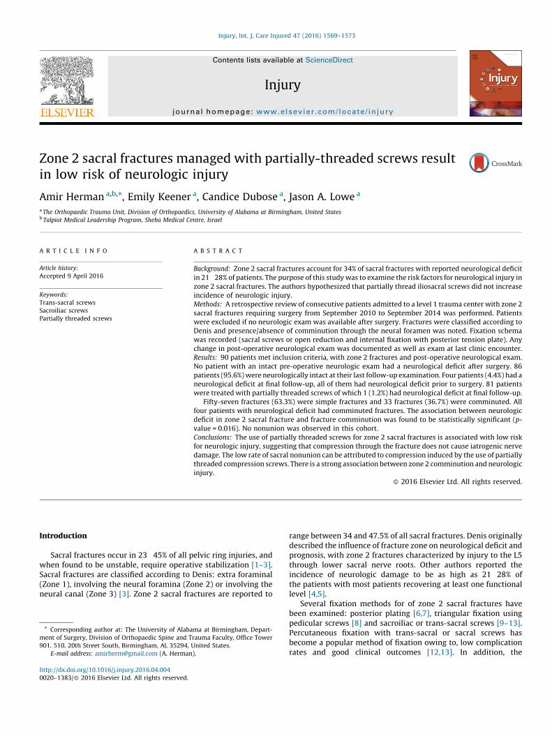
Zone 2 sacral fractures managed with partially-threaded screws resultin low risk of neurologic injury
Amir Herman a,b,*, Emily Keener a, Candice Dubose a, Jason A. Lowe a
a The Orthopaedic Trauma Unit, Division of Orthopaedics, University of Alabama at Birmingham, United Statesb Talpiot Medical Leadership Program, Sheba Medical Centre, Israel
Introduction
Sacral fractures occur in 23!45% of all pelvic ring injuries, andwhen found to be unstable, require operative stabilization [1–3].Sacral fractures are classified according to Denis: extra foraminal(Zone 1), involving the neural foramina (Zone 2) or involving theneural canal (Zone 3) [3]. Zone 2 sacral fractures are reported to
range between 34 and 47.5% of all sacral fractures. Denis originallydescribed the influence of fracture zone on neurological deficit andprognosis, with zone 2 fractures characterized by injury to the L5through lower sacral nerve roots. Other authors reported theincidence of neurologic damage to be as high as 21!28% ofthe patients with most patients recovering at least one functionallevel [4,5].
Several fixation methods for of zone 2 sacral fractures havebeen examined: posterior plating [6,7], triangular fixation usingpedicular screws [8] and sacroiliac or trans-sacral screws [9–13].Percutaneous fixation with trans-sacral or sacral screws hasbecome a popular method of fixation owing to, low complicationrates and good clinical outcomes [12,13]. In addition, the
Injury, Int. J. Care Injured 47 (2016) 1569–1573
A R T I C L E I N F O
Article history:Accepted 9 April 2016
Keywords:Trans-sacral screwsSacroiliac screwsPartially threaded screws
A B S T R A C T
Background: Zone 2 sacral fractures account for 34% of sacral fractures with reported neurological deficitin 21!28% of patients. The purpose of this study was to examine the risk factors for neurological injury inzone 2 sacral fractures. The authors hypothesized that partially thread iliosacral screws did not increaseincidence of neurologic injury.Methods: A retrospective review of consecutive patients admitted to a level 1 trauma center with zone 2sacral fractures requiring surgery from September 2010 to September 2014 was performed. Patientswere excluded if no neurologic exam was available after surgery. Fractures were classified according toDenis and presence/absence of comminution through the neural foramen was noted. Fixation schemawas recorded (sacral screws or open reduction and internal fixation with posterior tension plate). Anychange in post-operative neurological exam was documented as well as exam at last clinic encounter.Results: 90 patients met inclusion criteria, with zone 2 fractures and post-operative neurological exam.No patient with an intact pre-operative neurologic exam had a neurological deficit after surgery. 86patients (95.6%) were neurologically intact at their last follow-up examination. Four patients (4.4%) had aneurological deficit at final follow-up, all of them had neurological deficit prior to surgery. 81 patientswere treated with partially threaded screws of which 1 (1.2%) had neurological deficit at final follow-up.
Fifty-seven fractures (63.3%) were simple fractures and 33 fractures (36.7%) were comminuted. Allfour patients with neurological deficit had comminuted fractures. The association between neurologicdeficit in zone 2 sacral fracture and fracture comminution was found to be statistically significant (p-value = 0.016). No nonunion was observed in this cohort.Conclusions: The use of partially threaded screws for zone 2 sacral fractures is associated with low riskfor neurologic injury, suggesting that compression through the fracture does not cause iatrogenic nervedamage. The low rate of sacral nonunion can be attributed to compression induced by the use of partiallythreaded compression screws. There is a strong association between zone 2 comminution and neurologicinjury.
! 2016 Elsevier Ltd. All rights reserved.
* Corresponding author at: The University of Alabama at Birmingham, Depart-ment of Surgery, Division of Orthopaedic Spine and Trauma Faculty, Office Tower901, 510, 20th Street South, Birmingham, AL 35294, United States.
E-mail address: [email protected] (A. Herman).
Contents lists available at ScienceDirect
Injury
jo ur n al ho m epag e: ww w.els evier . c om / lo cat e/ in ju r y
http://dx.doi.org/10.1016/j.injury.2016.04.0040020–1383/! 2016 Elsevier Ltd. All rights reserved.
biomechanical properties of the sacral screws show 80!85% returnof pelvic biomechanical rigidity after instrumentation [14,15].
One potential concern with partially threaded screws is thepotential for compression and narrowing of the neural foramenparticularly through comminuted fractures, which may resultiatrogenic neurologic injury [16,17]. Fully threaded screwsrepresent an alternative to partially threaded screws as a meansto avoid this potential complication [18,19].
The purpose of this study was to define the incidence of post-operative neurologic injury and identify risk factors for neurologi-cal deficit after zone 2 sacral fractures. The authors hypothesizedthat partially threaded posterior screws (trans-sacral, sacral)would not result in iatrogenic neurologic injury, and that fracturecomminution through the neural foramen would predict injury-related neurologic compromise.
Methods
Following institution IRB approval, a retrospective review of allpelvic ring fractures and fracture dislocations fixed at a single level1 trauma academic center from September 2010 to September2014 was performed using CPT and ICD-9 codes. Patient inclusioncriteria included: age > 18 years at the time of injury and closedzone 2 sacral fracture according to Denis. Exclusion criteria were:lack of neurological examination after surgery, patients that didnot have neurologic examination prior to surgery and had aneurologic deficit after surgery, open fractures, and any patienttransferred after fixation to an outside facility.
We extracted patient demographic data including age andgender; as well as injury profiles (mechanism of injury, abbreviat-ed injury score (AIS) and the injury severity score (ISS)).Neurological status at arrival, first available neurologic exam aftersurgery and last clinical follow-up neurological examination wereobtained from consult, post-operative, in-patient progress, andclinic notes, respectively. Further data collection included:comminuted vs simple fractures, the surgical intervention of openvs closed reduction, and posterior tension band plate fixationversus percutaneous screws, as well as screw type (partiallythreaded vs fully threaded), and screw number.
Neurologic deficit at final follow up was defined as motorstrength of less than five (out of five) of the injured extremity.
Statistical analysis
Statistical analysis was performed by an experienced biostatis-tician (A.H.) using SPSS ! 23.0 (Chicago, IL, USA). Categorical dataare presented as count (percent). Continuous data are presented asmean ("standard deviation). The data were divided to patients withintact and deficient neurological examination at final follow-up. Allthe collected variables were compared between these two studygroups. Comparisons between continuous variables were done by theWilcoxon!Mann!Whitney rank sum test. Comparisons betweencategorical data were done with chi-square test or the Fisher exacttest. The later test was used if expected count was less than five in anycell. All p-values reported are two-sided.
Results
Initial data review included 100 patients with zone 2 sacralfractures. After reviewing the patients’ data, seven patients wereexcluded because of lack of neurological examination at follow-up.These included one patient that died shortly after admission, fivepatients that were transferred intubated and followed at anotherhospital and one patient that had an above knee amputation thesame side as the sacral fracture. Three additional patients were
excluded because they did not have a neurological examinationprior to surgery and had a neurologic deficit after surgery.
The study population included 90 patients, mean age was 39.10("15.03). Of the 90 patients, 36 patients (40.0%) were male and 54patients (60.0%) were female. The most common cause of injury wasmotor vehicle accident – 52 patients (57.8%) followed by fall fromheight – 12 patients (13.3%). There was no statistically significantdifference in the demographic characteristics between neurologicallyintact and deficient patients (Table 1).
Of the 90 patients, 86 (95.6%) had no neurological deficit at anytime point. Four patients (4.4%) had a neurological deficit at finalfollow-up. None of the four patients with final neurological deficithad an intact examination at time of admission. These fourpatients presented with weak but present initial motor examdefined as 2!3/5 motor strength in the tibialis anterior, extensorhallucis longus, flexor hallucis longus or gastrocnemius–soleuscomplex. No patient presenting with an initially intact neurologicexam developed a neurologic deficit following operative fixation(Table 2).
Table 1Demographic and injury related data.
Neurologicallyintact PostOp(N = 86)
Neurologicaldeficit PostOp(N = 4)
P-value
Age 39.21 ("15.22) 36.75 ("10.43) 0.887GenderMaleFemale
34 (39.5%)52 (60.5%)
2 (50.0%)2 (50.0%) 0.676
Mechanism of injuryAssaultCrush InjuryFall from heightFall from horseMCCMVCPedestrian vs MVTornado
1 (1.2%)5 (5.8%)12 (14.0%)2 (2.3%)5 (5.9%)48 (55.8%)10 (11.6%)3 (3.5%)
00 (00%)00 (00%)00 (00%)00 (00%)00 (00%)4 (100.0%)00 (00%)00 (00%)
0.931
AIS–Abdomen 2.38 ("0.61) 3.33 ("1.53) 0.226AIS–Extremities 3.02 ("0.88) 3.25 ("0.96) 0.595AIS–Injury severity score 22.34 ("10.62) 30.5 ("8.34) 0.087
AIS = Abbreviated Injury Score; MVC = Motor vehicle collision; MCC = motor cyclecollision; MV = motor vehicle.
Table 2Pelvic injury and surgery related data.
Neurologicallyintact PostOp(N = 86)
Neurologicaldeficit PostOp(N = 4)
P-value
Side of zone 2 fractureLeftRightBilateral
42 (48.8%)33 (38.4%)11 (12.8%)
2 (50.0%)2 (50.0%)0 (00.0%)
0.886
Fracture patternSimpleComminuted
57 (66.3%)29 (33.7%)
0 (0.0%)4 (100.0%) 0.016
Fixation methodORIF and plateCRPP and screw
5 (5.8%)81 (94.2%)
2 (50.0%)2 (50.0%) 0.001
Type of screwsSacroiliac screwsTrans-sacral screws
35 (43.2%)46 (56.8%)
0 (00.0%)2 (100.0%) 0.506
Number of screws1 screw2 screws3 screws4 screws
48 (59.3%)25 (30.9%)5 (6.2%)3 (3.7%)
2 (100.0%)0 (00.0%)0 (00.0%)0 (00.0%)
0.717
Type of screw threadsPartially threadedFully threaded
80 (98.8%)1 (1.2%)
1 (50.0%)1 (50.0%) 0.049
A. Herman et al. / Injury, Int. J. Care Injured 47 (2016) 1569–15731570
Of the four patients with deficit at final follow-up all had TibialisAnterior (TA) and extensor halucis longus (EHL) motor strength ofless than five (out of five). Two patients had either flexor hallucislongus or Gastrocnemius-soleus complex with strength of four orless (of five). One patient also experienced a sensory deficit. Mostpatients had some recovery of their initial examination (see Table 3).
Correlation of associated injuries and neurologic deficit aftersacral fracture was performed using the abbreviated injury score(AIS, Table 1). For the entire patient cohort the mean AIS !abdomen was 2.43 ("0.689). The mean ISS for the entire patientcohort was 22.72 ("10.63). There was a statistical trend for higher ISSin neurologically deficient patients (p-value = 0.087).
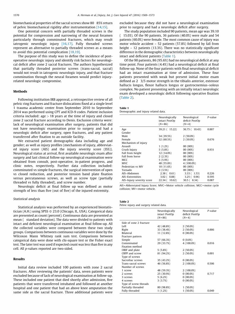
Fifty seven patients (66.3%) had simple pattern fractures (Fig. 1)and 33 patients (36.7%) had comminuted fractures (Fig. 2). All fourpatients with neurological deficit had a comminuted fracturepattern. Of the 86 patients without neurological deficit, 29 patients(33.7%) and 57 (66.3%) had comminuted or simple fracturepatterns, respectively. The association between comminutedfractures and neurological deficit was found to be statisticallysignificant. (p-value = 0.016, Table 2).
Of patients with comminuted fracture those with neurologicaldeficit were more commonly fixed open reduction and internalplate fixation (two patients 50.0%) than patients that wereneurologically intact (five patients, 5.8%). This difference wasfound to be statistically significant (p-value = 0.001).
Sacroiliac screws alone were used to fix the sacral fractures of35 patients (42.16%). In 48 patients (58.84%), trans-sacral screwswere used for fracture fixation, either combined with sacroiliacscrews or as sole fixation. No statistically significant difference wasfound between these two groups (p-value = 0.506, see Table 2).Percutaneous screw fixation was performed in 83 patients andpartially threaded screws were used in 81 (97.6%) of these. Onlyone (1.2%) of these 81 patients had neurological deficit at finalfollow-up, but this deficit was present pre-operatively. There wereno nonunions recorded in this cohort.
Discussion
The data presented supports a higher incidence of neurologicinjury with comminuted zone II fractures than simple fractures.Surgical fixation with partially threaded sacral and trans-sacralscrews did not result in iatrogenic neurologic injury even incommunited fractures.
Previous works described the incidence of neurological injuryafter sacral fractures to be as high as 21!28% [2,5]. The mostcommon injury pattern described was a sensory-motor deficit [5].Zone 2 sacral fractures are mostly associated with sciatic-likenerve injury and drop-foot – injury to the L5-S1 nerve roots. In thisseries, the incidence of neurologic injury (4.4%) is lower thanpreviously reported.
It has been reported that all patients show improvement of atleast one grade of muscle function and 53% have complete
Table 3Neurological examination of patients with neurological deficit.
First available examination Last follow-up examination
TA EHL FHL GS TA EHL FHL GSPatient 1 3 3 4 4 4 1 5 5Patient 2 2 2 NA NA 2 2 5 5Patient 3 3 NA NA 3 4 1 4 5Patient 4 3 3 3 3 4 4 4 4
NA = Not available. TA = Tibialis anterior, EHL = Extensor hallucis longus,FHL = Flexor hallucis longus, GS = Gastrocnemius-Soleus.
Fig. 1. A CT of a 54-year-old male that was injured falling from 10 feet. He had a zone two simple fracture – the foraminal involvement was not comminuted although somecomminution exists in zone 1. He was treated by closed reduction and internal fixation using a transacral screw that produced fixation. (b) (c) present the six months follow-up pelvis outlet and inlet x-rays. Both compression across the fracture and union can be seen. At this time, he is neurologically intact and pain free.
A. Herman et al. / Injury, Int. J. Care Injured 47 (2016) 1569–1573 1571
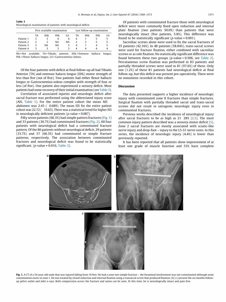
neurological recovery [5]. In our patient, cohort one patient hadneurological recovery and that after he had sacral laminectomyand decompression during his open reduction and internal fixation(Fig. 2).
Vaccaro et al. and Routt et al. have suggested that zone 2 sacralfractures should be fixed using fully threaded sacral screws to avoidneuroforaminal compression generated by partially threadedscrews and resultant iatrogenic nerve injury [16,17]. Concisescientific study of this assertion is lacking. Data presented in thiscohort does not support these prior concerns as no patient withcomminution developed a postoperative neurologic injury. Addi-tionally, this series supports that surgical stabilization with partiallythreaded sacral and trans-sacral screws yielded 100% union rate.
Min et al. reported on 35 patients with Zone 2 sacral fracturesthat were treated with partially threaded sacral screws. They didnot find any new neurological deficit that could be attributed to thecompression screws [20]. They did not study other risk factors suchas comminution and they had a small cohort of 35 patients that hadZone 2 sacral fracture.
Our data suggest that neurologic deficit is injury related andthat it depends on the comminution of the fracture that isdetermined at injury. Strengthening this conclusion is the fact thatneurologically injured patients had higher scores of abdomen AISand ISS. This suggests that these were more severely injuredpatients. We did not recognize a single patient that had an intactneurological examination upon admission and had a neurologicaldeficit after surgery.
Our study has several drawbacks; the first is that it is aretrospective study based on clinical records some of which somerecords might be incomplete or biased. Several patients wereintubated upon arrival which makes it impossible to determinetheir neurological status at arrival. However, intubation uponarrival is inherent to a population of severely injured patients at areferral trauma center. These patients cannot be excluded if a truerepresentation of the sacral fracture population is to be considered.
Further studies are required to confirm our results. Morespecifically, we would be interested in a randomized trailcomparing the results between partially threaded and fullythreaded screws, focusing both on neurologic status and unionrate after surgical fixation of zone 2 sacral fractures.
Conflicts of interest
None declared.
References
[1] Gansslen A, Pohlmann T, Paul CH, Lobenhoffer PH, Tscherne H. Epidemiology ofpelvic ring injuries. Injury 1996;27(Supp 1):13–20.
[2] Mehta S, Auerbach J, Born C, Chin K. Sacral fractures. J Am Acad Orthop Surg2006;14:656–65.
[3] Denis F, Davis S, Comfort T. Sacral fractures: an important problem. Retro-spective analysis of 236 cases. Clin Orthop Rel Res 1988;227:67–81.
[4] Gibbons K, Soloniuk D, Razack N. Neurological injury and patterns of sacralfractures. J Neurosurg 1990;72:889.
Fig. 2. A 39-year-old male that was injured in a motor vehicle collision. (a) an axial CT in which the comminution involves the neural foramin can be seen with a fragment inthe foramina. He initially presented with motor function of three out of five in TA, EHL, FHL and GS. He was treated by open reduction, decompression laminectomy andinternal fixation by two tension bands. (b) and (c) presents his inlet and outlet pelvis x-ray at 1 year follow-up. He recovered to four out of five in TA, EHL, FHL and GS butremained 0/5 in peroneal strength.
A. Herman et al. / Injury, Int. J. Care Injured 47 (2016) 1569–15731572
[5] Reilly M, Zinar D, Matta JM. Neurologic injuries in pelvic ring fracture. ClinOrthop Rel Res 1996;329:28–36.
[6] Pohlemann T, Angst M, Schneider E, Ganz R, Tscherne H. Fixation of transforaminalsacrum fractures a biomechanical study. J Ortho Trauma 1993;7(3):107–17.
[7] Suzuki T, Hak D, Ziran B, Adams S, Stahel PF, Morgan SJ, et al. Outcome andcomplications of posterior transiliac plating for vertically unstable sacralfractures. Injury 2009;40:405–9.
[8] Sagi C, Militano U, Caron T, Lindvall E. A comprehensive analysis with mini-mum 1-year, follow-up of vertically unstable transforaminal sacral fracturestreated with triangular osteosynthesis. J Ortho Trauma 2009;23(5):313–21.
[9] Gardner MJ, Morshed S, Nork SE, Ricci WM, Rout ML. Quantification of theupper and second sacral segment safe zones in normal and dysmorphic sacra. JOrtho Trauma 2010;24(10):622–9.
[10] Rout ML, Simonian PT, William J. Iliosacral screw fixation: early complicationsof the percutanous technique. J Ortho Trauma 1997;11(8):584–9.
[11] Rout Ml, Nork SE, Mills WJ. Percutaneous fixation of pelvic ring disruption. ClinOrthop Rel Res 2000;375:15–29.
[12] Gardner MJ, Routt ML. Transiliac–transsacral screws for posterior stabiliza-tion. J Ortho Trauma 2011;25(6):378–84.
[13] Templeman D, Schmidt A, Freese J, Weisman I. Proximity of Iliosacral screws toneurovascular structures after internal fixation. Clin Orthop Rel Res 1996;329:194–8.
[14] Comstock CP, van der Meulen MCH, Goodman SB. Biomechanical comparisonsof posterior internal fixation techniques for unstable pelvic, fractures. J OrthoTrauma 1996;10(8):517–22.
[15] Yinger K, Scalie J, Olson SA, Bay BK, Finkemeier CG. Biomechanical comparisonof posterior pelvic ring fixation. J Ortho Trauma 2003;17(7):481–7.
[16] Vaccaro AR, Kim DH, Brodke DS, Harris M, Chapman JR, Schildhauer T, et al.Diagnosis-and-managemetn-of-sacral-spine-fractures. Instr Course Lect 2004;53:375–85.
[17] Routt ML, Simonian T, Wiss DA. Posterior pelvic ring disruptions: iliosacralscrews. In master techniques in orthopaedic surgery, factures. Philadelphia:Lippincott Raven Publishers; 1998. p. 595–612.
[18] Keating JF. Early fixation of the vertically unstable pelvis: the role ofiliosacral screw fixation of the posterior lesion. J Ortho Trauma 1999;13(2):107–33.
[19] Reilly MC, Bono CM, Litkouhi B, Sirkin M, Behrens FF. The effect of sacralfracture malreduction on the safe placement of iliosacral screws. J OrthoTrauma 2003;17(2):88–94.
[20] Min W, Chambers M, Leslie MP, Lee MA, Ferguson TA. The clinicalefficacy of compressive transacral screw fixation for unstable pelvic ringinjuries. In: Orthopaedic Trauma Association conference; 2011. pre-sented paper #42.
A. Herman et al. / Injury, Int. J. Care Injured 47 (2016) 1569–1573 1573
This Paper Will Change Your
Practice, It Changed Mine: New
Information That You Need to
Know
OTA Specialty Day March
Kenneth A. Egol, M.D.

2

Closed Contact Casting Vs Surgery for Initial Treatment of
Unstable Ankle Fractures in Older Adults: Arandomized
Clinical Trial
3
•Randomized Clinical
Trial
•Published IN JAMA
•Took place at 24 centers
in the UK
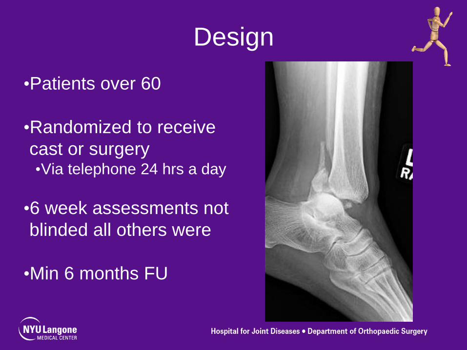
Design
•Patients over 60
•Randomized to receive
cast or surgery •Via telephone 24 hrs a day
•6 week assessments not
blinded all others were
•Min 6 months FU
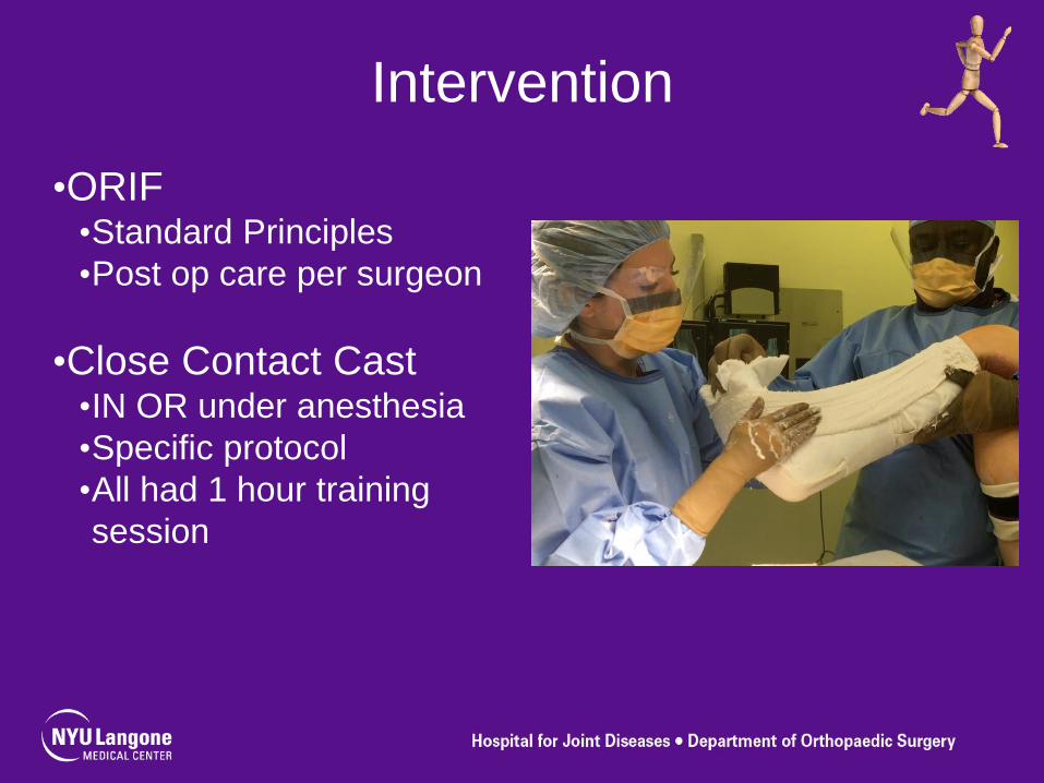
Intervention
•ORIF •Standard Principles
•Post op care per surgeon
•Close Contact Cast •IN OR under anesthesia
•Specific protocol
•All had 1 hour training
session
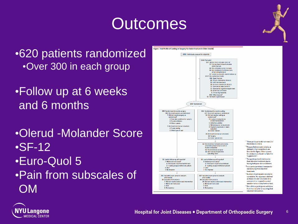
Outcomes
•620 patients randomized •Over 300 in each group
•Follow up at 6 weeks
and 6 months
•Olerud -Molander Score
•SF-12
•Euro-Quol 5
•Pain from subscales of
OM
6
Outcomes
•Patient reported time to
WB
•Timed get up and go
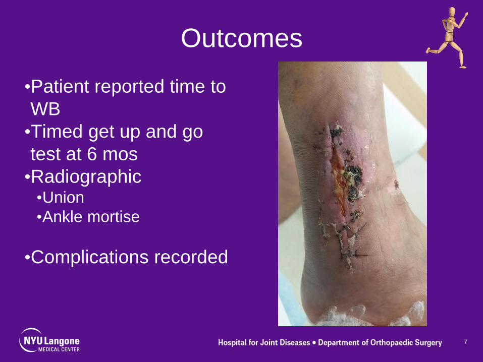
test at 6 mos
•Radiographic •Union
•Ankle mortise
•Complications recorded
7
Results
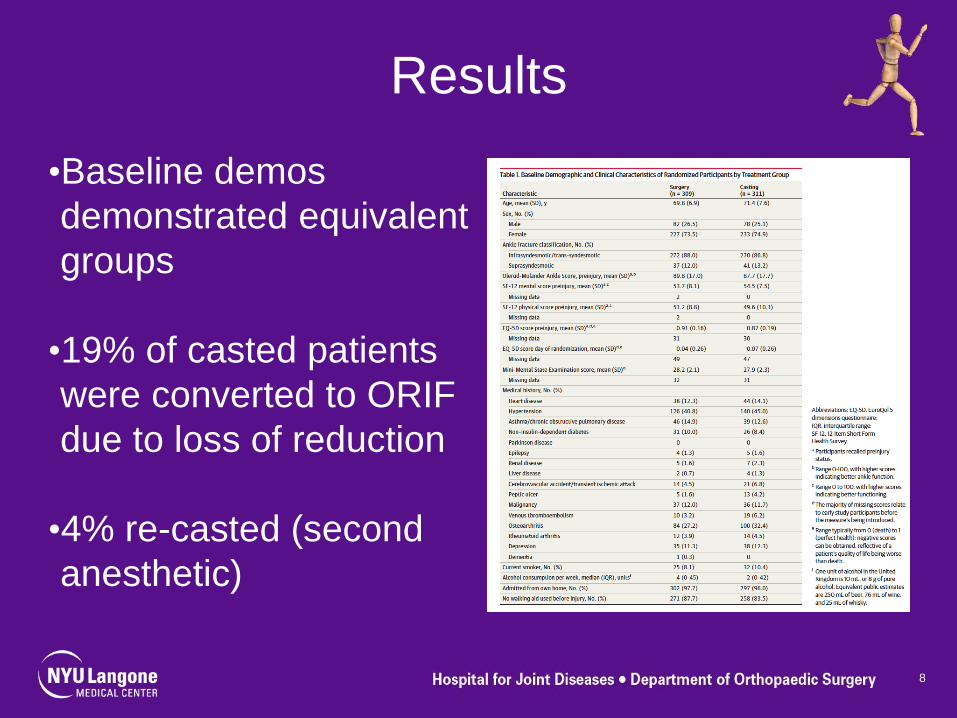
•Baseline demos
demonstrated equivalent
groups
•19% of casted patients
were converted to ORIF
due to loss of reduction
•4% re-casted (second
anesthetic)
8
Results
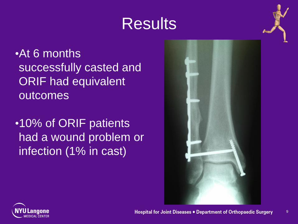
•At 6 months
successfully casted and
ORIF had equivalent
outcomes
•10% of ORIF patients
had a wound problem or
infection (1% in cast)
9
Limitations
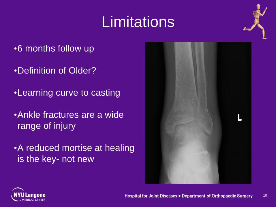
•6 months follow up
•Definition of Older?
•Learning curve to casting
•Ankle fractures are a wide
range of injury
•A reduced mortise at healing
is the key- not new
10
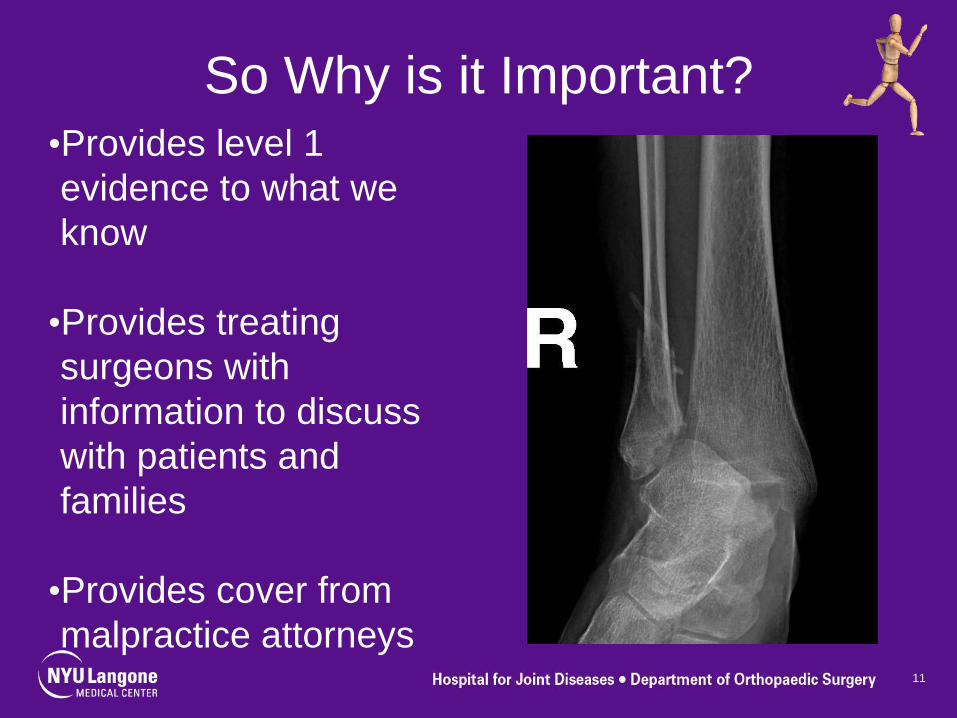
So Why is it Important?
•Provides level 1
evidence to what we
know
•Provides treating
surgeons with
information to discuss
with patients and
families
•Provides cover from
malpractice attorneys 11
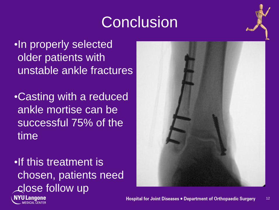
Conclusion
•In properly selected
older patients with
unstable ankle fractures
•Casting with a reduced
ankle mortise can be
successful 75% of the
time
•If this treatment is
chosen, patients need
close follow up 12
Thank You
This Paper Will Change Your Practice – It Changed Mine. New Information That You Need to Know: Basic Science Theodore Miclau, MD Department of Orthopaedic Surgery, University of California, San Francisco UCSF/ZSFG Orthopaedic Trauma Institute, San Francisco, USA The burden of musculoskeletal disease has surpassed cardiovascular disease as the major health burden in the world, and bone fractures contribute substantially to the overall burden of musculoskeletal disease. In the US, there are over 600,000 fractures per year with a substantial number of these fractures exhibiting delayed healing or non-union. The gold standard to stimulate bone union been autologous bone grafting, which although generally good, remains problematic due to limited graft supply, donor site morbidity, and potential complications. Therefore, understanding mechanisms of normal fracture healing to develop effective therapies to treat patients is imperative. Generally, fracture repair occurs through two processes: direct bone (intramembranous ossification) and the formation of bone through a cartilage intermediate (endochondral ossification). With the exception of an initial inflammatory process, adult healing is similar to that observed during bone development. Previous work suggested that in adult repair, stem cells in the periosteum and endosteum give rise to chondrocytes that form the soft callus during endochondral ossification, and subsequently, during vascular invasion of the cartilage callus, osteoprogenitor cells are delivered to the fracture site to form new bone. Recent findings, however, challenge this assumption. This presentation will summarize an article (Hinton et al., 2017) that reviews data suggesting that suggests that a significant number of bone cells are derived directly from the transformation of chondrocytes. They discuss other recent findings that show demonstrate this concept (see other references below). These finding are potentially paradigm-shifting. Traditionally, strategies for stimulating bone repair seek to stimulate the process of direct bone formation. However, given that the majority of long bone fractures heal with some degree of callus formation (with the exception of those treated with absolute stability) and bone is formed directly through the transformation of chondrocytes, successful fracture repair therapies might target the endochondral rather than intramembranous ossification process. These findings have the potential to affect the way fracture healing, bone incorporation, and bone tissue engineering strategies are developed and employed. Reference: Hinton RJ, Jing Y, Jing J, Feng JQ. Roles of Chondrocytes in Endochondral Bone Formation and Fracture Repair. J Dent Res. 2017 Jan;96(1):23-30. doi: 10.1177/0022034516668321. Other references: 1) Yang, L., et al., Hypertrophic chondrocytes can become osteoblasts and osteocytes in
endochondral bone formation. Proceedings of the National Academy of Sciences of the United States of America, 2014. 111(33): p. 12097-102.
2) Yang, G., et al., Osteogenic fate of hypertrophic chondrocytes. Cell Res, 2014. 24(10): p. 1266-9. 3) Zhou, X., et al., Chondrocytes transdifferentiate into osteoblasts in endochondral bone during
development, postnatal growth and fracture healing in mice. PLoS Genet, 2014. 10(12): p. e1004820.
4) Bahney CS, Hu DP, Taylor AJ, Ferro F, Britz HM, Hallgrimsson B, Johnstone B, Miclau T, Marcucio RS. 2014. Stem cell-derived endochondral cartilage stimulates bone healing by tissue transformation. J Bone Miner Res. 29(5):1269–1282.
5) Jing, Y., et al., Chondrocytes Directly Transform into Bone Cells in Mandibular Condyle Growth. J Dent Res, 2015. 94(12): p. 1668-75.
6) Park, J., et al., Dual pathways to endochondral osteoblasts: a novel chondrocyte-derived osteoprogenitor cell identified in hypertrophic cartilage. Biol Open, 2015. 4(5): p. 608-21.