TheProinflammatoryCytokine,Interleukin-6,Up-regulates ... · PDF...
16
The Proinflammatory Cytokine, Interleukin-6, Up-regulates Calcium-sensing Receptor Gene Transcription via Stat1/3 and Sp1/3 * Received for publication, September 27, 2007, and in revised form, February 13, 2008 Published, JBC Papers in Press, March 17, 2008, DOI 10.1074/jbc.M708087200 Lucie Canaff 1 , Xiang Zhou, and Geoffrey N. Hendy 2 From the Departments of Medicine, Physiology, and Human Genetics, McGill University and Calcium Research Laboratory and Hormones and Cancer Research Unit, Royal Victoria Hospital, Montreal, Quebec H3A 1A1, Canada Increased expression of the calcium-sensing receptor (CASR), which controls blood calcium homeostasis, leads to a decrease in the extracellular calcium set-point, thereby reducing parathy- roid hormone secretion and renal calcium reabsorption and increasing calcitonin secretion resulting in reduced circulating calcium levels. Critically ill patients with elevated proinflamma- tory cytokine levels commonly have hypocalcemia, although the mechanism is not known. After intraperitoneal injection of interleukin (IL)-6 in the rat, circulating levels of parathyroid hormone, 1,25-dihydroxyvitamin D, and calcium fell within hours and remained low at 24 h. Expression of CASR (mRNA and protein) increased within hours in parathyroid, thyroid, and kidney and remained elevated at 24 h. The CASR gene has two promoters (P1 and P2) yielding transcripts having alternative 5-untranslated regions but encoding the same receptor protein. Activities of P1 and P2 promoter/luciferase reporter constructs were stimulated 2–3-fold by IL-6 in proximal tubule HKC cells and TT thyroid C-cells. Studies with P1 deleted and mutated promoter-reporter and Stat1 and/or Stat3 dominant- negative constructs showed that a Stat1/3 element downstream of the P1 start site accounted for the IL-6 induction. There are no Stat elements in the P2 promoter, but Sp1/3 elements are clustered at the transcription start site. A series of transfection P2 promoter-reporter analyses showed that Sp1 together with Stat1/3 was critical for IL-6 responsiveness of P2. By oligonu- cleotide precipitation assay, IL-6 rapidly promoted a complex containing both Sp1/3 and Stat1/3 on the Sp1/3 elements. In conclusion, Stat1/3 directly controls promoter P1, and the Stats indirectly regulate promoter P2 via Sp1/3 in response to IL-6. By this mechanism, the cytokine likely contributes to altered extra- cellular calcium homeostasis. The calcium-sensing receptor (CASR) 3 is expressed in the parathyroid hormone (PTH) producing chief cells of the para- thyroid gland, the calcitonin-producing C-cells of the thyroid, and the cells lining the kidney tubule. The CASR, a plasma membrane G protein-coupled receptor, senses small changes in circulating calcium concentration and modulates intracellular pathways that alter PTH and calcitonin secretion or renal cation handling, thereby playing an essential role in blood mineral ion homeostasis. The relationship between extracellular ionized cal- cium and PTH concentrations is represented by an inverse sig- moidal curve. The activity and/or expression level of the CASR dictates the extracellular calcium set point (defined as the extracellular calcium concentration at which PTH secretion from the parathyroid gland or calcium reabsorption across the kidney tubule is half-maximal). Increases in extracellular cal- cium directly stimulate calcitonin secretion. The importance of the CASR in orchestrating the endocrine control of blood calcium concentrations has been underscored by the identification of naturally occurring mutations in the CASR gene that cause human disease. Inactivating mutations result in hypercalcemia, and activating mutations result in hypocalcemia (1–3). Hypocalcemia is common in critically ill patients, especially those with sepsis and major burn injury (4), and in nonacutely ill patients undergoing surgery (5). The mechanisms underlying the hypocalcemia are not known. Several factors may be involved, including decreased secretion of PTH and resistance to the action of PTH in kidney and bone. The metabolism and function of vitamin D are impaired. Calcitonin precursors are increased in the circulation of critically ill patients with sepsis and could contribute to the hypocalcemia (6, 7). Several studies of critically ill patients have shown that serum interleukin-6 (IL-6) levels increase within hours of severe burns and infection and can rise to very high levels (8, 9). In these patients the serum IL-6 levels are even more elevated than those of other proinflammatory cytokines, like interleukin-1 (IL-1) (10), and are inversely related to the serum calcium concentra- tion (6) and correlate with a poor prognosis (11–16). Carlstedt et al. (17) have shown that in vitro incubating isolated bovine parathyroid cells with IL-6 decreases PTH secretion at clinically * This work was supported in part by Canadian Institutes of Health Research Grants MOP-86581 and MOP-57730 (to G. N. H.). The costs of publication of this article were defrayed in part by the payment of page charges. This article must therefore be hereby marked “advertisement” in accordance with 18 U.S.C. Section 1734 solely to indicate this fact. 1 Recipient of a biomedical fellowship from the Kidney Foundation of Canada. 2 To whom correspondence should be addressed: Calcium Research Labora- tory, Rm. H4.67, Royal Victoria Hospital, 687 Pine Ave. West, Montreal, Que- bec H3A 1A1, Canada. Tel.: 514-843-1632; Fax: 514-843-1712; E-mail: [email protected]. 3 The abbreviations used are: CASR, calcium-sensing receptor; PTH, parathy- roid hormone; 1,25(OH) 2 D, 1,25-dihydroxyvitamin D; IL, interleukin; TT cells, human thyroid C-cells; HKC, human proximal tubule cells; IL-6R, inter- leukin-6 receptor; JAK, Janus kinase; STAT, signal transducers and activa- tors of transcription; Sp1/3, specificity protein 1/3; MAPK, mitogen-acti- vated protein kinase; SRF, serum-response factor; EMSA, electrophoretic mobility shift assay; GAPDH, glyceraldehyde-3-phosphate dehydrogen- ase; DMEM, Dulbecco’s modified Earle’s medium; PBS, phosphate-buff- ered saline; PMSF, phenylmethylsulfonyl fluoride; DTT, dithiothreitol; UTR, untranslated region; oligo, oligonucleotide. THE JOURNAL OF BIOLOGICAL CHEMISTRY VOL. 283, NO. 20, pp. 13586 –13600, May 16, 2008 © 2008 by The American Society for Biochemistry and Molecular Biology, Inc. Printed in the U.S.A. 13586 JOURNAL OF BIOLOGICAL CHEMISTRY VOLUME 283 • NUMBER 20 • MAY 16, 2008 by guest on May 6, 2018 http://www.jbc.org/ Downloaded from
Transcript of TheProinflammatoryCytokine,Interleukin-6,Up-regulates ... · PDF...
The Proinflammatory Cytokine, Interleukin-6, Up-regulatesCalcium-sensing Receptor Gene Transcription viaStat1/3 and Sp1/3*
Received for publication, September 27, 2007, and in revised form, February 13, 2008 Published, JBC Papers in Press, March 17, 2008, DOI 10.1074/jbc.M708087200
Lucie Canaff1, Xiang Zhou, and Geoffrey N. Hendy2
From the Departments of Medicine, Physiology, and Human Genetics, McGill University and Calcium Research Laboratory andHormones and Cancer Research Unit, Royal Victoria Hospital, Montreal, Quebec H3A 1A1, Canada
Increased expressionof the calcium-sensing receptor (CASR),which controls blood calciumhomeostasis, leads to adecrease inthe extracellular calcium set-point, thereby reducing parathy-roid hormone secretion and renal calcium reabsorption andincreasing calcitonin secretion resulting in reduced circulatingcalcium levels. Critically ill patients with elevated proinflamma-tory cytokine levels commonly have hypocalcemia, although themechanism is not known. After intraperitoneal injection ofinterleukin (IL)-6 in the rat, circulating levels of parathyroidhormone, 1,25-dihydroxyvitamin D, and calcium fell withinhours and remained low at 24 h. Expression of CASR (mRNAandprotein) increasedwithin hours in parathyroid, thyroid, andkidney and remained elevated at 24 h. The CASR gene has twopromoters (P1 and P2) yielding transcripts having alternative5�-untranslated regionsbut encoding the same receptor protein.Activities of P1 and P2 promoter/luciferase reporter constructswere stimulated �2–3-fold by IL-6 in proximal tubule HKCcells and TT thyroid C-cells. Studies with P1 deleted andmutated promoter-reporter and Stat1 and/or Stat3 dominant-negative constructs showed that a Stat1/3 element downstreamof the P1 start site accounted for the IL-6 induction. There areno Stat elements in the P2 promoter, but Sp1/3 elements areclustered at the transcription start site. A series of transfectionP2 promoter-reporter analyses showed that Sp1 together withStat1/3 was critical for IL-6 responsiveness of P2. By oligonu-cleotide precipitation assay, IL-6 rapidly promoted a complexcontaining both Sp1/3 and Stat1/3 on the Sp1/3 elements. Inconclusion, Stat1/3 directly controls promoter P1, and the Statsindirectly regulate promoter P2 via Sp1/3 in response to IL-6. Bythismechanism, the cytokine likely contributes to altered extra-cellular calcium homeostasis.
The calcium-sensing receptor (CASR)3 is expressed in theparathyroid hormone (PTH) producing chief cells of the para-
thyroid gland, the calcitonin-producing C-cells of the thyroid,and the cells lining the kidney tubule. The CASR, a plasmamembraneGprotein-coupled receptor, senses small changes incirculating calcium concentration and modulates intracellularpathways that alter PTH and calcitonin secretion or renal cationhandling, thereby playing an essential role in blood mineral ionhomeostasis. The relationship between extracellular ionized cal-cium and PTH concentrations is represented by an inverse sig-moidal curve. The activity and/or expression level of the CASRdictates the extracellular calcium set point (defined as theextracellular calcium concentration at which PTH secretionfrom the parathyroid gland or calcium reabsorption across thekidney tubule is half-maximal). Increases in extracellular cal-cium directly stimulate calcitonin secretion.The importance of the CASR in orchestrating the endocrine
control of blood calcium concentrations has been underscoredby the identification of naturally occurring mutations in theCASR gene that cause human disease. Inactivating mutationsresult in hypercalcemia, and activating mutations result inhypocalcemia (1–3).Hypocalcemia is common in critically ill patients, especially
those with sepsis and major burn injury (4), and in nonacutelyill patients undergoing surgery (5). Themechanisms underlyingthe hypocalcemia are not known. Several factors may beinvolved, including decreased secretion of PTH and resistanceto the action of PTH in kidney and bone. The metabolism andfunction of vitamin D are impaired. Calcitonin precursors areincreased in the circulation of critically ill patients with sepsisand could contribute to the hypocalcemia (6, 7).Several studies of critically ill patients have shown that serum
interleukin-6 (IL-6) levels increase within hours of severe burnsand infection and can rise to very high levels (8, 9). In thesepatients the serum IL-6 levels are evenmore elevated than thoseof other proinflammatory cytokines, like interleukin-1� (IL-1�)(10), and are inversely related to the serum calcium concentra-tion (6) and correlate with a poor prognosis (11–16). Carlstedtet al. (17) have shown that in vitro incubating isolated bovineparathyroid cellswith IL-6 decreases PTHsecretion at clinically
* This work was supported in part by Canadian Institutes of Health ResearchGrants MOP-86581 and MOP-57730 (to G. N. H.). The costs of publication ofthis article were defrayed in part by the payment of page charges. Thisarticle must therefore be hereby marked “advertisement” in accordancewith 18 U.S.C. Section 1734 solely to indicate this fact.
1 Recipient of a biomedical fellowship from the Kidney Foundation of Canada.2 To whom correspondence should be addressed: Calcium Research Labora-
tory, Rm. H4.67, Royal Victoria Hospital, 687 Pine Ave. West, Montreal, Que-bec H3A 1A1, Canada. Tel.: 514-843-1632; Fax: 514-843-1712; E-mail:[email protected].
3 The abbreviations used are: CASR, calcium-sensing receptor; PTH, parathy-roid hormone; 1,25(OH)2D, 1,25-dihydroxyvitamin D; IL, interleukin; TT
cells, human thyroid C-cells; HKC, human proximal tubule cells; IL-6R, inter-leukin-6 receptor; JAK, Janus kinase; STAT, signal transducers and activa-tors of transcription; Sp1/3, specificity protein 1/3; MAPK, mitogen-acti-vated protein kinase; SRF, serum-response factor; EMSA, electrophoreticmobility shift assay; GAPDH, glyceraldehyde-3-phosphate dehydrogen-ase; DMEM, Dulbecco’s modified Earle’s medium; PBS, phosphate-buff-ered saline; PMSF, phenylmethylsulfonyl fluoride; DTT, dithiothreitol; UTR,untranslated region; oligo, oligonucleotide.
THE JOURNAL OF BIOLOGICAL CHEMISTRY VOL. 283, NO. 20, pp. 13586 –13600, May 16, 2008© 2008 by The American Society for Biochemistry and Molecular Biology, Inc. Printed in the U.S.A.
13586 JOURNAL OF BIOLOGICAL CHEMISTRY VOLUME 283 • NUMBER 20 • MAY 16, 2008
by guest on May 6, 2018
http://ww
w.jbc.org/
Dow
nloaded from
relevant concentrations. Murphey et al. (18) found that in vivoparathyroid CASR levels were up-regulated in a sheepmodel ofburn injury in which circulating cytokine levels would be antic-ipated to be increased. However, further analyses are requiredto more fully understand the mechanisms linking the raisedIL-6 levels to altered calcium metabolism.Previously, we investigated the hypothesis that cytokines
such as IL-1� increase CASR expression in those tissues impor-tant for the control of systemic calcium homeostasis therebyleading to hypocalcemia and hypoparathyroidism. We showedin vivo in the rat that parathyroid, thyroid, and kidney CASRmRNA and protein increased after intraperitoneal injection ofIL-1� (19). This was associated with decreased circulatingPTH, 1,25-dihydroxyvitamin D (1,25(OH)2D), and calcium.The CASR gene has two promoters (P1 and P2) yielding alter-native transcripts containing either exon 1A or exon 1B 5�-un-translated region sequences that splice to exon 2 containing theATG translation start codon (20–22). We demonstrated thatboth the CASR gene promoters have functional �B elementsthat mediate up-regulation by IL-1� (19).
In this study, we postulated that IL-6, by up-regulating CASRexpression in parathyroid, thyroid C-cell, and kidney tubule,reducing PTH secretion and renal calcium reabsorption, andincreasing calcitonin secretion, contributes to altered calciumhomeostasis. Here we have demonstrated that IL-6 stimulatesCASR expression in vivo and defined, for the first time, themechanisms whereby the cytokine up-regulates CASR genepromoter activity. The resulting increases in CASR expressioncould be contributing to the hypocalcemia of critically illpatients.
EXPERIMENTAL PROCEDURES
Materials—Recombinant IL-6 was from Sigma. The humanmedullary thyroid carcinoma TT cell line was from the Amer-ican Type Culture Collection (Manassas, VA) and the humanproximal kidney tubule cells (HKC-8) were a gift of Dr. MartinHewison, Cedars-Sinai Medical Center, Los Angeles. Dulbec-co’s modified Earle’s medium (DMEM), Ham’s F-12 (F-12)medium, fetal bovine serum (FBS), and antibiotics were fromInvitrogen. [�-32P]ATP and [�-32P]dUTP were from MP Bio-medicals, Baie d’Urfe, Quebec. Restriction enzymes, polynucle-otide kinase, andMoloney murine leukemia virus reverse tran-scriptase were from MBI Fermentas, Burlington, Ontario,Canada.Hybond andReady-to-Go beadswere fromAmershamBiosciences. Monoclonal antibody against �-tubulin and oligo-nucleotides representing Stat1, Stat3, and Sp1 consensusresponse elements were from Santa Cruz Biotechnology, Inc.,Santa Cruz, CA.DNAconstructs were as follows: human Sp1 inpCMV6-X6 (catalog number SC101137; Origene Technolo-gies, Rockville, MD), human Sp1DN (dominant-negative con-struct) (pCI-neo-HA-MCS-Sp1(622–788)) (23), human Stat1�in pCMV6-XL5 (Origene catalog number SC115595), andhuman Stat3 (variant 1) in pCMV6-XL4 (Origene catalog num-ber SC124165). Dominant-negative Stats (Stat1Y701F andStat3Y705F) were prepared using the wild-type constructs astemplate with the QuikChange site-directed mutagenesis kit(Stratagene, La Jolla, CA). The following primers were used:Stat1DNY701F, forward primer 5�-GGCCCTAAAGGAACT-
GGATTTATCAAGACTGAGTTGATTTCT-3� and reverseprimer 5�-AGAAATCAACTCAGTCTTGATAAATCCAG-TTCCTTTAGGGCC-3�; Stat3DNY705F, forward primer5�-CCAGGTAGCGCTGCCCCATTCCTGAAGACCAAG-TTTATCTGT-3� and reverse primer 5�-ACAGATAAACTTG-GTCTTCAGGAATGGGGCAGCGCTACCTGG-3�. Mutatednucleotides are in boldface type.Animals and Experimental Procedures—Normal male Spra-
gue-Dawley rats (Charles River Laboratories, Inc., St. Constant,Quebec, Canada), weighing 180–200 g when received, were feda standard rodent chow (Ralston Purina Co., LaSalle, Quebec,Canada) containing 1.01% calcium, 0.74% phosphorus, and 3.3IU vitamin D3/g. All animal experiments were carried out incompliance with, and were approved by, the Institutional Ani-mal Care and Use Committee. Rats were injected intraperito-neally at 24, 16, 12, 8, or 4 h before death, with either vehicle(propylene glycol, 0.2 ml/100 g body weight) or 0.75 �g IL-6/100 g bodyweight. Bloodwas obtained by cardiac puncture, andthe serum was separated and stored at �20 °C. The rats wereanesthetized with pentobarbital; the kidneys were taken, andthe parathyroid and thyroid glands were microdissected sepa-rately and quick-frozen. Sera were analyzed for calcium andmagnesium (Sigma kits), PTH (Rat Intact PTH Elisa kit, Immu-topics, San Clemente, CA), and 1,25(OH)2D3 (immunoextrac-tion and radioimmunoassay kit, IDS Ltd., Bolton, UK).RNA Extraction—Total RNA was prepared from cells or tis-
sues using TRIzol (Invitrogen) according to the manufacturer’sinstructions.Ribonuclease Protection Assay of Rat CASR mRNA—For the
CASR riboprobe, a 232-bp fragment of a rat CASR cDNA (24)was PCR-amplified (forward primer, 5�-ACCTTGAGTTTTG-TTGCCCA-3� (in exon 3), and reverse primer, 5�-GGAATGG-TGCGGAGGAAGGATT-3� (in exon 4)) and cloned intoPCR2.1 vector. For the glyceraldehyde-3-phosphate dehydro-genase (GAPDH) riboprobe that protects a 316-nucleotidetranscript, the pTRI-GAPDH Rat vector (AM7432: AmbionInc., Austin, TX) was used. After linearization of the vectors,the antisense probeswere in vitro transcribedwithT7 polymer-ase incorporating [�-32P]UTP using a MAXI script kit, and thegel-purified riboprobes were used with a ribonuclease protec-tion analysis II kit (Ambion Inc.). Each probe (2.5 � 105 cpm)was hybridized overnight with 2–25 �g of total RNA followedby digestion with a ribonuclease A:T1 mix (25). Protected frag-
TABLE 1Oligonucleotides used for CASR gene promoter deletion constructsEach forward (F) primer is named according to the first CASR gene nucleotiderelative to the transcription start site (�1) of either promoter P1 or P2. The commonreverse primer anneals to the sequence immediately upstreamof theATG in exon 2.Restriction enzyme recognition sites (ACGCGT, MluI; AAGCTT, HindIII) are inboldface type. Nucleotides in lowercase type are those added to promote efficientrestriction enzyme digestion.
CASR P1F(�938) 5�-agtcACGCGTTCACCTTGTGATCCACTGTTATCT-3�F(�701) 5�-agtcACGCGTGCAGCCTCATTCGGGTTTTGAAC-3�F(�382) 5�-agtcACGCGTCTCAGCTCCTTCTCCTACCCT-3�F(�194) 5�-agtcACGCGTGGACCCCCTGAGGTAGATGTTA-3�
CASR P2F(�341) 5�-agtcACGCGTTCCATCGCCAAGGAGTAGGGGTC-3�
CASR P1 and P2Reverse 5�-tgcaAAGCTTGGTTCTGCCGTCTCTCCAGGGC-3�
Interleukin-6 Up-regulates CASR Gene Transcription
MAY 16, 2008 • VOLUME 283 • NUMBER 20 JOURNAL OF BIOLOGICAL CHEMISTRY 13587
by guest on May 6, 2018
http://ww
w.jbc.org/
Dow
nloaded from
ments were resolved on 6% acrylamide denaturing gels andexposed to x-ray film.Nuclear Run-on Transcription Assays—Relative transcrip-
tion rates were measured using a nuclear run-on assay (26).Nuclei were prepared from 10 to 20 � 106 HKC or TT cellsincubatedwith either 5 ng/ml IL-6 or 1% bovine serum albuminin PBS carrier alone. Cells were scraped into ice-cold PBS, pH7.4, pelleted at 4 °C, and lysed with Nonidet P-40 buffer (0.3 Msucrose, 60 mM KCl, 15 mMNaCl, 15 mMHEPES, pH 7.5, 2 mMEDTA, 0.5 mM EGTA, 0.15 mM spermine, 0.5 mM spermidine,14mM �-mercaptoethanol, 0.2%Nonidet P-40). After 8min onice, nuclei were pelleted at 800� g. They were rinsed once with1 ml of nuclei storage buffer (50% glycerol, 20 mM Tris, pH 7.9,75 mM NaCl, 0.5 mM EDTA, 0.85 mM DTT, 125 mM PMSF),snap-frozen in liquid nitrogen, and stored at�80 °C until assay.Run-on reactions (50 �l) were carried out at 30 °C in 300 mMNH4(SO4)2, 100mMTris-Cl, pH 7.9, 4mMMgCl2, 4mMMnCl2,50mMNaCl, 0.4mMEDTA, 1.2mMDTT, 0.1mMPMSF, 10mMcreatinine phosphate, 29% glycerol, Method 1, 150 �Ci of[32P]UTP, 3000 Ci/mmol (ICN,Mississauga, Ontario, Canada),or Method 2, 350 �M Biotin-16-UTP (Roche Diagnostics); 1.5mM each of CTP, ATP, and GTP (MBI Fermentas) for 45 min.RNA was extracted with TRIzol (Invitrogen) according to themanufacturer’s instructions. Five �g of plasmid DNA contain-ing specific gene inserts or no insert were NaOH-denaturedand slot-blotted (HybriSlot, Invitrogen) onto Nytran mem-branes. The gene-specific plasmids were as follows: 1) humanCASR exon 1A, a 280-bp AseI-StuI fragment cloned inpBluescript II KS; 2) human CASR exon 1B, 230-bp Not-StuIfragment cloned in pBluescript II KS; 3) human CASR exon
2, a 227-bp StuI-NotI fragmentcloned in pBluescript II KS; 4)human cyclooxygenase (COX)-2,a 488-bp fragment reverse tran-scription-PCR amplified from HKCRNA (forward primer 5�-CATCCC-TGATCCCCAGGGCTCA-3� andreverse primer 5�-TGCACAT-AATCTTCAATCACAA-3�) TAcloned into pCR2.1Topo; 5)humanGAPDH, a 469-bp fragmentreverse transcription-PCR ampli-fied from HKC RNA (forwardprimer 5�-CCCTTCATTGACCT-
CAACTACATGGT-3� and reverse primer 5�-GAGGGGGCC-ATCCACAGTCTTCTG-3�) TA-cloned in pGEM-T; 6)pUC18. Themembranes were hybridized withMethod 1, 2 �107 cpm of 32P-labeled transcripts/ or Method 2, 25 �l eachbiotinylated RNA in 50% formamide, 50 mM HEPES, pH 7.3,0.75 M NaCl, 2 mM EDTA, 0.5% SDS, 10� Denhardt’s, and 20�g/ml salmon sperm DNA for a minimum of 40 h. In anysingle experiment, equal amounts of labeled RNA were usedfor all conditions. Membranes were exposed to Method 1,autoradiographic film directly, or Method 2, after the signalwas revealed with streptavidin-alkaline phosphatase usingthe BrightStar BioDetect nonisotopic detection kit (Ambion,Austin, TX.) and quantitation of the relative rates of tran-scription was achieved by densitometry.Western Blot Analysis of the CASR—Tissues or cells were
lysed in triple detergent buffer (50 mM Tris-HCl, pH 8.0, 150mM NaCl, 0.02% NaN3, 0.1% SDS, 1 mM EDTA, 100 �g/mlPMSF, 2 �g/ml leupeptin, 2 �g/ml aprotinin, 0.1% NonidetP-40, 0.5% sodium deoxycholate) for 5 min at 0 °C. The lysateswere spun at 1200 � g for 2 min at 4 °C, and the supernatantswere stored at �80 °C. Aliquots were electrophoresed through4–12% SDS-polyacrylamide gels and blotted onto polyvinyli-dene difluoridemembranes (Bio-Rad).Membranes were rinsedin 10 mM Tris-HCl, pH 8.0, 150 mM NaCl, 0.05% Tween 20,blocked with 5% dried milk powder in TBST for 3 h, and incu-bated with CASR antibody, either an affinity-purified poly-clonal antibody, AS2011 (27), raised against a peptide compris-ing CASR amino acids 215–235 coupled to keyhole limpethemocyanin, or theADDmonoclonal antibody directed againstthe same epitope (Abcam, Cambridge, MA). As a control,
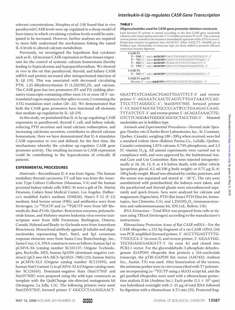
FIGURE 1. Serum PTH, 1,25(OH)2D, and calcium levels are decreased by IL-6. Rats were injected intraperi-toneally with IL-6 and sacrificed at the times shown, and serum PTH, 1,25(OH)2D, and calcium levels weredetermined as described under “Experimental Procedures.” Each value is the mean � S.E. (n � 3). The asterisksindicate a significant difference (p � 0.05) from the time 0 value.
TABLE 2Oligonucleotides used for EMSA and/or oligo pulldown experimentsThe Sp1, Stat1 and CRE sites are italic boldface and mutated residues are underlined.
Oligonucleotides SequencesConsensusSp1 5�-ATTCGATCGGGGGGGGGCGAGC-3�
CASR promotersP1 Stat1-WT 5�-CCCTGTATTTGAGGGAAAGGAAG-3�1A Stat1-WTa 5�-TATTTTGTTCTGGAAATTTCCGCAAGGGTGG-3�1A Stat1-Muta 5�-TATTTTGTTCTGGAAATTTAAACAAGAATGG-3�P2 Sp1-WTa 5�-GGCAAGCGGGCGGGGAGCAGGAGAGGGGCGGAGCTCCGGGGCCGGGGCCGGGA-3�P2 Sp1-Muta 5�-GGCAAGCGAAAGGGGAGCAGGAGAGGGAAAGAGCTCCGGGGAAAGGGCCGGGA-3�
OtherCgA-CRE-WTb 5�-GATCCACCGCTGACGTCATTTCCAGATC-3�
a For oligo pulldown experiments, a biotin moiety was present at the 5� end.b Cyclic AMP-response element sequence of the human chromogranin A gene is shown.
Interleukin-6 Up-regulates CASR Gene Transcription
13588 JOURNAL OF BIOLOGICAL CHEMISTRY VOLUME 283 • NUMBER 20 • MAY 16, 2008
by guest on May 6, 2018
http://ww
w.jbc.org/
Dow
nloaded from
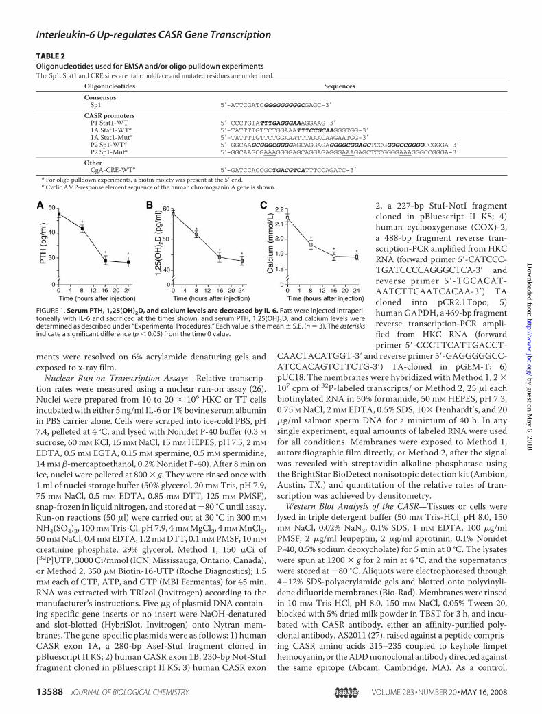
FIGURE 2. Induction of parathyroid, thyroid, and kidney CASR mRNA by IL-6. Rats were injected intraperitoneally with IL-6, sacrificed at the times shown, and CASRand GAPDH mRNA levels of parathyroid gland (A), thyroid gland (B), and kidney (C) were measured by ribonuclease protection assay as described under “ExperimentalProcedures.” Autoradiographs of representative ribonuclease protection analysis signals are shown for each time point. P � undigested probe. CASR and GAPDHmRNA levels were assessed by densitometry, and each value is the mean � S.E. (n � 3). Asterisks indicate a significant difference (p � 0.05) from the time 0 value.Injection of vehicle was without effect (data not shown). For induction of thyroid and kidney CASR protein by IL-6, rats were injected intraperitoneally with IL-6 andsacrificed at the times shown, and thyroid and kidney tissues were harvested and extracts made and subjected to CASR immunoblot analysis as described under“Experimental Procedures.” For each time point, representative samples (from two different rats) are shown. D, thyroid, immunoblot; E, densitometric analysis;F, kidney, immunoblot. The lane with asterisk is one in which the blot was developed with the CASR antibody preadsorbed with the peptide against which it was raised.G, densitometric analysis. E and G, values are mean � S.E. (n � 3). Asterisks indicate a significant difference (p � 0.05) from the time 0 value.
Interleukin-6 Up-regulates CASR Gene Transcription
MAY 16, 2008 • VOLUME 283 • NUMBER 20 JOURNAL OF BIOLOGICAL CHEMISTRY 13589
by guest on May 6, 2018
http://ww
w.jbc.org/
Dow
nloaded from
immunoblotting was carried out as described above with theantibody preadsorbed for 1 h with the peptide (10 �g/ml)against which it was raised. Antibody-antigen complexes weredetected by chemiluminescence using the Lumi-glo kit(Invitrogen). Duplicate membranes were probed with a �-tu-bulin antibody as control.Human CASRGene Promoter Constructs—The construction
of the P1-luciferase reporter plasmid (designated here asP1(�1438)) containing the P1 promoter, exon 1A, and the 5�part of exon 2 to nucleotide �1 (nucleotide �1 is the A of theATG initiation codon), upstreamof the luciferase reporter genein pGL3 basic, has been described previously (P1-WT; 22). Thedeletion constructs, P1(�938), P1(�701), P1(�382), andP1(�194) were prepared using standard techniques (see Table1 for primer sequences). A P1(-1438)StatMut construct inwhich a Stat element in exon 1A is mutated was preparedusing the wild-type construct, P1(�1438), as template withthe QuikChange site-directed mutagenesis kit (Stratagene)as described (28). The primers used were as follows: forwardprimer 5�-TATTATTTTGTTCTGGAAATTTAAACAAG-AATGGAATACTGCATTAAAG-3� and reverse primer 5�-CTTTAATGCAGTATTCCATTCTTGTTTAAATTTCC-AGAACAAAATAATA-3�. Mutated oligonucleotides are inboldface type.The construction of the P2-luciferase reporter plasmid (des-
ignated here as P2(�459)] containing the P2 promoter, exon1B, and the 5� part of exon 2 to nucleotide �1, upstream of theluciferase gene in pGL3 basic, has been described previously(P2-WT; 22). The deletion construct, P2(�341), was preparedby standard techniques (see Table 1 for primer sequences).P2(�459)Sp1Mut in which three Sp1 sites (one just before andtwo just after the transcription initiation site) are mutated wasgenerated by the overlap extension site-specific mutagenesismethod (29) using P2(�459) as template. The primers sets usedwere as follows: F1 (5�-AAGCGCCCTAAGCTTCTTTCCAT-
CGCC-3� with a HindIII site inboldface type), RM (5�-TCCCGGC-CCTTTCCCCGGAGCTCTTTC-CCTCTCCTGCTCCCCTTTCG-CTTGCC-3� with mutated residuesin boldface type to generate product1), and FM (5�-GGCAAGCGAAA-GGGGAGCAGGAGAGGGAAA-GAGCTCCGGGGAAAGGGCC-GGGA-3� with mutated residues inboldface type), and R2 (5�-TGCAA-AGCTTGGTTCTGCCGTCTCT-CCAGGGC-3� with HindIII site inboldface type to generate product2). The overlapping fragments weredenatured and amplified withprimers F1 and R2 to generate prod-uct 3 that was cleaved with HindIIIand cloned into the HindIII-di-gested P2(�459) plasmid to createP2(�459)Sp1Mut.Cell Culture—HKC cells were
grown in DMEM supplementedwith 10% FBS. TT cells were cultured in RPMI 1640 mediumwith 10% FBS and 5% horse serum. All maintenance mediacontained 100 units/ml penicillin, 100�g/ml streptomycin, and250 ng/ml amphotericin B. For mitogen-activated proteinkinase (MAPK) inhibitor studies, cells were grown in 6-wellplates to 75–80% confluence and cotransfected with eitherpGL3 or P1 or P2 promoter constructs and the Renilla lucifer-ase vector (internal control). Twelve to 15 h after transfection,cells were serum-starved for 8 h, then stimulated or not with 10ng/ml IL-6, and harvested 9 h later. Ten�Mof theMAPK inhib-itor U0126 (Promega, Madison, WI) or 0.1% DMSO vehiclewere added to the medium 30 min prior to stimulation. Aftercell lysis, supernatants were assayed for luciferase activity.Transfection and Reporter Assay—For transient transfection,
cells were trypsinized, plated in 6-well dishes in DMEM, 10%FBS (1–4 � 105 cells per well), and incubated overnight. Thenext day, cells were transfected with 30 �g/well of Superfectreagent with 1 �g of CASR promoter construct and 0.5 �g ofRenilla luciferase construct per well. The following day, cellswere serum-starved in DMEM overnight and cultured with orwithout cytokine for 10 h. The cells were washed in PBS andlysed in 250 �l of passive lysis buffer (Promega) on ice. Thelysates were vortexed for 30 s and supernatants collected bycentrifugation (12,000 rpm, 20 min, 4 °C). Luciferase activitywas measured in a Fluostar Optima luminometer (BMGLabtech) using 45 �l of cell lysate and D-Luciferin. Firefly lucif-erase activity was normalized to Renilla luciferase activity.Nuclear Extracts of HKC Cells—Cells were stimulated with
IL-6 (10 ng/ml) for 30 min, washed, scraped into 1 ml of PBS,and centrifuged at 1500 � g for 10min at 4 °C. Cell pellets wereprocessed in a loose Dounce tissue homogenizer in 2 volumesof buffer (25 mM Tris, pH 7.9, 0.3 mM DTT, 420 mM NaCl, 10mM EDTA, 0.5 mM PMSF, and protease inhibitors). Nuclearpellets were obtained by centrifugation (25,000 � g for 20 minat 4 °C), resuspended in 20 mM HEPES, pH 7.9, 25% glycerol,
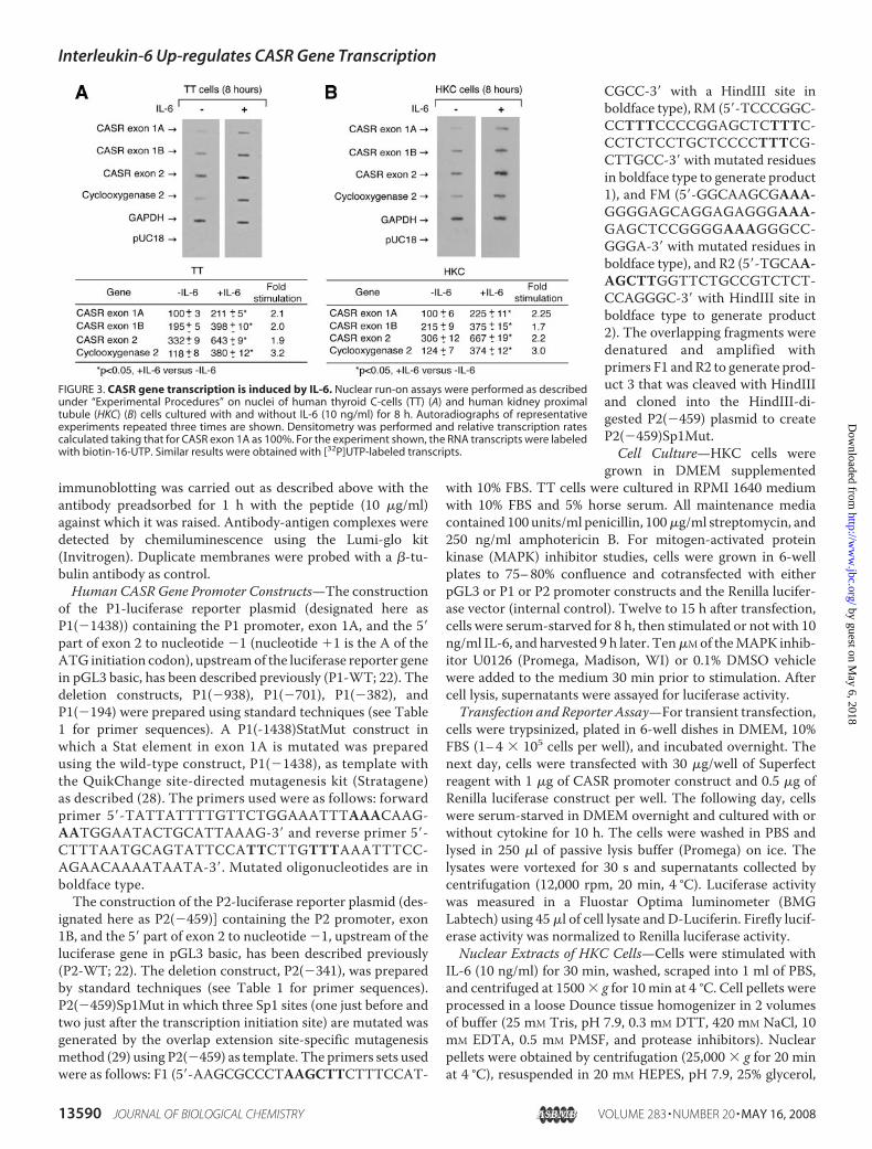
FIGURE 3. CASR gene transcription is induced by IL-6. Nuclear run-on assays were performed as describedunder “Experimental Procedures” on nuclei of human thyroid C-cells (TT) (A) and human kidney proximaltubule (HKC) (B) cells cultured with and without IL-6 (10 ng/ml) for 8 h. Autoradiographs of representativeexperiments repeated three times are shown. Densitometry was performed and relative transcription ratescalculated taking that for CASR exon 1A as 100%. For the experiment shown, the RNA transcripts were labeledwith biotin-16-UTP. Similar results were obtained with [32P]UTP-labeled transcripts.
Interleukin-6 Up-regulates CASR Gene Transcription
13590 JOURNAL OF BIOLOGICAL CHEMISTRY VOLUME 283 • NUMBER 20 • MAY 16, 2008
by guest on May 6, 2018
http://ww
w.jbc.org/
Dow
nloaded from
0.42 M NaCl, 1.5 mM MgCl2, 0.2 mM EDTA, 0.5 mM PMSF, 0.5mM DTT, and Dounce-homogenized. After a 20-min centrifu-gation at 25,000 � g, nuclear extracts (supernatants) were dia-lyzed for 5 h against 20 mM HEPES, pH 7.9, 20% glycerol, 0.1 MKCl, 0.2 mM EDTA, 0.5 mM PMSF, 0.5 mM DTT. Protein con-tent was determined, and aliquots were stored at �80 °C.Electrophoretic Mobility Shift Assay—Two micrograms of
nuclear extract were incubated for 20 min on ice with 1 �g ofpoly(dI-dC) in binding buffer (25 mM Tris-HCl, pH 8.0, 50 mMKCl, 15% glycerol, 0.5 mM DTT). Antibodies were then addedor not, and samples were incubated for 60min at room temper-ature. Five femtomoles of 32P-end-labeled double-stranded oli-gonucleotides were added and incubated for a further 20 min.Oligonucleotides used for EMSA are detailed in Table 2. Sam-ples were electrophoresed at 8 V/cm through 6% nondena-turing polyacrylamide gels equilibrated in 25 mM Tris, pH8.3, 190 mM glycine, 1 mM EDTA, which were then dried andautoradiographed.Oligonucleotide Precipitation Assay—HKCcells were treated
with IL-6 for up to 20 min and lysed by sonication in HKMGbuffer (10 mM HEPES, pH 7.9, 100 mM KCl, 5 mM MgCl2, 10%glycerol, 1 mM DTT, 0.1% Nonidet P-40) with phosphataseinhibitors and protease inhibitors. Cell debris was removed bycentrifugation (2 � 5 min, 10,000 � g, 4 °C). Cell extracts wereincubated with 1 �g of biotinylated double-stranded oligonu-cleotides corresponding to the wild-type or mutated CASR P1Stat1/3 or CASR P2 Sp1 elements (see Table 2 for sequences)for 16 h. DNA-protein complexes (1 �g of biotinylated oligoand 700 �g of protein) were added to Catch and Release SpinColumns (Upstate, Charlottesville, VA) with anti-biotin anti-body (1 �g, Abcam, Cambridge, MA) and 10 �l of antibodycapture affinity ligand with 1� wash buffer in a 500-�l totalvolume. The screw-cap columnswere rotated at 4 °C for 30minand then centrifuged (2000 � g; 30 s). Columns were washedthree times and eluted with denaturing buffer, and eluates weresubjected to immunoblotting with either Stat1 or Stat3 or Sp1or Sp3 antibodies.Statistics—Data are expressed as mean � S.E. The results
from the in vivo IL-6 response studies were initially subjected toanalysis of variance. The significance of differences from baseline was then determined using Dunnett’s multiple comparisontest. For the nuclear run-on assays and luciferase transienttransfection assays, comparisonswere performed by Student’s ttest. A p value of �0.05 was taken to indicate a statisticallysignificant difference.
RESULTS
Interleukin-6 Decreases Serum PTH, 1,25(OH)2D, and Cal-cium Levels in Vivo—To examine the effect of IL-6 on extracel-lular calcium homeostasis, the cytokine was administered torats, and circulating PTH, 1,25(OH)2D, and calcium levels weremonitored over a 24-h period. After a single intraperitonealinjection of IL-6 in rats, serum PTH, 1,25(OH)2D, and calciumlevels were significantly decreased at 8, 16, and 24 h (Fig. 1).Interleukin-6Up-regulates Parathyroid, Thyroid, and Kidney
CASR mRNA Levels in Vivo—To assess whether alterations incirculating PTH, 1,25(OH)2D and calcium levels brought aboutby IL-6 could be due to altered CASR expression in tissues
important for regulation of extracellular calcium homeostasis,we measured CASR mRNA levels by ribonuclease protectionassay throughout the 24-h period. After the injection of IL-6 inrats, parathyroid, thyroid, and kidney CASR mRNA levels rosesignificantly above basal level to peak at 16 h, and the levelswerestill elevated at 24 h (Fig. 2, A–C). The peak values relative tobasal levels were 4.0-fold (parathyroid and thyroid) and 3.5-fold(kidney). Injection of vehicle had no effect on CASR mRNAlevels (data not shown).Interleukin-6 Up-regulates Thyroid and Kidney CASR Pro-
tein Levels in Vivo—To evaluate whether the changes observedin CASRmRNA levels were reflected in corresponding changesin CASR protein levels, immunoblot analysis was conducted onextracts of rat thyroid and kidney. Under the conditions used,
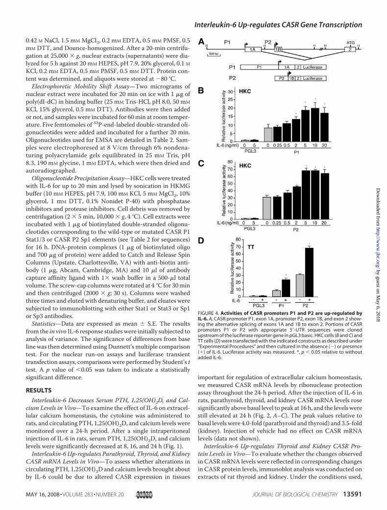
FIGURE 4. Activities of CASR promoters P1 and P2 are up-regulated byIL-6. A, CASR promoter P1, exon 1A, promoter P2, exon 1B, and exon 2 show-ing the alternative splicing of exons 1A and 1B to exon 2. Portions of CASRpromoters P1 or P2 with appropriate 5�-UTR sequences were clonedupstream of the luciferase reporter gene in pGL3 basic. HKC cells (B and C) andTT cells (D) were transfected with the indicated constructs as described under“Experimental Procedures” and then cultured in the absence (�) or presence(�) of IL-6. Luciferase activity was measured. *, p � 0.05 relative to withoutadded IL-6.
Interleukin-6 Up-regulates CASR Gene Transcription
MAY 16, 2008 • VOLUME 283 • NUMBER 20 JOURNAL OF BIOLOGICAL CHEMISTRY 13591
by guest on May 6, 2018
http://ww
w.jbc.org/
Dow
nloaded from
the CASR exists in both monomeric and dimeric forms. Themonomeric core-glycosylated (immature) species is 140 kDa,and mature, fully glycosylated species is 160 kDa (Fig. 2, D andF). The slower mobility forms (�300 kDa) are likely to bedimers (28). After injection of IL-6 in rats, thyroid and kidneyCASR protein levels (the 140- and 160-kDa species related to�-tubulin values) rose �3-fold over basal levels by 24 h (Fig. 2,E and G). Injection of vehicle had no effect on CASR proteinlevels (data not shown). Parathyroid CASR protein levels werenot assessed because of the insufficient amount of tissue.Interleukin-6 Increases CASRGene Transcription—To assess
whether the changes inCASRmRNA levels reflected changes atthe transcription level, nuclear run-on assays were performedon extracts of human TT cells and HKC cells cultured with andwithout IL-6 for 8 and 12 h. CASR gene exon 1A, exon 1B, andexon 2 transcripts were all stimulated �2-fold, and COX2 genetranscription �3-fold, in both cell types (Fig. 3; data not
shown). GAPDH gene transcriptionwas unaffected by IL-6. Therefore,IL-6 specifically stimulates endoge-nous CASR gene transcription.Transcriptional Activities of Hu-
man CASR P1 and P2 Are Up-reg-ulated by IL-6—To assess the effectof IL-6 on the transcriptional activ-ity of specific CASR gene promot-ers, constructs were used in whichhuman CASR P1 or P2 promotersdrive transcription of a luciferasereporter gene (Fig. 4A). Whentransfected intoHKC cells, the basalactivities of P1 and P2 were 8- and33-fold that of the promoterlesscontrol, respectively (Fig. 4, B andC). In the TT thyroid C-cells, thebasal activities of P1 and P2 were 9-and 32-fold that of the control,respectively (Fig. 4D). The additionof IL-6 stimulated reporter activityof P1 and P2 constructs in a dose-dependentmanner inHKC (Fig. 4,Band C) and TT cells (data notshown) with an �2-fold increaseover basal at 10 ng/ml (Fig. 4, B–D).Thus, the proinflammatory cyto-kine stimulates the activity of bothCASR gene promoters.IL-6 Up-regulates the Activities of
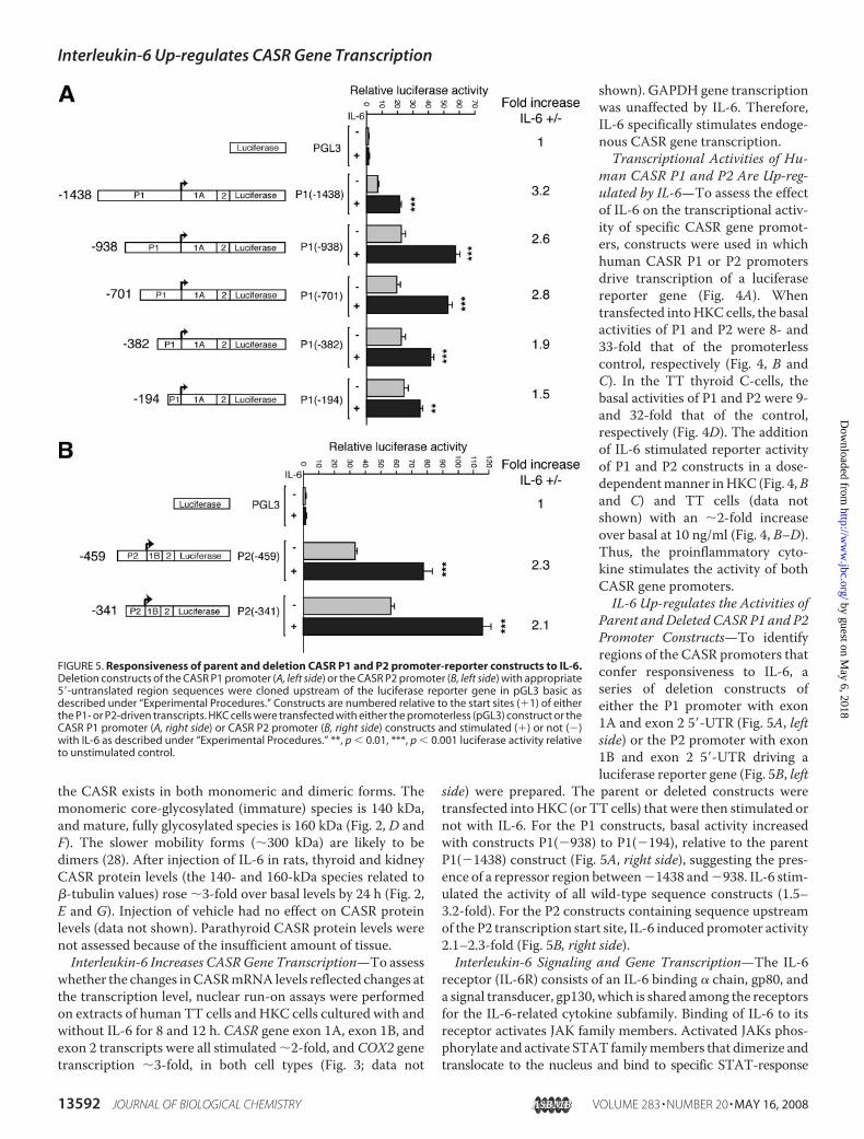
Parent andDeleted CASR P1 and P2Promoter Constructs—To identifyregions of the CASR promoters thatconfer responsiveness to IL-6, aseries of deletion constructs ofeither the P1 promoter with exon1A and exon 2 5�-UTR (Fig. 5A, leftside) or the P2 promoter with exon1B and exon 2 5�-UTR driving aluciferase reporter gene (Fig. 5B, left
side) were prepared. The parent or deleted constructs weretransfected intoHKC (or TT cells) that were then stimulated ornot with IL-6. For the P1 constructs, basal activity increasedwith constructs P1(�938) to P1(�194), relative to the parentP1(�1438) construct (Fig. 5A, right side), suggesting the pres-ence of a repressor region between�1438 and�938. IL-6 stim-ulated the activity of all wild-type sequence constructs (1.5–3.2-fold). For the P2 constructs containing sequence upstreamof the P2 transcription start site, IL-6 induced promoter activity2.1–2.3-fold (Fig. 5B, right side).Interleukin-6 Signaling and Gene Transcription—The IL-6
receptor (IL-6R) consists of an IL-6 binding � chain, gp80, anda signal transducer, gp130, which is shared among the receptorsfor the IL-6-related cytokine subfamily. Binding of IL-6 to itsreceptor activates JAK family members. Activated JAKs phos-phorylate and activate STAT familymembers that dimerize andtranslocate to the nucleus and bind to specific STAT-response
FIGURE 5. Responsiveness of parent and deletion CASR P1 and P2 promoter-reporter constructs to IL-6.Deletion constructs of the CASR P1 promoter (A, left side) or the CASR P2 promoter (B, left side) with appropriate5�-untranslated region sequences were cloned upstream of the luciferase reporter gene in pGL3 basic asdescribed under “Experimental Procedures.” Constructs are numbered relative to the start sites (�1) of eitherthe P1- or P2-driven transcripts. HKC cells were transfected with either the promoterless (pGL3) construct or theCASR P1 promoter (A, right side) or CASR P2 promoter (B, right side) constructs and stimulated (�) or not (�)with IL-6 as described under “Experimental Procedures.” **, p � 0.01, ***, p � 0.001 luciferase activity relativeto unstimulated control.
Interleukin-6 Up-regulates CASR Gene Transcription
13592 JOURNAL OF BIOLOGICAL CHEMISTRY VOLUME 283 • NUMBER 20 • MAY 16, 2008
by guest on May 6, 2018
http://ww
w.jbc.org/
Dow
nloaded from
elements thereby activating gene transcription (30, 31). Stat1 andStat3mediate IL-6 signaling. The JAKs can also couple to the Ras-Raf-MAPK pathway modifying the activity of a variety of tran-scription factors such as the STAT itself but also other regulatorsthat act through theirowncognate responseelements.Other tran-scription factors like AP-1, serum-response factor (SRF), and
Sp1/3 potentially can respond to signaling pathway factors acti-vated by IL-6 and regulate gene expression (32).Several Potential Elements Responsive to IL-6 Are Present in
the CASR Gene Regulatory Regions—Scanning of the CASRgene withMatInspector Release Professional 7.4.3 (GenomatixSoftware) (33) revealed potential STAT, AP-1, SRF, and Sp1
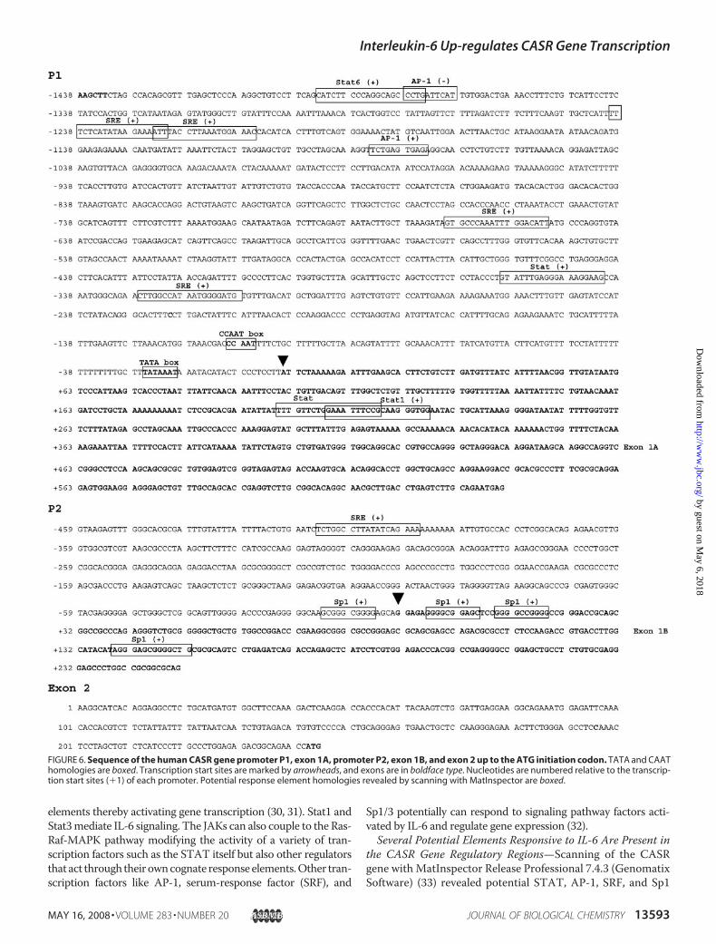
FIGURE 6. Sequence of the human CASR gene promoter P1, exon 1A, promoter P2, exon 1B, and exon 2 up to the ATG initiation codon. TATA and CAAThomologies are boxed. Transcription start sites are marked by arrowheads, and exons are in boldface type. Nucleotides are numbered relative to the transcrip-tion start sites (�1) of each promoter. Potential response element homologies revealed by scanning with MatInspector are boxed.
Interleukin-6 Up-regulates CASR Gene Transcription
MAY 16, 2008 • VOLUME 283 • NUMBER 20 JOURNAL OF BIOLOGICAL CHEMISTRY 13593
by guest on May 6, 2018
http://ww
w.jbc.org/
Dow
nloaded from
elements in P1 and/or P2 promoters and/or the corresponding5�-untranslated regions (UTR) (Fig. 6).Of special note, inCASRpromoter P1 there is a consensus Stat1 element in exon 1A andin CASR promoter P2 there are no Stat elements of any type.However, there are several Sp1 elements that cluster at thetranscription start site.The MAPK Pathway Does Not Markedly Affect the IL-6
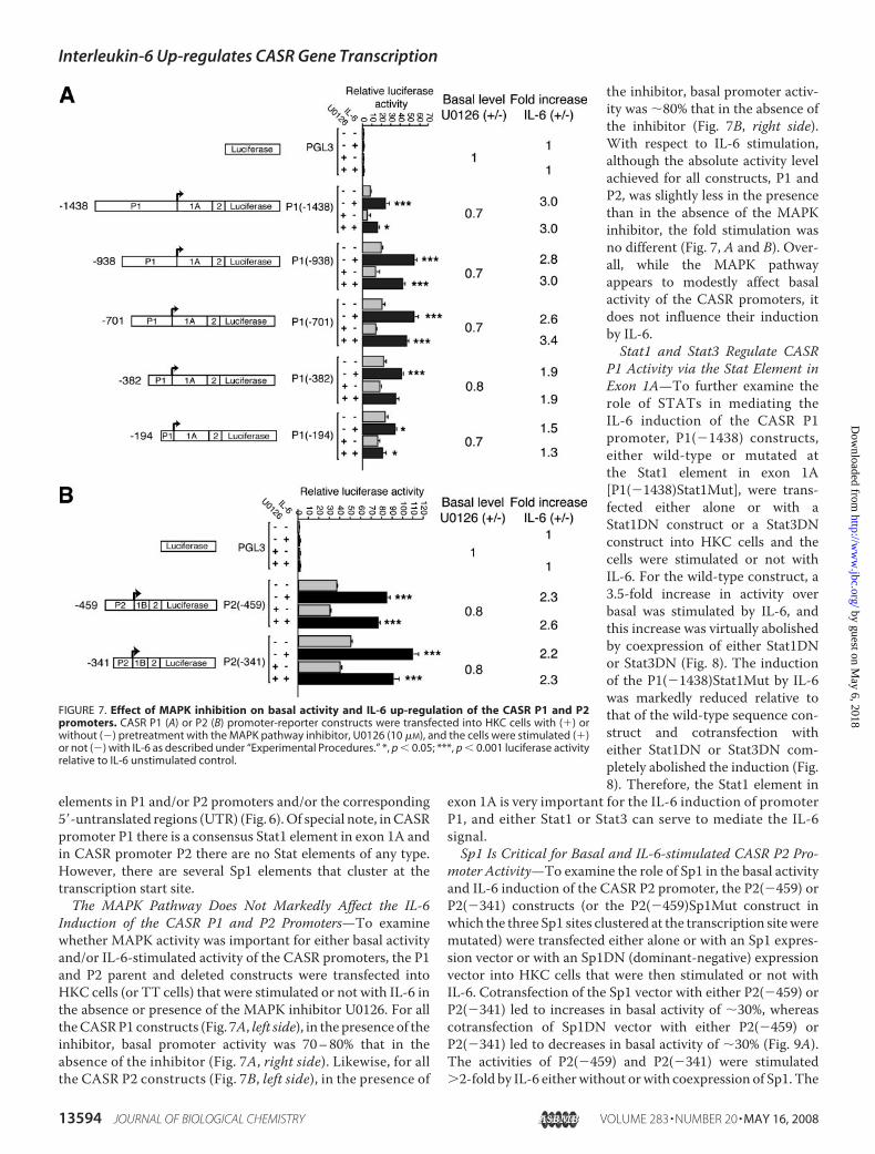
Induction of the CASR P1 and P2 Promoters—To examinewhether MAPK activity was important for either basal activityand/or IL-6-stimulated activity of the CASR promoters, the P1and P2 parent and deleted constructs were transfected intoHKC cells (or TT cells) that were stimulated or not with IL-6 inthe absence or presence of the MAPK inhibitor U0126. For alltheCASRP1 constructs (Fig. 7A, left side), in the presence of theinhibitor, basal promoter activity was 70–80% that in theabsence of the inhibitor (Fig. 7A, right side). Likewise, for allthe CASR P2 constructs (Fig. 7B, left side), in the presence of
the inhibitor, basal promoter activ-ity was �80% that in the absence ofthe inhibitor (Fig. 7B, right side).With respect to IL-6 stimulation,although the absolute activity levelachieved for all constructs, P1 andP2, was slightly less in the presencethan in the absence of the MAPKinhibitor, the fold stimulation wasno different (Fig. 7, A and B). Over-all, while the MAPK pathwayappears to modestly affect basalactivity of the CASR promoters, itdoes not influence their inductionby IL-6.Stat1 and Stat3 Regulate CASR
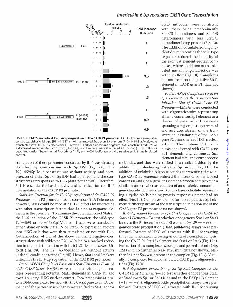
P1 Activity via the Stat Element inExon 1A—To further examine therole of STATs in mediating theIL-6 induction of the CASR P1promoter, P1(�1438) constructs,either wild-type or mutated atthe Stat1 element in exon 1A[P1(�1438)Stat1Mut], were trans-fected either alone or with aStat1DN construct or a Stat3DNconstruct into HKC cells and thecells were stimulated or not withIL-6. For the wild-type construct, a3.5-fold increase in activity overbasal was stimulated by IL-6, andthis increase was virtually abolishedby coexpression of either Stat1DNor Stat3DN (Fig. 8). The inductionof the P1(�1438)Stat1Mut by IL-6was markedly reduced relative tothat of the wild-type sequence con-struct and cotransfection witheither Stat1DN or Stat3DN com-pletely abolished the induction (Fig.8). Therefore, the Stat1 element in
exon 1A is very important for the IL-6 induction of promoterP1, and either Stat1 or Stat3 can serve to mediate the IL-6signal.Sp1 Is Critical for Basal and IL-6-stimulated CASR P2 Pro-
moter Activity—To examine the role of Sp1 in the basal activityand IL-6 induction of the CASR P2 promoter, the P2(�459) orP2(�341) constructs (or the P2(�459)Sp1Mut construct inwhich the three Sp1 sites clustered at the transcription siteweremutated) were transfected either alone or with an Sp1 expres-sion vector or with an Sp1DN (dominant-negative) expressionvector into HKC cells that were then stimulated or not withIL-6. Cotransfection of the Sp1 vector with either P2(�459) orP2(�341) led to increases in basal activity of �30%, whereascotransfection of Sp1DN vector with either P2(�459) orP2(�341) led to decreases in basal activity of �30% (Fig. 9A).The activities of P2(�459) and P2(�341) were stimulated2-fold by IL-6 eitherwithout orwith coexpression of Sp1. The
FIGURE 7. Effect of MAPK inhibition on basal activity and IL-6 up-regulation of the CASR P1 and P2promoters. CASR P1 (A) or P2 (B) promoter-reporter constructs were transfected into HKC cells with (�) orwithout (�) pretreatment with the MAPK pathway inhibitor, U0126 (10 �M), and the cells were stimulated (�)or not (�) with IL-6 as described under “Experimental Procedures.” *, p � 0.05; ***, p � 0.001 luciferase activityrelative to IL-6 unstimulated control.
Interleukin-6 Up-regulates CASR Gene Transcription
13594 JOURNAL OF BIOLOGICAL CHEMISTRY VOLUME 283 • NUMBER 20 • MAY 16, 2008
by guest on May 6, 2018
http://ww
w.jbc.org/
Dow
nloaded from
stimulation of these promoter constructs by IL-6 was virtuallyabolished by coexpression with Sp1DN (Fig. 9A). TheP2(�459)Sp1Mut construct was without activity, and coex-pression of either Sp1 or Sp1DN had no effect, and the con-struct was unresponsive to IL-6 (data not shown). Therefore,Sp1 is essential for basal activity and is critical for the IL-6up-regulation of the CASR P2 promoter.Stats Are Essential for the IL-6 Up-regulation of the CASR P2
Promoter—TheP2promoter has no consensus STATelements;however, Stats could be mediating IL-6 effects by interactingwith other transcription factors that do bind to response ele-ments in the promoter. To examine the potential role of Stats inthe IL-6 induction of the CASR P2 promoter, the wild-typeP2(�459) or P2(�459)Sp1Mut constructs were transfectedeither alone or with Stat1DN or Stat3DN expression vectorsinto HKC cells that were then stimulated or not with IL-6.Cotransfection of any of the above dominant-negative con-structs alone with wild-type P2(�459) led to a marked reduc-tion in the fold stimulation with IL-6 (1.2–1.4-fold versus 2.5-fold) (Fig. 9B). The P2(�459)Sp1Mut was without activityunder all conditions tested (Fig. 9B). Hence, Stat1 and Stat3 arecritical for the IL-6 up-regulation of the CASR P2 promoter.Protein-DNA Complexes Form on a Stat Element in Exon 1A
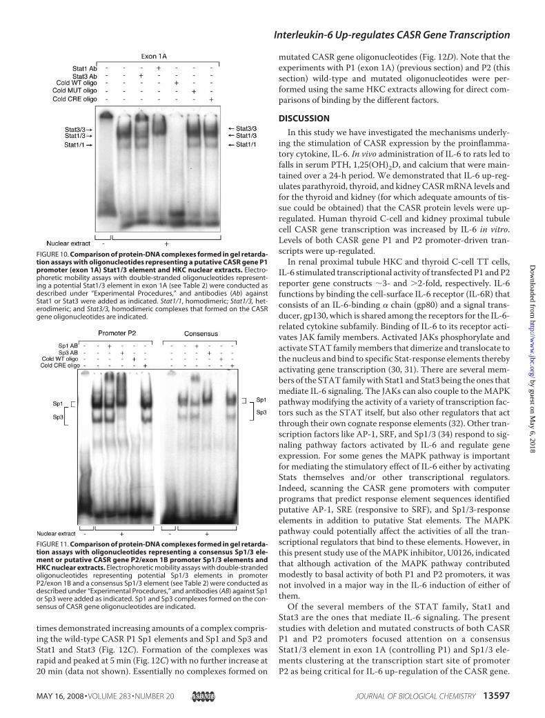
of the CASR Gene—EMSAs were conducted with oligonucleo-tides representing potential Stat1 elements in CASR P1 andexon 1A using HKC nuclear extract. Two predominant pro-tein-DNA complexes formed with the CASR gene exon 1A ele-ment and the pattern inwhich theywere shifted by Stat1 and/or
Stat3 antibodies were consistentwith them being predominantlyStat3/3 homodimers and Stat1/3heterodimers with less Stat1/1homodimer being present (Fig. 10).The addition of unlabeled oligonu-cleotides representing the wild-typesequence reduced the intensity ofthe exon 1A element-protein com-plexes, whereas addition of an unla-beled mutant oligonucleotide waswithout effect (Fig. 10). Complexesdid not form on the putative Stat1element in CASR gene P1 (data notshown).Protein-DNA Complexes Form on
Sp1 Elements at the TranscriptionInitiation Site of CASR Gene P2Promoter—EMSAs were conductedwith oligonucleotides representingeither a consensus Sp1 element or acluster of putative Sp1 elementsspanning a region just upstream ofand just downstream of the tran-scription initiation site of the CASRgene P2 promoter and HKC nuclearextract. The protein-DNA com-plexes that formed with CASR geneSp1 elements and consensus Sp1element had similar electrophoretic
mobilities, and they were shifted in a similar fashion by theaddition of antibodies against either Sp1 or Sp3 (Fig. 11). Theaddition of unlabeled oligonucleotides representing the wild-type CASR P2 sequence reduced the intensity of the labeledconsensus and CASR gene Sp1 element-protein complexes in asimilar manner, whereas addition of an unlabeled mutant oli-gonucleotide (data not shown) or an oligonucleotide represent-ing a cyclic AMP-binding protein response-element had noeffect (Fig. 11). Complexes did not form on a putative Sp1 ele-ment further upstream of the transcription initiation site of theCASR gene P2 promoter (data not shown).IL-6-dependent Formation of a Stat Complex on the CASR P1
Stat1/3 Element—To test whether endogenous Stat1 or Stat3binds to the P1 (exon 1A) Stat1/3 element (�1293�99), oli-gonucleotide precipitation (DNA pulldown) assays were per-formed. Extracts of HKC cells treated with IL-6 for varyingtimes demonstrated increasing amounts of a complex compris-ing the CASR P1 Stat1/3 element and Stat1 or Stat3 (Fig. 12A).Formation of the complexeswas rapid and peaked at 5min (Fig.12A) with no further increase at 20 min (data not shown). Nei-ther Sp1 nor Sp3 was present in the complex (Fig. 12A). Virtu-ally no complexes formed onmutated CASR gene oligonucleo-tides (Fig. 12B).IL-6-dependent Formation of an Sp-Stat Complex on the
CASR P2 Sp1 Elements—To test whether endogenous Stat1or Stat3 (with Sp1 or Sp3) is bound to the P2 Sp1/3 element(�19 3 �34), oligonucleotide precipitation assays were per-formed. Extracts of HKC cells treated with IL-6 for varying
FIGURE 8. STATS are critical for IL-6 up-regulation of the CASR P1 promoter. CASR P1 promoter-reporterconstructs, either wild-type [P1(�1438)] or with a mutated Stat exon 1A element [P1(�1438)StatMut], weretransfected into HKC cells either alone (�) or with (�) either a dominant-negative Stat1 construct (Stat1DN) ora dominant negative Stat3 construct (Stat3DN), and the cells were stimulated (�) or not (�) with IL-6 asdescribed under “Experimental Procedures.” ***, p � 0.001 luciferase activity relative to IL-6 unstimulatedcontrol.
Interleukin-6 Up-regulates CASR Gene Transcription
MAY 16, 2008 • VOLUME 283 • NUMBER 20 JOURNAL OF BIOLOGICAL CHEMISTRY 13595
by guest on May 6, 2018
http://ww
w.jbc.org/
Dow
nloaded from
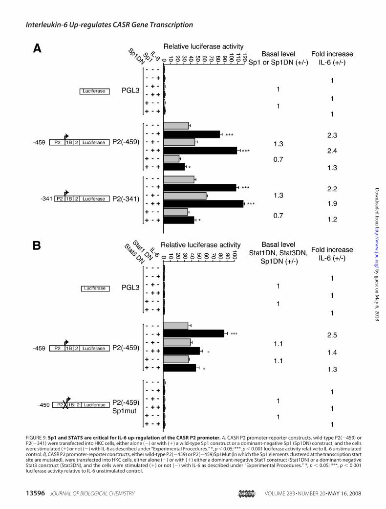
FIGURE 9. Sp1 and STATS are critical for IL-6 up-regulation of the CASR P2 promoter. A, CASR P2 promoter-reporter constructs, wild-type P2(�459) orP2(�341) were transfected into HKC cells, either alone (�) or with (�) a wild-type Sp1 construct or a dominant-negative Sp1 (Sp1DN) construct, and the cellswere stimulated (�) or not (�) with IL-6 as described under “Experimental Procedures.” *, p � 0.05; ***, p � 0.001 luciferase activity relative to IL-6 unstimulatedcontrol. B, CASR P2 promoter-reporter constructs, either wild-type P2(�459) or P2(�459)Sp1Mut (in which the Sp1 elements clustered at the transcription startsite are mutated), were transfected into HKC cells, either alone (�) or with (�) either a dominant-negative Stat1 construct (Stat1DN) or a dominant-negativeStat3 construct (Stat3DN), and the cells were stimulated (�) or not (�) with IL-6 as described under “Experimental Procedures.” *, p � 0.05; ***, p � 0.001luciferase activity relative to IL-6 unstimulated control.
Interleukin-6 Up-regulates CASR Gene Transcription
13596 JOURNAL OF BIOLOGICAL CHEMISTRY VOLUME 283 • NUMBER 20 • MAY 16, 2008
by guest on May 6, 2018
http://ww
w.jbc.org/
Dow
nloaded from
times demonstrated increasing amounts of a complex compris-ing the wild-type CASR P1 Sp1 elements and Sp1 and Sp3 andStat1 and Stat3 (Fig. 12C). Formation of the complexes wasrapid and peaked at 5min (Fig. 12C) with no further increase at20 min (data not shown). Essentially no complexes formed on
mutated CASR gene oligonucleotides (Fig. 12D). Note that theexperiments with P1 (exon 1A) (previous section) and P2 (thissection) wild-type and mutated oligonucleotides were per-formed using the same HKC extracts allowing for direct com-parisons of binding by the different factors.
DISCUSSION
In this study we have investigated the mechanisms underly-ing the stimulation of CASR expression by the proinflamma-tory cytokine, IL-6. In vivo administration of IL-6 to rats led tofalls in serum PTH, 1,25(OH)2D, and calcium that were main-tained over a 24-h period. We demonstrated that IL-6 up-reg-ulates parathyroid, thyroid, and kidney CASRmRNA levels andfor the thyroid and kidney (for which adequate amounts of tis-sue could be obtained) that the CASR protein levels were up-regulated. Human thyroid C-cell and kidney proximal tubulecell CASR gene transcription was increased by IL-6 in vitro.Levels of both CASR gene P1 and P2 promoter-driven tran-scripts were up-regulated.In renal proximal tubule HKC and thyroid C-cell TT cells,
IL-6 stimulated transcriptional activity of transfected P1 and P2reporter gene constructs �3- and 2-fold, respectively. IL-6functions by binding the cell-surface IL-6 receptor (IL-6R) thatconsists of an IL-6-binding � chain (gp80) and a signal trans-ducer, gp130, which is shared among the receptors for the IL-6-related cytokine subfamily. Binding of IL-6 to its receptor acti-vates JAK family members. Activated JAKs phosphorylate andactivate STAT familymembers that dimerize and translocate tothe nucleus and bind to specific Stat-response elements therebyactivating gene transcription (30, 31). There are several mem-bers of the STAT familywith Stat1 and Stat3 being the ones thatmediate IL-6 signaling. The JAKs can also couple to theMAPKpathway modifying the activity of a variety of transcription fac-tors such as the STAT itself, but also other regulators that actthrough their own cognate response elements (32). Other tran-scription factors like AP-1, SRF, and Sp1/3 (34) respond to sig-naling pathway factors activated by IL-6 and regulate geneexpression. For some genes the MAPK pathway is importantfor mediating the stimulatory effect of IL-6 either by activatingStats themselves and/or other transcriptional regulators.Indeed, scanning the CASR gene promoters with computerprograms that predict response element sequences identifiedputative AP-1, SRE (responsive to SRF), and Sp1/3-responseelements in addition to putative Stat elements. The MAPKpathway could potentially affect the activities of all the tran-scriptional regulators that bind to these elements. However, inthis present study use of theMAPK inhibitor, U0126, indicatedthat although activation of the MAPK pathway contributedmodestly to basal activity of both P1 and P2 promoters, it wasnot involved in a major way in the IL-6 induction of either ofthem.Of the several members of the STAT family, Stat1 and
Stat3 are the ones that mediate IL-6 signaling. The presentstudies with deletion and mutated constructs of both CASRP1 and P2 promoters focused attention on a consensusStat1/3 element in exon 1A (controlling P1) and Sp1/3 ele-ments clustering at the transcription start site of promoterP2 as being critical for IL-6 up-regulation of the CASR gene.
FIGURE 10. Comparison of protein-DNA complexes formed in gel retarda-tion assays with oligonucleotides representing a putative CASR gene P1promoter (exon 1A) Stat1/3 element and HKC nuclear extracts. Electro-phoretic mobility assays with double-stranded oligonucleotides represent-ing a potential Stat1/3 element in exon 1A (see Table 2) were conducted asdescribed under “Experimental Procedures,” and antibodies (Ab) againstStat1 or Stat3 were added as indicated. Stat1/1, homodimeric; Stat1/3, het-erodimeric; and Stat3/3, homodimeric complexes that formed on the CASRgene oligonucleotides are indicated.
FIGURE 11. Comparison of protein-DNA complexes formed in gel retarda-tion assays with oligonucleotides representing a consensus Sp1/3 ele-ment or putative CASR gene P2/exon 1B promoter Sp1/3 elements andHKC nuclear extracts. Electrophoretic mobility assays with double-strandedoligonucleotides representing potential Sp1/3 elements in promoterP2/exon 1B and a consensus Sp1/3 element (see Table 2) were conducted asdescribed under “Experimental Procedures,” and antibodies (AB) against Sp1or Sp3 were added as indicated. Sp1 and Sp3 complexes formed on the con-sensus of CASR gene oligonucleotides are indicated.
Interleukin-6 Up-regulates CASR Gene Transcription
MAY 16, 2008 • VOLUME 283 • NUMBER 20 JOURNAL OF BIOLOGICAL CHEMISTRY 13597
by guest on May 6, 2018
http://ww
w.jbc.org/
Dow
nloaded from
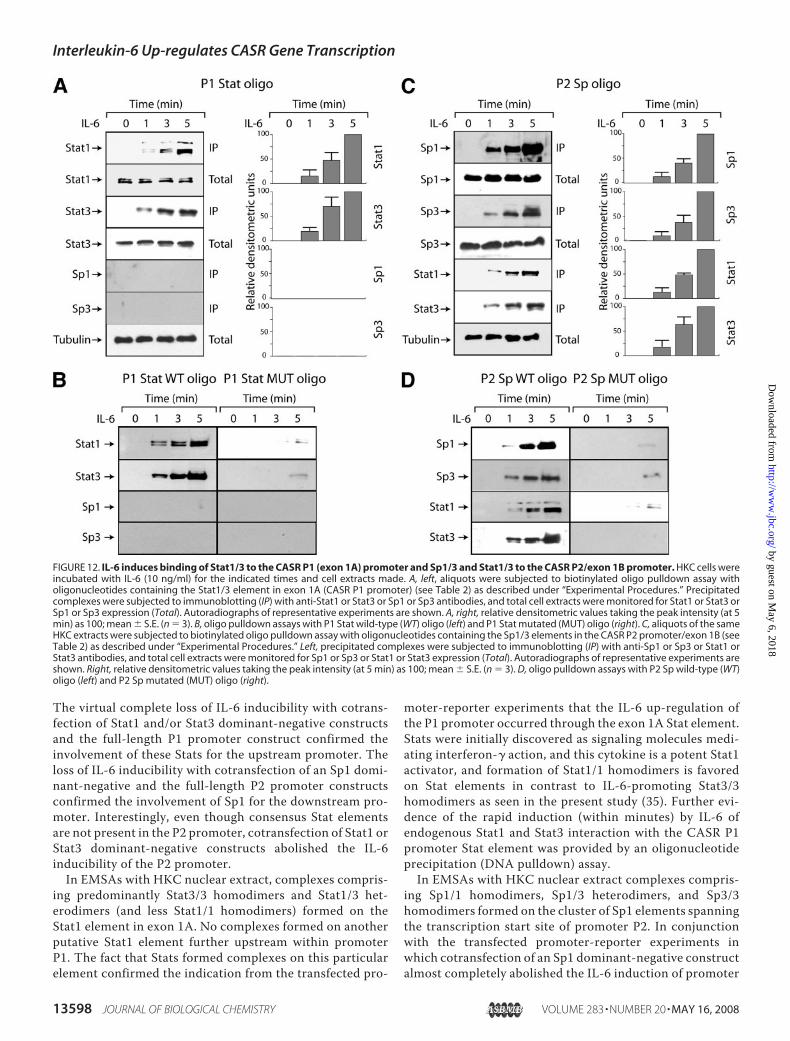
The virtual complete loss of IL-6 inducibility with cotrans-fection of Stat1 and/or Stat3 dominant-negative constructsand the full-length P1 promoter construct confirmed theinvolvement of these Stats for the upstream promoter. Theloss of IL-6 inducibility with cotransfection of an Sp1 domi-nant-negative and the full-length P2 promoter constructsconfirmed the involvement of Sp1 for the downstream pro-moter. Interestingly, even though consensus Stat elementsare not present in the P2 promoter, cotransfection of Stat1 orStat3 dominant-negative constructs abolished the IL-6inducibility of the P2 promoter.In EMSAs with HKC nuclear extract, complexes compris-
ing predominantly Stat3/3 homodimers and Stat1/3 het-erodimers (and less Stat1/1 homodimers) formed on theStat1 element in exon 1A. No complexes formed on anotherputative Stat1 element further upstream within promoterP1. The fact that Stats formed complexes on this particularelement confirmed the indication from the transfected pro-
moter-reporter experiments that the IL-6 up-regulation ofthe P1 promoter occurred through the exon 1A Stat element.Stats were initially discovered as signaling molecules medi-ating interferon-� action, and this cytokine is a potent Stat1activator, and formation of Stat1/1 homodimers is favoredon Stat elements in contrast to IL-6-promoting Stat3/3homodimers as seen in the present study (35). Further evi-dence of the rapid induction (within minutes) by IL-6 ofendogenous Stat1 and Stat3 interaction with the CASR P1promoter Stat element was provided by an oligonucleotideprecipitation (DNA pulldown) assay.In EMSAs with HKC nuclear extract complexes compris-
ing Sp1/1 homodimers, Sp1/3 heterodimers, and Sp3/3homodimers formed on the cluster of Sp1 elements spanningthe transcription start site of promoter P2. In conjunctionwith the transfected promoter-reporter experiments inwhich cotransfection of an Sp1 dominant-negative constructalmost completely abolished the IL-6 induction of promoter
FIGURE 12. IL-6 induces binding of Stat1/3 to the CASR P1 (exon 1A) promoter and Sp1/3 and Stat1/3 to the CASR P2/exon 1B promoter. HKC cells wereincubated with IL-6 (10 ng/ml) for the indicated times and cell extracts made. A, left, aliquots were subjected to biotinylated oligo pulldown assay witholigonucleotides containing the Stat1/3 element in exon 1A (CASR P1 promoter) (see Table 2) as described under “Experimental Procedures.” Precipitatedcomplexes were subjected to immunoblotting (IP) with anti-Stat1 or Stat3 or Sp1 or Sp3 antibodies, and total cell extracts were monitored for Stat1 or Stat3 orSp1 or Sp3 expression (Total). Autoradiographs of representative experiments are shown. A, right, relative densitometric values taking the peak intensity (at 5min) as 100; mean � S.E. (n � 3). B, oligo pulldown assays with P1 Stat wild-type (WT) oligo (left) and P1 Stat mutated (MUT) oligo (right). C, aliquots of the sameHKC extracts were subjected to biotinylated oligo pulldown assay with oligonucleotides containing the Sp1/3 elements in the CASR P2 promoter/exon 1B (seeTable 2) as described under “Experimental Procedures.” Left, precipitated complexes were subjected to immunoblotting (IP) with anti-Sp1 or Sp3 or Stat1 orStat3 antibodies, and total cell extracts were monitored for Sp1 or Sp3 or Stat1 or Stat3 expression (Total). Autoradiographs of representative experiments areshown. Right, relative densitometric values taking the peak intensity (at 5 min) as 100; mean � S.E. (n � 3). D, oligo pulldown assays with P2 Sp wild-type (WT)oligo (left) and P2 Sp mutated (MUT) oligo (right).
Interleukin-6 Up-regulates CASR Gene Transcription
13598 JOURNAL OF BIOLOGICAL CHEMISTRY VOLUME 283 • NUMBER 20 • MAY 16, 2008
by guest on May 6, 2018
http://ww
w.jbc.org/
Dow
nloaded from
P2, this firmly established the importance of the Sp1 clusterin mediating the up-regulation by the cytokine. In addition,the Sp1 elements are key for the basal activity of thepromoter.For some genes that contain both Stat and Sp1 elements in
their promoters, it appears that the effectiveness of the cyto-kine-mediated transcriptional activation is due to synergybetween the particular Stat and Sp1 acting at their cognateresponse elements. For example, this occurs with Stat1 andSp1 for interferon-� stimulation of the intercellular adhe-sion molecule-1 gene (36) and with Stat3 and Sp1 for theCCAAT/enhancer binding protein � gene (37). However,this is not the case with the CASR P2 promoter as it has noconsensus Stat elements. However, the virtual abolition ofthe IL-6 induction of P2 promoter reporter constructs byStat1 or Stat3 dominant-negative constructs established akey role for the Stats in the up-regulation of this promoter byIL-6. Again, use of the DNA pulldown assay was key in estab-lishing the interaction of not only endogenous Sp1 and Sp3but also Stat1 and Stat3 in a complex at the Sp1 elementcluster in the CASR P2 promoter. A similar mechanism hasbeen proposed for cytokine induction of other gene promot-ers that lack Stat elements but have Sp1 elements. For exam-ple, IL-6 stimulates transcriptional activation of the vascularendothelial growth factor gene via direct interactionbetween Stat3 and Sp1 (38), and Stat3 interaction with Sp1mediates the up-regulation by leptin of the tissue inhibitor ofmetalloproteinase 1 gene (39).In summary, our studies provide further insight into how
the altered CASR expression that affects the endocrine con-trol of blood calcium homeostasis may be achieved in meta-bolic alterations occurring in critically ill patients (and inother pathophysiological situations) where circulatingproinflammatory cytokine levels are increased. In these sit-uations, inflammation promotes local blood coagulation thatalthough beneficial carries the risk of increased systemiccoagulation. The mechanisms that we are uncovering mayunderlie a critical counter-regulatory system that minimizesthe deleterious effects of calcium and cytokines in promot-ing intravascular coagulation and atherosclerosis during theinflammatory response.Potentially, interventions that lessen the relative fall in serum
calcium may be helpful as in critically ill patients a greaterdegree of hypocalcemia is associated with a worse prognosis.The present study provides the framework to explore whetherthe administration of calcilytics, small molecules that targetand antagonize the CASR, would be beneficial in critically ill,hypocalcemic patients.
Acknowledgments—We thank Drs. Hugh P. J. Bennett and BernardTurcotte for critical review of the manuscript, Dr. Karin Schorr andYaroslava Chtompei for technical assistance, Dr. Martin Hewison(Cedars-Sinai Medical Center, Los Angeles) for the HKC-8 cells, andDrs. Cindy Goodyer (McGill University) and Hans Rotheneder (Uni-versity of Vienna, Austria) for the Sp1DN construct.
REFERENCES1. Pollak, M. R., Brown, E. M., Chou, Y.-W. H., Hebert, S. C., Marx, S. J.,
Steinmann, B., Levi, T., Seidman, C. E., and Seidman, J. G. (1993) Cell 75,1297–1303
2. Pollak, M. R., Brown, E. M., Estep, H. L., McLaine, P. N., Kifor, O., Park, J.,Hebert, S. C., Seidman, C. E., and Seidman, J. G. (1994) Nat. Genet. 8,303–307
3. Hendy, G. N., D’Souza-Li, L., Yang, B., Canaff, L., and Cole, D. E. C. (2000)Hum. Mutat. 16, 281–296
4. Zivin, J. R., Gooley, T., Zager, R. A., and Ryan, M. J. (2001) Am. J. KidneyDis. 37, 689–698
5. Lepage, R., Legare, G., Racicot, C., Brossard, J. H., Lapointe, R., Dagenais,M., and D’Amour, P. (1999) J. Clin. Endocrinol. Metab. 84, 2654–2658
6. Lind, L., Carlstedt, F., Rastad, J., Stiernstrom, H., Stridsberg, M., Ljungren,O., Wide, L., Larsson, A., Hellman, P., and Ljunghall, S. (2000) Crit. CareMed. 28, 93–99
7. Muller, B., Becker, K. L., Kranzlin, M., Schachinger, H., Huber, P. R.,Nylen, E. S., Snider, R. H., White, J. C., Schmidt-Gayk, H., Zimmerli, W.,and Ritz, R. (2000) Eur. J. Clin. Investig. 30, 823–831
8. Caldwell, F. T., Jr., Graves, D. B., and Wallace, B. H. (1997) J. Burn CareRehabil. 18, 525–530
9. Kowal-Vern, A., Walenga, J. M., Hoppensteadt, D., Sharp-Pucci, M., andGamelli, R. L. (1994) J. Am. Coll. Surg. 178, 357–362
10. Klein, G. L., Herndon, D. N., Goodman, W. G., Langman, C. B., Phillips,W. A., Dickson, I. R., Eastell, R., Naylor, K. E., Maloney, N. A., Desai, M.,Benjamin, D., and Alfrey, A. C. (1995) Bone 17, 455–460
11. Remick, D. G., Bolgos, G. R., Siddiqui, J., and Nemzek, J. A. (2002) Shock17, 463–467
12. Ohzato, H., Monden, M., Yoshizaki, K., Ogata, A., Nishimoto, N., Gotoh,M., Kishimoto, T., and Mori, T. (1993) Biochem. Biophys. Res. Commun.197, 1556–1562
13. Nijsten, M. W., Hack, C. E., Helle, M., ten Duis, H. J., Klasen, H. J., andAarden, L. A. (1991) Surgery 109, 761–767
14. Yamada, Y., Endo, S., and Inada, K. (1996) Burns 22, 587–59315. Schluter, B., Konig, B., Bergmann, U., Muller, F. E., and Konig, W. (1991)
J. Trauma 31, 1663–167016. Guo, Y., Dickerson, C., Chrest, F. J., Adler, W. H., Munster, A. M., and
Winchurch, R. A. (1990) Clin. Immunol. Immunopathol. 54, 361–37117. Carlstedt, E., Ridefelt, P., Lind, L., and Rastad, J. (1999) Biosci. Rep. 19,
35–4218. Murphey, E. D., Chattopadhyay, N., Bai, M., Kifor, O., Harper, D., Traber,
D. L., Hawkins, H. K., Brown, E.M., andKlein, G. L. (2000)Crit. CareMed.28, 3885–3890
19. Canaff, L., and Hendy, G. N. (2005) J. Biol. Chem. 280, 14177–1418620. Garrett, J. E., Capuano, I. V., Hammerland, L. G., Hung, B. C. P., Brown,
E. M., Hebert, S. C., Nemeth, E. F., and Fuller, F. (1995) J. Biol. Chem. 270,12919–12925
21. Chikatsu, N., Fukumoto, S., Takeuchi, Y., Suzawa, M., Obara, T., Mat-sumoto, T., and Fujita, T. (2000) J. Biol. Chem. 275, 7553–7557
22. Canaff, L., and Hendy, G. N. (2002) J. Biol. Chem. 277, 30337–3035023. Rotheneder, H., Geymayer, S., and Haldweger, E. (1999) J. Mol. Biol. 293,
1005–101524. Canaff, L., Petit, J.-L., Kiesel, M., Watson, P. H., Gascon-Barre, M., and
Hendy, G. N. (2001) J. Biol. Chem. 276, 4070–407925. D’Souza-Li, L., Canaff, L., Janicic, N., Cole, D. E. C., and Hendy, G. N.
(2001) Hum. Mutat. 18, 411–42126. Mouland, A. J., and Hendy, G. N. (1991) Endocrinology 128, 441–44927. D’Souza-Li, L., Yang, B., Canaff, L., Bai, M., Hanley, D. A., Bastepe, M.,
Salisbury, S. R., Brown, E. M., Cole, D. E. C., and Hendy, G. N. (2002)J. Clin. Endocrinol. Metab. 87, 1309–1318
28. Pidasheva, S., Grant,M., Canaff, L., Ercan,O., Kumar,U., andHendy,G.N.(2006) Hum. Mol. Genet. 15, 2200–2209
29. Ho, S. N., Hunt, H. D., Horton, R. M., Pullen, J. K., and Pease, L. R. (1999)Gene (Amst.) 77, 51–59
30. Horvath, C. M. (2000) Trends Biochem. Sci. 25, 496–50231. Levy, D. E., and Darnell, J. E., Jr. (2002) Nat. Rev. Mol. Cell Biol. 3,
651–662
Interleukin-6 Up-regulates CASR Gene Transcription
MAY 16, 2008 • VOLUME 283 • NUMBER 20 JOURNAL OF BIOLOGICAL CHEMISTRY 13599
by guest on May 6, 2018
http://ww
w.jbc.org/
Dow
nloaded from
32. Heinrich, P. C., Behrmann, I., Haan, S., Hermanns, H. M., Muller-Newen,G., and Schaper, F. (2003) Biochem. J. 374, 1–20
33. Cartharius, K., Frech, K., Grote, K., Klocke, B., Haltmeier,M., Klingenhoff,A., Frisch, M., Bayerlein, M., and Werner, T. (2005) Bioinformatics (Oxf.)21, 2933–2942
34. Suske, G. (1999) Gene (Amst.) 238, 291–30035. Haan, S., Keller, J. F., Behrmann, I., Heinrich, P. C., and Haan, C. (2005)
Cell. Signal. 17, 1542–1550
36. Look, D. C., Pelletier, M. R., Tidwell, R. M., Roswit, W. T., and Holtzman,M. J. (1995) J. Biol. Chem. 270, 30264–30267
37. Cantwell, C. A., Sterneck, E., and Johnson, P. F. (1998)Mol. Cell. Biol. 18,2108–2117
38. Loeffler, S., Fayard, B., Weis, J., andWeissenberger, J. (2005) Int. J. Cancer115, 202–213
39. Lin, S., Saxena, N. K., Ding, X., Stein, L. L., and Anania, F. A. (2006)Mol.Endocrinol. 20, 3376–3388
Interleukin-6 Up-regulates CASR Gene Transcription
13600 JOURNAL OF BIOLOGICAL CHEMISTRY VOLUME 283 • NUMBER 20 • MAY 16, 2008
by guest on May 6, 2018
http://ww
w.jbc.org/
Dow
nloaded from
Lucie Canaff, Xiang Zhou and Geoffrey N. HendyReceptor Gene Transcription via Stat1/3 and Sp1/3
The Proinflammatory Cytokine, Interleukin-6, Up-regulates Calcium-sensing
doi: 10.1074/jbc.M708087200 originally published online March 17, 20082008, 283:13586-13600.J. Biol. Chem.
10.1074/jbc.M708087200Access the most updated version of this article at doi:
Alerts:
When a correction for this article is posted•
When this article is cited•
to choose from all of JBC's e-mail alertsClick here
http://www.jbc.org/content/283/20/13586.full.html#ref-list-1
This article cites 39 references, 8 of which can be accessed free at
by guest on May 6, 2018
http://ww
w.jbc.org/
Dow
nloaded from