Cellular localization of NLRP3 infl ammasome · containing a CARD (ASC), which subsequently...
7
© Higher Education Press and Springer-Verlag Berlin Heidelberg 2013 June 2013 | Volume 4 | Issue 6 | 425 Protein Cell 2013, 4(6): 425–431 DOI 10.1007/s13238-013-2113-2 Protein Cell & Protein Cell & * These authors contributed equally to the work. Cellular localization of NLRP3 inflammasome OMMUNICATION C Yan Wang 1* , Chen Yang 2* , Kairui Mao 2 , Shuzhen Chen 2 , Guangxun Meng 1 , Bing Sun 1,2 1 Key Laboratory of Molecular Virology and Immunology, Institute Pasteur of Shanghai, Shanghai Institutes for Biological Sciences, Chinese Academy of Sciences, Shanghai 200025, China 2 State Key Laboratory of Cell Biology, Institute of Biochemistry and Cell Biology, Shanghai Institutes for Biological Sciences, Chinese Academy of Science, Shanghai 200031, China Correspondence: [email protected] (G. Meng), [email protected] (B. Sun) Received November 12, 2012 Accepted February 6, 2013 ABSTRACT Inflammasome is a large protein complex activated upon cellular stress or microbial infection, which triggers maturation of pro-inflammatory cytokines interleukin-1β and interleukin-18 through caspase-1 activation. Nod-like receptor family protein 3 (NLRP3) is the most character- ized inflammasome activated by various stimuli. However, the mechanism of its activation is unclear and its exact cellular localization is still unknown. We examined the potential co-localization of NLRP3 inflammasome with mi- tochondria and seven other organelles under adenosine triphosphate, nigericin or monosodium urate stimulation in mouse peritoneal macrophages using confocal micros- copy approach. Our results revealed that the activated endogenous apoptosis-associated speck-like protein containing a CARD (ASC) pyroptosome forms in the cyto- plasm and co-localizes with NLRP3 and caspase-1, but not with any of the organelles screened. This study indicates that the ASC pyroptosome universally localizes within the cytoplasm rather than with any specific organelles. KEYWORDS NLRP3, inflammasome, ASC pyroptosome, confocal microscopy, cytoplasmic INTRODUCTION Inflammasome is formed with the participation of certain pat- tern recognition receptors and senses various danger signals (Martinon et al., 2002; Agostini et al., 2004; Mariathasan et al., 2004; Poeck et al., 2010). As a large protein complex, in- flammasome controls the activation of the proteolytic enzyme caspase-1 through the apoptosis-associated speck-like protein containing a CARD (ASC), which subsequently regulates the maturation of the pro-inflammatory cytokines interleukin-1β (IL-1β) and IL-18 (Agostini et al., 2004). IL-1β and IL-18 are secreted into the extracellular space and function as versatile cytokines (Dinarello, 2009) that recruit more immune cells, educate lymphocytes, and eventually induce inflammatory ef- fects. The Nod-like receptor family protein 3 (NLRP3) is the most common and best studied inflammasome. NLRP3 is activated by a wide range of signals that cover both endogenous and pathogenic origins. Endogenous danger signals, such as adenosine triphosphate (ATP), amyloid-β fibris, and uric acid crystals, together with pathogens such as Listeria monocy- togenes, Canidida abicans, and influenza A virus, can activate the NLRP3 inflammasome (Martinon et al., 2006; Dostert et al., 2008; Eisenbarth et al., 2008; Allen et al., 2009; Gross et al., 2009; Thomas et al., 2009). However, the detailed mechanism of its activation is still unknown. Three distinct mechanisms have been reported, namely, reactive oxidative stress (ROS) (Schroder et al., 2010), lysosome damage (Hornung and Latz, 2010), and potassium leakage (Arlehamn et al., 2010). How- ever, none of them can explain all the observed phenomena, and the relationship between them need to be elucidated. In addition to caspase-1 activation and IL-1β secretion, NLRP3 inflammasome activation is also characterized by ASC pyroptosome formation, which is the aggregation of ASC as a pre-step to activate caspase-1 (Fernandes-Alnemri et al., 2007). Interestingly, only one pyroptosome is found to form per cell (Fernandes-Alnemri et al., 2007). Furthermore, to our knowledge, the ASC pyroptosome represents the assembled NLRP3 inflammasome. Several studies have investigated the localization of ASC in THP-1 human cell line and M1/M2 po- larized macrophages with different stimuli (Bryan et al., 2009; Pelegrin and Surprenant, 2009; Bryan et al., 2010). However, no exact localization of the pyroptosome in primary peritoneal macrophages has been reported. Detailed localization study of the pyroptosome may help us understand the activation
Transcript of Cellular localization of NLRP3 infl ammasome · containing a CARD (ASC), which subsequently...

© Higher Education Press and Springer-Verlag Berlin Heidelberg 2013 June 2013 | Volume 4 | Issue 6 | 425
Protein Cell 2013, 4(6): 425–431DOI 10.1007/s13238-013-2113-2 Protein Cell&
Prot
ein
C
ell
&
*These authors contributed equally to the work.
Cellular localization of NLRP3 infl ammasomeOMMUNICATIONC
Yan Wang1*, Chen Yang2*, Kairui Mao2, Shuzhen Chen2, Guangxun Meng1, Bing Sun1,2
1 Key Laboratory of Molecular Virology and Immunology, Institute Pasteur of Shanghai, Shanghai Institutes for Biological Sciences, Chinese Academy of Sciences, Shanghai 200025, China2 State Key Laboratory of Cell Biology, Institute of Biochemistry and Cell Biology, Shanghai Institutes for Biological Sciences, Chinese Academy of Science, Shanghai 200031, China Correspondence: [email protected] (G. Meng), [email protected] (B. Sun)Received November 12, 2012 Accepted February 6, 2013
ABSTRACT
Infl ammasome is a large protein complex activated upon cellular stress or microbial infection, which triggers maturation of pro-inflammatory cytokines interleukin-1β and interleukin-18 through caspase-1 activation. Nod-like receptor family protein 3 (NLRP3) is the most character-ized infl ammasome activated by various stimuli. However, the mechanism of its activation is unclear and its exact cellular localization is still unknown. We examined the potential co-localization of NLRP3 infl ammasome with mi-tochondria and seven other organelles under adenosine triphosphate, nigericin or monosodium urate stimulation in mouse peritoneal macrophages using confocal micros-copy approach. Our results revealed that the activated endogenous apoptosis-associated speck-like protein containing a CARD (ASC) pyroptosome forms in the cyto-plasm and co-localizes with NLRP3 and caspase-1, but not with any of the organelles screened. This study indicates that the ASC pyroptosome universally localizes within the cytoplasm rather than with any specifi c organelles.
KEYWORDS NLRP3, infl ammasome, ASC pyroptosome, confocal microscopy, cytoplasmic
INTRODUCTIONInfl ammasome is formed with the participation of certain pat-tern recognition receptors and senses various danger signals (Martinon et al., 2002; Agostini et al., 2004; Mariathasan et al., 2004; Poeck et al., 2010). As a large protein complex, in-fl ammasome controls the activation of the proteolytic enzyme caspase-1 through the apoptosis-associated speck-like protein containing a CARD (ASC), which subsequently regulates the maturation of the pro-inflammatory cytokines interleukin-1β
(IL-1β) and IL-18 (Agostini et al., 2004). IL-1β and IL-18 are secreted into the extracellular space and function as versatile cytokines (Dinarello, 2009) that recruit more immune cells, educate lymphocytes, and eventually induce infl ammatory ef-fects.
The Nod-like receptor family protein 3 (NLRP3) is the most common and best studied infl ammasome. NLRP3 is activated by a wide range of signals that cover both endogenous and pathogenic origins. Endogenous danger signals, such as adenosine triphosphate (ATP), amyloid-β fi bris, and uric acid crystals, together with pathogens such as Listeria monocy-togenes, Canidida abicans, and infl uenza A virus, can activate the NLRP3 infl ammasome (Martinon et al., 2006; Dostert et al., 2008; Eisenbarth et al., 2008; Allen et al., 2009; Gross et al., 2009; Thomas et al., 2009). However, the detailed mechanism of its activation is still unknown. Three distinct mechanisms have been reported, namely, reactive oxidative stress (ROS) (Schroder et al., 2010), lysosome damage (Hornung and Latz, 2010), and potassium leakage (Arlehamn et al., 2010). How-ever, none of them can explain all the observed phenomena, and the relationship between them need to be elucidated.
In addition to caspase-1 activation and IL-1β secretion, NLRP3 infl ammasome activation is also characterized by ASC pyroptosome formation, which is the aggregation of ASC as a pre-step to activate caspase-1 (Fernandes-Alnemri et al., 2007). Interestingly, only one pyroptosome is found to form per cell (Fernandes-Alnemri et al., 2007). Furthermore, to our knowledge, the ASC pyroptosome represents the assembled NLRP3 infl ammasome. Several studies have investigated the localization of ASC in THP-1 human cell line and M1/M2 po-larized macrophages with different stimuli (Bryan et al., 2009; Pelegrin and Surprenant, 2009; Bryan et al., 2010). However, no exact localization of the pyroptosome in primary peritoneal macrophages has been reported. Detailed localization study of the pyroptosome may help us understand the activation

Yan Wang et al.COMMUNICATION
426 | June 2013 | Volume 4 | Issue 6 © Higher Education Press and Springer-Verlag Berlin Heidelberg 2013
Prot
ein
C
ell
&
ASC pyroptosome was not localized to the mitochondria upon nigericin or MSU stimulation in mouse macrophages
Besides ATP, NLRP3 inflammasome is activated by various stimuli, including nigericin and MSU (Martinon et al., 2006;
process and provide evidence on the mechanism of NLRP3 infl ammasome activation. The objective of this study was to determine the potential localization of the NLRP3 infl amma-some under the activated state in peritoneal macrophages us-ing confocal microscopy approach. The immunofl uorescence results revealed the cytoplasmic localization of endogenous pyropotosome, which co-localized with NLRP3 and caspase-1 rather than to any detected organelles, indicating the organelle-free cytoplasmic localization of the NLRP3 infl ammasome in macrophages upon activation.
RESULTSASC pyroptosome was cytoplasmic but not localized to the mitochondria upon ATP stimulation in mouse macrophages
Mitochondria are the main sources of infl ammasome-activating ROS. NLRP3 inflammasome activation is largely impaired when the mitochondrial activity is inhibited (Nakahira et al., 2010; Zhou et al., 2011). Therefore, the mitochondria may be signal-integrating organelles, and are probably the organelles for NLRP3 infl ammasome activation. We stimulated low-dose lipopolysaccharide (LPS)-primed peritoneal macrophages with ATP, an NLRP3 infl ammasome activator, and stained the mitochondria with MitoTracker to investigate the hypothesis. The confocal results showed that the ASC dispersed over the nucleus and cytoplasm in the LPS-primed cells. The ASC ag-gregated across the whole cell and formed in the cytoplasm (green foci) upon further stimulation with ATP, but did not local-ize to the mitochondria (Fig. 1A). The ASC pyroptosome co-localized with NLRP3 and caspase-1, although most NLRP3 were not aggregated in the foci (Fig. 1B and 1C), consistent with previous studies. The activation state was also confi rmed by abundant IL-1β secretion from the same set of cells (Fig. 1D). These results suggested that the ASC pyroptosome is normally formed in ATP-activated macrophages together with NLRP3 infl ammasome activation, but localizes in the cytoplasm and not in the mitochondria.
ASC pyroptosome did not co-localize with other detected organelles upon ATP stimulation in mouse macrophages
Seven other organelles were detected upon ATP stimulation to determine the exact localization of ASC pyroptosome. The peritoneal macrophages were primed and stimulated as de-scribed in MATERIALS AND METHODS. GM-130, calnexin, α-tubulin, early endosome antigen 1 (EEA1), vimentin, and γ-tubulin were used as markers of Golgi apparatus, endoplas-mic reticulum, microtubule, endosome, phagosome, and cen-tromere, respectively (Webb et al., 2001; Latz et al., 2004; Eng et al., 2007; David et al., 2010; Wolff et al., 2011; Yuan et al., 2012). Lysotracker was applied for lysosome detection. The confocal results suggested that the ASC pyroptosome not co-localize with any of the organelles (Fig. 2A–G), but co-localizes with NLRP3 and caspase-1 (Fig. 2H and 2I). NLRP3 infl am-masome is normally activated (Fig. 2J).
Figure 1. ASC pyroptosome localized in the cytoplasm, but not in the mitochondria, upon ATP stimulation. (A) Immuno-fl uorescence microscopy of LPS-primed peritoneal macrophages stained with MitoTracker for 40 min and left unstimulated (upper panel) or stimulated with 5 mmol/L ATP for 30 min (lower panel). Scale bar, 10 μm. (B and C) Immunofl uorescence microscopy of LPS-primed peritoneal macrophages stained with MitoTracker and left unstimulated (upper panel) or stimulated with ATP (lower panel). NLRP3 (B) or caspase-1 (C), ASC, and DNA (with DAPI) were separately stained. Scale bar, 10 μm. (D) ELISA results of IL-1β production in peritoneal macrophages treated similar to A–C. Data represent at least three experiments (mean ± SD in D).
A
BMergeDAPIMitoASCNLRP3
ATP
Moc
k
MergeDAPIMitoASC
ATP
Moc
k
MergeDAPIMitoASCCaspase-1
ATP
Moc
k
C
D
ATPMock
IL-1
β (n
g/m
L)
100
80
60
40
20
0

Cellular localization of NLRP3 infl ammasome
© Higher Education Press and Springer-Verlag Berlin Heidelberg 2013 June 2013 | Volume 4 | Issue 6 | 427
COMMUNICATION
Prot
ein
C
ell
&
Figure 2. ASC pyroptosome co-localized with none of the detected organelles in ATP-simulated cells. (A–G) Immunofl uorescence microscopy of LPS-primed peritoneal macrophages left unstimulated (upper panel) or stimulated with 5 mmol/L ATP for 30 min (lower panel). Anti-GM-130, anti-calnexin, anti-α-tubulin, anti-EEA1, anti-vimentin, and anti-γ-tubulin were used for detection of corresponding organelles. LysoTracker was added 40 min before ATP stimulation. Cells were stained of ASC and DNA (with DAPI). Scale bar, 10 μm. (H and I) Immunofl uorescence microscopy of LPS-primed peritoneal macrophages stained with LysoTracker and left unstimulated (upper panel) or stimulated with ATP (lower panel). NLRP3 (H) or caspase-1 (I), ASC, and DNA (with DAPI) were separately stained. Scale bar, 10 μm. (J) ELISA results of IL-1β production in peritoneal macrophages treated similar to A–I. Data represent at least three experiments (mean ± SD in J).
GM130 DAPI MergeASC EEA1 DAPI MergeASC
Vimentin DAPI MergeASC
γ-tubulin DAPI MergeASC
L yso DAPI MergeASCNLRP3
DAPI MergeASC Calnexin
DAPI MergeASC Lyso
DAPI MergeASC α-tubulin
DAPI MergeASCCaspase-1 Lyso
ATP
Moc
kA
TPM
ock
ATP
Moc
kA
TPM
ock
ATP
Moc
kA
TPM
ock
ATP
Moc
k
ATP
Moc
k
ATP
Moc
k
A
B
C
D
E
F
G
H
I
J 100
80
60
40
20
0
Mock
GM130
Calnex
in
Lyso
track
er
α-tub
ulin
γ-tub
ulin
EEA1
Vimen
tin
ATP
IL-1
β (n
g/m
L)

Yan Wang et al.COMMUNICATION
428 | June 2013 | Volume 4 | Issue 6 © Higher Education Press and Springer-Verlag Berlin Heidelberg 2013
Prot
ein
C
ell
&
Hu et al., 2010). We used nigericin or MSU and detected ASC pyroptosome localization to investigate whether the ASC pyroptosome location varied upon different stimuli. The distri-bution of ASC was found to be similar as the ATP-stimulated macrophages. No co-localization of ASC pyroptosome with mitochondria was found upon nigericin or MSU stimulation (Fig. 3A), but IL-1β was normally secreted (Fig. 3D). Both NLRP3 and caspase-1 showed co-localization with ASC pyroptosome upon stimulation (Fig. 3B and 3C).
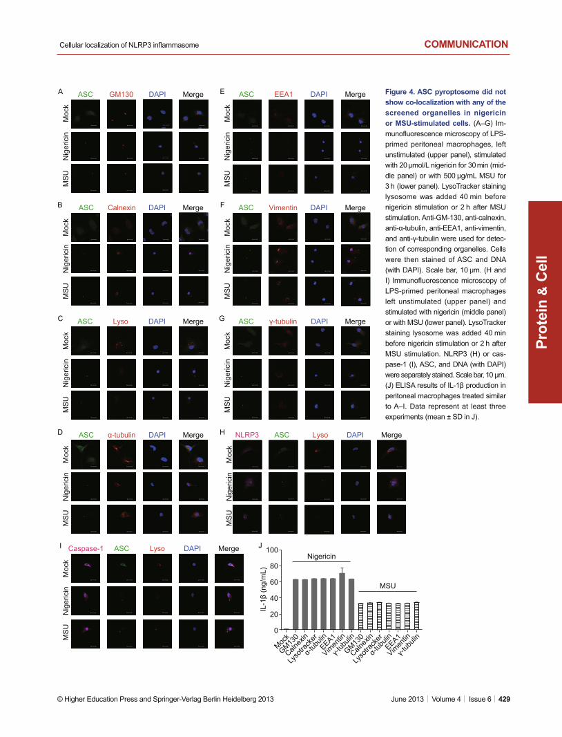
ASC pyroptosome showed no co-localization with other screened organelles upon nigericin or MSU stimulation
Seven organelles were also screened in nigericin or MSU stim-ulation. The confocal results suggested that none of them is the organelle for ASC pyroptosome localization (Fig. 4A–G). Enzyme-linked immunosorbent assay (ELISA) results of IL-1β proved the activation of NLRP3 infl ammasome (Fig. 4J). These results again confi rmed the co-localization of ASC pyroptosome with NLRP3 and caspase-1 (Fig. 4H and 4I).
DISCUSSIONThe NLRP3 inflammasome is critical for protection against pathogens and induction of adaptive immune responses (Eisenbarth et al., 2008; Ichinohe et al., 2009). Deregulated NLRP3 inflammasome activation is associated with multiple diseases such as gout, Crohn’s disease, atherosclerosis, and type II diabetes (Martinon et al., 2006; Duewell et al., 2010; Zaki et al., 2010; Wen et al., 2011). However, the mechanism of NLRP3 infl ammasome activation and its exact localization in the cell remain unclear. The determination of the locus of the activated infl ammasome will help identify the possible activa-tion process and the precise activation mechanism.
Mitochondria are potential organelles for NLRP3 inflam-masome activation because of their vital role in the process. Mitochondrial ROS production and mitochondria DNA (mtDNA) release are required for NLRP3 inflammasome activation. Meanwhile, mitochondrial dysfunctions lead to NLRP3 infl am-masome activation (Nakahira et al., 2010; Zhou et al., 2011). Therefore, the mitochondria were fi rst chosen for the localiza-tion exploration. ASC aggregation is believed to recruit pro-caspase-1 for its activation and is supposed to be the locus for NLRP3 inflammasome activation. Thus, the ASC focus was used as readout for the NLRP3 inflammasome localization study. Peritoneal macrophages were used as targeting cells because of their sensitivity to infl ammasome induction (Mari-athasan et al., 2004). We observed that the ASC dispersed across the cell, both in the nucleus and in the cytoplasm, in the unstimulated LPS-primed macrophages. All of the ASC were gathered in the cytoplasm and formed dots upon ATP stimula-tion, but were not localized to the mitochondria.
Several organelles are involved in the ASC activation pro-cess, such as the endoplasmic reticulum, which is believed to co-localize with exogenous NLRP3 (Zhou et al., 2011). Thus,
Figure 3. ASC pyroptosome did not localize in the mito-chondria upon nigericin or MSU stimulation. (A) Immunofl uo-rescence microscopy of LPS-primed peritoneal macrophages stained with MitoTracker for 40 min and left unstimulated (upper panel), stimulated with 20 μmol/L nigericin for 30 min (middle pan-el) or with 500 μg/mL MSU for 3 h (lower panel), followed by stain-ing for ASC and DNA (with DAPI). (B and C) Immunofl uorescence microscopy of LPS-primed peritoneal macrophages stained with MitoTracker, left unstimulated (upper panel), stimulated with nigericin (middle panel) or with MSU (lower panel), followed by staining for NLRP3 (B) or caspase-1 (C), ASC and DNA(with DAPI). Scale bar, 10 μm. (D) ELISA results of IL-1β production in peritoneal macrophages treated similar to A–C. Data represent at least three experiments (mean ± SD in D).
A
B
C
D
MergeDAPIMitoASCNLRP3
MergeDAPIMitoASC
Nig
eric
inM
ock
MS
UN
iger
icin
Moc
kM
SU
Nig
eric
inM
ock
MS
UMergeDAPIMitoASCCaspase-1
NigericinMock MSU
80
60
40
20
0
IL-1
β (n
g/m
L)
Cellular localization of NLRP3 infl ammasome
© Higher Education Press and Springer-Verlag Berlin Heidelberg 2013 June 2013 | Volume 4 | Issue 6 | 429
COMMUNICATION
Prot
ein
C
ell
&
Figure 4. ASC pyroptosome did not show co-localization with any of the screened organelles in nigericin or MSU-stimulated cells. (A–G) Im-munofl uorescence microscopy of LPS-primed peritoneal macrophages, left unstimulated (upper panel), stimulated with 20 μmol/L nigericin for 30 min (mid-dle panel) or with 500 μg/mL MSU for 3 h (lower panel). LysoTracker staining lysosome was added 40 min before nigericin stimulation or 2 h after MSU stimulation. Anti-GM-130, anti-calnexin, anti-α-tubulin, anti-EEA1, anti-vimentin, and anti-γ-tubulin were used for detec-tion of corresponding organelles. Cells were then stained of ASC and DNA (with DAPI). Scale bar, 10 μm. (H and I) Immunofl uorescence microscopy of LPS-primed peritoneal macrophages left unstimulated (upper panel) and stimulated with nigericin (middle panel) or with MSU (lower panel). LysoTracker staining lysosome was added 40 min before nigericin stimulation or 2 h after MSU stimulation. NLRP3 (H) or cas-pase-1 (I), ASC, and DNA (with DAPI) were separately stained. Scale bar, 10 μm. (J) ELISA results of IL-1β production in peritoneal macrophages treated similar to A–I. Data represent at least three experiments (mean ± SD in J).
Nig
eric
inM
ock
MS
U
Nig
eric
inM
ock
MS
U
Nig
eric
inM
ock
MS
U
Nig
eric
inM
ock
MS
U
Nig
eric
inM
ock
MS
UN
iger
icin
Moc
kM
SU
Nig
eric
inM
ock
MS
U
Nig
eric
inM
ock
MS
UN
iger
icin
Moc
kM
SU
GM130 DAPI MergeASC EEA1 DAPI MergeASC
Vimentin DAPI MergeASC
γ-tubulin DAPI MergeASC
L yso DAPI MergeASCNLRP3
DAPI MergeASC Calnexin
DAPI MergeASC Lyso
DAPI MergeASC α-tubulin
DAPI MergeASCCaspase-1 Lyso
A
B
C
D
E
F
G
H
I J
Mock
GM130
Calnex
in
Lyso
track
er
α-tub
ulin
γ-tub
ulin
EEA1
Vimen
tin
GM130
Calnex
in
Lyso
track
er
α-tub
ulin
γ-tub
ulin
EEA1
Vimen
tin
100
80
60
40
20
0
IL-1
β (n
g/m
L)
Nigericin
MSU

Yan Wang et al.COMMUNICATION
430 | June 2013 | Volume 4 | Issue 6 © Higher Education Press and Springer-Verlag Berlin Heidelberg 2013
Prot
ein
C
ell
&
ASC antibody for 1.5 h, and rinsed with PBS. Mitochondrial and lysosome detection was performed by staining
cells with MitoTracker (Molecular Probes) or LysoTracker (Molecular Probes) for 40 min before ATP or nigericin stimulation and incubation with FITC-conjugated anti-ASC antibody for 1.5 h. NLRP3 and cas-pase-1 detection was performed by incubating cells with anti-NLRP3 (Enzo Life Sciences) or anti-caspase-1 (Santa Cruz) antibodies for 2 h before incubation with anti-ASC antibody. Finally, all cells were stained with 4′,6-diamidino-2-phenylindole (DAPI). Confocal microscopic anal-yses were performed using Leica TCS SP2. Anti-vimentin and anti-γ-tubulin were provided by Xueliang Zhu’s laboratory.
ELISA
Mouse IL-1β in culture supernatants were measured using an ELISA kit (R&D Systems) according to the manufacturer’s protocol.
Statistical analysis
Data were presented as mean ± standard deviation of three independ-ent experiments. Statistical comparisons between different treatments were performed using an unpaired Student’s t-test. P < 0.01 was con-sidered signifi cant and P < 0.001 was highly signifi cant.
ACKNOWLEDGEMENTS
We thank Xueliang Zhu for providing the experimental materials from his laboratory. This work was supported by grants from the National Basic Research Program (973 Program) (No. 2013CB530504), the National Natural Science Foundation of China (Grant Nos. 31230024, 31030029, 31100662, 91029707 and 31170868), the Shanghai Natural Science Foundation (No. 11ZR1442600), the National Ministry of Sci-ence and Technology (No. 2007DFC31700), the National Science and Technology Major Project (Nos. 2008ZX10004-002, 2008ZX10002-014, 2009ZX10004-105, 2009ZX10004-016, 2011ZX10004-001 and 2012ZX10002007), the Shanghai Pasteur Health Research Founda-tion (SPHRF2008001 and SPHRF2009001), the Novo Nordisk-CAS Research Foundation, the SA-SIBS Discovery Innovation Grant, the Li Kha Shing Foundation, and the 100 Talent Program of the Chinese Academy of Sciences (to G.M.).
ABBREVIATIONS
ASC, apoptosis-associated speck-like protein containing a CARD; IL-1β, interleukin-1β; LPS, lipopolysaccharide; MSU, monosodium urate; NLRP3, Nod-like receptor family protein 3; ROS, reactive oxida-tive stress
REFERENCE S
Agostini, L., Martinon, F., Burns, K., McDermott, M.F., Hawkins, P.N., and Tschopp, J. (2004). NALP3 forms an IL-1beta-processing in-fl ammasome with increased activity in Muckle-Wells autoinfl amma-tory disorder. Immunity 20, 319–325 .
Allen, I.C., Scull, M.A., Moore, C.B., Holl, E.K., McElvania-TeKippe, E., Taxman, D.J., Guthrie, E.H., Pickles, R.J., and Ting, J.P. (2009). The NLRP3 inflammasome mediates in vivo innate immunity to infl uenza A virus through recognition of viral RNA. Immunity 30, 556–565 .
we tested seven other organelles including the Golgi appara-tus, endoplasmic reticulum, microtubule, endosome, phago-some, centromere, and lysosome. However, none of them exhibited co-localization with ASC pyroptosome upon ATP stimulation.
We used two other activators of NLRP3 infl ammasome, ni-gericin and MSU, to determine whether the cytoplasmic locali-zation of ASC pyroptosome was universal for different stimuli. Similar results were observed among three different stimula-tions, that is, the ASC pyroptosome was not co-localized with any of the tested organelles. Although NLRP3 showed limited aggregation into the ASC speck, the ASC pyroptosome was co-localized with caspase-1 and NLRP3 upon different stimula-tions, consistent with previous studies.
Our study aimed to explore the endogenous localization of activated ASC pyroptosome in primary peritoneal macrophag-es and provide evidence on its localization in the cytoplasm, but not to specifi c organelles. The results were similar among ATP, nigericin, and MSU stimulations, indicating the universal localization of ASC with different activators. Although localiza-tion with other organelles is also possible, our results eliminate the most probable organelles. Further study needs to be con-ducted to detect the dynamic activity of NLRP3 infl ammasome. The candidate proteins interacting with NLRP3 infl ammasome should also be tested to determine the activation mechanism and possible regulation process.
MATERIALS AND METHODS
Cells and stimulation
Peritoneal macrophages from C57BL/6 were prepared as follows. Briefl y, mice were intraperitoneally injected with 1 mL of 4% thioglycol-late (Sigma). Peritoneal exudates at the fourth day post-infection were isolated from the peritoneal cavity. Subsequently, cells were incubated at 37°C for 6 h with Dulbecco’s modifi ed eagle medium (DMEM) con-taining 10% fetal bovine serum (Gibco) and washed twice with DMEM. After additional overnight culture, the adherent cells were used as the peritoneal macrophages. The peritoneal macrophages were primed with 200 ng/mL LPS from Escherichia coli 0111:B4 (Sigma) for 5 h before stimulation with 5 mmol/L ATP (Sigma) for 30 min, 20 μmol/L nigericin (Sigma) for 30 min or 500 μg/mL MSU (Sigma) for 3 h.
Confocal microscopy
Peritoneal macrophages were plated overnight on coverslips and stimulated as described above. After stimulation, cells were washed with phosphate-buffered saline (PBS), fixed with 4% paraformalde-hyde in PBS for 15 min, permeabilized with Triton X-100 in PBS for 5 min, and blocked with 1% bovine serum albumin in PBS for 30 min. Subsequently, cells were incubated with antibodies for various or-ganelles, including anti-GM130 (Golgi apparatus, BD), anti-calnexin (endoplasmic reticulum, Sigma), anti-EEA1 (endosome, BD), anti-α-tubulin (microtubule, Sigma), anti-vimentin (phagosome), and anti-γ-tubulin (centromere). After incubation with antibodies for 2 h, cells were washed and incubated with Alexa 561 goat-anti-mouse antibody (BD) for 1 h, added with fl uorescein isothiocyanate (FITC)-conjugated anti-

Cellular localization of NLRP3 infl ammasome
© Higher Education Press and Springer-Verlag Berlin Heidelberg 2013 June 2013 | Volume 4 | Issue 6 | 431
COMMUNICATION
Prot
ein
C
ell
&
in the lysosome. Nat Immunol 5, 190–198 .Mariathasan, S., Newton, K., Monack, D.M., Vucic, D., French, D.M.,
Lee, W.P., Roose-Girma, M., Erickson, S., and Dixit, V.M. (2004). Differential activation of the infl ammasome by caspase-1 adaptors ASC and Ipaf. Nature 430, 213–218 .
Martinon, F., Burns, K., and Tschopp, J. (2002). The infl ammasome: a molecular platform triggering activation of infl ammatory caspases and processing of proIL-beta. Mol Cell 10, 417–426 .
Martinon, F., Petrilli, V., Mayor, A., Tardivel, A., and Tschopp, J. (2006). Gout-associated uric acid crystals activate the NALP3 infl amma-some. Nature 440, 237–241 .
Nakahira, K., Haspel, J.A., Rathinam, V.A.K., Lee, S.-J., Dolinay, T., Lam, H.C., Englert, J.A., Rabinovitch, M., Cernadas, M., Kim, H.P., et al. (2010). Autophagy proteins regulate innate immune respons-es by inhibiting the release of mitochondrial DNA mediated by the NALP3 infl ammasome. Nat Immunol 12, 222–230 .
Pelegrin, P., and Surprenant, A. (2009). Dynamics of macrophage po-larization reveal new mechanism to inhibit IL-1beta release through pyrophosphates. EMBO J 28, 2114–2127 .
Poeck, H., Bscheider, M., Gross, O., Finger, K., Roth, S., Rebsamen, M., Hannesschlager, N., Schlee, M., Rothenfusser, S., Barchet, W., et al. (2010). Recognition of RNA virus by RIG-I results in activation of CARD9 and infl ammasome signaling for interleukin 1 beta pro-duction. Nat Immunol 11, 63–69 .
Schroder, K., Zhou, R., and Tschopp, J. (2010). The NLRP3 infl amma-some: a sensor for metabolic danger? Science 327, 296–300 .
Thomas, P.G., Dash, P., Aldridge, J.R., Ellebedy, A.H., Reynolds, C., Funk, A.J., Martin, W.J., Lamkanfi, M., Webby, R.J., Boyd, K.L., et al. (2009). The intracellular sensor NLRP3 mediates key innate and healing responses to infl uenza A virus via the regulation of cas-pase-1. Immunity 30, 566–575 .
Webb, J.L., Harvey, M.W., Holden, D.W., and Evans, T.J. (2001). Mac-rophage nitric oxide synthase associates with cortical actin but is not recruited to phagosomes. Infect Immun 69, 6391–6400 .
Wen, H., Gris, D., Lei, Y., Jha, S., Zhang, L., Huang, M.T., Brickey, W.J., and Ting, J.P. (2011). Fatty acid-induced NLRP3-ASC infl amma-some activation interferes with insulin signaling. Nat Immunol 12, 408–415 .
Wolff, N.A., Lee, W.K., and Thevenod, F. (2011). Role of Arf1 in endo-somal traffi cking of protein-metal complexes and cadmium-metal-lothionein-1 toxicity in kidney proximal tubule cells. Toxicol Lett 203, 210–218 .
Yuan, R.T., Young, S., Liang, J., Schmid, M.C., Mielgo, A., and Stu-pack, D.G. (2012). Caspase-8 isoform 6 promotes death effector filament formation independent of microtubules. Apoptosis 17, 229–235 .
Zaki, M.H., Boyd, K.L., Vogel, P., Kastan, M.B., Lamkanfi , M., and Kan-neganti, T.D. (2010). The NLRP3 infl ammasome protects against loss of epithelial integrity and mortality during experimental colitis. Immunity 32, 379–391 .
Zhou, R., Yazdi, A.S., Menu, P., and Tschopp, J. (2011). A role for mitochondria in NLRP3 inflammasome activation. Nature 469, 221–225.
Arlehamn, C.S., Petrilli, V., Gross, O., Tschopp, J., and Evans, T.J. (2010). The role of potassium in infl ammasome activation by bacte-ria. J Biol Chem 285, 10508–10518 .
Bryan, N.B., Dorfl eutner, A., Kramer, S.J., Yun, C., Rojanasakul, Y., and Stehlik, C. (2010). Differential splicing of the apoptosis-asso-ciated speck like protein containing a caspase recruitment domain (ASC) regulates infl ammasomes. J Infl amm (Lond) 7, 23 .
Bryan, N.B., Dorfl eutner, A., Rojanasakul, Y., and Stehlik, C. (2009). Activation of infl ammasomes requires intracellular redistribution of the apoptotic speck-like protein containing a caspase recruitment domain. J Immunol 182, 3173–3182 .
David, E., Kaufman, J.L., Flowers, C.R., Schafer-Hales, K., Torre, C., Chen, J., Marcus, A.I., Sun, S.Y., Boise, L.H., and Lonial, S. (2010). Tipifarnib sensitizes cells to proteasome inhibition by block-ing degradation of bortezomib-induced aggresomes. Blood 116, 5285–5288 .
Dinarello, C.A. (2009). Immunological and infl ammatory functions of the interleukin-1 family. Annu Rev Immunol 27, 519–550 .
Dostert, C., Petrilli, V., Van Bruggen, R., Steele, C., Mossman, B.T., and Tschopp, J. (2008). Innate immune activation through Nalp3 in-fl ammasome sensing of asbestos and silica. Science 320, 674–677 .
Duewell, P., Kono, H., Rayner, K.J., Sirois, C.M., Vladimer, G., Bauern-feind, F.G., Abela, G.S., Franchi, L., Nunez, G., Schnurr, M., et al. (2010). NLRP3 infl ammasomes are required for atherogenesis and activated by cholesterol crystals. Nature 464, 1357–1361 .
Eisenbarth, S.C., Colegio, O.R., O’Connor, W., Sutterwala, F.S., and Flavell, R.A. (2008). Crucial role for the Nalp3 infl ammasome in the immunostimulatory properties of aluminium adjuvants. Nature 453, 1122–1126 .
Eng, E.W., Bettio, A., Ibrahim, J., and Harrison, R.E. (2007). MTOC reorientation occurs during FcgammaR-mediated phagocytosis in macrophages. Mol Biol Cell 18, 2389–2399 .
Fernandes-Alnemri, T., Wu, J., Yu, J.W., Datta, P., Miller, B., Jankows-ki, W., Rosenberg, S., Zhang, J., and Alnemri, E.S. (2007). The py-roptosome: a supramolecular assembly of ASC dimers mediating infl ammatory cell death via caspase-1 activation. Cell Death Differ 14, 1590–1604 .
Gross, O., Poeck, H., Bscheider, M., Dostert, C., Hannesschlager, N., Endres, S., Hartmann, G., Tardivel, A., Schweighoffer, E., Tybul-ewicz, V., et al. (2009). Syk kinase signalling couples to the Nlrp3 infl ammasome for anti-fungal host defence. Nature 459, 433–436 .
Hornung, V., and Latz, E. (2010). Critical functions of priming and lyso-somal damage for NLRP3 activation. Eur J Immunol 40, 620–623 .
Hu, Y., Mao, K., Zeng, Y., Chen, S., Tao, Z., Yang, C., Sun, S., Wu, X., Meng, G., and Sun, B. (2010). Tripartite-motif protein 30 negatively regulates NLRP3 infl ammasome activation by modulating reactive oxygen species production. J Immunol 185, 7699–7705 .
Ichinohe, T., Lee, H.K., Ogura, Y., Flavell, R., and Iwasaki, A. (2009). Infl ammasome recognition of infl uenza virus is essential for adap-tive immune responses. J Exp Med 206, 79–87 .
Latz, E., Schoenemeyer, A., Visintin, A., Fitzgerald, K.A., Monks, B.G., Knetter, C.F., Lien, E., Nilsen, N.J., Espevik, T., and Golenbock, D.T. (2004). TLR9 signals after translocating from the ER to CpG DNA


![Cell stress increases ATP release in NLRP3 …nyleneiodonium [DPI (13)] impair pro-IL-1β synthesis and NLRP3 priming (14, 15) in primary human monocytes, pro-IL-1β bio-synthesis](https://static.fdocuments.in/doc/165x107/5f2c218e05ddda39661ee653/cell-stress-increases-atp-release-in-nlrp3-nyleneiodonium-dpi-13-impair-pro-il-1.jpg)