The Werner Syndrome Helicase/Exonuclease...
15
The Werner Syndrome Helicase/Exonuclease Processes Mobile D-Loops through Branch Migration and Degradation Patricia L. Opresko 1 *, Gregory Sowd 1 , Hong Wang 2,3 1 Department of Environmental and Occupational Health, University of Pittsburgh Graduate School of Public Health, Pittsburgh, Pennsylvania, United States of America, 2 Department of Pharmacology and Chemical Biology, University of Pittsburgh School of Medicine, Pittsburgh, Pennsylvania, United States of America, 3 The University of Pittsburgh Cancer Institute, Hillman Cancer Center, Pittsburgh, Pennsylvania, United States of America Abstract RecQ DNA helicases are critical for preserving genome integrity. Of the five RecQ family members identified in humans, only the Werner syndrome protein (WRN) possesses exonuclease activity. Loss of WRN causes the progeroid disorder Werner syndrome which is marked by cancer predisposition. Cellular evidence indicates that WRN disrupts potentially deleterious intermediates in homologous recombination (HR) that arise in genomic and telomeric regions during DNA replication and repair. Precisely how the WRN biochemical activities process these structures is unknown, especially since the DNA unwinding activity is poorly processive. We generated biologically relevant mobile D-loops which mimic the initial DNA strand invasion step in HR to investigate whether WRN biochemical activities can disrupt this joint molecule. We show that WRN helicase alone can promote branch migration through an 84 base pair duplex region to completely displace the invading strand from the D-loop. However, substrate processing is altered in the presence of the WRN exonuclease activity which degrades the invading strand both prior to and after release from the D-loop. Furthermore, telomeric D-loops are more refractory to disruption by WRN, which has implications for tighter regulation of D-loop processing at telomeres. Finally, we show that WRN can recognize and initiate branch migration from both the 59 and 39 ends of the invading strand in the D-loops. These findings led us to propose a novel model for WRN D-loop disruption. Our biochemical results offer an explanation for the cellular studies that indicate both WRN activities function in processing HR intermediates. Citation: Opresko PL, Sowd G, Wang H (2009) The Werner Syndrome Helicase/Exonuclease Processes Mobile D-Loops through Branch Migration and Degradation. PLoS ONE 4(3): e4825. doi:10.1371/journal.pone.0004825 Editor: Mikhail V. Blagosklonny, Ordway Research Institute, United States of America Received December 9, 2008; Accepted January 16, 2009; Published March 13, 2009 Copyright: ß 2009 Opresko et al. This is an open-access article distributed under the terms of the Creative Commons Attribution License, which permits unrestricted use, distribution, and reproduction in any medium, provided the original author and source are credited. Funding: This work was funded by NIH grant ES0515052 (P.L.O.) and the Ellison Medical Foundation (P.L.O.). The funders had no role in study design, data collection and analysis, decision to publish, or preparation of the manuscript. Competing Interests: The authors have declared that no competing interests exist. * E-mail: [email protected] Introduction Werner syndrome (WS) is an autosomal recessive disorder marked by the premature onset of numerous features associated with aging and a predisposition to mesenchymal cancers [1]. WS is caused by loss of the DNA repair protein WRN which is a member of the RecQ family of DNA helicases [2]. E. coli and S. cerevisiase each have a single family member, whereas five members exist in humans: RECQ1, RECQ4, RECQ5, BLM and WRN [3,4]. Mutations in the human RecQ helicases BLM and RECQ4 also give rise to the cancer predisposition disorders Bloom syndrome (BS) and Rothmund-Thomson syndrome (RTS), respectively [5]. Despite the common feature of genomic instability, these disorders are clinically distinct from WS. While there are no known human disorders caused by defects in RECQ1 or RECQ5, cellular and transgenic mouse studies indicate that loss of these proteins also leads to genomic instability [5]. Thus, RecQ helicases have been classified as ‘‘caretakers’’ of the genome [3]. RecQ helicases have critical roles in regulating homologous recombination (HR) pathways. HR functions in repair of DNA double strand breaks, restoration of collapsed replication forks, and the alternative lengthening of telomeres (ALT) pathway [6–9]. Inappropriate HR can lead to loss of heterozygosity due to DNA strand crossovers, chromosome translocations, telomere loss, and tangled DNA intermediates that are potentially toxic if left unresolved [6]. In HR broken DNA ends are processed to generate single stranded tails that are coated by Rad51, which catalyzes invasion of the ssDNA tail into homologous duplex sequence and promotes base pairing between the ssDNA tail and one strand of the duplex [10]. The result is a joint molecule termed a displacement loop (D-loop). If the invading ssDNA terminates in a39OH it primes DNA polymerase extension and copying of the homologous sequence [8,11]. Dissociation of the D-loop can lead to re-annealing of the displaced strand to the ssDNA tail of the other broken end in the synthesis-dependent strand annealing (SDSA) pathway [12]. Alternatively, the tail of the other broken end may also be captured leading to the formation of a double Holliday junction (HJ), which is processed into strand crossover or non-crossover products [13]. RecQ helicases regulate HR by preventing inappropriate events through displacing Rad51 (BLM, RECQ1, RECQ5) [14–16] and by dissociating recombination intermediates including various HJ and D-loop constructs [16–20]. Unwinding of duplex DNA is catalyzed by RecQ helicases with 39 to 59 polarity and requires nucleotide hydrolysis. WRN is unique among the human RecQ helicases in that the protein also has 39 to 59 exonuclease activity [21], and both PLoS ONE | www.plosone.org 1 March 2009 | Volume 4 | Issue 3 | e4825
Transcript of The Werner Syndrome Helicase/Exonuclease...
The Werner Syndrome Helicase/Exonuclease ProcessesMobile D-Loops through Branch Migration andDegradationPatricia L. Opresko1*, Gregory Sowd1, Hong Wang2,3
1 Department of Environmental and Occupational Health, University of Pittsburgh Graduate School of Public Health, Pittsburgh, Pennsylvania, United States of America,
2 Department of Pharmacology and Chemical Biology, University of Pittsburgh School of Medicine, Pittsburgh, Pennsylvania, United States of America, 3 The University of
Pittsburgh Cancer Institute, Hillman Cancer Center, Pittsburgh, Pennsylvania, United States of America
Abstract
RecQ DNA helicases are critical for preserving genome integrity. Of the five RecQ family members identified in humans, onlythe Werner syndrome protein (WRN) possesses exonuclease activity. Loss of WRN causes the progeroid disorder Wernersyndrome which is marked by cancer predisposition. Cellular evidence indicates that WRN disrupts potentially deleteriousintermediates in homologous recombination (HR) that arise in genomic and telomeric regions during DNA replication andrepair. Precisely how the WRN biochemical activities process these structures is unknown, especially since the DNAunwinding activity is poorly processive. We generated biologically relevant mobile D-loops which mimic the initial DNAstrand invasion step in HR to investigate whether WRN biochemical activities can disrupt this joint molecule. We show thatWRN helicase alone can promote branch migration through an 84 base pair duplex region to completely displace theinvading strand from the D-loop. However, substrate processing is altered in the presence of the WRN exonuclease activitywhich degrades the invading strand both prior to and after release from the D-loop. Furthermore, telomeric D-loops aremore refractory to disruption by WRN, which has implications for tighter regulation of D-loop processing at telomeres.Finally, we show that WRN can recognize and initiate branch migration from both the 59 and 39 ends of the invading strandin the D-loops. These findings led us to propose a novel model for WRN D-loop disruption. Our biochemical results offer anexplanation for the cellular studies that indicate both WRN activities function in processing HR intermediates.
Citation: Opresko PL, Sowd G, Wang H (2009) The Werner Syndrome Helicase/Exonuclease Processes Mobile D-Loops through Branch Migration andDegradation. PLoS ONE 4(3): e4825. doi:10.1371/journal.pone.0004825
Editor: Mikhail V. Blagosklonny, Ordway Research Institute, United States of America
Received December 9, 2008; Accepted January 16, 2009; Published March 13, 2009
Copyright: � 2009 Opresko et al. This is an open-access article distributed under the terms of the Creative Commons Attribution License, which permitsunrestricted use, distribution, and reproduction in any medium, provided the original author and source are credited.
Funding: This work was funded by NIH grant ES0515052 (P.L.O.) and the Ellison Medical Foundation (P.L.O.). The funders had no role in study design, datacollection and analysis, decision to publish, or preparation of the manuscript.
Competing Interests: The authors have declared that no competing interests exist.
* E-mail: [email protected]
Introduction
Werner syndrome (WS) is an autosomal recessive disorder
marked by the premature onset of numerous features associated
with aging and a predisposition to mesenchymal cancers [1]. WS is
caused by loss of the DNA repair protein WRN which is a
member of the RecQ family of DNA helicases [2]. E. coli and S.
cerevisiase each have a single family member, whereas five members
exist in humans: RECQ1, RECQ4, RECQ5, BLM and WRN
[3,4]. Mutations in the human RecQ helicases BLM and RECQ4
also give rise to the cancer predisposition disorders Bloom
syndrome (BS) and Rothmund-Thomson syndrome (RTS),
respectively [5]. Despite the common feature of genomic
instability, these disorders are clinically distinct from WS. While
there are no known human disorders caused by defects in RECQ1
or RECQ5, cellular and transgenic mouse studies indicate that loss
of these proteins also leads to genomic instability [5]. Thus, RecQ
helicases have been classified as ‘‘caretakers’’ of the genome [3].
RecQ helicases have critical roles in regulating homologous
recombination (HR) pathways. HR functions in repair of DNA
double strand breaks, restoration of collapsed replication forks,
and the alternative lengthening of telomeres (ALT) pathway [6–9].
Inappropriate HR can lead to loss of heterozygosity due to DNA
strand crossovers, chromosome translocations, telomere loss, and
tangled DNA intermediates that are potentially toxic if left
unresolved [6]. In HR broken DNA ends are processed to
generate single stranded tails that are coated by Rad51, which
catalyzes invasion of the ssDNA tail into homologous duplex
sequence and promotes base pairing between the ssDNA tail and
one strand of the duplex [10]. The result is a joint molecule termed
a displacement loop (D-loop). If the invading ssDNA terminates in
a 39OH it primes DNA polymerase extension and copying of the
homologous sequence [8,11]. Dissociation of the D-loop can lead
to re-annealing of the displaced strand to the ssDNA tail of the
other broken end in the synthesis-dependent strand annealing
(SDSA) pathway [12]. Alternatively, the tail of the other broken
end may also be captured leading to the formation of a double
Holliday junction (HJ), which is processed into strand crossover or
non-crossover products [13]. RecQ helicases regulate HR by
preventing inappropriate events through displacing Rad51 (BLM,
RECQ1, RECQ5) [14–16] and by dissociating recombination
intermediates including various HJ and D-loop constructs [16–20].
Unwinding of duplex DNA is catalyzed by RecQ helicases with 39
to 59 polarity and requires nucleotide hydrolysis.
WRN is unique among the human RecQ helicases in that the
protein also has 39 to 59 exonuclease activity [21], and both
PLoS ONE | www.plosone.org 1 March 2009 | Volume 4 | Issue 3 | e4825
helicase and exonuclease activities are implicated in the dissoci-
ation of HR intermediates. WS cells exhibit defects in resolving
HR intermediates that form in response to stalled replication forks,
whether induced by blocking lesions or by depleted dNTP pools
[22,23]. The HR defect can be rescued by inhibiting Rad51 [24],
and thus the formation of recombination intermediates, but is not
rescued by expressing WRN mutants that lack either helicase or
exonuclease activities [25]. WRN deficient cells are also prone to
abnormal HR at telomeric ends, and exhibit increased telomere
loss, telomeric sister chromatid exchanges and spontaneous extra-
chromosomal telomeric circles [26–29]. Telomeres contain a 39
single strand (ssDNA) tail that is ,50 to 150 nt long in vivo [30]
and forms protective intra-telomeric D-loops that stabilize the
large t-loop [31]. Aberrant HR involving the natural D-loop/t-
loop or inappropriate strand invasion into telomeric sequences in
other chromatids or chromosomes can lead to telomere loss and
abnormal telomere structures [32,33]. RecQ helicases are also
implicated in the resolution step of the recombination-based ALT
pathway to lengthen telomeres [34,35].
WRN acts on many of the same substrates as other human
RecQ helicases, but has the potential to process these substrates
differently due to the exonuclease activity. Precisely how both
WRN biochemical activities contribute to processing HR
intermediates is not clear. We and others observed previously
that oligomeric non-mobile D-loops are substrates for both the
WRN helicase and exonuclease activities, but these constructs
could not test for branch migration activity [36,37]. WRN helicase
activity is poorly processive and is unable to completely unwind a
33-bp oligomeric telomeric D-loop in the absence of accessory
proteins POT1 or RPA, or assistance from the WRN exonuclease
activity [37,38]. Exonucleolytic degradation shortens the duplex
length to be unwound by digesting the invading strand in a 39 to 59
direction [37]. Here we extended our previous studies by
constructing more biologically relevant mobile D-loops. They
have a much longer invading strand that pairs with one strand in a
plasmid to form an 84 bp duplex, and can be branch migrated.
Based on evidence that WRN regulates recombination at
telomeres [39], we also constructed telomeric D-loops for analysis.
In this study we identified mobile plasmid-based D-loops as novel
substrates for both WRN helicase driven branch migration and
exonuclease activities, and found that telomeric D-loops were
disrupted less efficiently than non-telomeric D-loops. In contrast to
oligomeric constructs, we show that WRN helicase alone can
completely dissociate the 84 bp duplex to release the invading
strand from the plasmid D-loop. However, the WRN exonuclease
activity alters processing by degrading the long invading strand both
prior to and after release from the plasmid D-loop. WRN disrupts
plasmid D-loops with a protruding 39 or 59 single stranded tail, but a
protruding tail is not required for either branch migration or
exonuclease activities. Furthermore, we demonstrate that WRN
recognizes both the 59 and 39 ends of the D-loop duplex, which led
us to propose a novel model for WRN loading and processing of
biologically relevant D-loop recombination intermediates.
Results
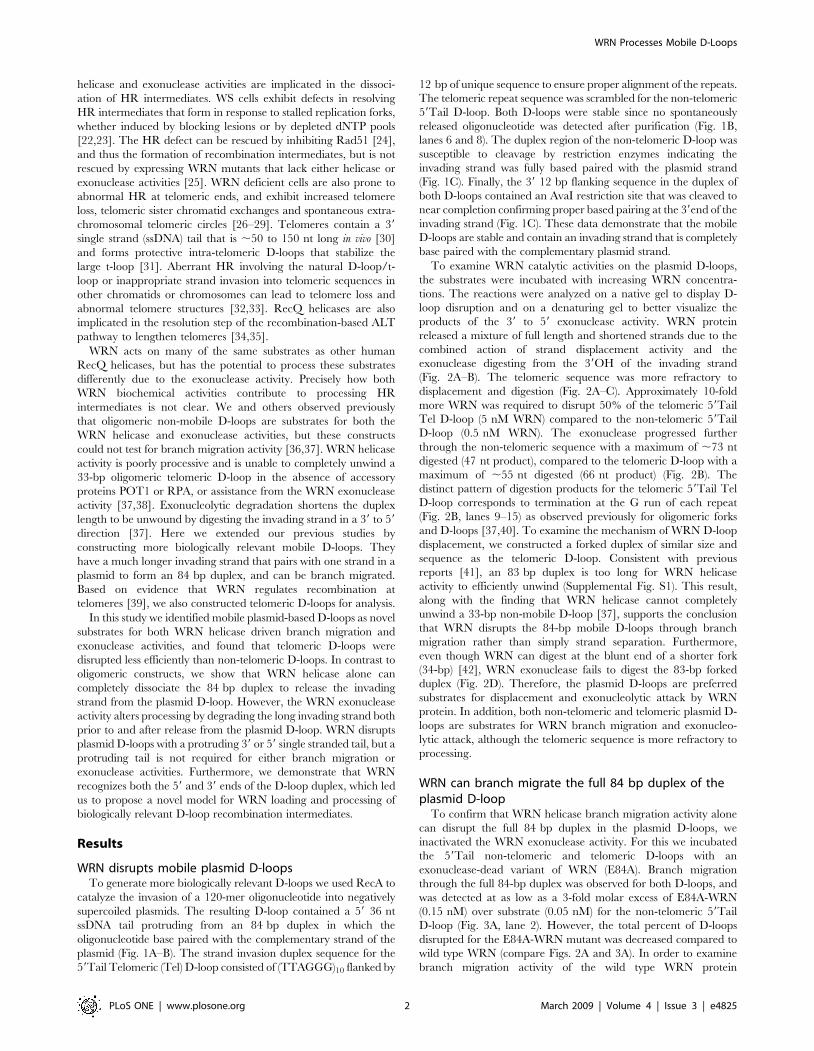
WRN disrupts mobile plasmid D-loopsTo generate more biologically relevant D-loops we used RecA to
catalyze the invasion of a 120-mer oligonucleotide into negatively
supercoiled plasmids. The resulting D-loop contained a 59 36 nt
ssDNA tail protruding from an 84 bp duplex in which the
oligonucleotide base paired with the complementary strand of the
plasmid (Fig. 1A–B). The strand invasion duplex sequence for the
59Tail Telomeric (Tel) D-loop consisted of (TTAGGG)10 flanked by
12 bp of unique sequence to ensure proper alignment of the repeats.
The telomeric repeat sequence was scrambled for the non-telomeric
59Tail D-loop. Both D-loops were stable since no spontaneously
released oligonucleotide was detected after purification (Fig. 1B,
lanes 6 and 8). The duplex region of the non-telomeric D-loop was
susceptible to cleavage by restriction enzymes indicating the
invading strand was fully based paired with the plasmid strand
(Fig. 1C). Finally, the 39 12 bp flanking sequence in the duplex of
both D-loops contained an AvaI restriction site that was cleaved to
near completion confirming proper based pairing at the 39end of the
invading strand (Fig. 1C). These data demonstrate that the mobile
D-loops are stable and contain an invading strand that is completely
base paired with the complementary plasmid strand.
To examine WRN catalytic activities on the plasmid D-loops,
the substrates were incubated with increasing WRN concentra-
tions. The reactions were analyzed on a native gel to display D-
loop disruption and on a denaturing gel to better visualize the
products of the 39 to 59 exonuclease activity. WRN protein
released a mixture of full length and shortened strands due to the
combined action of strand displacement activity and the
exonuclease digesting from the 39OH of the invading strand
(Fig. 2A–B). The telomeric sequence was more refractory to
displacement and digestion (Fig. 2A–C). Approximately 10-fold
more WRN was required to disrupt 50% of the telomeric 59Tail
Tel D-loop (5 nM WRN) compared to the non-telomeric 59Tail
D-loop (0.5 nM WRN). The exonuclease progressed further
through the non-telomeric sequence with a maximum of ,73 nt
digested (47 nt product), compared to the telomeric D-loop with a
maximum of ,55 nt digested (66 nt product) (Fig. 2B). The
distinct pattern of digestion products for the telomeric 59Tail Tel
D-loop corresponds to termination at the G run of each repeat
(Fig. 2B, lanes 9–15) as observed previously for oligomeric forks
and D-loops [37,40]. To examine the mechanism of WRN D-loop
displacement, we constructed a forked duplex of similar size and
sequence as the telomeric D-loop. Consistent with previous
reports [41], an 83 bp duplex is too long for WRN helicase
activity to efficiently unwind (Supplemental Fig. S1). This result,
along with the finding that WRN helicase cannot completely
unwind a 33-bp non-mobile D-loop [37], supports the conclusion
that WRN disrupts the 84-bp mobile D-loops through branch
migration rather than simply strand separation. Furthermore,
even though WRN can digest at the blunt end of a shorter fork
(34-bp) [42], WRN exonuclease fails to digest the 83-bp forked
duplex (Fig. 2D). Therefore, the plasmid D-loops are preferred
substrates for displacement and exonucleolytic attack by WRN
protein. In addition, both non-telomeric and telomeric plasmid D-
loops are substrates for WRN branch migration and exonucleo-
lytic attack, although the telomeric sequence is more refractory to
processing.
WRN can branch migrate the full 84 bp duplex of theplasmid D-loop
To confirm that WRN helicase branch migration activity alone
can disrupt the full 84 bp duplex in the plasmid D-loops, we
inactivated the WRN exonuclease activity. For this we incubated
the 59Tail non-telomeric and telomeric D-loops with an
exonuclease-dead variant of WRN (E84A). Branch migration
through the full 84-bp duplex was observed for both D-loops, and
was detected at as low as a 3-fold molar excess of E84A-WRN
(0.15 nM) over substrate (0.05 nM) for the non-telomeric 59Tail
D-loop (Fig. 3A, lane 2). However, the total percent of D-loops
disrupted for the E84A-WRN mutant was decreased compared to
wild type WRN (compare Figs. 2A and 3A). In order to examine
branch migration activity of the wild type WRN protein
WRN Processes Mobile D-Loops
PLoS ONE | www.plosone.org 2 March 2009 | Volume 4 | Issue 3 | e4825
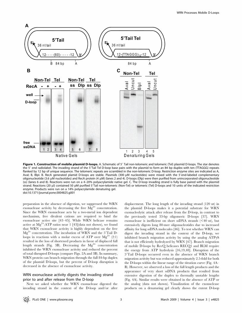
preparation in the absence of digestion, we suppressed the WRN
exonuclease activity by decreasing the free Mg2+ concentration.
Since the WRN exonuclease acts by a two-metal ion dependent
mechanism, free divalent cations are required to bind the
exonuclease active site [43–45]. While WRN helicase remains
active at Mg2+:ATP ratios near 1 [43](data not shown), we found
that WRN exonuclease activity is highly dependent on the free
Mg2+ concentration. The incubation of WRN and the 59Tail D-
loops in reactions with a molar excess of ATP over Mg2+ (2:1)
resulted in the loss of shortened products in favor of displaced full
length strands (Fig. 3B). Decreasing the Mg2+ concentration
inhibited the WRN exonuclease activity and reduced the percent
of total disrupted D-loops (compare Figs. 2A and 3B). In summary,
WRN protein can branch migration through the full 84-bp duplex
of the plasmid D-loops, but the percent of D-loop disruption is
decreased in the absence of exonuclease activity.
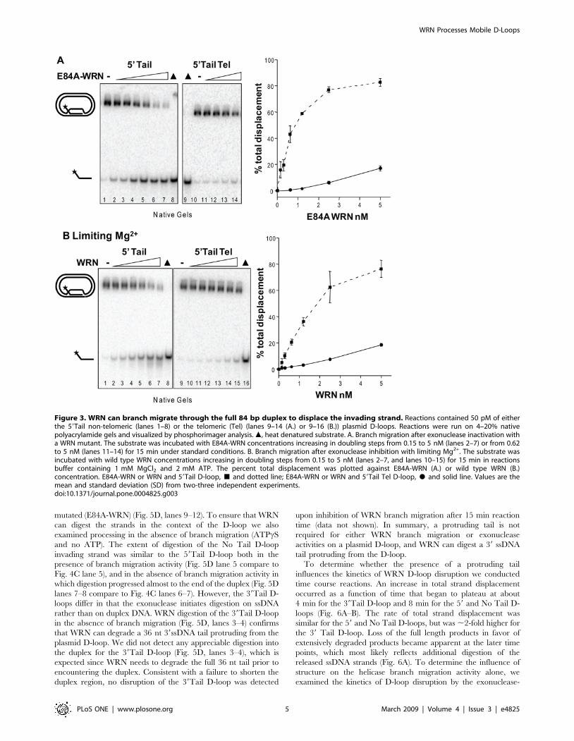
WRN exonuclease activity digests the invading strandprior to and after release from the D-loop
Next we asked whether the WRN exonuclease digested the
invading strand in the context of the D-loop and/or after
displacement. The long length of the invading strand (120 nt) in
the plasmid D-loops makes it a potential substrate for WRN
exonucleolytic attack after release from the D-loop, in contrast to
the previously tested 33-bp oligomeric D-loops [37]. WRN
exonuclease is inefficient on short ssDNA strands (,40 nt), but
extensively digests long 80-mer oligonucleotides due to increased
affinity for long ssDNA molecules [46]. To test whether WRN can
digest the invading strand in the context of the D-loop, we
inhibited branch migration activity by using the analog ATPcS
that is not efficiently hydrolyzed by WRN [47]. Branch migration
of mobile D-loops by RecQ helicases RECQ1 and BLM require
the energy from ATP hydrolysis [16,19,48]. Disruption of the
59Tail D-loops occurred even in the absence of WRN branch
migration activity but was reduced approximately 2.5-fold for both
the D-loops within the linear range of the titration curve (Fig. 4A–
B). However, we observed a loss of the full length products and the
appearance of very short ssDNA products that resulted from
extensive digestion of the duplex to thermally unstable lengths
(Fig. 4A). Similar results were obtained in the absence of ATP or
the analog (data not shown). Visualization of the exonuclease
products on a denaturing gel clearly shows the extent D-loop
Figure 1. Construction of mobile plasmid D-loops. A. Schematic of 59 Tail non-telomeric and telomeric (Tel) plasmid D-loops. The star denotesthe 59 end radiolabel. The invading strand of the 59Tail Tel D-loop base pairs with the plasmid to form an 84 bp duplex with ten (TTAGGG) repeatsflanked by 12 bp of unique sequence. The telomeric repeats are scrambled in the non-telomeric D-loop. Restriction enzyme sites are indicated as A,AvaI; B, BlpI. B. RecA generated plamid D-loops are stable. Plasmids (300 mM nucleotides) were mixed with the 59end-labeled complementaryoligonucleotide (3.6 mM nucleotides) and RecA protein (4 mM) (lanes 2 and 4). D-loops (Dlp) were then purified from unincorporated oligonucleotide(ss) (lanes 6 and 8). Reactions were run on a 4–20% polyacrylamide native gel. C. The D-loop invading strand is fully base paired with the plasmidstrand. Reactions (20 ml) contained 50 pM purified 59Tail non-telomeric (Non-Tel) or telomeric (Tel) D-loops and 10 units of the indicated restrictionenzyme. Products were run on a 14% polyacrylamide denaturing gel.doi:10.1371/journal.pone.0004825.g001
WRN Processes Mobile D-Loops
PLoS ONE | www.plosone.org 3 March 2009 | Volume 4 | Issue 3 | e4825

digestion progressed further when ATP hydrolysis is inhibited
(ATPcS and no ATP) (Fig. 4C, compare lanes 5 to 6 and 7, Fig. 4D
compare lanes 8 to 9) for some molecules. The exonuclease-dead
WRN mutant (E84A) yielded no detectable digestion products
after incubation with either D-loop (Fig. 4C lanes 1–3, Fig. 4D
lanes 10–12).
Next we asked if WRN can digest the long invading strands
after branch migration released them from the D-loop, by
incubating WRN with the 120 nt oligomers used to construct
the 59Tail D-loops (Table 1). WRN digestion of the long oligomers
was apparent (Figs. 4C lanes 11–14, and 4D lanes 1–4), dose
dependent and eliminated upon mutation of the WRN exonucle-
ase domain (Supplemental Fig. S2). The extent of WRN digestion
of the long ssDNA molecules was not increased in the absence of
ATP hydrolysis (Fig. 4C, lanes 12–14 and Fig. 4D, lanes 2–4), in
contrast to digestion of the 59Tail D-loops. In summary, WRN
exonucleolytic attack of the D-loop invading strand does not
require branch migration activity and occurs prior to and after
release from the D-loop.
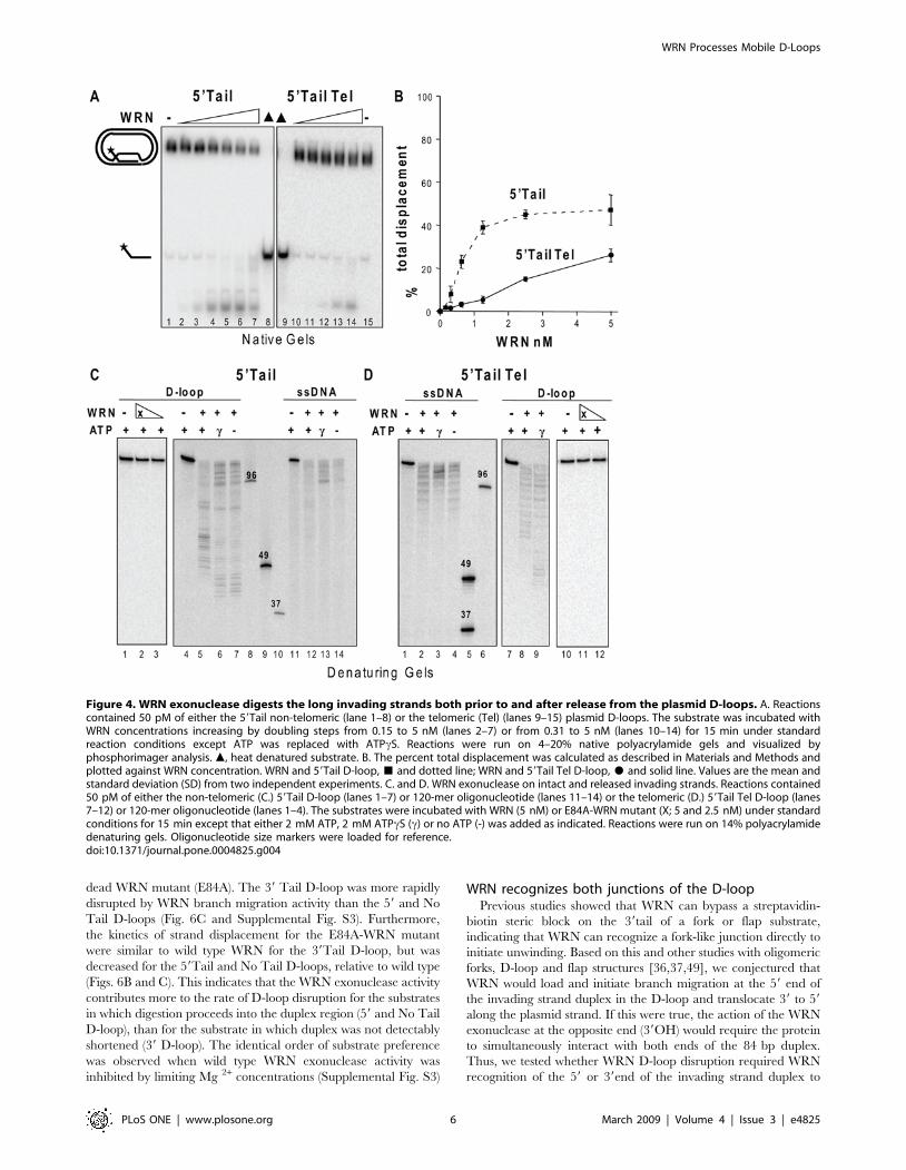
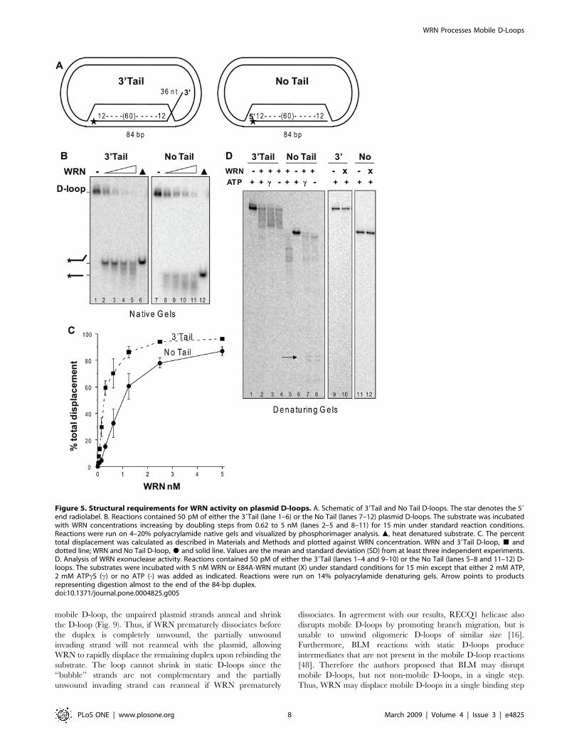
Structural requirements of WRN D-loop branch migrationand exonuclease activities
To determine the structural requirements for WRN catalytic
activities on the plasmid D-loops, we constructed D-loops with
either a protruding 39 ssDNA tail or no protruding tail (Fig. 5A).
WRN disrupted about 50% of the D-loops at ,0.25 nM and
1 nM protein for the 39Tail D-loop and No Tail D-loop,
respectively, and released a mix of full length and shortened
strands (Fig. 5A–C). Importantly, disruption of the 39Tail D-loop
(7.6%) was detected at near equal molar WRN (0.04 nM) and
substrate (0.05 nM) concentrations (Fig. 5C). No digestion of the
D-loops is detected when the WRN exonuclease domain is
Figure 2. WRN displaces and digests the invading strand of plasmid D-loops. A. Reactions contained 50 pM of either the 59Tail non-telomeric (lane 1–6) or the telomeric (Tel) (lanes 7–12) plasmid D-loops. The substrate was incubated with WRN concentrations increasing in doublingsteps from 0.62 to 5 nM (lanes 2–5 or 8–11, respectively) for 15 min under standard reaction conditions and run on a 4–20% native polyacrylamidegel. m, heat denatured substrate. B. The 59 Tail non-telomeric (lanes 1–8) or telomeric (lanes 9–15) were incubated with WRN concentrationsincreasing in doubling steps from 0.078 to 5 nM (lanes 2–8) and 0.15 to 5 nM (lanes 10–15) and run on a 14% denaturing polyacrylamide gel.Numbers represent oligonucleotide size markers. C. The percent total D-loop displacement from reactions in panel A were calculated as described inMaterials and Methods and plotted against WRN concentration. WRN and 59Tail plasmid D-loop, & and dotted line; WRN and 59Tail Tel plasmid D-loop, $ and solid line. Values represent the mean and standard deviation (SD) from at least three independent experiments. D. WRN exonuclease isinactive on an 83 bp forked duplex. Reactions contained 0.25 nM of the 83-bp forked duplex containing ten telomeric repeats (thick black line) with15 and 8 bp of flanking sequence. The substrate was incubated with WRN concentrations increasing in doubling steps from 0.19 to 25 nM for 15 minunder standard reaction conditions. Reactions were run on a 14% denaturing gel.doi:10.1371/journal.pone.0004825.g002
WRN Processes Mobile D-Loops
PLoS ONE | www.plosone.org 4 March 2009 | Volume 4 | Issue 3 | e4825
mutated (E84A-WRN) (Fig. 5D, lanes 9–12). To ensure that WRN
can digest the strands in the context of the D-loop we also
examined processing in the absence of branch migration (ATPcS
and no ATP). The extent of digestion of the No Tail D-loop
invading strand was similar to the 59Tail D-loop both in the
presence of branch migration activity (Fig. 5D lane 5 compare to
Fig. 4C lane 5), and in the absence of branch migration activity in
which digestion progressed almost to the end of the duplex (Fig. 5D
lanes 7–8 compare to Fig. 4C lanes 6–7). However, the 39Tail D-
loops differ in that the exonuclease initiates digestion on ssDNA
rather than on duplex DNA. WRN digestion of the 39Tail D-loop
in the absence of branch migration (Fig. 5D, lanes 3–4) confirms
that WRN can degrade a 36 nt 39ssDNA tail protruding from the
plasmid D-loop. We did not detect any appreciable digestion into
the duplex for the 39Tail D-loop (Fig. 5D, lanes 3–4), which is
expected since WRN needs to degrade the full 36 nt tail prior to
encountering the duplex. Consistent with a failure to shorten the
duplex region, no disruption of the 39Tail D-loop was detected
upon inhibition of WRN branch migration after 15 min reaction
time (data not shown). In summary, a protruding tail is not
required for either WRN branch migration or exonuclease
activities on a plasmid D-loop, and WRN can digest a 39 ssDNA
tail protruding from the D-loop.
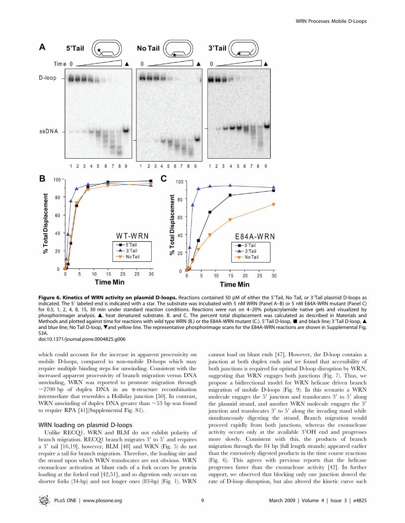
To determine whether the presence of a protruding tail
influences the kinetics of WRN D-loop disruption we conducted
time course reactions. An increase in total strand displacement
occurred as a function of time that began to plateau at about
4 min for the 39Tail D-loop and 8 min for the 59 and No Tail D-
loops (Fig. 6A–B). The rate of total strand displacement was
similar for the 59 and No Tail D-loops, but was ,2-fold higher for
the 39 Tail D-loop. Loss of the full length products in favor of
extensively degraded products became apparent at the later time
points, which most likely reflects additional digestion of the
released ssDNA strands (Fig. 6A). To determine the influence of
structure on the helicase branch migration activity alone, we
examined the kinetics of D-loop disruption by the exonuclease-
Figure 3. WRN can branch migrate through the full 84 bp duplex to displace the invading strand. Reactions contained 50 pM of eitherthe 59Tail non-telomeric (lanes 1–8) or the telomeric (Tel) (lanes 9–14 (A.) or 9–16 (B.)) plasmid D-loops. Reactions were run on 4–20% nativepolyacrylamide gels and visualized by phosphorimager analysis. m, heat denatured substrate. A. Branch migration after exonuclease inactivation witha WRN mutant. The substrate was incubated with E84A-WRN concentrations increasing in doubling steps from 0.15 to 5 nM (lanes 2–7) or from 0.62to 5 nM (lanes 11–14) for 15 min under standard conditions. B. Branch migration after exonuclease inhibition with limiting Mg2+. The substrate wasincubated with wild type WRN concentrations increasing in doubling steps from 0.15 to 5 nM (lanes 2–7, and lanes 10–15) for 15 min in reactionsbuffer containing 1 mM MgCl2 and 2 mM ATP. The percent total displacement was plotted against E84A-WRN (A.) or wild type WRN (B.)concentration. E84A-WRN or WRN and 59Tail D-loop, & and dotted line; E84A-WRN or WRN and 59Tail Tel D-loop, $ and solid line. Values are themean and standard deviation (SD) from two-three independent experiments.doi:10.1371/journal.pone.0004825.g003
WRN Processes Mobile D-Loops
PLoS ONE | www.plosone.org 5 March 2009 | Volume 4 | Issue 3 | e4825
dead WRN mutant (E84A). The 39 Tail D-loop was more rapidly
disrupted by WRN branch migration activity than the 59 and No
Tail D-loops (Fig. 6C and Supplemental Fig. S3). Furthermore,
the kinetics of strand displacement for the E84A-WRN mutant
were similar to wild type WRN for the 39Tail D-loop, but was
decreased for the 59Tail and No Tail D-loops, relative to wild type
(Figs. 6B and C). This indicates that the WRN exonuclease activity
contributes more to the rate of D-loop disruption for the substrates
in which digestion proceeds into the duplex region (59 and No Tail
D-loop), than for the substrate in which duplex was not detectably
shortened (39 D-loop). The identical order of substrate preference
was observed when wild type WRN exonuclease activity was
inhibited by limiting Mg 2+ concentrations (Supplemental Fig. S3)
WRN recognizes both junctions of the D-loopPrevious studies showed that WRN can bypass a streptavidin-
biotin steric block on the 39tail of a fork or flap substrate,
indicating that WRN can recognize a fork-like junction directly to
initiate unwinding. Based on this and other studies with oligomeric
forks, D-loop and flap structures [36,37,49], we conjectured that
WRN would load and initiate branch migration at the 59 end of
the invading strand duplex in the D-loop and translocate 39 to 59
along the plasmid strand. If this were true, the action of the WRN
exonuclease at the opposite end (39OH) would require the protein
to simultaneously interact with both ends of the 84 bp duplex.
Thus, we tested whether WRN D-loop disruption required WRN
recognition of the 59 or 39end of the invading strand duplex to
Figure 4. WRN exonuclease digests the long invading strands both prior to and after release from the plasmid D-loops. A. Reactionscontained 50 pM of either the 59Tail non-telomeric (lane 1–8) or the telomeric (Tel) (lanes 9–15) plasmid D-loops. The substrate was incubated withWRN concentrations increasing by doubling steps from 0.15 to 5 nM (lanes 2–7) or from 0.31 to 5 nM (lanes 10–14) for 15 min under standardreaction conditions except ATP was replaced with ATPcS. Reactions were run on 4–20% native polyacrylamide gels and visualized byphosphorimager analysis. m, heat denatured substrate. B. The percent total displacement was calculated as described in Materials and Methods andplotted against WRN concentration. WRN and 59Tail D-loop, & and dotted line; WRN and 59Tail Tel D-loop, $ and solid line. Values are the mean andstandard deviation (SD) from two independent experiments. C. and D. WRN exonuclease on intact and released invading strands. Reactions contained50 pM of either the non-telomeric (C.) 59Tail D-loop (lanes 1–7) or 120-mer oligonucleotide (lanes 11–14) or the telomeric (D.) 59Tail Tel D-loop (lanes7–12) or 120-mer oligonucleotide (lanes 1–4). The substrates were incubated with WRN (5 nM) or E84A-WRN mutant (X; 5 and 2.5 nM) under standardconditions for 15 min except that either 2 mM ATP, 2 mM ATPcS (c) or no ATP (-) was added as indicated. Reactions were run on 14% polyacrylamidedenaturing gels. Oligonucleotide size markers were loaded for reference.doi:10.1371/journal.pone.0004825.g004
WRN Processes Mobile D-Loops
PLoS ONE | www.plosone.org 6 March 2009 | Volume 4 | Issue 3 | e4825
initiate displacement. We constructed plasmid D-loops containing
an invading strand with both a 59 and 39 18 nt ssDNA tail
protruding from a 60 bp duplex (Fig. 7). A biotinylated nucleotide
was incorporated in the 59 and/or 39 tail of the invading strand
2 nt away from the duplex region (junction). To impart a steric
block at either the 59 or 39 junction or both, the D-loops were pre-
incubated with streptavidin to bind the biotin groups. Free
streptavidin protein does not inhibit WRN [49]. Importantly, the
biotin moiety alone did not inhibit WRN branch migration
activity as indicated by rapid disruption of the plasmid D-loops
(Fig. 7). However, the WRN exonuclease cannot progress past the
biotin (Fig. 7D) which explains the lack of detectable shortened
products on the native gels for the D-loops with a biotin near the
39end (Fig. 7 A and C). Blocking both the 59 and 39 junctions
resulted in a dramatic reduction in D-loop disruption (Fig. 7A),
indicating that the streptavidin-biotin complex effectively blocked
WRN recognition of the junctions. In contrast, blocking only the
59 junction slowed the rate of D-loop disruption by about 4.8-fold
but did not prevent branch migration, suggesting that WRN can
recognize the 39 junction (Fig. 7B). Consistent with this, blocking
only the 39 junction also slowed the rate of D-loop disruption, in
this case 2.7 fold (Fig. 7C). When either the 39 or 59 junction
blocked, nearly the same maximal amount of D-loop disruption
was eventually achieved as for the corresponding non-blocked D-
loop (Fig. 7B–C). Furthermore, a WRN exonuclease-dead mutant
can disrupt a D-loop containing a blocked 59 junction that lacks a
39 tail, indicating that WRN helicase branch migration from the
accessible 39junction does not require a 39 tail for loading (Fig. 8).
Importantly, the displaced strand exhibited slower mobility in the
presence of streptavidin (Fig. 7–8). This confirms that streptavidin
remained bound to DNA throughout the reactions, consistent with
previous reports that WRN helicase does not dissociate the
streptavidin from the DNA [49]. These data indicate that WRN
protein can recognize both junction ends of the invading strand
duplex, and can effectively disrupt D-loops containing a steric
block at one junction but not when both junctions are blocked.
Discussion
Increasing evidence supports an important role for WRN
protein in mediating the proper dissociation of joint DNA
molecules that arise in genomic and telomeric regions during
homologous recombination and repair of breaks at collapsed
replication forks. WRN was previously shown to displace and
degrade the invading strand of static oligomeric-based D-loops
with a 59 tail or no tail, which were designed to mimic an
important HR intermediate [36,37]. However, WRN helicase was
unable to efficiently unwind the full 33-bp invaded duplex in a
telomeric D-loop without assistance from single strand binding
proteins RPA and POT1 or the exonuclease to shorten the duplex
[37,38]. Here, we demonstrate that WRN helicase alone is capable
of displacing a much longer telomeric and non-telomeric invading
strand (84-bp) from plasmid based D-loop, presumably by
promoting branch migration. However, D-loop processing is
altered in the presence of WRN exonuclease activity which digests
the invading strand both before and after release from the D-loop.
We propose that mobile D-loops are particularly suitable HR
structures for processing by WRN because they are substrates for
both the helicase and exonuclease activities, unlike Holliday
junction recombination intermediate. The increased activity of
WRN on the more biologically relevant plasmid D-loops,
compared to oligomeric D-loops, greatly strengthens the argument
that WRN is capable of processing such structures at telomeric
ends and during HR in vivo.
WRN processing of static versus mobile D-loopsOur results indicate that WRN helicase and exonuclease process
mobile D-loops and static D-loops by different mechanisms. We
found that WRN helicase apparent processivity is increased on
mobile D-loops, since WRN helicase alone can disrupt mobile D-
loops with duplexes that are much longer (84 bp) (Figs. 2–3) than
static D-loops that cannot be unwound (33 bp) [37]. We propose
that by WRN helicase promoting branch migration through the
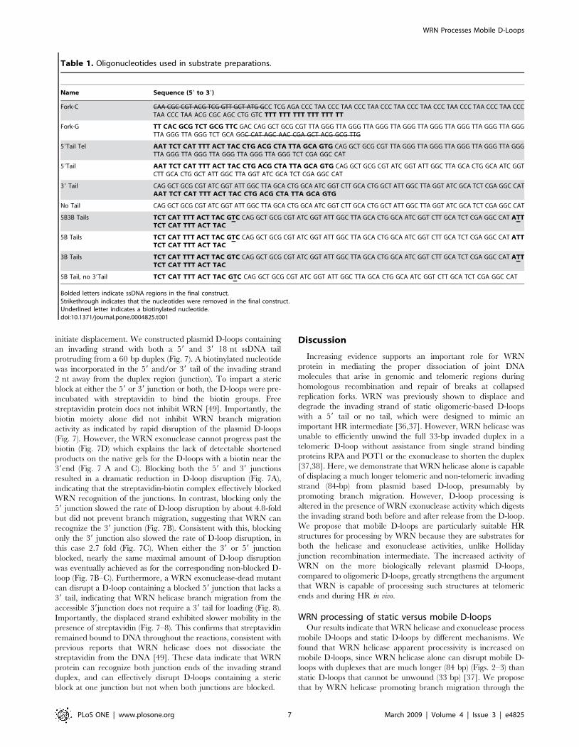
Table 1. Oligonucleotides used in substrate preparations.
Name Sequence (59 to 39)
Fork-C CAA CGC CGT ACG TCG GTT GCT ATG GCC TCG AGA CCC TAA CCC TAA CCC TAA CCC TAA CCC TAA CCC TAA CCC TAA CCC TAA CCCTAA CCC TAA ACG CGC AGC CTG GTC TTT TTT TTT TTT TTT TT
Fork-G TT CAC GCG TCT GCG TTC GAC CAG GCT GCG CGT TTA GGG TTA GGG TTA GGG TTA GGG TTA GGG TTA GGG TTA GGG TTA GGGTTA GGG TTA GGG TCT GCA GGC CAT AGC AAC CGA GCT ACG GCG TTG
59Tail Tel AAT TCT CAT TTT ACT TAC CTG ACG CTA TTA GCA GTG CAG GCT GCG CGT TTA GGG TTA GGG TTA GGG TTA GGG TTA GGGTTA GGG TTA GGG TTA GGG TTA GGG TTA GGG TCT CGA GGC CAT
59Tail AAT TCT CAT TTT ACT TAC CTG ACG CTA TTA GCA GTG CAG GCT GCG CGT ATC GGT ATT GGC TTA GCA CTG GCA ATC GGTCTT GCA CTG GCT ATT GGC TTA GGT ATC GCA TCT CGA GGC CAT
39 Tail CAG GCT GCG CGT ATC GGT ATT GGC TTA GCA CTG GCA ATC GGT CTT GCA CTG GCT ATT GGC TTA GGT ATC GCA TCT CGA GGC CATAAT TCT CAT TTT ACT TAC CTG ACG CTA TTA GCA GTG
No Tail CAG GCT GCG CGT ATC GGT ATT GGC TTA GCA CTG GCA ATC GGT CTT GCA CTG GCT ATT GGC TTA GGT ATC GCA TCT CGA GGC CAT
5B3B Tails TCT CAT TTT ACT TAC GTC CAG GCT GCG CGT ATC GGT ATT GGC TTA GCA CTG GCA ATC GGT CTT GCA TCT CGA GGC CAT ATTTCT CAT TTT ACT TAC
5B Tails TCT CAT TTT ACT TAC GTC CAG GCT GCG CGT ATC GGT ATT GGC TTA GCA CTG GCA ATC GGT CTT GCA TCT CGA GGC CAT ATTTCT CAT TTT ACT TAC
3B Tails TCT CAT TTT ACT TAC GTC CAG GCT GCG CGT ATC GGT ATT GGC TTA GCA CTG GCA ATC GGT CTT GCA TCT CGA GGC CAT ATTTCT CAT TTT ACT TAC
5B Tail, no 39Tail TCT CAT TTT ACT TAC GTC CAG GCT GCG CGT ATC GGT ATT GGC TTA GCA CTG GCA ATC GGT CTT GCA TCT CGA GGC CAT
Bolded letters indicate ssDNA regions in the final construct.Strikethrough indicates that the nucleotides were removed in the final construct.Underlined letter indicates a biotinylated nucleotide.doi:10.1371/journal.pone.0004825.t001
WRN Processes Mobile D-Loops
PLoS ONE | www.plosone.org 7 March 2009 | Volume 4 | Issue 3 | e4825
mobile D-loop, the unpaired plasmid strands anneal and shrink
the D-loop (Fig. 9). Thus, if WRN prematurely dissociates before
the duplex is completely unwound, the partially unwound
invading strand will not reanneal with the plasmid, allowing
WRN to rapidly displace the remaining duplex upon rebinding the
substrate. The loop cannot shrink in static D-loops since the
‘‘bubble’’ strands are not complementary and the partially
unwound invading strand can reanneal if WRN prematurely
dissociates. In agreement with our results, RECQ1 helicase also
disrupts mobile D-loops by promoting branch migration, but is
unable to unwind oligomeric D-loops of similar size [16].
Furthermore, BLM reactions with static D-loops produce
intermediates that are not present in the mobile D-loop reactions
[48]. Therefore the authors proposed that BLM may disrupt
mobile D-loops, but not non-mobile D-loops, in a single step.
Thus, WRN may displace mobile D-loops in a single binding step
Figure 5. Structural requirements for WRN activity on plasmid D-loops. A. Schematic of 39Tail and No Tail D-loops. The star denotes the 59end radiolabel. B. Reactions contained 50 pM of either the 39Tail (lane 1–6) or the No Tail (lanes 7–12) plasmid D-loops. The substrate was incubatedwith WRN concentrations increasing by doubling steps from 0.62 to 5 nM (lanes 2–5 and 8–11) for 15 min under standard reaction conditions.Reactions were run on 4–20% polyacrylamide native gels and visualized by phosphorimager analysis. m, heat denatured substrate. C. The percenttotal displacement was calculated as described in Materials and Methods and plotted against WRN concentration. WRN and 39Tail D-loop, & anddotted line; WRN and No Tail D-loop, $ and solid line. Values are the mean and standard deviation (SD) from at least three independent experiments.D. Analysis of WRN exonuclease activity. Reactions contained 50 pM of either the 39Tail (lanes 1–4 and 9–10) or the No Tail (lanes 5–8 and 11–12) D-loops. The substrates were incubated with 5 nM WRN or E84A-WRN mutant (X) under standard conditions for 15 min except that either 2 mM ATP,2 mM ATPcS (c) or no ATP (-) was added as indicated. Reactions were run on 14% polyacrylamide denaturing gels. Arrow points to productsrepresenting digestion almost to the end of the 84-bp duplex.doi:10.1371/journal.pone.0004825.g005
WRN Processes Mobile D-Loops
PLoS ONE | www.plosone.org 8 March 2009 | Volume 4 | Issue 3 | e4825
which could account for the increase in apparent processivity on
mobile D-loops, compared to non-mobile D-loops which may
require multiple binding steps for unwinding. Consistent with the
increased apparent processivity of branch migration versus DNA
unwinding, WRN was reported to promote migration through
,2700 bp of duplex DNA in an a-structure recombination
intermediate that resembles a Holliday junction [50]. In contrast,
WRN unwinding of duplex DNA greater than ,53 bp was found
to require RPA [41](Supplemental Fig. S1).
WRN loading on plasmid D-loopsUnlike RECQ1, WRN and BLM do not exhibit polarity of
branch migration. RECQ1 branch migrates 39 to 59 and requires
a 39 tail [16,19], however, BLM [48] and WRN (Fig. 5) do not
require a tail for branch migration. Therefore, the loading site and
the strand upon which WRN translocates are not obvious. WRN
exonuclease activation at blunt ends of a fork occurs by protein
loading at the forked end [42,51], and so digestion only occurs on
shorter forks (34-bp) and not longer ones (83-bp) (Fig. 1). WRN
cannot load on blunt ends [47]. However, the D-loop contains a
junction at both duplex ends and we found that accessibility of
both junctions is required for optimal D-loop disruption by WRN,
suggesting that WRN engages both junctions (Fig. 7). Thus, we
propose a bidirectional model for WRN helicase driven branch
migration of mobile D-loops (Fig. 9). In this scenario a WRN
molecule engages the 59 junction and translocates 39 to 59 along
the plasmid strand, and another WRN molecule engages the 39
junction and translocates 39 to 59 along the invading stand while
simultaneously digesting the strand. Branch migration would
proceed rapidly from both junctions, whereas the exonuclease
activity occurs only at the available 39OH end and progresses
more slowly. Consistent with this, the products of branch
migration through the 84 bp (full length strands) appeared earlier
than the extensively digested products in the time course reactions
(Fig. 6). This agrees with previous reports that the helicase
progresses faster than the exonuclease activity [42]. In further
support, we observed that blocking only one junction slowed the
rate of D-loop disruption, but also altered the kinetic curve such
Figure 6. Kinetics of WRN activity on plasmid D-loops. Reactions contained 50 pM of either the 59Tail, No Tail, or 39Tail plasmid D-loops asindicated. The 59 labeled end is indicated with a star. The substrate was incubated with 5 nM WRN (Panel A–B) or 5 nM E84A-WRN mutant (Panel C)for 0.5, 1, 2, 4, 8, 15, 30 min under standard reaction conditions. Reactions were run on 4–20% polyacrylamide native gels and visualized byphosphorimager analysis. m, heat denatured substrate. B. and C. The percent total displacement was calculated as described in Materials andMethods and plotted against time for reactions with wild type WRN (B.) or the E84A-WRN mutant (C.). 59Tail D-loop, & and black line; 39Tail D-loop, mand blue line; No Tail D-loop, .and yellow line. The representative phosphorimage scans for the E84A-WRN reactions are shown in Supplemental Fig.S3A.doi:10.1371/journal.pone.0004825.g006
WRN Processes Mobile D-Loops
PLoS ONE | www.plosone.org 9 March 2009 | Volume 4 | Issue 3 | e4825
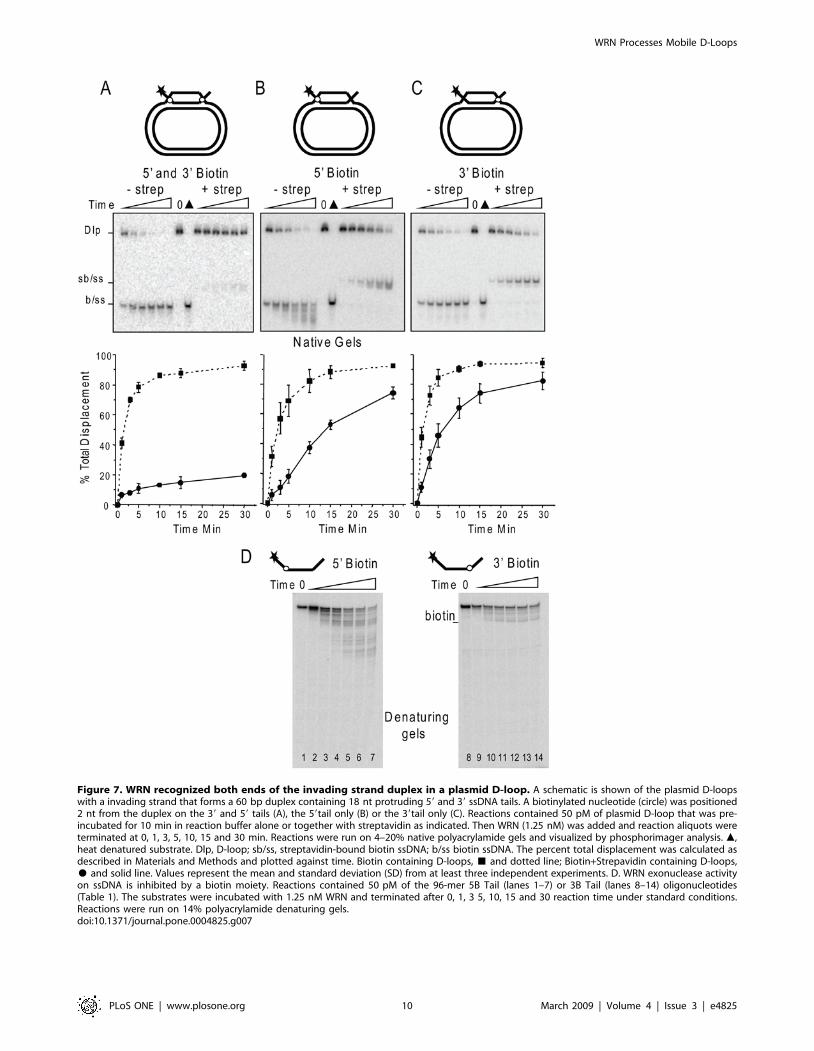
Figure 7. WRN recognized both ends of the invading strand duplex in a plasmid D-loop. A schematic is shown of the plasmid D-loopswith a invading strand that forms a 60 bp duplex containing 18 nt protruding 59 and 39 ssDNA tails. A biotinylated nucleotide (circle) was positioned2 nt from the duplex on the 39 and 59 tails (A), the 59tail only (B) or the 39tail only (C). Reactions contained 50 pM of plasmid D-loop that was pre-incubated for 10 min in reaction buffer alone or together with streptavidin as indicated. Then WRN (1.25 nM) was added and reaction aliquots wereterminated at 0, 1, 3, 5, 10, 15 and 30 min. Reactions were run on 4–20% native polyacrylamide gels and visualized by phosphorimager analysis. m,heat denatured substrate. Dlp, D-loop; sb/ss, streptavidin-bound biotin ssDNA; b/ss biotin ssDNA. The percent total displacement was calculated asdescribed in Materials and Methods and plotted against time. Biotin containing D-loops, & and dotted line; Biotin+Strepavidin containing D-loops,$ and solid line. Values represent the mean and standard deviation (SD) from at least three independent experiments. D. WRN exonuclease activityon ssDNA is inhibited by a biotin moiety. Reactions contained 50 pM of the 96-mer 5B Tail (lanes 1–7) or 3B Tail (lanes 8–14) oligonucleotides(Table 1). The substrates were incubated with 1.25 nM WRN and terminated after 0, 1, 3 5, 10, 15 and 30 reaction time under standard conditions.Reactions were run on 14% polyacrylamide denaturing gels.doi:10.1371/journal.pone.0004825.g007
WRN Processes Mobile D-Loops
PLoS ONE | www.plosone.org 10 March 2009 | Volume 4 | Issue 3 | e4825
that the reaction plateau was delayed (Fig. 7B–C). WRN
eventually achieved near maximal D-loop disruption when one
junction was blocked, but required more time than was needed for
the corresponding unblocked D-loop (Fig. 7B–C). This suggests
that WRN can achieve more rapid D-loop disruption when
branch migration proceeds from both ends rather than one end.
When both junctions were blocked, D-loop strand displacement
was barely detectable even after 30 min (Fig. 7A). This suggests
that initiation of branch migration was inhibited. However, we
cannot distinguish whether separate WRN molecules engage the
junction, or if different WRN monomers of a higher oligomeric
complex engage the junctions. WRN was recently found to bind
model replication forks and Holliday junctions [52] as a tetramer.
Structural analysis and single molecule imaging will be key to
deciphering these models.
Alternatively, WRN may only recognize and initiate branch
migration from one junction in a unidirectional model. For
example, the steric block at the 59 junction might inhibit initiation
whereas the steric block downstream at the 39 junction might
inhibit progression. We believe a biotin-streptavidin complex is
more likely to inhibit WRN initiation rather than progression for
several reasons. The downstream block is outside the duplex
region and is also present on the opposite strand that WRN is
presumed to track along in a unidirectional model. Bulky DNA
adducts in duplex DNA strongly inhibit WRN when present on
the translocating strand [53]. Furthermore, once the duplex is
shortened to unstable lengths (,12 bp) it will thermally melt [42],
so a downstream block would be ,14 nt away from WRN when
strand displacement occurred. A biotin-streptavidin complex on
oligomeric fork and flap substrates did not inhibit WRN
unwinding unless the complex was placed at the junction [49].
Thus, we do not favor a unidirectional model.
Comparison with other RecQ helicasesCaution should be applied when directly comparing our results
with WRN to studies with other human RecQ helicases due to
differences in the D-loop constructs examined (size and sequence)
and reaction conditions. Nevertheless, WRN is at least as efficient
as RECQ1 and BLM in disrupting various plasmid D-loops. In
reactions for equivalent times and with similar amounts of 59 tailed
D-loops (,50 pM), disruption of 50% of the D-loop is achieved
with 7.4 nM BLM [48], compared to 0.5 nM WRN (Fig. 2) or
,1 nM WRN if the exonuclease is inactivated (Fig. 3). RECQ1
preferentially unwinds 39tailed D-loops, and disruption of
approximately 50% of the substrate is achieved by 7.5 nM
RECQ1 within ,2.5 min [16], and by 5 nM WRN within
,1.5 min even if the exonuclease is inactivated (Fig. 6). Thus,
WRN is at least as efficient as BLM and RECQ1 but has the
added feature of trimming the invading tails.
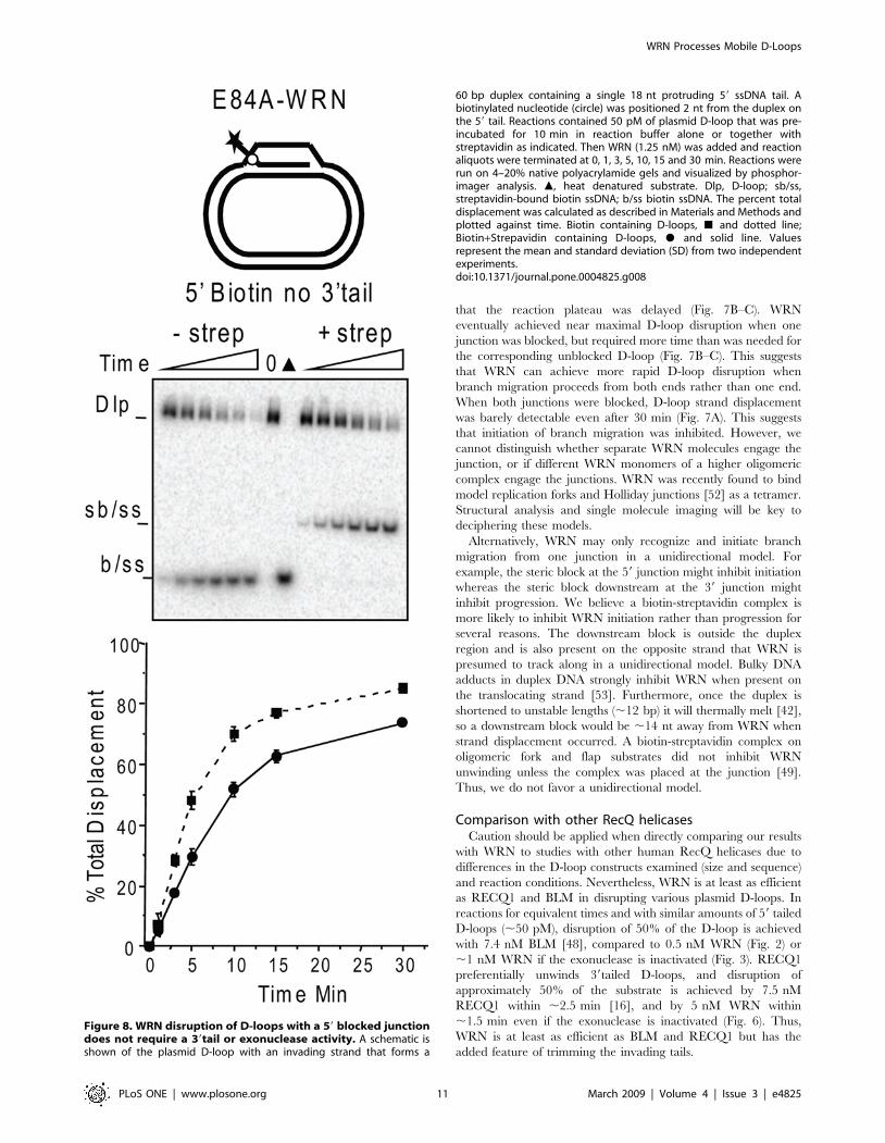
Figure 8. WRN disruption of D-loops with a 59 blocked junctiondoes not require a 39tail or exonuclease activity. A schematic isshown of the plasmid D-loop with an invading strand that forms a
60 bp duplex containing a single 18 nt protruding 59 ssDNA tail. Abiotinylated nucleotide (circle) was positioned 2 nt from the duplex onthe 59 tail. Reactions contained 50 pM of plasmid D-loop that was pre-incubated for 10 min in reaction buffer alone or together withstreptavidin as indicated. Then WRN (1.25 nM) was added and reactionaliquots were terminated at 0, 1, 3, 5, 10, 15 and 30 min. Reactions wererun on 4–20% native polyacrylamide gels and visualized by phosphor-imager analysis. m, heat denatured substrate. Dlp, D-loop; sb/ss,streptavidin-bound biotin ssDNA; b/ss biotin ssDNA. The percent totaldisplacement was calculated as described in Materials and Methods andplotted against time. Biotin containing D-loops, & and dotted line;Biotin+Strepavidin containing D-loops, $ and solid line. Valuesrepresent the mean and standard deviation (SD) from two independentexperiments.doi:10.1371/journal.pone.0004825.g008
WRN Processes Mobile D-Loops
PLoS ONE | www.plosone.org 11 March 2009 | Volume 4 | Issue 3 | e4825
Roles for WRN digestion of D-loop recombinationintermediates
The activity of WRN exonuclease on plasmid D-loops affords
WRN the ability to process these structures differently than other
human RecQ helicases. Both the WRN exonuclease and helicase
are required for the survival of recombinant progeny after the
induction of Rad51-dependent HR intermediates to restore
blocked or stalled DNA replication forks [22–24]. Our data
indicate that both the WRN exonuclease and helicase are active
on biologically relevant Rad51-dependent recombination inter-
mediates. In addition to disrupting 39 end invaded D-loops, we
demonstrate for the first time that WRN can also disrupt 59 end
invaded D-loops (39Tail D-loop) (Fig. 5). Rad51 promotes both 39
and 59 end invasion for D-loop production [54,55], however, the
latter is potentially toxic because it is unproductive. A 59 end
invaded D-loop cannot be extended by a polymerase to complete
the next steps of HR repair of replication forks, SDSA, or the ALT
pathways [6,11]. While the role for the helicase in branch
migration is clear, the role for the exonuclease has several
possibilities. First, shortening of the invading strand results in fewer
sites for Rad51 re-nucleation after strand release, which would
inhibit repeated strand invasion events. Second, if the Rad51
mediated D-loop formation is incomplete such that the 39 end is
not fully paired with the template strand, it cannot be extended by
a polymerase in the next steps of HR replication restart, SDSA or
ALT pathways. WRN can rescue this unproductive intermediate
by degrading the 39 protruding ssDNA (Fig. 5).
WRN activity on Telomeric D-loopsThe decrease in WRN branch migration and exonuclease
digestion of the telomeric sequence compared to the non-telomeric
sequence have both biochemical and biological implications.
Helicase processivity is influenced by base pair stability [56],
therefore, differences could be related to duplex stability.
However, we detected no obvious differences in thermal melting
temperatures for the telomeric sequence compared to the non-
telomeric sequence used in this study (Table 1) according to the
HyTher@ web-based program [57]. The ssDNA TTAGGG
repeats in the plasmid D-loop have the potential to form G-
quadruplex DNA which could impede branch migration and
would require resolution by the helicase [58]. The G-quadruplex
DNA might also sequester WRN since WRN binds G4 DNA with
high affinity [58]. The increased resistance of the telomeric D-loop
to branch migration and digestion might also have implications for
tighter regulation of processing at the telomeres in vivo.
Inappropriate processing of telomeric ends by DNA repair
proteins can have dire consequences for the cell and lead to
senescence, apoptosis or genomic instability [33]. Untimely release
of the telomeric t-loop/D-loop structure at the chromosome ends
can activate a DNA damage response [59]. Therefore, resolution
of telomeric D-loops might be more dependent on stimulation by
telomeric proteins TRF2 and POT1, which regulate recombina-
tion at telomeric ends [33]. Consistent with this, POT1 and TRF2
interaction with WRN and were found to regulate WRN activities
in vitro [37,60].
In summary, we report that biologically relevant plasmid based
D-loops with relatively long strand invasion duplex regions (84-bp)
are substrates for both WRN branch migration and WRN
exonuclease activity. WRN activity is not dependent on a
protruding ssDNA tail, but the exonuclease is highly dependent
on the free Mg2+ concentration. Our results offer a potential
explanation for the cellular studies that indicate both WRN
activities are required for dissociation of Rad51 dependent
recombination intermediates to complete repair and suppress
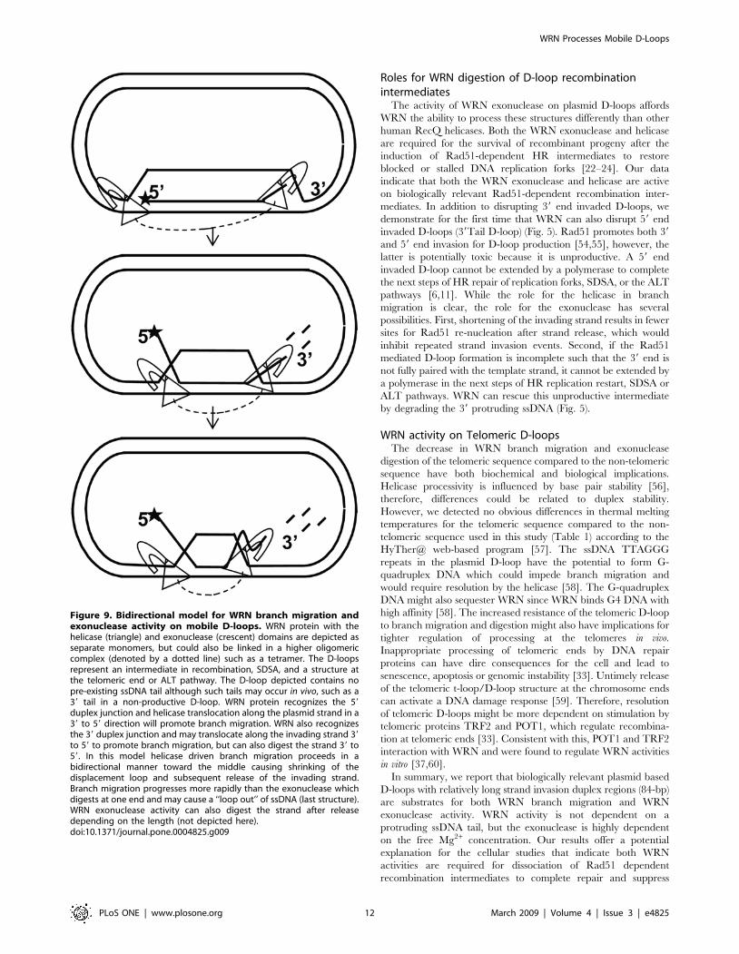
Figure 9. Bidirectional model for WRN branch migration andexonuclease activity on mobile D-loops. WRN protein with thehelicase (triangle) and exonuclease (crescent) domains are depicted asseparate monomers, but could also be linked in a higher oligomericcomplex (denoted by a dotted line) such as a tetramer. The D-loopsrepresent an intermediate in recombination, SDSA, and a structure atthe telomeric end or ALT pathway. The D-loop depicted contains nopre-existing ssDNA tail although such tails may occur in vivo, such as a39 tail in a non-productive D-loop. WRN protein recognizes the 59duplex junction and helicase translocation along the plasmid strand in a39 to 59 direction will promote branch migration. WRN also recognizesthe 39 duplex junction and may translocate along the invading strand 39to 59 to promote branch migration, but can also digest the strand 39 to59. In this model helicase driven branch migration proceeds in abidirectional manner toward the middle causing shrinking of thedisplacement loop and subsequent release of the invading strand.Branch migration progresses more rapidly than the exonuclease whichdigests at one end and may cause a ‘‘loop out’’ of ssDNA (last structure).WRN exonuclease activity can also digest the strand after releasedepending on the length (not depicted here).doi:10.1371/journal.pone.0004825.g009
WRN Processes Mobile D-Loops
PLoS ONE | www.plosone.org 12 March 2009 | Volume 4 | Issue 3 | e4825
inappropriate recombination at stalled replication forks [25] and
telomeric ends [29].
Materials and Methods
ProteinsRecombinant histidine-tagged WRN protein and the exonucle-
ase-dead E84A mutant (X-WRN) were purified using a baculo-
virus/insect cell expression system and an AKTA Explorer FLPC
(GE Healthcare, Piscataway, NJ) as previously described [60]
except the HiLoad 16/60 Superdex 200 pg column was replaced
with a HiTrap Phenyl HP column. Briefly, fractions containing
WRN eluted off the HisTrap FF column were diluted to obtain
final concentrations of 2 M NaCl in 20 mM phosphate buffer
pH 7.4, 10% glycerol and 0.05% igepal CA-630. The eluent was
loaded onto a HiTrap Phenyl HP column equilibrated with HIC
buffer (20 mM phosphate buffer pH 7.4, 10% glycerol and 0.05%
igepal CA-630) and washed once with HIC buffer containing
1.55 M NaCl prior to elution with 950 mM NaCl in HIC buffer.
The eluted fractions containing WRN were loaded onto a HiTrap
Desalt 5 ml column to exchange the buffer before loading onto a
1 ml Resource Q column for removal of remaining contaminants
as described previously [60]. The concentration of active protein
was determined by Bradford Assay (BioRad, Hercules, CA) and
standard helicase reactions with a 16-bp forked duplex. Purity was
determined by SDS-PAGE and Coomassie staining. All restriction
enzymes were from New England Biolabs (Ipswich, MA). RecA
protein was from USB Corporation (Cleveland, OH).
DNA SubstratesAll oligonucleotides used in this study (Table 1) were from
Integrated DNA Technologies (Coralville, IA) and were purified
by PAGE or HPLC by the manufacturer. Oligonucleotides were
59 end-labeled with [c-32P] ATP (3000 Ci/mmol) (Perkin Elmer,
Waltham, MA) using T4 polynucleotide kinase (New England
BioLabs, Ipswich, MA), according to the manufacturer’s instruc-
tions. The plasimds used for making the various 84-bp non-
telomeric and telomeric (Tel) D-loops were constructed by cloning
the sequence (TTAGGG)10 (Tel10 plasmid) or the scrambled
sequence 59- ATC GGT ATT GGC TTA GCA CTG GCA ATC
GGT CTT GCA CTG GCT ATT GGC TTA GGT ATC GCA
(Non-tel 10 plasmid) between the bases 111 and 112 of the HSV-tk
gene in the previously described pGTK4 plasmid [61]. The
plasmids for constructing the 60-bp D-loops used in Figs. 7–8
contained the shorter insert sequence 59-ATC GGT ATT GGC
TTA GCA CTG GCA ATC GGT CTT GCA. The 6 kB
plasmids were reduced to 3.6 kB to facilitate gel migration by
removing 2363 bp through restriction with AccI followed by re-
ligation. The plasmids were purified by two rounds of ethidium-
bromide saturated CsCl equilibrium gradient ultracentrifugation
(Loftstrand Labs, Gaithersburg, MD). The plasmid based D-loop
substrates were constructed as described previously [62]. Briefly
RecA (4 mM) was incubated with the invading strand oligonucle-
otide (3.6 mM nucleotide) for 5 min at 37uC, then the supercoiled
plasmid (300 mM nucleotides) was added and incubated for 3 min
more. The reactions were terminated by incubation with
Proteinase K and SDS for 30 min as described previously [62].
The D-loop constructs were PAGE purified by using 4.5% (37.5:1)
polyacrylamide gels. After the bands were excised the gel slices
were placed in dialysis D-tubes (Novagen, Madison, WI) and were
subjected to two rounds of electroelution in 16TBE for 2–4 hrs at
4uC and 120 V. The D-loops were concentrated and exchanged
into storage buffer (10 mM Tris-HCl (pH 7.5), 10 mM MgCl2)
using micron-30 devices (Amicon). Purification quality and yields
were determined by analysis on 4–20% native polyacrylamide gels,
followed by visualization and quantitation with a Typhoon
phosphorimager and ImageQuant software (GE Healthcare,
Piscataway, NJ). Restriction digest analysis of the invading strand
in the plasmid D-loops was conducted in reactions (20 ml)
containing 50 pM purified D-loops and 10 units of the restriction
enzyme indicated in Fig. 1 for 4–5 hr at 37uC, according to the
manufacturer’s protocol. Products were analyzed on a 14%
denaturing polyacrylamide gel.
The 83-bp long forked duplex was constructed by annealing
9 pmol of the 59end labeled Fork-G oligonucleotide (Table 1) with
12 pmol of the Fork-C complementary strand in 50 ml with
50 mM LiCl at 95uC for 5 min, followed by cooling to room
temperature. In order to achieve the final 83-bp length, the fork
was restricted with HaeIII and then PAGE purified by running on
8% native polyacrylamide gel and using Qiaex II Gel Extraction
Kit (Qiagen, Valencia, CA). Product purity and yield were
determined by native PAGE and phosphorimager analysis.
Helicase branch migration and exonuclease reactionsReactions were performed in standard reaction buffer contain-
ing 40 mM Tris-HCl, pH 8.0, 4 mM MgCl2, 5 mM DTT,
100 mg/ml BSA, 14–28 ng/ml yeast tRNA, and 20 mM ATP
[42] unless otherwise indicated. DNA substrate and protein
concentrations were as indicated in the figure legends. The
reactions were initiated by adding WRN protein and were
incubated at 37uC for 15 minutes, unless otherwise indicated.
For reactions with D-loops containing a biotinylated nucleotide
the substrate was pre-incubated at 37uC for 10 minutes in buffer
alone or with 1.5 nM streptavidin prior to WRN addition. For
analysis of radiolabeled molecules on 4–20% native polyacryl-
amide gels, the reactions (10 mL) were stopped with 5 mL of 36stop dye supplemented with 10 mg/mL proteinase K [42] and de-
proteinized for 15 min at 37uC. For analysis of the radio-labeled
molecules on 14% denaturing gels, the reactions were terminated
with an equal volume of formamide stop dye [42]. The helicase
reactions with the 83-bp forked duplex were terminated in 36stop
dye with a 10-fold molar excess of cold competitor oligonucleotide
to prevent reannealing of the unwound products. After drying the
gels, the reactions were visualized using a Typhoon phosphor-
imager and quantified using ImageQuant software (GE Health-
care, Piscataway, NJ).
For quantitation of strand displacement the percent of displaced
products (full length and shortened) were calculated as a function
of the total radioactivity in the reaction lane [37]. All values were
corrected for background in the no enzyme control and heat
denatured substrate lanes.
Supporting Information
Figure S1 WRN helicase cannot completely unwind an 83 bp
telomeric forked duplex. Reactions contained 50 pM of the 84-bp
forked duplex containing ten telomeric repeats (thick black line)
flanked by 15 and 8 bp of unique sequence. The substrate was
incubated with WRN concentrations increasing by doubling steps
from 0.039 to 5 nM for 15 min under standard reaction
conditions. Reactions were run on an 8% native gel.
Found at: doi:10.1371/journal.pone.0004825.s001 (1.17 MB EPS)
Figure S2 WRN exonuclease digests free 120 nt single strands.
Reactions contained 50 pM of the 59Tail non-telomeric oligonu-
cleotide (panel A, lanes 1–7; panel B, lanes 1–4) or the telomeric
(59Tail Tel) oligonucleotide (panel A, lanes 8–14; panel B, lanes 5–
8) (see Table 1). The substrate was incubated with increasing
WRN concentrations (0, 0.15, 0.31, 0.62, 1.2, 2.5 or 5 nM) (panel
WRN Processes Mobile D-Loops
PLoS ONE | www.plosone.org 13 March 2009 | Volume 4 | Issue 3 | e4825
A) or E84A-WRN mutant (0, 1.2, 2.5, or 5 nM) (panel B) for
15 min under standard reaction conditions. Reactions were run on
a 14% denaturing polyacrylamide gel.
Found at: doi:10.1371/journal.pone.0004825.s002 (3.57 MB EPS)
Figure S3 Kinetics of WRN branch migration activity on
plasmid D-loops. Reactions contained 50 pM of either the 59Tail,
No Tail, or 39 Tail plasmid D-loops as indicated. The 59 labeled
end is indicated with a star. The substrate was incubated with
5 nM E84A-WRN under standard reaction conditions (Panel A)
or wild type WRN (Panel B) under modified conditions in which
Mg2+ was reduced to 1 mM. Aliquots were removed at 0, 0.5, 1, 2,
4, 8, 15, 30 min and terminated. Reactions were run on 4–20%
polyacrylamide native gels and visualized by phosphorimager
analysis. D, heat denatured substrate. The quantification of the
reactions in Panel A is shown in Fig. 6C. B. The percent total
displacement was calculated as described in Materials and
Methods and plotted against time for reactions with wild type
WRN and limiting Mg2+. 59Tail D-loop, square and black line; 39
Tail D-loop, triangle and blue line; No Tail D-loop, inverted
triangle and yellow line.
Found at: doi:10.1371/journal.pone.0004825.s003 (2.53 MB EPS)
Acknowledgments
We thank the Opresko lab and Dr. Bennett Van Houten for critical
reading of the manuscript.
Author Contributions
Conceived and designed the experiments: PLO GS HW. Performed the
experiments: PLO GS HW. Analyzed the data: PLO GS HW. Contributed
reagents/materials/analysis tools: PLO GS HW. Wrote the paper: PLO.
References
1. Kudlow BA, Kennedy BK, Monnat RJ Jr (2007) Werner and Hutchinson-
Gilford progeria syndromes: mechanistic basis of human progeroid diseases. Nat
Rev Mol Cell Biol 8: 394–404.
2. Yu CE, Oshima J, Fu YH, Wijsman EM, Hisama F, et al. (1996) Positional
cloning of the Werner’s syndrome gene. Science 272: 258–262.
3. Hickson ID (2003) RecQ helicases: caretakers of the genome. Nat Rev Cancer 3:
169–178.
4. Opresko PL, Cheng WH, Bohr VA (2004) Junction of RecQ helicase
biochemistry and human disease. J Biol Chem 279: 18099–18102.
5. Brosh RM Jr, Bohr VA (2007) Human premature aging, DNA repair and RecQ
helicases. Nucleic Acids Res 35: 7527–7544.
6. Sung P, Klein H (2006) Mechanism of homologous recombination: mediators
and helicases take on regulatory functions. Nat Rev Mol Cell Biol 7: 739–750.
7. Wu L, Hickson ID (2006) DNA helicases required for homologous recombina-
tion and repair of damaged replication forks. Annu Rev Genet 40: 279–306.
8. Heller RC, Marians KJ (2006) Replisome assembly and the direct restart of
stalled replication forks. Nat Rev Mol Cell Biol 7: 932–943.
9. Tarsounas M, West SC (2005) Recombination at mammalian telomeres: an
alternative mechanism for telomere protection and elongation. Cell Cycle 4:
672–674.
10. Sung P (1994) Catalysis of ATP-dependent homologous DNA pairing and strand
exchange by yeast RAD51 protein. Science 265: 1241–1243.
11. McIlwraith MJ, Vaisman A, Liu Y, Fanning E, Woodgate R, et al. (2005)
Human DNA polymerase eta promotes DNA synthesis from strand invasion
intermediates of homologous recombination. Mol Cell 20: 783–792.
12. Adams MD, McVey M, Sekelsky JJ (2003) Drosophila BLM in double-strand
break repair by synthesis-dependent strand annealing. Science 299: 265–
267.
13. Schwacha A, Kleckner N (1995) Identification of double Holliday junctions as
intermediates in meiotic recombination. Cell 83: 783–791.
14. Bugreev DV, Yu X, Egelman EH, Mazin AV (2007) Novel pro- and anti-
recombination activities of the Bloom’s syndrome helicase. Genes Dev 21:
3085–3094.
15. Hu Y, Raynard S, Sehorn MG, Lu X, Bussen W, et al. (2007) RECQL5/Recql5
helicase regulates homologous recombination and suppresses tumor formation
via disruption of Rad51 presynaptic filaments. Genes Dev 21: 3073–3084.
16. Bugreev DV, Brosh RM Jr, Mazin AV (2008) RECQ1 Possesses DNA Branch
Migration Activity. J Biol Chem 283: 20231–20242.
17. Bachrati CZ, Hickson ID (2003) RecQ helicases: suppressors of tumorigenesis
and premature aging. Biochem J 374: 577–606.
18. Ozsoy AZ, Ragonese HM, Matson SW (2003) Analysis of helicase activity and
substrate specificity of Drosophila RECQ5. Nucleic Acids Res 31: 1554–1564.
19. Popuri V, Bachrati CZ, Muzzolini L, Mosedale G, Costantini S, et al. (2008)
The Human RecQ helicases, BLM and RECQ1, display distinct DNA substrate
specificities. J Biol Chem 283: 17766–17776.
20. Wu L, Hickson ID (2003) The Bloom’s syndrome helicase suppresses crossing
over during homologous recombination. Nature 426: 870–874.
21. Huang S, Li B, Gray MD, Oshima J, Mian IS, et al. (1998) The premature
ageing syndrome protein, WRN, is a 39–.59 exonuclease. Nat Genet 20:
114–116.
22. Saintigny Y, Makienko K, Swanson C, Emond MJ, Monnat JR Jr (2002)
Homologous recombination resolution defect in werner syndrome. Mol Cell Biol
22: 6971–6978.
23. Dhillon KK, Sidorova J, Saintigny Y, Poot M, Gollahon K, et al. (2007)
Functional role of the Werner syndrome RecQ helicase in human fibroblasts.
Aging Cell 6: 53–61.
24. Prince PR, Emond MJ, Monnat RJ Jr (2001) Loss of Werner syndrome protein
function promotes aberrant mitotic recombination. Genes Dev 15: 933–938.
25. Swanson C, Saintigny Y, Emond MJ, Monnat RJ Jr (2004) The Werner
syndrome protein has separable recombination and survival functions. DNA
Repair 3: 475–482.
26. Crabbe L, Verdun RE, Haggblom CI, Karlseder J (2004) Defective telomere
lagging strand synthesis in cells lacking WRN helicase activity. Science 306:
1951–1953.
27. Bai Y, Murnane JP (2003) Telomere instability in a human tumor cell line
expressing a dominant-negative WRN protein. Hum Genet 113: 337–347.
28. Laud PR, Multani AS, Bailey SM, Wu L, Ma J, et al. (2005) Elevated telomere-
telomere recombination in WRN-deficient, telomere dysfunctional cells
promotes escape from senescence and engagement of the ALT pathway. Genes
Dev 19: 2560–2570.
29. Li B, Jog SP, Reddy S, Comai L (2008) WRN controls formation of
extrachromosomal telomeric circles and is required for TRF2DeltaB-mediated
telomere shortening. Mol Cell Biol 28: 1892–1904.
30. Chai W, Du Q, Shay JW, Wright WE (2006) Human telomeres have different
overhang sizes at leading versus lagging strands. Mol Cell 21: 427–435.
31. Griffith JD, Comeau L, Rosenfield S, Stansel RM, Bianchi A, et al. (1999)
Mammalian telomeres end in a large duplex loop. Cell 97: 503–514.
32. Wang RC, Smogorzewska A, De Lange T (2004) Homologous recombination
generates T-loop-sized deletions at human telomeres. Cell 119: 355–368.
33. de LT (2005) Shelterin: the protein complex that shapes and safeguards human
telomeres. Genes Dev 19: 2100–2110.
34. Johnson FB, Marciniak RA, McVey M, Stewart SA, Hahn WC, et al. (2001)
The Saccharomyces cerevisiae WRN homolog Sgs1p participates in telomere
maintenance in cells lacking telomerase. EMBO J 20: 905–913.
35. Cohen H, Sinclair DA (2001) Recombination-mediated lengthening of terminal
telomeric repeats requires the Sgs1 DNA helicase. Proc Natl Acad Sci 98:
3174–3179.
36. Orren DK, Theodore S, Machwe A (2002) The Werner Syndrome Helicase/
Exonuclease (WRN) Disrupts and Degrades D- Loops in Vitro. Biochemistry 41:
13483–13488.
37. Opresko PL, Otterlei M, Graakjaer J, Bruheim P, Dawut L, et al. (2004) The
Werner Syndrome Helicase and Exonuclease Cooperate to Resolve Telomeric
D Loops in a Manner Regulated by TRF1 and TRF2. Mol Cell 14: 763–774.
38. Opresko PL, Mason PA, Podell ER, Lei M, Hickson ID, et al. (2005) POT1
stimulates RecQ helicases WRN and BLM to unwind telomeric DNA substrates.
J Biol Chem 280: 32069–32080.
39. Opresko PL (2007) Telomere ResQue and preservation-Roles for the Werner
syndrome protein and other RecQ helicases. Mech Ageing Dev 129: 79–90.
40. Opresko PL, von Kobbe C, Laine JP, Harrigan J, Hickson ID, et al. (2002)
Telomere-binding Protein TRF2 Binds to and Stimulates the Werner and
Bloom Syndrome Helicases. J Biol Chem 277: 41110–41119.
41. Brosh RM Jr, Orren DK, Nehlin JO, Ravn PH, Kenny MK, et al. (1999)
Functional and physical interaction between WRN helicase and human
replication protein A. J Biol Chem 274: 18341–18350.
42. Opresko PL, Laine JP, Brosh RM Jr, Seidman MM, Bohr VA (2001) Coordinate
Action of the Helicase and 39 to 59 Exonuclease of Werner Syndrome Protein.
J Biol Chem 276: 44677–44687.
43. Choudhary S, Sommers JA, Brosh RM Jr (2004) Biochemical and kinetic
characterization of the DNA helicase and exonuclease activities of werner
syndrome protein. J Biol Chem 279: 34603–34613.
44. Perry JJ, Yannone SM, Holden LG, Hitomi C, Asaithamby A, et al. (2006)
WRN exonuclease structure and molecular mechanism imply an editing role in
DNA end processing. Nat Struct Mol Biol 13: 414–422.
45. Choi JM, Kang SY, Bae WJ, Jin KS, Ree M, et al. (2007) Probing the roles of
active site residues in the 39-59 exonuclease of the Werner syndrome protein.
J Biol Chem 282: 9941–9951.
WRN Processes Mobile D-Loops
PLoS ONE | www.plosone.org 14 March 2009 | Volume 4 | Issue 3 | e4825
46. Machwe A, Xiao L, Orren DK (2006) Length-dependent degradation of single-
stranded 39 ends by the Werner syndrome protein (WRN): implications for
spatial orientation and coordinated 39 to 59 movement of its ATPase/helicase
and exonuclease domains. BMC Mol Biol 7: 6.
47. Kamath-Loeb AS, Shen JC, Loeb LA, Fry M (1998) Werner Syndrome Protein.
Ii. characterization of the integral 39–.59 dna exonuclease. J Biol Chem 273:
34145–34150.
48. Bachrati CZ, Borts RH, Hickson ID (2006) Mobile D-loops are a preferred
substrate for the Bloom’s syndrome helicase. Nucleic Acids Res 34: 2269–2279.
49. Brosh RM Jr, Waheed J, Sommers JA (2002) Biochemical characterization of the
DNA substrate specificity of werner syndrome helicase. J Biol Chem 277:
23236–23245.
50. Constantinou A, Tarsounas M, Karow JK, Brosh RM, Bohr VA, et al. (2000)
Werner’s syndrome protein (WRN) migrates Holliday junctions and co- localizes
with RPA upon replication arrest. EMBO Rep 1: 80–84.
51. Machwe A, Xiao L, Theodore S, Orren DK (2002) DNase I footprinting and
enhanced exonuclease function of the bipartite Werner syndrome protein
(WRN) bound to partially melted duplex DNA. J Biol Chem 277: 4492–4504.
52. Compton SA, Tolun G, Kamath-Loeb AS, Loeb LA, Griffith JD (2008) The
Werner syndrome protein binds replication fork and holliday junction DNAs as
an oligomer. J Biol Chem 283: 24478–24483.
53. Driscoll HC, Matson SW, Sayer JM, Kroth H, Jerina DM, et al. (2003)
Inhibition of Werner syndrome helicase activity by benzo[c]phenanthrene diol
epoxide dA adducts in DNA is both strand-and stereoisomer-dependent. J Biol
Chem 278: 41126–41135.
54. Baumann P, Benson FE, West SC (1996) Human Rad51 protein promotes ATP-
dependent homologous pairing and strand transfer reactions in vitro. Cell 87:757–766.
55. McIlwraith MJ, Van DE, Masson JY, Stasiak AZ, Stasiak A, et al. (2000)
Reconstitution of the strand invasion step of double-strand break repair usinghuman Rad51 Rad52 and RPA proteins. J Mol Biol 304: 151–164.
56. Donmez I, Rajagopal V, Jeong YJ, Patel SS (2007) Nucleic acid unwinding byhepatitis C virus and bacteriophage t7 helicases is sensitive to base pair stability.
J Biol Chem 282: 21116–21123.
57. SantaLucia J, Jr. (1998) A unified view of polymer, dumbbell, andoligonucleotide DNA nearest-neighbor thermodynamics. Proc Natl Acad
Sci U S A 95: 1460–1465.58. Mohaghegh P, Karow JK, Brosh JR Jr, Bohr VA, Hickson ID (2001) The
Bloom’s and Werner’s syndrome proteins are DNA structure-specific helicases.Nucleic Acids Res 29: 2843–2849.
59. Verdun RE, Crabbe L, Haggblom C, Karlseder J (2005) Functional human
telomeres are recognized as DNA damage in g2 of the cell cycle. Mol Cell 20:551–561.
60. Sowd G, Lei M, Opresko PL (2008) Mechanism and substrate specificity oftelomeric protein POT1 stimulation of the Werner syndrome helicase. Nucleic
Acids Res 36: 4242–4256.
61. Hile SE, Yan G, Eckert KA (2000) Somatic mutation rates and specificities atTC/AG and GT/CA microsatellite sequences in nontumorigenic human
lymphoblastoid cells. Cancer Res 60: 1698–1703.62. Bachrati CZ, Hickson ID (2006) Analysis of the DNA unwinding activity of
RecQ family helicases. Methods Enzymol 409: 86–100.
WRN Processes Mobile D-Loops
PLoS ONE | www.plosone.org 15 March 2009 | Volume 4 | Issue 3 | e4825


![The Role of WRN Helicase/Exonuclease in DNA Replication€¦ · upon DNA damage, when it is redistributed to sites of DNA replication or repair ([17-19]. Of the five human RecQ proteins,](https://static.fdocuments.in/doc/165x107/5f0be0ba7e708231d432a89d/the-role-of-wrn-helicaseexonuclease-in-dna-replication-upon-dna-damage-when-it.jpg)