The Use of a Custom Earmold to Prevent Recurrent · PDF filedescribes a custom canal earmold...
4
J Am Acad Audiol 19:233–236 (2008) *Department of Otorhinolaryngology/Audiology, Mayo Clinic Florida; †North Florida Otolaryngology Associates, P.A. Christopher Garvey, M.D., Mayo Clinic Florida, Department of Otorhinolaryngology/Audiology, 4500 San Pablo Rd., Jacksonville, FL 32224; Phone: (904) 953-2217; Fax: (904) 953-2489; E-mail: [email protected] Abstract The treatment of external auditory canal (EAC) stenoses often requires the prolonged use of a stent or splint. Traditional stents often occlude the EAC, resulting in a temporary conductive hearing loss. This case report describes a custom canal earmold with a large bore used as a stent in a patient with EAC stenosis. The customized earmold stent successfully pre- vented restenosis, while the large bore provided ventilation and improved hearing subjectively during the stenting phase. Key Words: Acquired stenosis, earmold, external auditory canal, stent Abbreviations: BCC = basal cell carcinoma; EAC = external auditory canal Sumario El tratamiento de la estenosis del conducto auditivo externo (EAC) a menudo requiere del uso prolongado de un stent o una férula. Los stents tradicionales a menudo ocluyen el EAC, provocando una hipoacusia con- ductiva temporal. Este reporte describe un caso un molde auditivo hecho a la medida con un agujero grande utilizado como stent en un paciente con estenosis del EAC. El stent de molde auditivo a la medida fue exitoso en prevenir la re-estenosis, mientras que el gran agujero aportó ventilación y mejoró subjetivamente la audición durante la fase de mantenimiento del stent. Palabras Clave: Estenosis adquirida, molde auditivo, conducto auditivo externo, stent Abreviaturas: BCC = carcinoma basocelular; EAC = conducto auditivo externo Christopher Garvey* Heather Turner† The Use of a Custom Earmold to Prevent Recurrent External Auditory Canal Stenosis DOI: 10.3766/jaaa.19.3.7
Transcript of The Use of a Custom Earmold to Prevent Recurrent · PDF filedescribes a custom canal earmold...
J Am Acad Audiol 19:233–236 (2008)
*Department of Otorhinolaryngology/Audiology, Mayo Clinic Florida; †North Florida Otolaryngology Associates, P.A.
Christopher Garvey, M.D., Mayo Clinic Florida, Department of Otorhinolaryngology/Audiology, 4500 San Pablo Rd., Jacksonville, FL 32224; Phone: (904) 953-2217; Fax: (904) 953-2489; E-mail: [email protected]
Abstract
The treatment of external auditory canal (EAC) stenoses often requires the prolonged use of a stent or splint. Traditional stents often occlude the EAC, resulting in a temporary conductive hearing loss. This case report describes a custom canal earmold with a large bore used as a stent in a patient with EAC stenosis. The customized earmold stent successfully pre-vented restenosis, while the large bore provided ventilation and improved hearing subjectively during the stenting phase.
Key Words: Acquired stenosis, earmold, external auditory canal, stent
Abbreviations: BCC = basal cell carcinoma; EAC = external auditory canal
Sumario
El tratamiento de la estenosis del conducto auditivo externo (EAC) a menudo requiere del uso prolongado de un stent o una férula. Los stents tradicionales a menudo ocluyen el EAC, provocando una hipoacusia con-ductiva temporal. Este reporte describe un caso un molde auditivo hecho a la medida con un agujero grande utilizado como stent en un paciente con estenosis del EAC. El stent de molde auditivo a la medida fue exitoso en prevenir la re-estenosis, mientras que el gran agujero aportó ventilación y mejoró subjetivamente la audición durante la fase de mantenimiento del stent.
Palabras Clave: Estenosis adquirida, molde auditivo, conducto auditivo externo, stent
Abreviaturas: BCC = carcinoma basocelular; EAC = conducto auditivo externo
Christopher Garvey*Heather Turner†
The Use of a Custom Earmold to Prevent Recurrent External Auditory Canal StenosisDOI: 10.3766/jaaa.19.3.7
Journal of the American Academy of Audiology / Volume 19, Number 3, 2008
234
Acquired external auditory canal (EAC) stenosis or fibrosis is a narrowing of the EAC caused by a variety of
factors. It can occur at the meatus (open-ing) or along the cartilaginous or bony seg-ment of the EAC. It is an uncommon occur-rence, developing in a reported 0.6 cases per 100,000 people (Becker and Tos, 1998; Luong and Roland, 2005). The most common causes include tumor, infection, trauma, and surgery. All these factors induce an inflam-matory response, resulting in the formation of acute granulation tissue (soft, fleshy reac-tionary tissue). If left untreated, granulation tissue can epithelize (skin from the edges of the injury grows over the granulation tissue) into a soft membranous stenosis. Eventually, this soft membrane will remodel and mature into firm, fibrotic scar tissue or stenosis (Bonding and Tos, 1975; Luong and Roland, 2005).
Treatment of an acquired EAC stenosis depends on the maturity of the stenotic region. Immature stenoses, which are still soft and have not developed into firm dense scar tissue, can be treated with nonsurgical methods such as frequent aural cleansing, ototopical antibiotic/steroid drops, serial local steroid injections, and prolonged stent-ing (Luong and Roland, 2005).
Mature stenoses are resistant to these conservative measures and require sur-gical excision. Once the stenotic region is surgically removed, canalplasty, meato-plasty, and split-thickness skin grafting are commonly performed (McCary et al, 1995). These surgical procedures are used to widen the EAC, open the EAC meatus, and cover the resected area with skin to prevent restenosis. Incomplete excision of the fibrotic scar tissue results in higher restenosis rates (Soliman et al, 1980; Luong and Roland, 2005).
Postoperative EAC stents are almost always used. Common/traditional stenting materials include plain and coated gauze such as Iodoform or Xeroform, expandable ear wicks, and absorbable gelatin sponges (Gelfoam). Silicone dental-impression com-pounds (Optosil and Reprosil) have also been used on occasion as short-term stents, with conversion to custom acrylic stents for long-term use (Sela et al, 1986; Weber et al, 1999). Stenting time frames vary from one week to three months depending on surgeon preference and degree of stenosis.
This article presents an interesting case where stenting was accomplished through the use of a custom canal earmold with a large bore. This approach was successful and may warrant consideration in other cases where EAC stenting is required.
CASE REPORT
A 66-year-old Caucasian male presented in February 2006 with an 18-year histo-
ry of extensive sun-damaged skin. Multiple cutaneous precancerous and cancerous lesions of his head and neck region had been previously treated with antineoplastic creams and several surgical resections.
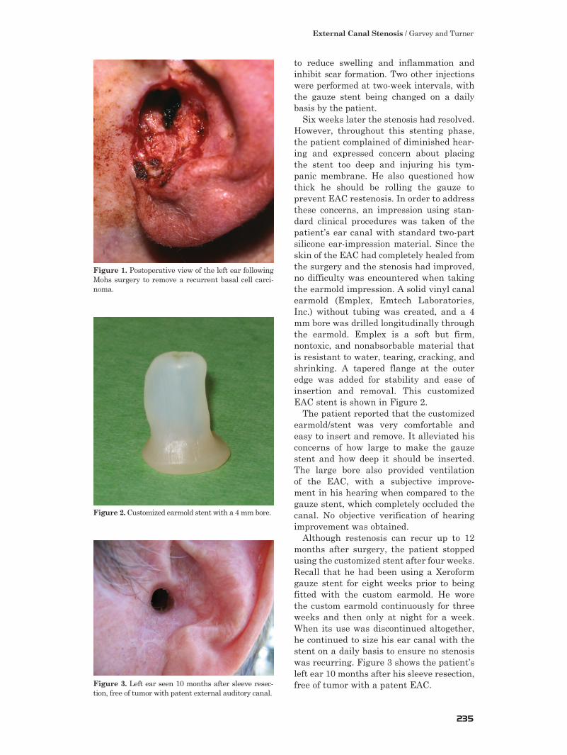
In April 1995, basal cell carcinoma (BCC), the most common form of skin cancer, was detected on the patient’s left ear. A spe-cialized dermatologic surgical procedure designed to preserve as much of the sur-rounding normal tissue, known as Mohs surgery, was performed. A 2.5 × 2.1 cm defect around the external auditory meatus was left open to heal by secondary intention (or left to heal on its own) and is shown in Figure 1. Three months later a near total lateral EAC stenosis developed, leaving only a pinhead opening with daily intermittent drainage. The stenotic region was completely excised, and a complicated closure using adjacent skin flaps was performed to reestablish EAC pat-ency. Xeroform gauze packing was used as a stent postoperatively for two weeks.
The patient was followed closely for 10 years by several dermatologists and did well until February 2006, when another left EAC BCC was identified. A local Mohs surgeon was unable to clear margins, or completely excise the skin cancer, as the tumor spread deep into the EAC. The patient was subse-quently referred to the first author, and a sleeve resection (removal of the skin lining his entire EAC) was performed in March 2006, with the resected margins now free of tumor. A split-thickness skin graft was used to line the EAC. Xeroform gauze was used as a stent postoperatively for a month and then removed.
Three weeks after removing the stent, a soft stenosis developed, narrowing the patient’s EAC meatus and lateral canal by 50 percent. This immature stenotic area was injected circumferentially with 0.5 ml of tri-amcinolone (40 mg/ml), and a Xeroform gauze stent was inserted. Triamcinolone is a corti-costeroid commonly injected intralesionally
235
External Canal Stenosis / Garvey and Turner
to reduce swelling and inflammation and inhibit scar formation. Two other injections were performed at two-week intervals, with the gauze stent being changed on a daily basis by the patient.
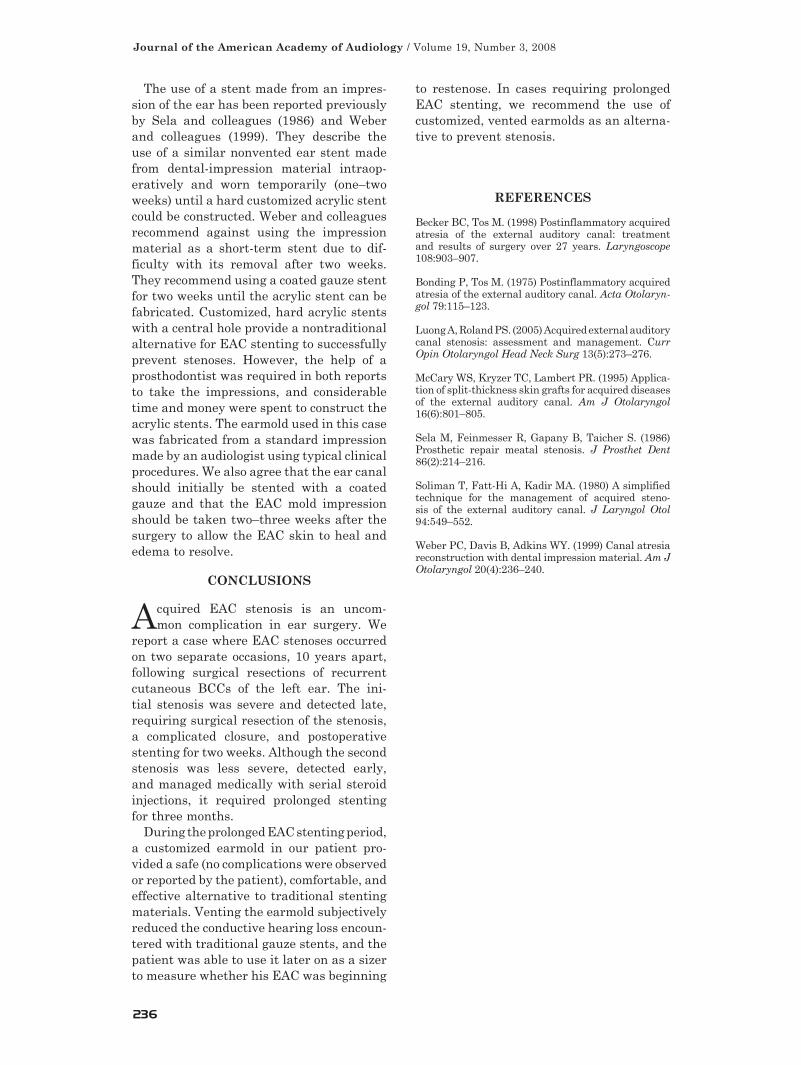
Six weeks later the stenosis had resolved. However, throughout this stenting phase, the patient complained of diminished hear-ing and expressed concern about placing the stent too deep and injuring his tym-panic membrane. He also questioned how thick he should be rolling the gauze to prevent EAC restenosis. In order to address these concerns, an impression using stan-dard clinical procedures was taken of the patient’s ear canal with standard two-part silicone ear-impression material. Since the skin of the EAC had completely healed from the surgery and the stenosis had improved, no difficulty was encountered when taking the earmold impression. A solid vinyl canal earmold (Emplex, Emtech Laboratories, Inc.) without tubing was created, and a 4 mm bore was drilled longitudinally through the earmold. Emplex is a soft but firm, nontoxic, and nonabsorbable material that is resistant to water, tearing, cracking, and shrinking. A tapered flange at the outer edge was added for stability and ease of insertion and removal. This customized EAC stent is shown in Figure 2.
The patient reported that the customized earmold/stent was very comfortable and easy to insert and remove. It alleviated his concerns of how large to make the gauze stent and how deep it should be inserted. The large bore also provided ventilation of the EAC, with a subjective improve-ment in his hearing when compared to the gauze stent, which completely occluded the canal. No objective verification of hearing improvement was obtained.
Although restenosis can recur up to 12 months after surgery, the patient stopped using the customized stent after four weeks. Recall that he had been using a Xeroform gauze stent for eight weeks prior to being fitted with the custom earmold. He wore the custom earmold continuously for three weeks and then only at night for a week. When its use was discontinued altogether, he continued to size his ear canal with the stent on a daily basis to ensure no stenosis was recurring. Figure 3 shows the patient’s left ear 10 months after his sleeve resection, free of tumor with a patent EAC.
Figure 2. Customized earmold stent with a 4 mm bore.
Figure 3. Left ear seen 10 months after sleeve resec-tion, free of tumor with patent external auditory canal.
Figure 1. Postoperative view of the left ear following Mohs surgery to remove a recurrent basal cell carci-noma.
Journal of the American Academy of Audiology / Volume 19, Number 3, 2008
236
The use of a stent made from an impres-sion of the ear has been reported previously by Sela and colleagues (1986) and Weber and colleagues (1999). They describe the use of a similar nonvented ear stent made from dental-impression material intraop-eratively and worn temporarily (one–two weeks) until a hard customized acrylic stent could be constructed. Weber and colleagues recommend against using the impression material as a short-term stent due to dif-ficulty with its removal after two weeks. They recommend using a coated gauze stent for two weeks until the acrylic stent can be fabricated. Customized, hard acrylic stents with a central hole provide a nontraditional alternative for EAC stenting to successfully prevent stenoses. However, the help of a prosthodontist was required in both reports to take the impressions, and considerable time and money were spent to construct the acrylic stents. The earmold used in this case was fabricated from a standard impression made by an audiologist using typical clinical procedures. We also agree that the ear canal should initially be stented with a coated gauze and that the EAC mold impression should be taken two–three weeks after the surgery to allow the EAC skin to heal and edema to resolve.
CONCLUSIONS
Acquired EAC stenosis is an uncom-mon complication in ear surgery. We
report a case where EAC stenoses occurred on two separate occasions, 10 years apart, following surgical resections of recurrent cutaneous BCCs of the left ear. The ini-tial stenosis was severe and detected late, requiring surgical resection of the stenosis, a complicated closure, and postoperative stenting for two weeks. Although the second stenosis was less severe, detected early, and managed medically with serial steroid injections, it required prolonged stenting for three months.
During the prolonged EAC stenting period, a customized earmold in our patient pro-vided a safe (no complications were observed or reported by the patient), comfortable, and effective alternative to traditional stenting materials. Venting the earmold subjectively reduced the conductive hearing loss encoun-tered with traditional gauze stents, and the patient was able to use it later on as a sizer to measure whether his EAC was beginning
to restenose. In cases requiring prolonged EAC stenting, we recommend the use of customized, vented earmolds as an alterna-tive to prevent stenosis.
REFERENCES
Becker BC, Tos M. (1998) Postinflammatory acquired atresia of the external auditory canal: treatment and results of surgery over 27 years. Laryngoscope 108:903–907.
Bonding P, Tos M. (1975) Postinflammatory acquired atresia of the external auditory canal. Acta Otolaryn-gol 79:115–123.
Luong A, Roland PS. (2005) Acquired external auditory canal stenosis: assessment and management. Curr Opin Otolaryngol Head Neck Surg 13(5):273–276.
McCary WS, Kryzer TC, Lambert PR. (1995) Applica-tion of split-thickness skin grafts for acquired diseases of the external auditory canal. Am J Otolaryngol 16(6):801–805.
Sela M, Feinmesser R, Gapany B, Taicher S. (1986) Prosthetic repair meatal stenosis. J Prosthet Dent 86(2):214–216.
Soliman T, Fatt-Hi A, Kadir MA. (1980) A simplified technique for the management of acquired steno-sis of the external auditory canal. J Laryngol Otol 94:549–552.
Weber PC, Davis B, Adkins WY. (1999) Canal atresia reconstruction with dental impression material. Am J Otolaryngol 20(4):236–240.