The transcriptional responsiveness of LKB1 to STAT-mediated signaling is differentially modulated by...
19
RESEARCH ARTICLE Open Access The transcriptional responsiveness of LKB1 to STAT-mediated signaling is differentially modulated by prolactin in human breast cancer cells Katja Linher-Melville and Gurmit Singh * Abstract Background: Liver kinase 1 (LKB1) is an important multi-tasking protein linked with metabolic signaling, also controlling polarity and cytoskeletal rearrangements in diverse cell types including cancer cells. Prolactin (PRL) and Signal transducer and activator of transcription (STAT) proteins have been associated with breast cancer progression. The current investigation examines the effect of PRL and STAT-mediated signaling on the transcriptional regulation of LKB1 expression in human breast cancer cells. Methods: MDA-MB-231, MCF-7, and T47D human breast cancer cells, and CHO-K1 cells transiently expressing the PRL receptor (long form), were treated with 100 ng/ml of PRL for 24 hours. A LKB1 promoter-luciferase construct and its truncations were used to assess transcriptional changes in response to specific siRNAs or inhibitors targeting Janus activated kinase 2 (JAK2), STAT3, and STAT5A. Real-time PCR and Western blotting were applied to quantify changes in mRNA and protein levels. Electrophoretic mobility shift (EMSA) and chromatin immunoprecipitation (ChIP) assays were used to examine STAT3 and STAT5A binding to the LKB1 promoter. Results: Consistent with increases in mRNA, the LKB1 promoter was up-regulated by PRL in MDA-MB-231 cells, a response that was lost upon distal promoter truncation. A putative GAS element that could provide a STAT binding site mapped to this region, and its mutation decreased PRL-responsiveness. PRL-mediated increases in promoter activity required signaling through STAT3 and STAT5A, also involving JAK2. Both STATs imparted basally repressive effects in MDA-MB-231 cells. PRL increased in vivo binding of STAT3, and more definitively, STAT5A, to the LKB1 promoter region containing the GAS site. In T47D cells, PRL down-regulated LKB1 transcriptional activity, an effect that was reversed upon culture in phenol red-free media. Interleukin 6, a cytokine activating STAT signaling in diverse cell types, also increased LKB1 mRNA levels and promoter activity in MDA-MB-231 cells. Conclusions: LKB1 is differentially regulated by PRL at the level of transcription in representative human breast cancer cells. Its promoter is targeted by STAT proteins, and the cellular estrogen receptor status may affect PRL-responsiveness. The hormonal and possibly cytokine-mediated control of LKB1 expression is particularly relevant in aggressive breast cancer cells, potentially promoting survival under energetically unfavorable conditions. Keywords: Breast cancer, STAT3, STAT5, LKB1, Prolactin, Interleukin 6, Promoter, Transcriptional regulation * Correspondence: [email protected] Department of Pathology and Molecular Medicine, McMaster University, Hamilton, Ontario, Canada © 2014 Linher-Melville and Singh; licensee BioMed Central Ltd. This is an Open Access article distributed under the terms of the Creative Commons Attribution License (http://creativecommons.org/licenses/by/2.0), which permits unrestricted use, distribution, and reproduction in any medium, provided the original work is properly credited. The Creative Commons Public Domain Dedication waiver (http://creativecommons.org/publicdomain/zero/1.0/) applies to the data made available in this article, unless otherwise stated. Linher-Melville and Singh BMC Cancer 2014, 14:415 http://www.biomedcentral.com/1471-2407/14/415
-
Upload
gurmit-singh -
Category
Documents
-
view
215 -
download
3
Transcript of The transcriptional responsiveness of LKB1 to STAT-mediated signaling is differentially modulated by...

RESEARCH ARTICLE Open Access
The transcriptional responsiveness of LKB1 toSTAT-mediated signaling is differentiallymodulated by prolactin in human breast cancercellsKatja Linher-Melville and Gurmit Singh*
Abstract
Background: Liver kinase 1 (LKB1) is an important multi-tasking protein linked with metabolic signaling, alsocontrolling polarity and cytoskeletal rearrangements in diverse cell types including cancer cells. Prolactin (PRL) andSignal transducer and activator of transcription (STAT) proteins have been associated with breast cancer progression.The current investigation examines the effect of PRL and STAT-mediated signaling on the transcriptional regulationof LKB1 expression in human breast cancer cells.
Methods: MDA-MB-231, MCF-7, and T47D human breast cancer cells, and CHO-K1 cells transiently expressing thePRL receptor (long form), were treated with 100 ng/ml of PRL for 24 hours. A LKB1 promoter-luciferase constructand its truncations were used to assess transcriptional changes in response to specific siRNAs or inhibitors targetingJanus activated kinase 2 (JAK2), STAT3, and STAT5A. Real-time PCR and Western blotting were applied to quantifychanges in mRNA and protein levels. Electrophoretic mobility shift (EMSA) and chromatin immunoprecipitation(ChIP) assays were used to examine STAT3 and STAT5A binding to the LKB1 promoter.
Results: Consistent with increases in mRNA, the LKB1 promoter was up-regulated by PRL in MDA-MB-231 cells, aresponse that was lost upon distal promoter truncation. A putative GAS element that could provide a STAT bindingsite mapped to this region, and its mutation decreased PRL-responsiveness. PRL-mediated increases in promoteractivity required signaling through STAT3 and STAT5A, also involving JAK2. Both STATs imparted basally repressiveeffects in MDA-MB-231 cells. PRL increased in vivo binding of STAT3, and more definitively, STAT5A, to the LKB1promoter region containing the GAS site. In T47D cells, PRL down-regulated LKB1 transcriptional activity, an effectthat was reversed upon culture in phenol red-free media. Interleukin 6, a cytokine activating STAT signaling indiverse cell types, also increased LKB1 mRNA levels and promoter activity in MDA-MB-231 cells.
Conclusions: LKB1 is differentially regulated by PRL at the level of transcription in representative human breastcancer cells. Its promoter is targeted by STAT proteins, and the cellular estrogen receptor status may affectPRL-responsiveness. The hormonal and possibly cytokine-mediated control of LKB1 expression is particularly relevantin aggressive breast cancer cells, potentially promoting survival under energetically unfavorable conditions.
Keywords: Breast cancer, STAT3, STAT5, LKB1, Prolactin, Interleukin 6, Promoter, Transcriptional regulation
* Correspondence: [email protected] of Pathology and Molecular Medicine, McMaster University,Hamilton, Ontario, Canada
© 2014 Linher-Melville and Singh; licensee BioMed Central Ltd. This is an Open Access article distributed under the terms ofthe Creative Commons Attribution License (http://creativecommons.org/licenses/by/2.0), which permits unrestricted use,distribution, and reproduction in any medium, provided the original work is properly credited. The Creative Commons PublicDomain Dedication waiver (http://creativecommons.org/publicdomain/zero/1.0/) applies to the data made available in thisarticle, unless otherwise stated.
Linher-Melville and Singh BMC Cancer 2014, 14:415http://www.biomedcentral.com/1471-2407/14/415

BackgroundProlactin (PRL) affects a range of physiological processesto maintain homeostasis, playing important roles inthe mammary gland (reviewed in [1]) and influencingreproduction, maternal behavior, the immune system,osteogenesis, blood vessel development, ion transport,and metabolism, among other diverse functions (reviewedin [2-5]). PRL has been definitively associated with the on-set and progression of human breast cancer by increasingcell proliferation (reviewed in [6-8]), and may contributeto metastasis by inducing the motility of human breastcancer cells [9]. The human PRL receptor (PRLR) iswidely expressed in diverse tissues, and signaling throughPRLR initiates activation of several intracellular pathways,the most well-characterized being the Janus activated kin-ase (JAK)/signal transducer and activator of transcription(STAT) pathway (reviewed in [3,10]). Some of the keyevents that occur in the normal mammary gland duringpregnancy, lactation, and involution, as well as in adipo-cytes and during tumorigenesis in the breast, are regulatedby STAT proteins [2-4,7,10]. The activation of cytokinereceptors, including PRLR, in response to ligand bind-ing typically results in phosphorylation and activationof JAK/STAT. STATs dimerize, translocate to the nucleus,and bind to specific recognition sequences in the pro-moter regions of select target genes, thereby activatingor repressing transcription [11,12]. Seven mammalianSTAT proteins have been identified. STAT2 is activated byα/β interferon, STAT4 by interleukin (IL)-12, and STAT6by IL-4 to IL-13, while STAT1, STAT3, STAT5A, andSTAT5B are activated by a range of stimuli, including PRLand IL-6 [13,14]. Targeting Jak2 may protect againstthe onset of mammary tumorigenesis in mice [15,16], andvarious STAT proteins have also been associated withbreast cancer. In particular, STAT3 and STAT5 are gener-ally thought to mediate opposite effects in mammary car-cinoma cells [17]. Several negative regulators of JAK/STATsignaling have been identified that are induced differentlyin a cell type-dependent manner. STAT activation mayupregulate the expression of members of the Suppressorsof cytokine signalling (SOCS) family [18,19]. Other inhibi-tors include the phosphatase SHP-1 and Protein inhibitorsof activated STAT (PIAS), which specifically targets STAT3[20], providing another level of complexity in regulatingJAK/STAT signal transduction.A novel mechanism by which PRL may contribute to
breast cancer progression is through its action on liver kin-ase 1 (LKB1). Acting either as a kinase or by changing itssubcellular localization, LKB1 has been associated with pro-liferation, cell cycle arrest, apoptosis, polarity/motility, andenergy metabolism (reviewed in [21]), and has been de-scribed as a tumor suppressor during pulmonary tumorigen-esis [22]. However, it has also been suggested that LKB1 isrequired to protect cells from apoptosis during energy stress
by initiating adenosine monophosphate-activated proteinkinase (AMPK) signaling, leading to suppression of mTORand the activation of ATP-producing pathways [23-25].The LKB1-AMPK pathway has been described as a meansto rescue cancer cells from metabolic collapse [21]. We havepreviously shown that PRL activates the AMPK pathway inan LKB1-dependent manner in specific human breast cancercell lines, most notably MDA-MB-231 cells [26].Little is currently known regarding how the expression of
LKB1 is regulated. One means of repression is through pro-moter methylation [27,28], and the LKB1 promoter hasbeen reported to be hypermethylated in colorectal carcin-omas and testicular tumors, although out of 51 cancer celllines analyzed in vitro, only one cervical carcinoma andthree colorectal cell lines were methylated at the LKB1locus, also corresponding to loss of expression [27]. Estro-gen may be an important regulator, as multiple estrogen re-sponse elements (EREs) within the human LKB1 promoterregion confer a repressive action in estrogen receptor (ER)-positive MCF-7 human breast cancer cells [29]. We haveshown previously that levels of total LKB1 mRNA and pro-tein increase in MDA-MB-231 cells cultured in the pres-ence of PRL [26]. Similar to PRL-responsive promoters thatcontain potential STAT binding sites, such as those control-ling expression of the β-casein [30,31], cyclin D1 [32,33],fatty acid synthase [34], and pyruvate dehydrogenase kinase(PDK4) genes [35], a putative STAT binding/interferongamma-activated sequence (GAS) motif in the distal hu-man LKB1 promoter region was identified by computa-tional analysis. The presence of this putative site suggestedthat LKB1 transcriptional activity could be regulated bySTAT proteins. Others have shown that PRL, throughJAK2, induces binding of STAT5 to a distal GAS site in thecyclin D1 promoter, thereby enhancing promoter activity inChinese hamster ovary (CHO-K1) cells transfected with thelong form (LF) of PRLR [32]. In adipocytes, STAT5A bindsto a putative STAT site in the PDK4 promoter in responseto PRL stimulation [35]. In the current investigation, weaimed to investigate the importance of the GAS site in thedistal human LKB1 promoter region, and the potentialmechanisms underlying the responsiveness of LKB1 toPRL, in a representative triple-negative breast cancer cellline. Our findings demonstrate that changes in LKB1 ex-pression are, at least in part, transcriptionally regulated bySTAT3, as well as STAT5A. Identifying the mechanismsthat underlie the regulation of LKB1 expression in differentbreast cancer cells may provide new insights into how thisprotein responds to different stimuli, including PRL orother cytokines such as IL-6.
MethodsMaterialsAntibodies for total LKB1, total and phospho-JAK2,STAT3, STAT5, and ACC, and β-tubulin, β-catenin, and
Linher-Melville and Singh BMC Cancer 2014, 14:415 Page 2 of 19http://www.biomedcentral.com/1471-2407/14/415

calnexin were obtained from Cell Signaling Technologies,Inc, and Actin was from MP Biochemicals. The humanPRLR antibody was purchased from R&D Systems.Individual aliquots of recombinant human PRL (Cedarlane,Lot #608PRL01) or recombinant human IL-6 (R&DSystems) were prepared at a concentration of 100 μg/mLby reconstituting the lyophilates in sterile water or sterilePBS with 0.1% BSA, respectively, and stored at −20°C. TheSTAT3 pathway inhibitor (E)-3(6-bromopyridin-2-yl)-2-cyano-N-((S0-1-phenylethyl)acrylamide) (WP1066) (Sigma),STAT5 inhibitor (Calbiochem), and MEK1/2 inhibitorPD098059 (NEB) were reconstituted in DMSO, individualaliquots were stored at −20°C, and cells were pretreatedwith vehicle or an appropriate working concentration for1 hr at 37°C in 5% CO2 prior to addition of PRL for 24 hr.Cells were pretreated with 5 μM of WP1066, a concentra-tion that was experimentally determined to be effective atdegrading JAK2 protein and blocking STAT3 phosphoryl-ation in MDA-MB-231 cells. The STAT5 inhibitor was usedto treat cells at a 50 μM final concentration (Calbiochem),whilePD098059 was used at 20 μM [32]. Cells were pre-treated with 10 μg of Actinomycin D (Sigma) for 1 hrprior to culture in the presence of PRL for 24 hr.
Plasmid constructsThe cloning of the full-length LKB1 construct from −1889/+1109 into pGL3-Basic (Promega) and construction of theLKB1Δ-1083 truncation reporter construct were describedpreviously [29]. The pRL-TK Renilla luciferase constructwas obtained from Dr. Julang Li (University of Guelph).Mutation of the GAS site (5’-TTCCAAGAA-3’) within thedistal LKB1 promoter region at -1152 was accomplishedusing the Site-Directed Mutagenesis kit (Stratagene)and complementary mutant oligonucleotides correspondingto the sequence 5′-CCAGCATTATCTCCAGATTagtttAAGTTGGGGTGTGAGCCAG-3′ (the GAS site is italics;mutated base pairs in lowercase letters). Mutations wereconfirmed by bi-directional sequencing. The human PRLRLF (1869 bp of the coding sequence, GeneBank AccessionM31661.1, GI:190361) [36] was PCR amplified from cDNAderived from MDA-MB-231 cells using the primers PRLR-LF-FOR (5’-ATGAAGGAAAATGTGGCATCTGC-3’) andPRLR-LF-REV (5’-TCAGTGAAAGGAGTGTGTAAAACATG-3’), and the resulting product was confirmed bysequencing and expressed in pcDNA3.1.
Cell culture and transient transfectionsAll human cell lines were used in accordance with in-stitutional biosafety guidelines. MDA-MB-231 humanbreast cancer cells at low passage (less than 20 passages,ATCC #HTB-26) were maintained in DMEM supplementedwith 10% FBS, and Chinese hamster ovary (CHO-K1) cells(ATCC #CCL-61) were cultured in DMEM/F12 containing5% FBS and penicillin/streptomycin. T47D cells were
maintained in RPMI-1640 with 10% FBS, in eithermedia containing phenol red or without phenol red.For assays, cells were plated into 6-well tissue culture-treated plates (Falcon) at 2.5 × 10⁵ cells/well 24 hr prior tomanipulation. Cells were transfected using Lipofectamine2000 (Invitrogen) as described previously [29]. To assessviable cell proliferation, cells were counted using a haemo-cytometer and trypan blue staining.
Reporter gene assaysLuciferase activity of cell lysates was determined aspreviously described [29] using the Dual LuciferaseAssay (Promega) and a Berthold luminometer. Luciferasevalues were corrected for transfection efficiency by de-termining the ratio of firefly/Renilla luciferase activityand expressed as relative units. All data were normalizedto untreated pGL3-Basic.
siRNAsExperimentally verified siRNAs for JAK2 (Hs_JAK2_7),STAT3 (Hs_STAT3_7), STAT5A (Hs_STAT5A_2), LKB1(Hs_STK11_7), and a negative control (Ctrl_Control_1)were obtained from Qiagen. Transient transfections werecarried out as described previously using Hiperfect re-agent (Qiagen). MDA-MB-231 cells plated into 6-wellplates at 1.25 × 10⁵ cells/well 3 hr prior to treatmentwith siRNAs [26,29].
Real time PCRcDNA was prepared and quantitative real time PCR wascarried out using primers to amplify human LKB1 andthe RNA polymerase II housekeeping genes, which werepreviously optimized [26]. Primers described by others[37,38], resulting in a 200 bp product, were used toquantify mRNA levels of the human PRLR LF. RelativemRNA levels were calculated using the 2-[Δ][Δ]Ct method[39], and results are presented as fold changes relative tountreated controls.
Western blottingTotal cell lysates were prepared as described previously[26,29]. 50 μg of protein was subjected to SDS-PAGEelectrophoresis on 10% polyacrylamide gels and trans-ferred onto PVDF membranes, which were blocked innon-fat dry milk, incubated in 1:1000 diluted primaryantibody, followed by incubation with the appropriateanti-rabbit IgG horseradish peroxidise (HRP) secondaryantibody (1:3000, Cell Signaling Technology). Signalswere detected using the ECL Plus Western BlottingDetection System (Amersham Biosciences) and exposedto film. Stripped membranes were re-probed with primaryanti-Actin antibody and anti-mouse IgG-HRP.
Linher-Melville and Singh BMC Cancer 2014, 14:415 Page 3 of 19http://www.biomedcentral.com/1471-2407/14/415

DensitometryDensitometric analyses of blots were performed usingImage J analysis software. Values were expressed as apercent change over the control value and are repre-sented as the mean ± SE of at least 3 independent exper-iments. For total and phosphorylated proteins, valueswere corrected relative to actin and relative to totalprotein/actin, respectively.
Co-ImmunoprecipitationFollowing various treatments, cells were lysed in 1X lysisbuffer supplemented with protease inhibitors. 100 μg ofnon-sonicated, cleared lysate in a final volume of 200 μl(following a protocol provided by Cell Signaling Technology)were incubated with 2 μl of antibody against total JAK2overnight at 4°C with end-over-end rotation, followed bythe addition of 20 μl of protein A/G agarose (Invitrogen)and further incubation at 4°C for 3 hr. Samples werewashed 5 times with lysis buffer prior to adding 4XSDS-sample buffer and boiling. The signal was detectedfollowing Western blotting with anti-JAK2 or anti-phospho-JAK2 primary antibodies and incubation with anti-rabbitIgG-HRP. As a negative control, normal rabbit IgG(SC-2027; Santa Cruz Biotechnology, Inc.) was used insteadof specific antibody in one IP for each group of cells. Apositive control was included during Western blotting, re-ferred to as input, which represented 10% of cleared lysate.
Preparation of nuclear extractsCells were cultured in 10-cm dishes in the absence andpresence of 100 ng/mL of PRL for 24 hr before harvest-ing nuclear extracts using the NE-PER Cytoplasmic andNuclear Extraction Reagents kit (Pierce) following themanufacturer’s protocol. Protein concentrations of nuclearextracts were determined using a Bradford assay.
EMSAProbe preparation and EMSAs were performed as previ-ously described [40] using the DNA 3’ End Biotinylationkit (Pierce) and the LightShift Chemiluminescent EMSAkit (Pierce). EMSA probes consisted of biotinylated double-stranded oligonucleotides. Probe sequences are listed inTable 1, with the GAS and GASmut sequences in bolditalics. For competitor assays, 200-fold molar excess ofunlabeled, double-stranded probe, corresponding to 4pmol, was included in EMSA reactions.
ChIP assaysChIP assays were carried out using the ChIP-IT ExpressEnzymatic kit (Active Motif ) using a dounce homogini-zer to lyse cells. Optimal enzymatic digestion of chroma-tin from MDA-MB-231 cells was empirically determinedto occur after 10 min, yielding sheared chromatin thatmigrated between 200 and 1500 bp on an agarose gel.Equal DNA concentrations corresponding to 1.5 μg wereapplied to each set of immunoprecipitation reactions,which included either normal rabbit IgG, STAT3, orSTAT5A antibody (sc-2027, sc-7179X, or sc-1081X, re-spectively; Santa Cruz Biotechnology). Samples wereincubated with magnetic beads overnight at 4°C withend-over-end rotation. After reversal of cross-links, DNAprecipitation, and clean-up, enriched DNA and input wereanalyzed by quantitative real time PCR with primers span-ning the predicted GAS site, as well as primers specific to aregion of the LKB1 promoter that does not contain a puta-tive STAT binding motif (Table 2). The efficiency of eachprimer set was tested by producing a standard curve fromtwo-fold dilutions of input, and the integrity of productswas verified by agarose gel electrophoresis. Fold enrichmentrelative to IgG was calculated for immunoprecipitated sam-ples, and data are presented normalized to values obtainedfor the negative binding region.
Statistical analysesResults represent the mean ± SEM of at least threeindependent replicates, and were analyzed by t-test(denoted by stars) or 1-way ANOVA with a Tukey’spost-test (denoted by different letters) to assess statis-tical differences between groups using GraphPad Prismsoftware. Results were considered significant at p <0.05.For qualitative assays, including Western blots and EMSAs,the results shown are representative of at least two in-dependent experiments.
ResultsLKB1 plays an important role in MDA-MB-231 humanbreast cancer cellsWe previously showed that LKB1 contributes to AMPKpathway activation in human breast cancer cells [26]. Inthe current study, we demonstrated that, beyond modu-lating cellular metabolism, LKB1 may also be importantin regulating cell morphology. When cultured in DMEMsupplemented with 10% FBS, untreated MDA-MB-231
Table 1 EMSA probes
Probe Sequence (5’-3’) Length
GAS AGCATTATCTCCAGATACCAAGGGGTTGGGGTGTGAGCCA 40 bp
GASmut AGCATTATCTCCAGATTAGTTTAAGTTGGGGTGTGAGCCA 40 bp
Oct1 (non-specific) AGAGGATCCATGCAAATGGACGTACG 26 bp
Linher-Melville and Singh BMC Cancer 2014, 14:415 Page 4 of 19http://www.biomedcentral.com/1471-2407/14/415

cells display two distinct cell types, one spindle-shapedand the other more rounded. Knocking down LKB1 re-sulted in distinct morphological changes, with cells be-coming more rounded compared to cells treated with anon-specific negative control siRNA (Figure 1A). Cellnumber or viability, which was assessed by trypan blueexclusion, were not affected (Figure 1A). LKB1 is known toaffect cell polarity and motility, and interestingly, its knock-down resulted in decreased expression of β-tubulin, an im-portant component of the cytoskeleton, at the protein level,without affecting the expression of other proteins, includ-ing actin and calnexin (Figure 1B). In addition, levels of β
catenin, an epithelial marker that has also been implicatedin WNT signaling, were also decreased (Figure 1B). Itappears that LKB1 regulates several important cellularprocesses in human breast cancer cells, warranting fur-ther investigation into how its expression is controlled.
MDA-MB-231 cells express the PRLR and are responsiveto PRLOur previous work demonstrated that PRL activatesLKB1-AMPK-ACC signaling in MDA-MB-231 cells. PRLelicits cellular responses through the PRLR, with differ-ent receptor isoforms sharing common extracellular lig-and binding and transmembrane domains, differingonly in their intracellular regions due to alternative spli-cing. In humans, the known PRLR isoforms include theLF, as well as the delta S1, intermediate, and short forms(ΔS1, IF, SF1a and SF1b, respectively) and the PRLRbinding protein (reviewed in [10]). We verified that PRLhas the potential to directly signal through the PRLR inMDA-MB-231 cells by examining receptor mRNA and
Table 2 Primers for ChIP
Probe Sequence (5’-3’) Product size
LKB1-GAS-FOR GGACCTACCGATGCCAATTA 184 bp
LKB1-GAS-REV TGGGCAATAAGAGCGAAACT
LKB1-Neg-FOR GAGGACGAAGTTGACCCTGA 208 bp
LKB1-Neg-REV CAACAAAAACCCCAAAAGGA
NS LA
B
NS L0
1
2
3
4C
ell N
um
ber
(x1
05 )
NS L
Total LKB1
Actin
54 kDa
42 kDa
50 kDaβ-Tubulin
125 kDa
79 kDa
Total JAK2
Total STAT3
β-Catenin 92 kDa
Calnexin 90 kDa
Figure 1 LKB1 is functionally important in MDA-MB-231 human breast cancer cells. (A) Knock-down of LKB1 using a specific siRNA inMDA-MB-231 cells results in distinct morphological changes without affecting the total number of viable cells compared to cells treated with anon-specific (NS) siRNA. 10X magnification of live cells using a Leica DMIL microscope. (B) A representative Western blot demonstrating that lossof LKB1 reduces β-tubulin and β-catenin protein levels without affecting the expression of other proteins.
Linher-Melville and Singh BMC Cancer 2014, 14:415 Page 5 of 19http://www.biomedcentral.com/1471-2407/14/415

protein levels using T47D cells as a positive control forhigh expression of the LF. PRLR LF mRNA was detectedin MDA-MB-231 cells (Figure 2A), consistent with reportsby others [37,38]. Its expression at the protein level was
assessed using the monoclonal anti-human PRLR anti-body, which specifically recognizes the extracellular do-main common to all known isoforms (R&D Systems, Inc.).Differences in mRNA levels were reflected at the protein
Figure 2 PRL has the potential to directly signal to LKB1 in MDA-MB-231 cells. (A) The PRLR LF is expressed at the mRNA level in representativebreast cancer cells including MDA-MB-231 cells and 184B5 normal breast epithelial cells, while levels are close to undetectable in A549 lung cancercells, as assessed by quantitative real time PCR. (B) Various isoforms of the PRLR are potentially expressed at the protein level in 184B5, MCF-7, andMDA-MB-231 cells. The LF migrates at the expected molecular weight of 85-90 kDa, similar to the band obtained in T47D cells, which express highlevels of the LF, and (C) is comparable to migration in CHO-K1 cells transiently transfected with an expression vector encoding the LF of PRLR. (D)Representative Western blots of a time-course demonstrating that JAK2, STAT3, and STAT5 are phosphorylated in MDA-MB-231 cells cultured with100 ng/mL of PRL for 24 hr. (E) Co-immunoprecipitations (IPs) were carried out using equal amounts of total cell lysates followed by Western blotting(WB). IPs with total JAK2 followed by WB with phospho- and total JAK2 were performed on lysates from 184B5, MCF-7, and MDA-MB-231 cells. I: 10%of total non-IP lysate or “input” as a positive control, -: no treatment, +: treated with 100 ng/mL of PRL for 24 hr, ++: pre-treated with 5 μM WP1066 for2 hr followed by the addition of PRL for 24 hr. (F) PRL also temporally induced inactivation (phosphorylation) of ACC.
Linher-Melville and Singh BMC Cancer 2014, 14:415 Page 6 of 19http://www.biomedcentral.com/1471-2407/14/415

level, with the LF migrating at approximately 85–90 kDa(Figure 2B). Additional bands were also present, whichcould either be non-specifics or other PRLR isoforms. It ispossible that breast cancer cells could also express ΔS1, IF,SF1a, SF1b, or PRLRBP, as bands that correspond to theirexpected molecular weights were detected at 70, 50, 56,42, and 32 kDa, respectively. To confirm the functionalpresence of PRLR in MDA-MB-231 cells, we comparedprotein levels to exogenously introduced PRLR LF expres-sion in CHO-K1 cells, which exhibit low levels of en-dogenous PRLR (reviewed in [10]). Transient transfectionof CHO-K1s with a mammalian expression vector encod-ing the full-length coding sequence of the human PRLRLF resulted in an approximately 2-fold increase in recep-tor levels compared to cells transfected with either emptyvector (pcDNA3.1) or PRLR-SF1b encoding a shortisoform (Figure 2C). Bands for the LF were detected at85–90 kDa, consistent with migration of the endogenousband present at a similar molecular weight in MDA-MB-231 cells (Figure 2C).We next examined potential signaling through STATs,
as these proteins are commonly activated in response toPRL stimulation in cells that express the PRLR. A timecourse revealed that PRL induces a gradual increase inJAK2 and STAT3 phosphorylation in MDA-MB-231cells in the presence of 100 ng/mL of PRL (Figure 2D).Densitometric analysis revealed that at 24 hr, the presenceof PRL in the culture media increased phospho-JAK2levels by 1.5-fold (p < 0.02) and phospho-STAT3 levelsby 2.8-fold (p < 0.01) relative to time 0 (Figure 2D). Anincrease in phospho-STAT5 levels also occurred in re-sponse to PRL in MDA-MB-231 cells, although levels werevery low. To confirm the phosphorylation of JAK2, we per-formed an immunoprecipitation (IP) for total JAK2 on ly-sates derived from 184B5, MCF-7, and MDA-MB-231 cellstreated without and with PRL for 24 hr, or pretreatedwith WP1066, a drug that degrades total JAK2 protein,followed by Western blotting to detect both phospho-and total JAK2 (Figure 2E). IP of JAK2 in MDA-MB-231cells confirmed its increased activation in the presenceof PRL. Consistent with our previous findings [26], PRLinactivated ACC, temporally increasing its phosphorylationby 2.8-fold at 24 hr (p < 0.02) (Figure 2F).
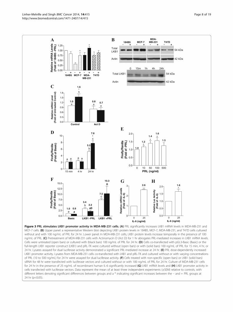
The LKB1 promoter is a target for PRL-mediated signalingWe have shown previously that PRL is able to up-regulateLKB1 protein levels in MDA-MB-231 cells [26]. A sig-nificant increase in LKB1 expression at the mRNAlevel was observed in MCF-7 and MDA-MB-231 cellsfollowing sustained PRL treatment, although no changeswere observed in 184B5 normal breast epithelial cells,and only a very minor increase occurred in T47D cells(Figure 3A). These changes were reflected at the proteinlevel (Figure 3B), and a time course in MDA-MB-231
cells revealed that maximal increases in LKB1 protein levelsoccurred after a 24 hr culture in the presence of PRL(Figure 3B). We therefore examined the potential in-volvement of PRL in regulating LKB1 expression at thetranscriptional level. As shown in Figure 3C, 100 ng/mLof PRL significantly increased LKB1 mRNA levels by ap-proximately 1.5-fold relative to the untreated control inMDA-MB-231 cells (p < 0.01), consistent with results inFigure 3A, while pretreatment with Actinomycin D com-pletely abolished this effect. The transcriptional regulationof LKB1 by PRL was examined further using a humanLKB1 promoter reporter construct, which included theregulatory region spanning −1889 to +1109 cloned up-stream of a firefly luciferase gene [29]. A time courserevealed that cotransfection of MDA-MB-231 cellswith the full-length LKB1 promoter construct signifi-cantly increased luciferase activity by approximately1.5-fold (p < 0.02) after a 24 hr culture in the presenceof 100 ng/mL of PRL (Figure 3D). The effect on LKB1promoter activity was dose-dependent, with a maximal1.6-fold stimulation obtained using 100 ng/mL of PRLfor 24 hr (p < 0.05; Figure 3E). Treatment with PRL alsoincreased LKB1 transcriptional activity in MDA-MB-231cells in which LKB1 was knocked down using a specificsiRNA (Figure 3F), consistent with our previous findings[26]. In addition to PRL, we also examined the responsive-ness of the LKB1 promoter to IL-6, which is also able toactivate JAK/STAT signaling. Treating MDA-MB-231cells with 25 ng/ml of recombinant human IL-6 for 24 hrsignificantly increased LKB1 mRNA levels by 2.6-fold(p < 0.001; Figure 3G), also significantly increasing pro-moter activity by 1.7-fold (p < 0.02; Figure 3H).Computational analysis using NSITE software (Softberry
Inc.) revealed that, in addition to several EREs that wepreviously characterized in MCF-7 cells [29], the LKB1promoter also contains a putative STAT/consensusGAS binding site (TTCNNNGAA) at −1152 bp, as wellas a hypoxia-inducible factor 1 alpha (HIF1α), an acti-vator protein 1 (AP-1), and two octamer-binding tran-scription factor 1 (OCT-1) sites (Figure 4A). The distalGAS site was of particular interest, given that PRL andcytokine stimulation are known to involve the activa-tion and nuclear translocation of STATs, and STATproteins mediate the action of cytokines at similar sitesin other systems. Most STATs bind to consensus GASsites, TTCNmGAA, where m = 4 for STAT6 and m = 3for the optimal binding of all other STATs [41,42]. Thesequence of the putative GAS site present in the LKB1promoter, when reverse complemented, was found to beidentical to both a PRL-responsive distal GAS site locatedin the human cyclin D1 promoter (TTCTTGGAA) [32,33]and a canonical STAT5 binding site (PRE) within theβ-casein promoter [30,31], differing by only one base pairfrom a binding site described for STAT3 (TTCTGGGAA)
Linher-Melville and Singh BMC Cancer 2014, 14:415 Page 7 of 19http://www.biomedcentral.com/1471-2407/14/415
Basic LKB1 -PRL LKB1 +PRL0.0
2.5
5.0
7.5
10.0
12.5
15.0
a
b
c
aab
b
Fir
efly
/Ren
illa
(Fo
ld o
f B
asic
)
1.0 0.9
3.4
7.5
2.2
10.3
D
0 10 50 100 500
0.0
0.5
1.0
1.5
2.0
*
*
*
PRL (ng/ml)
Fire
fly/R
enill
a(F
old
of -
PR
L)
1.0
1.2
1.41.6
1.3
A
Control Act D0.0
0.3
0.6
0.9
1.2
1.5
1.8
a
b
aa
Rel
ativ
e m
RN
A L
evel
(Fo
ld o
f U
ntr
eate
d C
on
tro
l)
1.0
1.6
0.8 0.7
*
Total LKB1
Actin
0 15m 1h 4h 24h
54 kDa
42 kDa
C
F
E
15 min 4 hr 24 hr
0
2
4
6
8
10
a a a a
b
c*
Fir
efly
/Ren
illa
(Fo
ld o
f B
asic
)
5.2
7.8
G
0 25
0
1
2
3
***
IL-6 (ng/ml)
Rel
ativ
e m
RN
A L
evel
s(F
old
of
-IL
-6)
0 250.0
0.5
1.0
1.5
2.0
*
IL-6 (ng/ml)
Rel
ativ
e L
uc
Act
ivit
y(F
old
of
-IL
-6)
H2.6 1.6
0.00
0.25
0.50
0.75
1.00
1.25
Rel
ativ
e m
RN
A L
evel
s(F
old
of
184B
5 -P
RL
)*
*
- + - + - + - +184B5 MCF-7 T47DMDA-
MB-231
B
Total
LKB1
Actin
- +
54 kDa
42 kDa
- + - + - +184B5 MCF-7 T47D
MDA-MB-231
Figure 3 PRL stimulates LKB1 promoter activity in MDA-MB-231 cells. (A) PRL significantly increases LKB1 mRNA levels in MDA-MB-231 andMCF-7 cells. (B) Upper panel: a representative Western blot depicting LKB1 protein levels in 184B5, MCF-7, MDA-MB-231, and T47D cells culturedwithout and with 100 ng/mL of PRL for 24 hr. Lower panel: In MDA-MB-231 cells, LKB1 protein levels increase temporally in the presence of 100ng/mL of PRL. (C) Pretreatment of MDA-MB-231 cells with Actinomycin D (Act D) for 1 hr abrogates PRL-mediated increases in LKB1 mRNA levels.Cells were untreated (open bars) or cultured with (black bars) 100 ng/mL of PRL for 24 hr. (D) Cells co-transfected with pGL3-Basic (Basic) or thefull-length LKB1 reporter construct (LKB1) and pRL-TK were cultured without (open bars) or with (solid bars) 100 ng/mL of PRL for 15 min, 4 hr, or24 hr. Lysates assayed for dual luciferase activity demonstrated a significant PRL-mediated increase at 24 hr. (E) PRL dose-dependently increasedLKB1 promoter activity. Lysates from MDA-MB-231 cells co-transfected with LKB1 and pRL-TK and cultured without or with varying concentrationsof PRL (10 to 500 ng/mL) for 24 hr were assayed for dual luciferase activity. (F) Cells treated with non-specific (open bars) or LKB1 (solid bars)siRNA for 48 hr were transfected with luciferase vectors and cultured without or with 100 ng/mL of PRL for 24 hr. Culture of MDA-MB-231 cellsfor 24 hr in the presence of 25 ng/mL of recombinant human IL-6 significantly increased (G) LKB1 mRNA levels and (H) LKB1 promoter activity incells transfected with luciferase vectors. Data represent the mean of at least three independent experiments (±SEM) relative to controls, withdifferent letters denoting significant differences between groups and a * indicating significant increases between the – and + PRL groups at24 hr (p<0.05).
Linher-Melville and Singh BMC Cancer 2014, 14:415 Page 8 of 19http://www.biomedcentral.com/1471-2407/14/415

[43]. Truncation analysis of the promoter region in MDA-MB-231 cells revealed the presence of a potential silencerelement in the region spanning −1889 to −1083, as loss ofthis 800 bp fragment led to a significant 2-fold increase inpromoter activity (Figure 4B), consistent with our previous
findings reported in MCF-7 cells [29] and results obtainedin T47D cells (Figure 4C). PRL-responsiveness was lost inMDA-MB-231 cells transiently transfected with LKB1Δ-1083, a truncated luciferase reporter construct lacking theputative GAS site (Figure 4D). As shown in Figure 4E, in
0
5
10
15
20
25
a
b
b
c
Fir
efly
/Ren
illa
(Fo
ld o
f B
asic
)
Basic
LKB1 -1889
-1083
LKB1-436
LKB1 +270
LKB1+696
LKB1+923
LKB1
0
5
10
15
20
a
ab
bc
a a a
c
Fir
efly
/Ren
illa
(Fo
ld o
f B
asic
)
Basic LKB1 LKB1 -10830.00
0.25
0.50
0.75
1.00
1.25
1.50
1.75 ***b
a a
Rel
ativ
e L
uc
Act
ivit
y(F
old
of
-PR
L)
MDA-MB-231
1.1
1.0
1.5
1.0
D
A+1-1889 -1083
LKB1 LKB1Δ+1109
5’-TTCCAAGAA-3’
-1152
GASOct-1AP-1
E
B
HIF1α
MDA-MB-231 C
F
1.0
5.6
17.6
7.2
G
Basic LKB1 GASmut0.00
0.25
0.50
0.75
1.00
1.25
1.50
a a
b***
Rela
tive L
uc A
cti
vit
y(F
old
of
-PR
L)
1.1
1.4
1.0
1.0
Basic
LKB1 -1889
-1083
LKB1-436
LKB1 +270
LKB1+696
LKB1+923
LKB1
0
2
4
6
8
10
a
a
bb
a
a
a
Fir
efly
/Ren
illa
(Fo
ld o
f B
asic
)
T47D
Basic LKB1 LKB1Δ-1083
GASmut
CHO-K1
1.1
1.0
1.4
1.1
Basic LKB1 LKB1 -10830.0
0.2
0.4
0.6
0.8
1.0
1.2
1.4
***b
a a
Rel
ativ
e L
uc
Act
ivit
y(F
old
of
-PR
L)
Figure 4 Truncating a region from −1889 to −1083 or mutating a distal GAS site abrogate PRL-responsiveness of the LKB1 promoter.(A) A diagrammatic representation of the human LKB1 promoter from -1889 to +1109 bp. A GAS consensus site (TTCCAAGAA), which may potentiallybe bound by STAT proteins, is located at -1152. In addition, putative binding sites for HIF1α (-1562), AP-1 (-1233), and OCT-1 (-1183, -1165) areindicated. The location of the LKB1Δ-1083 truncation is also shown. (B) MDA-MB-231 or (C) T47D cells were transiently co-transfected with either Basic,LKB1, or various promoter-luciferase truncation constructs (LKB1Δ-1083, -436, +270, +696, or +923) and pRL-TK and assayed for dual luciferase activity.(D) MDA-MB-231 cells were co-transfected with either LKB1 or LKB1Δ-1083 and pRL-TK, while (E) CHO-K1 cells were co-transfected with the PRLR LF,in addition to the constructs listed in (D), and both cell types were cultured without (open bars) or with (solid bars) 100 ng/mL of PRL for 24 hr beforemeasuring dual luciferase activity. Data are presented relative to untreated controls. (F) MDA-MB-231 cells were co-transfected with LKB1, LKB1Δ-1083,or the LKB1 promoter-luciferase construct containing a mutated GAS site (GASmut) and pRL-TK, and lysates were assayed for dual luciferase activity.Data is presented relative to Basic. (G) Transfected cells were cultured without (open bars) or with (solid bars) 100 ng/mL of PRL for 24 hr beforemeasuring dual luciferase activity, which is presented relative to the –PRL group. Data represent the mean of at least three independent experiments(±SEM). Different letters denote significant differences between groups (p<0.05), while a star (*) indicates statistically significant increases in PRL-treatedLKB1 promoter activity compared to untreated controls.
Linher-Melville and Singh BMC Cancer 2014, 14:415 Page 9 of 19http://www.biomedcentral.com/1471-2407/14/415

CHO-K1 cells transiently co-transfected with the PRLRLF and the full-length LKB1 luciferase construct, 100 ng/mLof PRL significantly increased promoter activity by 1.4-fold(p < 0.0005), which was also lost when the promoter wastruncated. The putative GAS site in the distal LKB1 pro-moter region was mutated to assess its contribution tothe stimulatory effect of PRL on transcriptional activityin MDA-MB-231 cells. Compared to the significant in-crease on basal LKB1 promoter activity obtained usingLKB1Δ-1083, mutation of the GAS site had only a mildrepressive effect, a change that was not statistically sig-nificant (Figure 4F). Importantly, the LKB1 full-lengthpromoter with the mutated GAS site did not respond toPRL (Figure 4G).
STAT signaling is important for basal and PRL-mediatedactivation of the LKB1 promoterTo assess the contribution of the STAT pathway in MDA-MB-231 cells, we employed an siRNA approach. Transientknock-down of each target with a specific siRNA was firstconfirmed at the protein level compared to cells treatedwith a non-specific (NS) siRNA (Figure 5A). Transfectionwith JAK2 siRNA significantly up-regulated basal LKB1promoter activity by approximately 3.8-fold relative to theNS control (p < 0.0001), an effect similar to that obtainedusing the LKB1Δ-1083 reporter construct (Figure 5B).Although knock-down of STAT3 increased basal pro-moter activity, the effect was not statistically significant(p = 0.08), while STAT5A knock-down significantly in-creased basal LKB1 promoter activity by approximately3-fold (p < 0.05; Figure 5B). Decreasing the levels of eitherSTAT3 or STAT5A using an siRNA approach resembledthe effect observed with the GASmut reporter construct.Basal increases in LKB1 transcriptional activity were largelyreflected at the protein level (Figure 5C). Knock-downof JAK2, STAT3, or STAT5A completely abolished thePRL-mediated induction of LKB1 promoter activitycompared to the NS siRNA (Figure 5D). In MCF-7 cells,in which PRL treatment also increased LKB1 mRNA andprotein levels (Figure 3A and B), the LKB1 promoter wasmildly but significantly activated in response to treatmentwith PRL (by approximately 1.2-fold, p < 0.001), althoughnot to the same level as observed in MDA-MB-231 cells(Figure 5E). Similar to MDA-MB-231 cells, knock-downof STAT3 in MCF-7 cells abolished PRL-responsiveness,although no effect was observed with the STAT5A siRNA(Figure 5E).Pretreatment of MDA-MB-231 cells with the STAT3
pathway inhibitor WP1066 significantly abolished PRL-mediated increases in promoter activity to levels com-parable to the untreated control (Figure 6A). Althoughthe STAT5 inhibitor did not significantly alter PRL-responsiveness compared to the untreated control, therewas a trend toward reducing transcriptional activity
mediated by PRL. PD098059, a MAPK pathway inhibitor,also completely abolished the effect of PRL (Figure 6A).WP1066 effectively blocked STAT3 phosphorylationinduced by PRL after 24 hr, from a 2.3-fold increase to0.54-fold (Figure 6B). Consistent with reports by others[44], it also degraded total JAK2 protein, as well as re-ducing the levels of total LKB1 (Figure 6B).
PRL down-regulates LKB1 promoter activity in T47D humanbreast cancer cellsBecause T47D cells express high endogenous levels of thePRLR LF, but do not exhibit increases in LKB1 mRNA orprotein following treatment with PRL, we evaluated theresponsiveness of the LKB1 promoter to PRL in thisbreast cancer cell line. PRL induced the expected rapidactivation of STAT5 (within 15 min, results not shown),and T47D cells were therefore treated with PRL for 15 minto assess the effect of knocking down JAK2, STAT3, andSTAT5A on LKB1 transcriptional activity. Interestingly,PRL significantly down-regulated promoter activity inthe NS siRNA control group by 40% (Figure 7A). In cells inwhich JAK2 or STAT3 were knocked down, PRL-inducedpromoter activity increased by approximately 1.7- or 2-foldin the presence of PRL (compare the results for NS at 0.61-fold to J↓ at 1.04-fold and S3↓ at 1.22-fold), while knock-down of STAT5A did not produce any significant changes(Figure 7A). These results are distinct from those observedusing a similar siRNA approach in MDA-MB-231 or MCF-7 cells, which express low levels of PRLR LF. As we previ-ously showed that EREs present in the promoter regionmay be important in regulating LKB1 expression in MCF-7cells, and T47D cells are also ER-positive, we evaluatedthe effect of treating T47D cells with PRL under phenolred-free conditions. When the estrogen-like propertiesconferred by phenol red were withdrawn from the culturemedium, treatment with PRL increased LKB1 promoteractivity in a manner similar to what was observed inMDA-MB-231 cells (Figure 7B). Knock-down of STAT3and STAT5A abolished PRL-responsiveness under theseconditions (Figure 7B). Pretreatment with WP1066 or theSTAT5 inhibitor produced results that were comparableto those obtained using siRNAs in either media containingphenol red or under phenol red-free culture conditions(Figures 7C and D, respectively).
PRL induces binding of STATs to the GAS site in the distalLKB1 promoter regionTo demonstrate that nuclear proteins present in MDA-MB-231 cells bind to the putative GAS site in the distalLKB1 promoter, EMSAs were carried out. Gel shift ex-periments revealed the formation of specific complexesin the presence of the GAS probe (Figure 8A). Nuclearextracts isolated from cells treated with PRL for 24 hrshowed that specific complex 1 was reduced while complex
Linher-Melville and Singh BMC Cancer 2014, 14:415 Page 10 of 19http://www.biomedcentral.com/1471-2407/14/415

2 increased compared to complexes formed in extractsderived from untreated cells (Figure 8A). An unlabeledGAS probe effectively competed with formation of complex2, while unlabeled oligonucleotides containing either a mu-tated GAS sequence or an unrelated nonspecific probe se-quence were unable to compete for complex formation.
Pretreatment with WP1066 prior to stimulation with PRLreduced formation of complex 2 (Figure 8B).To definitively demonstrate that PRL increased the bind-
ing of STAT3 and/or STAT5A to the GAS site, ChIP assayslinked with quantitative real time PCR were carried out onchromatin isolated from unstimulated and PRL-stimulated
A
B
D
E
C
Figure 5 JAK2, STAT3, and STAT5A differentially affect basal and PRL-stimulated LKB1 promoter activity in MDA-MB-231 cells.MDA-MB-231 cells were transfected with non-specific siRNA (NS) or specific siRNAs targeting JAK2 (J2), STAT3 (S3), or STAT5A (S5A). (A) After 48hr, knock-down was confirmed at the protein level by Western blotting. (B) Cells treated with siRNAs were co-transfected with Basic or LKB1 andpRL-TK, and lysates were assayed for dual luciferase activity. Data are presented relative to Basic. (C) Changes elicited by each siRNA at the basaltranscriptional level were also assessed by examining total LKB1 protein levels. (D) Knock-down cells transfected with luciferase constructs as in(B) were cultured without or with 100 ng/mL of PRL for 24 hr, and lysates were analyzed using the dual luciferase assay. Changes in firefly/renillarelative to Basic are shown in the left panel, while the resulting fold changes in PRL-responsiveness are shown in the right panel (-PRL = openbars, +PRL = solid bars). (E) MCF-7 cells were transfected with the indicated siRNAs followed by transfection with the luciferase constructs. Resultsrepresent the mean of at least three independent experiments (±SEM). Different letters denote significant differences between the +PRL groups(p<0.05), and a star (*) indicates statistically significant increases in PRL-treated LKB1 promoter activity (p<0.05) compared to untreated NS siRNA.
Linher-Melville and Singh BMC Cancer 2014, 14:415 Page 11 of 19http://www.biomedcentral.com/1471-2407/14/415

MDA-MB-231 cells. Quantitatively, the significant 4-foldenrichment of STAT5A binding to the LKB1 promoter re-gion containing the GAS site in response to PRL treatmentwas significantly reduced by pretreating cells with WP1066
or the STAT5 inhibitor (Figure 8C). Although not statis-tically significant, STAT3 binding at this site was alsoincreased by PRL by approximately 2-fold, an effect thatwas abrogated by pretreatment with WP1066 but not
A
B
Figure 6 WP1066, STAT5 inhibitor, and PD098059 affect PRL signaling to the LKB1 promoter in MDA-MB-231 cells. (A) MDA-MB-231 cellswere co-transfected with Basic or LKB1 and pRL-TK. Cells were cultured without (-, top panel; open bars, bottom panel) or with (+, top panel; solidbars, bottom panel) 100 ng/mL of PRL for 24 hr, and parallel groups of cells were pre-treated with WP1066, STAT5 inhibitor, or PD098059 for 2 hrprior to adding PRL for an additional 24 hr (++, top panel). Cell lysates were assayed for dual luciferase activity. Data in the top panel is presentedrelative to Basic, while the lower panel represents data normalized to the –PRL group. Results represent the mean of at least three independentexperiments (±SEM), with different letters denoting significant differences between the PRL-treated groups (p<0.05) and a star (*) indicatingstatistically significant increases in PRL-treated LKB1 promoter activity (p<0.01) compared with the non-PRL-treated control. (B) A representativeWestern blot and densitometric analyses showing that the STAT3 pathway inhibitor WP1066 effectively degrades total JAK2 protein, blocksPRL-stimulated STAT3 phosphorylation, and reduces total levels of LKB1 protein.
Linher-Melville and Singh BMC Cancer 2014, 14:415 Page 12 of 19http://www.biomedcentral.com/1471-2407/14/415

the STAT5 inhibitor (Figure 8C). Gel eletrophoresis ofthe real-time PCR reactions visually showed that, com-pared to IgG, STAT3 and STAT5A binding was higherfollowing PRL treatment (Figure 8D).
DiscussionCurrent research suggests that loss of LKB1, an importantmulti-tasking protein, is linked with changes in cell polarityand cytoskeletal rearrangements, and that these changesmay drive tumor growth when the cellular metabolic
balance is disrupted in response to energetically unfavor-able conditions. We previously showed that activation ofthe AMPK pathway involves LKB1 in human breast cancercells. In the current investigation, we suggest that LKB1may also control specific structural changes that couldpotentially be important during disease progression, asits knockdown in MDA-MB-231 cells produced markedmorphological changes, warranting further investigationinto the mechanisms that control its expression in breastcancer cells. Similar to results observed in our study
A
B
C D
Figure 7 Phenol red modulates PRL-responsiveness of the LKB1 promoter in T47D cells. T47D cells were co-transfected with LKB1 andpRL-TK, followed by culture without (open bars) or with (solid bars) 100 ng/mL of PRL for 24 hr in (A) media containing phenol red or (B) phenolred-free media. Cells in (A) and (B) were first transfected with non-specific siRNA (NS) or specific siRNAs targeting JAK2 (J2), STAT3 (S3), or STAT5A(S5A) for 48 hr. Transfected T47D cells in (C) media with phenol red or (D) phenol red-free media were pretreated for 2 hr with WP1066 or theSTAT5 inhibitor prior to adding PRL for an additional 24 hr. Lysates were assayed for dual luciferase activity. Data represent the mean of threeindependent experiments (±SEM) calculated relative to untreated controls, with different letters denoting significant differences between thePRL-treated groups and a star (*) indicating statistically significant increases in PRL-treated LKB1 promoter activity (p<0.05) compared withuntreated controls.
Linher-Melville and Singh BMC Cancer 2014, 14:415 Page 13 of 19http://www.biomedcentral.com/1471-2407/14/415

following knock-down of LKB1, knock-down of WNTin MDA-MB-231 cells altered their morphology, indi-cated by loss of the typical spindle shape, with cells be-coming rounded [45]. LKB1 has been linked with the
WNT pathway (reviewed in [46]), and assays carriedout in Xenopus and mammalian cells demonstrate thatLKB1 upregulates β-catenin only in the presence of WNT[47]. Furthermore, in Peutz-Jeghers syndrome polyps, the
A
C
D
B
Figure 8 PRL induces binding of STAT3 and STAT5A to the GAS site in the distal LKB1 promoter region. (A) Nuclear extracts fromMDA-MB-231 cells cultured for 24 hr without or with 100 ng/mL of PRL were added to binding reactions with a labeled LKB1 promoter probespanning the GAS site and subjected to EMSA. Arrows indicated the formation of two specific complexes (SC1 and SC2), with PRL enhancingSC2 and decreasing SC1 (lanes 2 and 3). Nuclear extracts were pretreated with unlabeled GAS probe ranging from 1-4 pmol (lanes 4, 5, 6),unlabeled mutated GAS probe (GASmut) (lane 7), or unlabeled nonspecific (NS) probe (lane 9). (B) Nuclear extracts from cells pretreatedwith WP1066 for 2 hr prior to adding PRL for 24 hr were incubated with labeled probe, demonstrating reduced SC2 formation. Arrowsindicate free probe (F) and a non-specific (NS) from probe alone. EMSAs in (A) and (B) represent results from at least two independent experiments.(C) and (D) represent ChIPs with anti-STAT3 and anti-STAT5A antibodies. A region spanning the putative GAS site in the distal LKB1 promoter regionwas PCR amplified from input, antibody-, or normal rabbit IgG-immunoprecipitated chromatin from untreated (-PRL) or treated (+100 ng/mL of PRL for24 hr) MDA-MB-231 cells. (C) ChIP-quantitative real-time PCR validated the effects of PRL on STAT binding to the GAS site in the LKB1 promoter. STAT3binding was reduced by WP1066, and PRL-enriched STAT5A binding was reduced by WP1066 and the STAT5 inhibitor. Results are expressed as foldenrichment relative to IgG normalized to a negative binding region. Different letters denote significant differences between treatment groups (p<0.05),representing results from two independent experiments. (D) ChIP PCR products analyzed by agarose gel electrophoresis confirmed the presence ofone specific band at 184 bp enriched in the +PRL group.
Linher-Melville and Singh BMC Cancer 2014, 14:415 Page 14 of 19http://www.biomedcentral.com/1471-2407/14/415

expression of LKB1 and β-catenin were positively cor-related [48]. We report that knock-down of LKB1 inMDA-MB-231 cells is associated with decreased levelsof β-catenin and β-tubulin, a key component of micro-tubules. In mice, knockdown of Lkb1 results in disintegra-tion of neurofilaments and microtubules in the spinal cord,with decreased staining for β-tubulin III [49], and lossof pancreatic Lkb1 deregulates AMPK and protein fam-ily members that establish tight junctions and mediatetubulin dynamics, leading to acinar polarity defects andcystic neoplasms [50]. Furthermore, in another studyidentifying LKB1 as a critical mediator in the WNT path-way, microtubules were affected in Lkb1 knockout cellsundergoing excessive cilia disassembly [51]. Loss of polar-ity and cytoskeletal rearrangements are generally associ-ated with tumor progression, and these changes are linkedwith the epithelial-to-mesenchymal transition. Alteredlevels of LKB1 could change expression of β-catenin andother key markers of this process, thereby driving asym-metric cell division and shifting the balance between self-renewal, differentiation, and de-differentiation [52]. Othershave shown that by activating JAK2 in MDA-MB-231cells, PRL regulates the morphogenic program, suppress-ing metastatic potential and acting as an invasion suppres-sor [53], and long-term administration of PRL to culturedneonatal rat pancreatic islet cells increases β-catenin levels[54]. While the molecular basis underlying how LKB1 af-fects cell polarity and cytoskeletal arrangements in breastcancer cells remains to be determined, our study focusedon gaining a better understanding of how LKB1 expres-sion is regulated, which may vary depending on the mo-lecular signature of different breast cancer cells.We previously reported that LKB1 protein levels in-
crease in response to PRL in MDA-MB-231 cells [26],suggesting that LKB1 expression could be transcription-ally regulated. While variable levels of LKB1 have beenreported in MDA-MB-231 cells [55,56], a recent studycorroborates our finding that LKB1 is present and func-tional in this particular human breast cancer cell line[57]. These cells are commonly used in experimentalmodels to represent aggressive, basal-like, triple-negativehuman breast cancer cells. To determine whether PRLcould directly alter LKB1 expression, we examined thePRLR status in MDA-MB-231 cells, as well as severalother cell lines. Seventy to 95% of human breast cancersexpress the PRLR [58,59]. It has been suggested that,compared to MCF-7 cells, the PRLR is not expressed inMDA-MB-231 cells due to DNA hypermethylation of itspromoter region [60], although expression at the proteinlevel was not assessed. Others have shown that severalisoforms of PRLR, including the LF, SF1a, and SF1b, areexpressed at the protein level in both MCF-7 and MDA-MB-231 xenografts [37]. Furthermore, changes in theexpression of several different homo- and heterodimeric
PRLR pairs consisting of the long and short forms wereobserved in MDA-MB-231 cells over the course of pro-longed PRL stimulation [61]. Activation of JAK2 and sig-naling to STATs has been reported for the LF, as well asseveral other splice variants (reviewed in [62]). In thecurrent investigation, we show that PRLR LF, and poten-tially several other isoforms that also support signalingthrough STATs, are expressed in MDA-MB-231 cells,and that JAK2 and STAT3, as well as STAT5, are acti-vated following sustained PRL treatment.PRL has been shown to up-regulate the transcription
of numerous target genes by promoting signaling toGAS sites that are bound by STAT proteins, includingcyclin D1 [32,33] and β-casein [30,31]. The activity of aLKB1 promoter-luciferase reporter construct was signifi-cantly enhanced by PRL in MDA-MB-231 cells, an effectthat was lost upon truncation of the distal promoter re-gion containing a putative GAS/STAT binding site. ThisGAS site was confirmed to be important in mediatingtranscriptional activity, and JAK2, STAT3, and STAT5Awere shown to be required for PRL to stimulate theLKB1 promoter in MDA-MB-231 cells. Furthermore,in vivo binding of STAT3 and STAT5A to the GAS sitewas enriched in MDA-MB-231 cells following treatmentwith PRL. The contribution of STAT5A in regulatingPRL-responsiveness was unexpected, given that STAT5phosphorylation was very low in this cell line. Its im-portance was, however, definitive, as both chemical andsiRNA-mediated inhibition blocked PRL-responsivenessof the LKB1 promoter. The effect of PRL on STAT acti-vation was not observed until 24 hours post-stimulation.A similar time frame has been described for assessingSTAT5A-mediated reporter gene activity of other pro-moters in breast cancer cells stimulated with a similarconcentration of PRL [63]. However, it is possible thatsustained treatment with PRL activates other proteinsfirst, particularly given the low levels of PRLR LF inMDA-MB-231 cells. These proteins could potentially in-duce the synthesis of factors that in turn activate JAK/STAT signaling, thereby indirectly contributing to LKB1transcriptional activity. It is possible, for example, thatthe action of phosphatases is inhibited, the effects ofwhich would accumulate over time. Indeed, others haveshown that levels of the JAK2 phosphatase, PTP1B, areinversely correlated with nuclear levels of phosphorylatedSTAT5A and B in human breast cancer and that PTP1Bsuppressed the levels of PRL-induced phosphorylatedSTAT5A [64]. The lack of STAT5 phosphorylation in thepresence of continued total STAT5 protein expressionin clinical breast cancer samples suggests that tyrosinephosphatases are important regulators, and Johnsonet al. (2010) show that PTP1B protein levels may behigher in MCF-7 and MDA-MB-231 cells compared toT47D cells [64]. Our results indicate that total levels of
Linher-Melville and Singh BMC Cancer 2014, 14:415 Page 15 of 19http://www.biomedcentral.com/1471-2407/14/415

STAT5 are relatively abundant in MDA-MB-231 cells,and changes in PTP1B levels may therefore be of rele-vance to our study. We aim to investigate the mechan-ism(s) underlying the delayed response reported in thecurrent investigation in future studies. Nevertheless, it isclear that STAT3 and STAT5 both play a role in regulatingLKB1, and that PRL and other cytokines known to induceSTAT signaling, such as IL-6, modulate its expression ina cell type-dependent manner. Interestingly, PRL hasbeen shown to induce the production of IL-6 in murinedendritic cells in vitro and in vivo [65], and MDA-MB-231cells have been shown to secrete IL-6 in vitro [66]. It istherefore possible that the longer time frame required forPRL to activate JAK/STAT3 and elicit its effect on LKB1in MDA-MB-231 cells may require up-regulated produc-tion of IL-6, which, via signaling through the IL-6 receptorcomposed of IL6Rα and GP130 heterodimers, then stimu-lates the LKB1 promoter through autocrine activationof the JAK/STAT pathway. STAT5 is phosphorylated inendothelial cells treated with IL-3, which suggests aninvolvement in angiogenesis and cell motility [67], andit is therefore also possible that IL-3 may play a role inbreast cancer cells. It will be of considerable interest toexplore whether PRL induces IL-6 or IL-3 expression inMDA-MB-231 cells, and whether depleting these cytokinesfrom conditioned media or blocking their receptors affectsLKB1 expression.Truncation of the region spanning −1889 to −1083
dramatically increased basal transcriptional activity, whilemutation of the GAS site only mildly lifted basal repression,suggesting that (an)other site(s) within these 800 basepairs likely confers the major inhibitory effect. Knock-down of STAT3 and STAT5, similar to GAS mutation,did not lift basal repression to the same extent as promotertruncation. In contrast, knockdown of JAK2 produceda dramatic effect similar to truncation, suggesting thatbroader JAK2-mediated signaling contributes to basaltranscriptional repression at the LKB1 locus. Whileknockdown of one STAT family member could poten-tially lead to a compensatory action by other familymembers, it is also possible that STATs, in particularSTAT5A, are not repressive on their own, but interactwith or enhance the action of (an)other repressor(s) inthe absence of PRL. For example, in the case of cyclinD1, PRL stimulation decreased constitutive binding ofOCT-1 to a specific site in the promoter region, therebylifting basal transcriptional repression, and PRL-mediatedcyclin D1 promoter activity increased in response toJAK2/STAT5 signaling involving an adjacent GAS site[33]. Interestingly, we identified two putative OCT-1sites in close proximity to the GAS site within the dis-tal LKB1 promoter, and this potential mechanism ofregulating basal LKB1 transcription will be explored infuture studies, particularly given that EMSAs indicated
the presence of a specific complex that is reduced whencells are treated with PRL.PRL may potentially promote synergism or induce an-
tagonism between STATs and other signaling compo-nents. In particular, contributions through the MAPKpathway cannot be discounted, given that a putative AP-1site also maps to the distal LKB1 promoter region. PRLhas been shown, in various cell types, to activate JNK, p38MAPK, and ERK1/2, thereby inducing DNA binding atAP-1 sites (reviewed in [32]), and PRL RAS-dependentlymodifies the composition and activity of complexes at adistal AP-1 site in the cyclin D1 promoter [68]. In additionto JAK-mediated signaling, activation of the RAS-MAPKpathway leads to the specific phosphorylation of a serinenear the C-terminus of most STATs, and, while not re-quired for STAT activity, this change may enhanceSTAT-mediated transcriptional activation [69]. We foundthat PD098059, a specific MEK1/2 inhibitor, repressedboth basal and PRL-stimulated LKB1 promoter activity. Inaddition, a putative early growth response 1 (EGR-1) bind-ing site is also present in the LKB1 promoter, and it hasbeen shown that PRL stimulates expression of vascularendothelial growth factor (VEGF) via Egr-1 in a JAK2 andMAPK-dependent manner in murine mammary epithelialcells [70]. Another interesting putative site mapping to thedistal LKB1 promoter is a HIF1α binding motif. HIF1α,together with STAT3, has been implicated in transcrip-tionally regulating VEGF expression via SRC in pancreaticand prostrate carcinomas [71], suppression of HIF1αand STAT3 is associated with anti-angiogenic activity inhypoxic prostate cancer cells [72], and PRL increasesVEGF expression in bovine mammary cells [73]. Of note,LKB1 is required for angiogenesis in endothelial cells [74],and it is therefore possible that STATs and HIF1α togethercontrol the transcriptional activity of LKB1 in breast can-cer cells under certain conditions.Similar to MDA-MB-231 cells, truncating the distal
LKB1 promoter region containing the putative GAS sitein T47D cells increased basal transcriptional activity.In the presence of phenol red, which has estrogenicproperties [75], PRL down-regulated LKB1 promoteractivity in T47D cells, reciprocal to its action in MDA-MB-231 cells. Blocking signaling through STAT3, butnot STAT5A, reversed this effect, as did culture of T47Dcells in phenol red-free conditions. In the absence ofphenol red, LKB1 promoter activity in response to PRLwas also affected by STAT3. These findings suggestthat up-regulation of LKB1 transcriptional activity byPRL is cell type-dependent, and may be influenced byestrogen, as well as STAT3, in ER-positive breast can-cer cells. PRL increases ERα expression in the ovary[76], and this could potentially be a mechanism thatdown-regulates LKB1 transcriptional activity in T47Dcells in our study. Nuclear receptors, including ER, are
Linher-Melville and Singh BMC Cancer 2014, 14:415 Page 16 of 19http://www.biomedcentral.com/1471-2407/14/415

negative modulators of STAT3 in multiple myelomacells [77]. Activation of STAT3 by IL-6 and subsequentchanges in target gene expression are suppressed by17β-estradiol in MCF-7 cells, an effect attributed to thedirect interaction between ER and STAT3 that preventsthe DNA binding activity of STAT3 [78]. Consistentwith the findings in T47D cells reported here, we andothers have previously shown that LKB1 expression maybe transcriptionally altered by 17β-estradiol in MCF-7cells [29,79], and while PRL does increase LKB1 promoteractivity in MCF-7 cells, the effect is significantly bluntedcompared to MDA-MB-231 cells. There appears to be amechanistic relationship between PRL, ERα, and STAT3in regulating LKB1 expression, the details of which remainto be determined.Cancer cells commonly develop resistance to therapies
over the course of treatment, and it is therefore advan-tageous to simultaneously target several signaling pathwaysto provide effective therapeutic intervention. Recently,it has been shown that methylsulfonylmethane (MSM), anatural compound without any known toxicities, effectivelyinhibits the STAT3/VEGF and STAT5B/insulin-like growthfactor receptor (IGF-1R) pathways in human breast cancercells [80]. A proposed mechanism driving MSM actionin MDA-MB-231 cells is its prevention of STAT bindingto sites within target gene promoters [80]. We have notexamined the contribution of STAT5B in our study, al-though it has been suggested that the balance betweenSTAT5A and B expression may be important in breastcancer progression [81]. A recent report has suggestedtherapeutically targeting phosphoinositide 3 kinase (PI3K)/mTOR signaling in conjunction with suppression of JAK2/STAT5 in certain triple-negative breast cancers [82]. Treat-ment of triple-negative breast tumors with PI3K inhibitorsresulted in upregulation of the JAK2/STAT5 pathway,leading to increased rates of metastasis, but when micewere treated with drugs that blocked both PI3K andJAK2/STAT5, tumor cells proliferated more slowly andmetastasized less readily, and the survival rate of theanimals increased [82]. Activated Stat5 has also been shownto increase metastases of prostate cancer cells in nude mice,promoting migration and invasion, also inducing rearrange-ments of the microtubule network [83]. The importance oftargeting more than one pathway, or more than one STATprotein, is underscored by the finding that STAT3 sup-presses the transcription of proapoptotic genes in breastcancer cells [84]. Feedback may also play a role, as loss ofSTAT5A using SRC inhibitors facilitates the recovery ofSTAT3-mediated signaling, thereby improving cell survivalin head and neck squamous carcinomas [85].
ConclusionsUnderstanding how PRL and other extracellular stimulisignal to key sites in the LKB1 promoter will provide
important insight into the cellular responses that changeduring breast cancer progression. Other factors of interestare cytokines, particularly IL-6, which plays a role in epi-thelial tumors and is linked with differential STAT3 sig-naling [86]. A mechanistic approach is relevant, given thatLKB1 acts either as an inducer or suppressor of apoptosisin a cell-type dependent manner that is linked with theseverity of energy stress [23-25], and activation of theLKB1-AMPK pathway decreases ATP-consuming pro-cesses while increasing ATP production, which fits wellwith the energy-compromised status of aggressive can-cer cells. Upregulation of LKB1 may provide a meansfor cancer cells to survive under energetically unfavor-able conditions, and hormones/cytokines may differen-tially alter their metastatic potential due to cytoskeletalchanges linked to LKB1. It is becoming apparent thatbreast cancer therapies need to be “tailored” to the in-dividual patient in a manner dependent on the uniquecharacteristics of the originating cancer cells. Examin-ing the contribution of STAT proteins in regulatingkey cellular proteins like LKB1, and their relationshipwith different levels of hormone-responsiveness, is anintegral component of this process.
Competing interestsThe authors declare that they have no competing interests.
Authors’ contributionsKL-M conceived and designed the study, conducted all experiments,performed statistical analyses, prepared figures, and drafted the manuscript.GS provided funding and critically reviewed the manuscript. Both authorshave read and approved the final manuscript.
Authors’ informationKatja Linher-Melville supported by a Canadian Breast Cancer Foundation(CBCF) fellowship.Gurmit Singh research supported by an operating grant from CanadianInstitutes for Health Research (CIHR).
AcknowledgementsThe authors thank Dr. Eric Seidlitz for proof-reading the manuscript, theCanadian Breast Cancer Foundation (CBCF) for providing fellowship funding,and Canada Institutes for Health Research (CIHR) for funding the research.
Received: 13 December 2013 Accepted: 27 May 2014Published: 9 June 2014
References1. Trott JF, Schennink A, Petrie WK, Manjarin R, VanKlompenberg MK, Hovey
RC: Triennial lactation symposium: prolactin: the multifaceted potentiatorof mammary growth and function. J Anim Sci 2012, 90(5):1674–1686.
2. Ben-Jonathan N, Hugo ER, Brandebourg TD, LaPensee CR: Focus onprolactin as a metabolic hormone. Trends Endocrinol Metab 2006,17(3):110–116.
3. Bole-Feysot C, Goffin V, Edery M, Binart N, Kelly PA: Prolactin (PRL) and itsreceptor: actions, signal transduction pathways and phenotypesobserved in PRL receptor knockout mice. Endocr Rev 1998, 19(3):225–268.
4. Brandebourg T, Hugo E, Ben-Jonathan N: Adipocyte prolactin: regulationof release and putative functions. Diabetes Obes Metab 2007, 9(4):464–476.
5. Clevenger CV, Freier DO, Kline JB: Prolactin receptor signal transduction incells of the immune system. J Endocrinol 1998, 157(2):187–197.
6. Ben-Jonathan N, Liby K, McFarland M, Zinger M: Prolactin as an autocrine/paracrine growth factor in human cancer. Trends Endocrinol Metab 2002,13(6):245–250.
Linher-Melville and Singh BMC Cancer 2014, 14:415 Page 17 of 19http://www.biomedcentral.com/1471-2407/14/415

7. Carver KC, Arendt LM, Schuler LA: Complex prolactin crosstalk in breastcancer: new therapeutic implications. Mol Cell Endocrinol 2009, 307(1–2):1–7.
8. Harvey PW: Hypothesis: prolactin is tumorigenic to human breast: dispellingthe myth that prolactin-induced mammary tumors are rodent-specific.J Appl Toxicol 2012, 32(1):1–9.
9. Maus MV, Reilly SC, Clevenger CV: Prolactin as a chemoattractant forhuman breast carcinoma. Endocrinology 1999, 140(11):5447–5450.
10. Clevenger CV, Furth PA, Hankinson SE, Schuler LA: The role of prolactin inmammary carcinoma. Endocr Rev 2003, 24(1):1–27.
11. Luo G, Yu-Lee L: Transcriptional inhibition by Stat5. differential activitiesat growth-related versus differentiation-specific promoters. J Biol Chem1997, 272(43):26841–26849.
12. O’Shea JJ, Gadina M, Schreiber RD: Cytokine signaling in 2002: newsurprises in the Jak/Stat pathway. Cell 2002, 109(Suppl):S121–S131.
13. Darnell JE Jr: STATs and gene regulation. Science 1997, 277(5332):1630–1635.14. Ihle JN: STATs: signal transducers and activators of transcription. Cell
1996, 84(3):331–334.15. Sakamoto K, Lin WC, Triplett AA, Wagner KU: Targeting janus kinase 2 in
Her2/neu-expressing mammary cancer: implications for cancerprevention and therapy. Cancer Res 2009, 69(16):6642–6650.
16. Sakamoto K, Triplett AA, Schuler LA, Wagner KU: Janus kinase 2 is requiredfor the initiation but not maintenance of prolactin-induced mammarycancer. Oncogene 2010, 29(39):5359–5369.
17. Walker SR, Nelson EA, Zou L, Chaudhury M, Signoretti S, Richardson A, FrankDA: Reciprocal effects of STAT5 and STAT3 in breast cancer. Mol CancerRes 2009, 7(6):966–976.
18. Endo TA, Masuhara M, Yokouchi M, Suzuki R, Sakamoto H, Mitsui K,Matsumoto A, Tanimura S, Ohtsubo M, Misawa H, Miyazaki T, Leonor N,Taniguchi T, Fujita T, Kanakura Y, Komiya S, Yoshimura A: A new proteincontaining an SH2 domain that inhibits JAK kinases. Nature 1997,387(6636):921–924.
19. Starr R, Willson TA, Viney EM, Murray LJ, Rayner JR, Jenkins BJ, Gonda TJ,Alexander WS, Metcalf D, Nicola NA, Hilton DJ: A family of cytokine-inducibleinhibitors of signalling. Nature 1997, 387(6636):917–921.
20. Chung CD, Liao J, Liu B, Rao X, Jay P, Berta P, Shuai K: Specific inhibition ofStat3 signal transduction by PIAS3. Science 1997, 278(5344):1803–1805.
21. Fan D, Ma C, Zhang H: The molecular mechanisms that underlie thetumor suppressor function of LKB1. Acta Biochim Biophys Sin 2009,41(2):97–107.
22. Ji H, Ramsey MR, Hayes DN, Fan C, McNamara K, Kozlowski P, Torrice C, WuMC, Shimamura T, Perera SA, Liang MC, Cai D, Naumov GN, Bao L, ContrerasCM, Li D, Chen L, Krishnamurthy J, Koivunen J, Chirieac LR, Padera RF,Bronson RT, Lindeman NI, Christiani DC, Lin X, Shapiro GI, Janne PA,Johnson BE, Meyerson M, Kwiatkowski DJ: LKB1 modulates lung cancerdifferentiation and metastasis. Nature 2007, 448(7155):807–810.
23. Alessi DR, Sakamoto K, Bayascas JR: LKB1-dependent signaling pathways.Annu Rev Biochem 2006, 75:137–163.
24. Mukherjee P, Mulrooney TJ, Marsh J, Blair D, Chiles TC, Seyfried TN: Differentialeffects of energy stress on AMPK phosphorylation and apoptosis inexperimental brain tumor and normal brain. Mol Cancer 2008, 7:37.
25. Shaw RJ, Kosmatka M, Bardeesy N, Hurley RL, Witters LA, DePinho RA,Cantley LC: The tumor suppressor LKB1 kinase directly activatesAMP-activated kinase and regulates apoptosis in response to energystress. Proc Natl Acad Sci U S A 2004, 101(10):3329–3335.
26. Linher-Melville K, Zantinge S, Sanli T, Gerstein H, Tsakiridis T, Singh G:Establishing a relationship between prolactin and altered fatty acidbeta-oxidation via carnitine palmitoyl transferase 1 in breast cancer cells.BMC Cancer 2011, 11:56.
27. Esteller M, Avizienyte E, Corn PG, Lothe RA, Baylin SB, Aaltonen LA, HermanJG: Epigenetic inactivation of LKB1 in primary tumors associated withthe Peutz-Jeghers syndrome. Oncogene 2000, 19(1):164–168.
28. Trojan J, Brieger A, Raedle J, Esteller M, Zeuzem S: 5'-CpG islandmethylation of the LKB1/STK11 promoter and allelic loss at chromosome19p13.3 in sporadic colorectal cancer. Gut 2000, 47(2):272–276.
29. Linher-Melville K, Zantinge S, Singh G: Liver kinase B1 expression (LKB1) isrepressed by estrogen receptor alpha (ERalpha) in MCF-7 human breastcancer cells. Biochem Biophys Res Commun 2012, 417(3):1063–1068.
30. Schmitt-Ney M, Doppler W, Ball RK, Groner B: Beta-casein gene promoteractivity is regulated by the hormone-mediated relief of transcriptionalrepression and a mammary-gland-specific nuclear factor. Mol Cell Biol1991, 11(7):3745–3755.
31. Schmitt-Ney M, Happ B, Ball RK, Groner B: Developmental and environmentalregulation of a mammary gland-specific nuclear factor essential fortranscription of the gene encoding beta-casein. Proc Natl Acad Sci U S A1992, 89(7):3130–3134.
32. Brockman JL, Schroeder MD, Schuler LA: PRL activates the cyclin D1promoter via the Jak2/Stat pathway. Mol Endocrinol 2002, 16(4):774–784.
33. Brockman JL, Schuler LA: Prolactin signals via Stat5 and Oct-1 to theproximal cyclin D1 promoter. Mol Cell Endocrinol 2005, 239(1–2):45–53.
34. Hogan JC, Stephens JM: The regulation of fatty acid synthase by STAT5A.Diabetes 2005, 54(7):1968–1975.
35. White UA, Coulter AA, Miles TK, Stephens JM: The STAT5A-mediatedinduction of pyruvate dehydrogenase kinase 4 expression by prolactinor growth hormone in adipocytes. Diabetes 2007, 56(6):1623–1629.
36. Boutin JM, Edery M, Shirota M, Jolicoeur C, Lesueur L, Ali S, Gould D, DjianeJ, Kelly PA: Identification of a cDNA encoding a long form of prolactinreceptor in human hepatoma and breast cancer cells. Mol Endocrinol1989, 3(9):1455–1461.
37. Ginsburg E, Alexander S, Lieber S, Tarplin S, Jenkins L, Pang L, Heger CD,Goldsmith P, Vonderhaar BK: Characterization of ductal and lobular breastcarcinomas using novel prolactin receptor isoform specific antibodies.BMC Cancer 2010, 10:678.
38. Ueda E, Ozerdem U, Chen YH, Yao M, Huang KT, Sun H, Martins-Green M,Bartolini P, Walker AM: A molecular mimic demonstrates thatphosphorylated human prolactin is a potent anti-angiogenic hormone.Endocr Relat Cancer 2006, 13(1):95–111.
39. Livak KJ, Schmittgen TD: Analysis of relative gene expression data usingreal-time quantitative PCR and the 2(-Delta Delta C(T)) Method.Methods 2001, 25(4):402–408.
40. Linher K, Cheung Q, Baker P, Bedecarrats G, Shiota K, Li J: An epigeneticmechanism regulates germ cell-specific expression of the porcineDeleted in Azoospermia-Like (DAZL) gene. Differentiation; research inbiological diversity 2009, 77(4):335–349.
41. Horvath CM, Wen Z, Darnell JE Jr: A STAT protein domain that determinesDNA sequence recognition suggests a novel DNA-binding domain. GenesDev 1995, 9(8):984–994.
42. Schindler U, Wu P, Rothe M, Brasseur M, McKnight SL: Components of aStat recognition code: evidence for two layers of molecular selectivity.Immunity 1995, 2(6):689–697.
43. Moon C, Yoo JY, Matarazzo V, Sung YK, Kim EJ, Ronnett GV: Leukemiainhibitory factor inhibits neuronal terminal differentiation through STAT3activation. Proc Natl Acad Sci U S A 2002, 99(13):9015–9020.
44. Ferrajoli A, Faderl S, Van Q, Koch P, Harris D, Liu Z, Hazan-Halevy I, Wang Y,Kantarjian HM, Priebe W, Estrov Z: WP1066 disrupts Janus kinase-2 andinduces caspase-dependent apoptosis in acute myelogenous leukemiacells. Cancer Res 2007, 67(23):11291–11299.
45. Yang L, Perez AA, Fujie S, Warden C, Li J, Wang Y, Yung B, Chen YR, Liu X,Zhang H, Zheng S, Liu Z, Ann D, Yen Y: Wnt modulates MCL1 to controlcell survival in triple negative breast cancer. BMC Cancer 2014, 14:124.
46. Martin-Belmonte F, Perez-Moreno M: Epithelial cell polarity, stem cells andcancer. Nat Rev Cancer 2012, 12(1):23–38.
47. Ossipova O, Bardeesy N, DePinho RA, Green JB: LKB1 (XEEK1) regulatesWnt signalling in vertebrate development. Nat Cell Biol 2003,5(10):889–894.
48. Ma Y, Zhang G, Fu X, Xia O, Zhan C, Li L, Wang Z, Wu B: Wnt signaling maybe activated in a subset of Peutz-Jeghers syndrome polyps closelycorrelating to LKB1 expression. Oncol Rep 2010, 23(6):1569–1576.
49. Sun G, Reynolds R, Leclerc I, Rutter GA: RIP2-mediated LKB1 deletioncauses axon degeneration in the spinal cord and hind-limb paralysis.Dis Model Mech 2011, 4(2):193–202.
50. Hezel AF, Gurumurthy S, Granot Z, Swisa A, Chu GC, Bailey G, Dor Y,Bardeesy N, Depinho RA: Pancreatic LKB1 deletion leads to acinar polaritydefects and cystic neoplasms. Mol Cell Biol 2008, 28(7):2414–2425.
51. Jacob LS, Wu X, Dodge ME, Fan CW, Kulak O, Chen B, Tang W, Wang B,Amatruda JF, Lum L: Genome-wide RNAi screen reveals disease-associatedgenes that are common to Hedgehog and Wnt signaling. Sci Signal 2011,4(157):ra4.
52. Green JB: Lkb1 and GSK3-beta: kinases at the center and poles of theaction. Cell Cycle 2004, 3(1):12–14.
53. Nouhi Z, Chughtai N, Hartley S, Cocolakis E, Lebrun JJ, Ali S: Defining therole of prolactin as an invasion suppressor hormone in breast cancercells. Cancer Res 2006, 66(3):1824–1832.
Linher-Melville and Singh BMC Cancer 2014, 14:415 Page 18 of 19http://www.biomedcentral.com/1471-2407/14/415

54. Collares-Buzato CB, Leite AR, Boschero AC: Modulation of gap andadherens junctional proteins in cultured neonatal pancreatic islets.Pancreas 2001, 23(2):177–185.
55. Phoenix KN, Vumbaca F, Claffey KP: Therapeutic metformin/AMPKactivation promotes the angiogenic phenotype in the ERalpha negativeMDA-MB-435 breast cancer model. Breast Cancer Res Treat 2009,113(1):101–111.
56. Shen Z, Wen XF, Lan F, Shen ZZ, Shao ZM: The tumor suppressor geneLKB1 is associated with prognosis in human breast carcinoma.Clin Cancer Res 2002, 8(7):2085–2090.
57. Nagalingam A, Arbiser JL, Bonner MY, Saxena NK, Sharma D: Honokiolactivates AMP-activated protein kinase in breast cancer cells via anLKB1-dependent pathway and inhibits breast carcinogenesis.Breast Cancer Res 2012, 14(1):R35.
58. Gill S, Peston D, Vonderhaar BK, Shousha S: Expression of prolactinreceptors in normal, benign, and malignant breast tissue: animmunohistological study. J Clin Pathol 2001, 54(12):956–960.
59. Reynolds C, Montone KT, Powell CM, Tomaszewski JE, Clevenger CV:Expression of prolactin and its receptor in human breast carcinoma.Endocrinology 1997, 138(12):5555–5560.
60. Ballestar E, Paz MF, Valle L, Wei S, Fraga MF, Espada J, Cigudosa JC, HuangTH, Esteller M: Methyl-CpG binding proteins identify novel sites ofepigenetic inactivation in human cancer. EMBO J 2003, 22(23):6335–6345.
61. Ginsburg E, Heger CD, Goldsmith P, Vonderhaar BK: Prolactin receptorisoforms in human breast cancer. In Prolactin. Edited by Nagy GM. 2013.http://www.intechopen.com/books/prolactin/prolactin-receptor-isoforms-in-human-breast-cancer: InTech; 2013.
62. Ding W, Wu W: Multiple human prolactin receptors and signaling.Afr J Biotechnol 2010, 9(7):940–949.
63. Fang F, Rycyzyn MA, Clevenger CV: Role of c-Myb during prolactin-inducedsignal transducer and activator of transcription 5a signaling in breastcancer cells. Endocrinology 2009, 150(4):1597–1606.
64. Johnson KJ, Peck AR, Liu C, Tran TH, Utama FE, Sjolund AB, Schaber JD,Witkiewicz AK, Rui H: PTP1B suppresses prolactin activation of Stat5 inbreast cancer cells. Am J Pathol 2010, 177(6):2971–2983.
65. Wu W, Sun M, Zhang HP, Chen T, Wu R, Liu C, Yang G, Geng XR, Feng BS,Liu Z, Yang PC: Prolactin mediates psychological stress-induced dysfunctionof regulatory T cells to facilitate intestinal inflammation. Gut 2014, Epubahead of print.
66. Yin K: The Mesenchymal-Like Phenotype of the MDA-MB-231 Cell Line. InBreast Cancer - Focusing Tumor Microenvironment, Stem cells and Metastasis.18th edition. Edited by Gunduz MG. InTech: 2011:584. http://www.intechopen.com/books/breast-cancer-focusing-tumor-microenvironment-stem-cells-and-metastasis/the-mesenchymal-like-phenotype-of-the-mda-mb-231-cell-line.
67. Wakao H, Gouilleux F, Groner B: Mammary gland factor (MGF) is a novelmember of the cytokine regulated transcription factor gene family andconfers the prolactin response. EMBO J 1994, 13(9):2182–2191.
68. Zhu T, Lobie PE: Janus kinase 2-dependent activation of p38mitogen-activated protein kinase by growth hormone. Resultanttranscriptional activation of ATF-2 and CHOP, cytoskeletal re-organizationand mitogenesis. J Biol Chem 2000, 275(3):2103–2114.
69. Decker T, Kovarik P: Serine phosphorylation of STATs. Oncogene 2000,19(21):2628–2637.
70. Goldhar AS, Vonderhaar BK, Trott JF, Hovey RC: Prolactin-inducedexpression of vascular endothelial growth factor via Egr-1. Mol CellEndocrinol 2005, 232(1–2):9–19.
71. Gray MJ, Zhang J, Ellis LM, Semenza GL, Evans DB, Watowich SS, Gallick GE:HIF-1alpha, STAT3, CBP/p300 and Ref-1/APE are components of atranscriptional complex that regulates Src-dependent hypoxia-inducedexpression of VEGF in pancreatic and prostate carcinomas. Oncogene2005, 24(19):3110–3120.
72. Shin J, Lee HJ, Jung DB, Jung JH, Lee EO, Lee SG, Shim BS, Choi SH, Ko SG,Ahn KS, Jeong SJ, Kim SH: Suppression of STAT3 and HIF-1 alpha mediatesanti-angiogenic activity of betulinic acid in hypoxic PC-3 prostate cancercells. PLoS One 2011, 6(6):e21492.
73. Nakajima KI, Nakamura M, Ishisaki A, Kozakai T: Synergistic effect ofDexamethasone and Prolactin on VEGF expression in Bovine MammaryEpithelial Cells via p44/p42 MAP Kinase. Asian Austral J Anim 2009,22(6):788–793.
74. Londesborough A, Vaahtomeri K, Tiainen M, Katajisto P, Ekman N, ValleniusT, Makela TP: LKB1 in endothelial cells is required for angiogenesis and
TGFbeta-mediated vascular smooth muscle cell recruitment. Development2008, 135(13):2331–2338.
75. Rajendran KG, Lopez T, Parikh I: Estrogenic effect of phenol red in MCF-7cells is achieved through activation of estrogen receptor by interactingwith a site distinct from the steroid binding site. Biochem Biophys ResCommun 1987, 142(3):724–731.
76. Frasor J, Gibori G: Prolactin regulation of estrogen receptor expression.Trends Endocrinol Metab 2003, 14(3):118–123.
77. Wang LH, Yang XY, Zhang X, Farrar WL: Nuclear receptors as negativemodulators of STAT3 in multiple myeloma. Cell Cycle 2005, 4(2):242–245.
78. Yamamoto T, Matsuda T, Junicho A, Kishi H, Saatcioglu F, Muraguchi A:Cross-talk between signal transducer and activator of transcription 3 andestrogen receptor signaling. FEBS Lett 2000, 486(2):143–148.
79. Brown KA, McInnes KJ, Takagi K, Ono K, Hunger NI, Wang L, Sasano H,Simpson ER: LKB1 expression is inhibited by estradiol-17beta in MCF-7cells. J Steroid Biochem Mol Biol 2011, 127(3–5):439–443.
80. Lim EJ, Hong DY, Park JH, Joung YH, Darvin P, Kim SY, Na YM, Hwang TS, Ye SK,Moon ES, Cho BW, Do Park K, Lee HK, Park T, Yang YM: Methylsulfonylmethanesuppresses breast cancer growth by down-regulating STAT3 and STAT5bpathways. PLoS One 2012, 7(4):e33361.
81. Peck AR, Witkiewicz AK, Liu C, Klimowicz AC, Stringer GA, Pequignot E,Freydin B, Yang N, Ertel A, Tran TH, Girondo MA, Rosenberg AL, Hooke JA,Kovatich AJ, Shriver CD, Rimm DL, Magliocco AM, Hyslop T, Rui H: Low levelsof Stat5a protein in breast cancer are associated with tumor progressionand unfavorable clinical outcomes. Breast Cancer Res 2012, 14(5):R130.
82. Britschgi A, Andraos R, Brinkhaus H, Klebba I, Romanet V, Muller U, MurakamiM, Radimerski T, Bentires-Alj M: JAK2/STAT5 inhibition circumvents resistanceto PI3K/mTOR blockade: a rationale for cotargeting these pathways inmetastatic breast cancer. Cancer Cell 2012, 22(6):796–811.
83. Gu L, Vogiatzi P, Puhr M, Dagvadorj A, Lutz J, Ryder A, Addya S, Fortina P,Cooper C, Leiby B, Dasgupta A, Hyslop T, Bubendorf L, Alanen K, Mirtti T,Nevalainen MT: Stat5 promotes metastatic behavior of human prostatecancer cells in vitro and in vivo. Endocr Relat Cancer 2010, 17(2):481–493.
84. Timofeeva OA, Tarasova NI, Zhang X, Chasovskikh S, Cheema AK, Wang H,Brown ML, Dritschilo A: STAT3 suppresses transcription of proapoptoticgenes in cancer cells with the involvement of its N-terminal domain.Proc Natl Acad Sci U S A 2013, 110(4):1267–1272.
85. Sen B, Peng S, Woods DM, Wistuba I, Bell D, El-Naggar AK, Lai SY, JohnsonFM: STAT5A-mediated SOCS2 expression regulates Jak2 and STAT3activity following c-Src inhibition in head and neck squamous carcinoma.Clin Cancer Res 2012, 18(1):127–139.
86. Leslie K, Gao SP, Berishaj M, Podsypanina K, Ho H, Ivashkiv L, Bromberg J:Differential interleukin-6/Stat3 signaling as a function of cellular contextmediates Ras-induced transformation. Breast Cancer Res 2010, 12(5):R80.
doi:10.1186/1471-2407-14-415Cite this article as: Linher-Melville and Singh: The transcriptionalresponsiveness of LKB1 to STAT-mediated signaling is differentiallymodulated by prolactin in human breast cancer cells. BMC Cancer2014 14:415.
Submit your next manuscript to BioMed Centraland take full advantage of:
• Convenient online submission
• Thorough peer review
• No space constraints or color figure charges
• Immediate publication on acceptance
• Inclusion in PubMed, CAS, Scopus and Google Scholar
• Research which is freely available for redistribution
Submit your manuscript at www.biomedcentral.com/submit
Linher-Melville and Singh BMC Cancer 2014, 14:415 Page 19 of 19http://www.biomedcentral.com/1471-2407/14/415