TECNIS ® Multifocal IOL MD to MD Speaker Presentation 2009.12.28-CT1551.

Patient Brochure for the TECNIS® Toric Intraocular Lens (IOL)
The TECNIS® Toric IOL Patient Information Brochure
Patient Brochure for the TECNIS® Toric Intraocular Lens (IOL)
Table of Contents Introduction ..................................................................................................................... 1 What is corneal astigmatism? ....................................................................................... 1 What is a cataract? ......................................................................................................... 1 What to expect during cataract surgery ....................................................................... 2 What to expect after cataract surgery ........................................................................... 3 Choosing the implant best for your vision ................................................................... 3 Risks ................................................................................................................................. 3 Warnings .......................................................................................................................... 3 Precautions ...................................................................................................................... 4 Making the right choice .................................................................................................. 4 What this means to you .................................................................................................. 7

Patient Brochure for the TECNIS® Toric Intraocular Lens (IOL)
Page 1
Introduction If you have a cataract, don’t worry. You’re not alone. Every year, nearly 2,500,000 Americans have cataract surgery. It is one of today’s safest and most successful procedures. This brochure is designed to help you and your eye doctor decide on the best type of treatment choice for you. If you have questions about cataract surgery or any of the information in this brochure, please ask your eye doctor.
What is corneal astigmatism? Astigmatism is a focusing error in the eye. It results in blurred distance and/or near vision. In a normal eye, the cornea (clear front cover of the eye) has a round shape (like a basketball). This allows the light rays that enter the eye to focus on the back of the eye (retina) and make a clear image. In an eye with corneal astigmatism, the cornea has an oblong shape (like an American football). As a result, the light rays do not focus at the same point on the retina and parts of an object may not appear clear. High levels of corneal astigmatism may also be associated with visual distortions (e.g. objects appear tilted or misshapen or floors appear curved). During your eye exam, your eye doctor will be able to tell you if you have corneal astigmatism.
What is a cataract? Inside your eye is a natural lens that helps focus light. The lens creates images in the back of your eye like a camera focuses images on film (Figure 1). As people age, the lens can become less clear, even cloudy. This cloudiness in the lens is called a cataract. Just as a dirty camera lens can spoil a picture, a cataract can prevent light from focusing clearly inside the eye. Typical signs of cataracts are blurred vision and sensitivity to light. For example, you may have trouble reading, or driving at night or at dusk. Colors may seem less vivid and it may be difficult to thread a needle, shave or put on makeup.

Patient Brochure for the TECNIS® Toric Intraocular Lens (IOL)
Page 2
Figure 1: Diagram of eye with intraocular lens implant
What to expect during cataract surgery The most common treatment today is to remove the clouded natural lens and replace it with an artificial lens. The artificial lens is called an intraocular lens, or “IOL”. Figure 2 compares the size of the TECNIS® Toric IOL to a U.S. penny.
Figure 2: Size comparison of TECNIS® Toric IOL and U.S. penny
When you and your eye doctor agree to proceed with your cataract surgery, you will have an evaluation before surgery. This includes checking for any eye diseases and measuring your eye to select the correct lens power. Be sure to tell your eye doctor if you have any health conditions that may affect your surgery or vision and provide an updated list of medications to your doctor. Cataract surgery is usually done as an outpatient procedure. You will be given anesthesia in the form of eye drops to numb your eye. Typically, you will be fully awake during the surgery but you will be comfortable and should feel little or no discomfort. To remove the cataract, your surgeon will first make a tiny incision in your eye. Then, a very small probe will be inserted so the cataract can be broken into little pieces. Next, the probe will be used to vacuum out the cataract pieces. Now there will be room for the intraocular lens to be placed in your eye. The surgeon will insert the lens through the same tiny incision. When the surgery is complete, your eye doctor may place a protective patch or shield over your eye. Right after surgery, you should remain in the recovery area for a short time. You should make plans to have someone else drive you home.

Patient Brochure for the TECNIS® Toric Intraocular Lens (IOL)
Page 3
What to expect after cataract surgery After your operation, your eye doctor should give you an identification card to keep in your wallet. This card shows the type of implant in your eye. You should present this card to any eye doctor who examines your eyes after your surgery. You will be given a date and time for a follow-up appointment with your eye doctor. Typically, your eye doctor will examine you the following day. Your doctor will examine you several more times following your surgery. Many patients may begin to see better within 1 to 2 days, more are stable at 10 to 14 days. Some may take 4 to 6 weeks to recover from surgery. Improvements in vision are different for each individual. Your doctor may prescribe eye drops and/or medicines after surgery. Take all prescribed medicines and apply antibiotic eye drops as instructed by your eye doctor. Be sure to speak with your eye doctor if you have any questions or concerns as a result of your cataract surgery.
Choosing the implant best for your vision Your eye doctor has a choice of IOLs that may be used to improve your vision. You may want to discuss with your eye doctor whether a monofocal IOL or Toric IOL is best for you. AMO’s Monofocal IOLs AMO’s Monofocal IOLs are designed to restore distance vision. They do not correct corneal astigmatism. The TECNIS® Toric IOL The TECNIS® Toric IOL is designed to correct corneal astigmatism. It can also improve distance vision. There are different types of the TECNIS® Toric IOL for different degrees of corneal astigmatism.
Risks No matter what lens you choose, there are risks or problems that can happen with cataract surgery. The problems could be minor, temporary, or affect your vision long term. Complications are rare and may include the worsening of your vision, bleeding, or infection. Call your eye doctor right away if you experience any itching, pain, flashing lights, “floaters”, redness, severe headache, upset stomach, light sensitivity, or watery eyes after surgery.
Warnings A Toric IOL corrects astigmatism when it is placed correctly in the eye. There is a chance that the Toric IOL could be placed incorrectly or could move within the eye. Your doctor may need to move the lens to the right position following surgery. If the Toric lens is not placed correctly, you may have visual distortions. A second surgery may be needed to properly position the lens. You should not receive this device if you have had previous trauma to your eye. Also, children under the age of 2 should not receive this device.

Patient Brochure for the TECNIS® Toric Intraocular Lens (IOL)
Page 4
Precautions 1. If your eye is not healthy (including glaucoma), your vision may not be good even
after your cataract is removed. In this case, you may not get the full benefit of the Toric IOL. Before surgery, your eye doctor will check to see if you have any eye diseases.
2. There is a chance that you still may need glasses for distance vision with a Toric
IOL. 3. Take all prescribed medicines and apply eye drops as instructed. 4. You should avoid any activity that could harm your eye while you are recovering
from surgery. Your eye doctor will tell you what activities you should avoid. 5. If you wear contact lenses, your eye doctor may ask you to stop wearing them
before being tested for the Toric IOL.
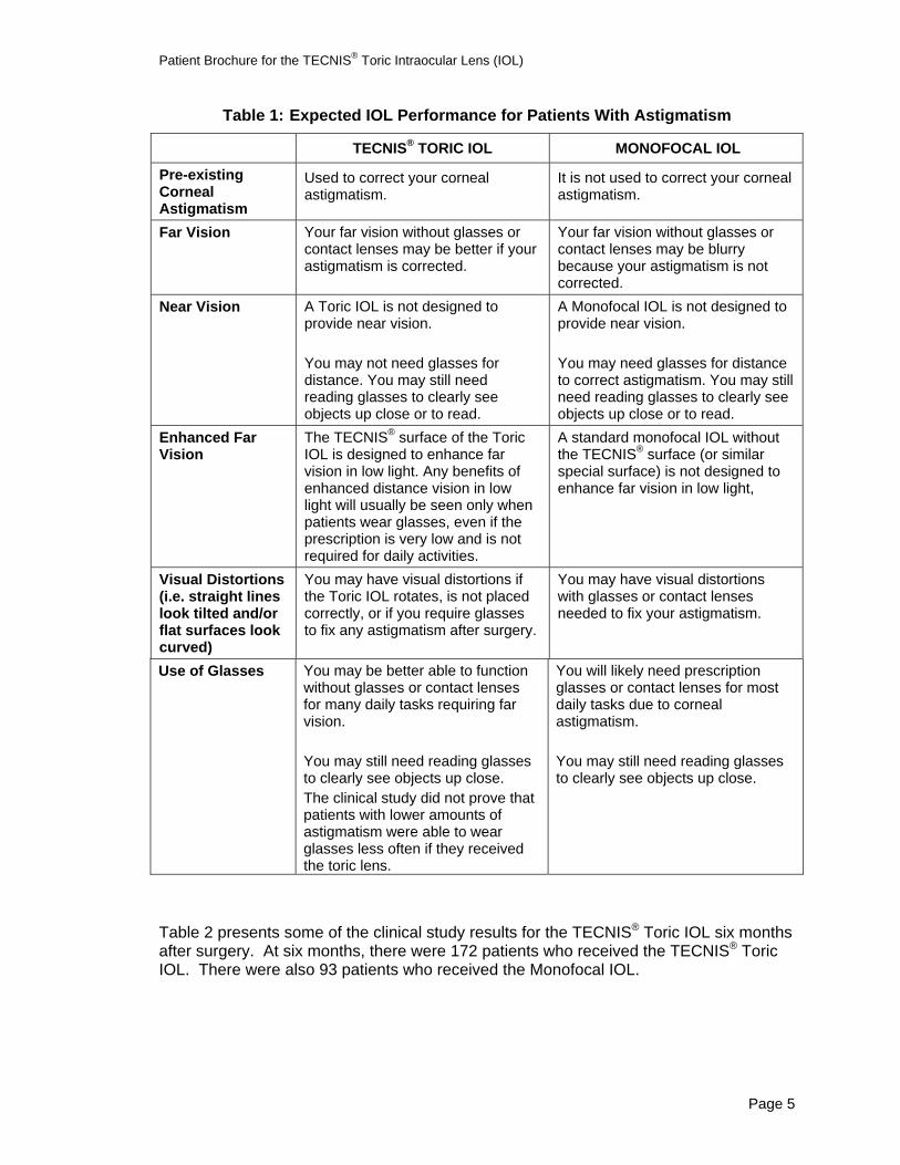
Making the right choice AMO’s Monofocal IOLs and TECNIS® Toric IOL have been well studied. Both are used to replace the natural lens of the eye. If you have corneal astigmatism, the TECNIS® Toric IOL may be a good choice for you. It may improve your distance vision and allow you to be less dependent on glasses at distance. Table 1 will help you compare the Monofocal IOL and the TECNIS® Toric IOL.
Patient Brochure for the TECNIS® Toric Intraocular Lens (IOL)
Page 5
Table 1: Expected IOL Performance for Patients With Astigmatism
TECNIS® TORIC IOL MONOFOCAL IOL
Pre-existing Corneal Astigmatism
Used to correct your corneal astigmatism.
It is not used to correct your corneal astigmatism.
Far Vision Your far vision without glasses or contact lenses may be better if your astigmatism is corrected.
Your far vision without glasses or contact lenses may be blurry because your astigmatism is not corrected.
Near Vision A Toric IOL is not designed to provide near vision. You may not need glasses for distance. You may still need reading glasses to clearly see objects up close or to read.
A Monofocal IOL is not designed to provide near vision. You may need glasses for distance to correct astigmatism. You may still need reading glasses to clearly see objects up close or to read.
Enhanced Far Vision
The TECNIS® surface of the Toric IOL is designed to enhance far vision in low light. Any benefits of enhanced distance vision in low light will usually be seen only when patients wear glasses, even if the prescription is very low and is not required for daily activities.
A standard monofocal IOL without the TECNIS® surface (or similar special surface) is not designed to enhance far vision in low light,
Visual Distortions (i.e. straight lines look tilted and/or flat surfaces look curved)
You may have visual distortions if the Toric IOL rotates, is not placed correctly, or if you require glasses to fix any astigmatism after surgery.
You may have visual distortions with glasses or contact lenses needed to fix your astigmatism.
Use of Glasses
You may be better able to function without glasses or contact lenses for many daily tasks requiring far vision. You may still need reading glasses to clearly see objects up close. The clinical study did not prove that patients with lower amounts of astigmatism were able to wear glasses less often if they received the toric lens.
You will likely need prescription glasses or contact lenses for most daily tasks due to corneal astigmatism. You may still need reading glasses to clearly see objects up close.
Table 2 presents some of the clinical study results for the TECNIS® Toric IOL six months after surgery. At six months, there were 172 patients who received the TECNIS® Toric IOL. There were also 93 patients who received the Monofocal IOL.
Patient Brochure for the TECNIS® Toric Intraocular Lens (IOL)
Page 6
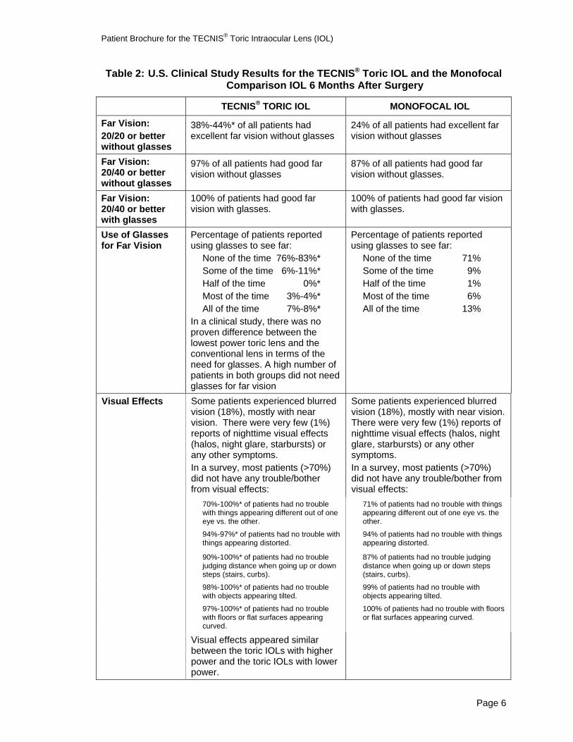
Table 2: U.S. Clinical Study Results for the TECNIS® Toric IOL and the Monofocal Comparison IOL 6 Months After Surgery
TECNIS® TORIC IOL MONOFOCAL IOL
Far Vision: 20/20 or better without glasses
38%-44%* of all patients had excellent far vision without glasses
24% of all patients had excellent far vision without glasses
Far Vision: 20/40 or better without glasses
97% of all patients had good far vision without glasses
87% of all patients had good far vision without glasses.
Far Vision: 20/40 or better with glasses
100% of patients had good far vision with glasses.
100% of patients had good far vision with glasses.
Use of Glasses for Far Vision
Percentage of patients reported using glasses to see far:
None of the time 76%-83%* Some of the time 6%-11%* Half of the time 0%* Most of the time 3%-4%* All of the time 7%-8%*
In a clinical study, there was no proven difference between the lowest power toric lens and the conventional lens in terms of the need for glasses. A high number of patients in both groups did not need glasses for far vision
Percentage of patients reported using glasses to see far:
None of the time 71% Some of the time 9% Half of the time 1% Most of the time 6% All of the time 13%
Visual Effects
Some patients experienced blurred vision (18%), mostly with near vision. There were very few (1%) reports of nighttime visual effects (halos, night glare, starbursts) or any other symptoms. In a survey, most patients (>70%) did not have any trouble/bother from visual effects:
Some patients experienced blurred vision (18%), mostly with near vision. There were very few (1%) reports of nighttime visual effects (halos, night glare, starbursts) or any other symptoms. In a survey, most patients (>70%) did not have any trouble/bother from visual effects:
70%-100%* of patients had no trouble with things appearing different out of one eye vs. the other.
71% of patients had no trouble with things appearing different out of one eye vs. the other.
94%-97%* of patients had no trouble with things appearing distorted.
94% of patients had no trouble with things appearing distorted.
90%-100%* of patients had no trouble judging distance when going up or down steps (stairs, curbs).
87% of patients had no trouble judging distance when going up or down steps (stairs, curbs).
98%-100%* of patients had no trouble with objects appearing tilted.
99% of patients had no trouble with objects appearing tilted.
97%-100%* of patients had no trouble with floors or flat surfaces appearing curved.
100% of patients had no trouble with floors or flat surfaces appearing curved.
Visual effects appeared similar between the toric IOLs with higher power and the toric IOLs with lower power.

Patient Brochure for the TECNIS® Toric Intraocular Lens (IOL)
Page 7
TECNIS® TORIC IOL MONOFOCAL IOL
Patient Satisfaction with the Lens
In a survey, patients were asked if they would choose to have the same lens again, if they were given a choice. Almost all patients (95%-99%) said they would choose this Toric lens again. Patients with the Toric lens rated their satisfaction without glasses as 9.0-9.2 (average) on a scale of 0-10 with 10 being best.
In a survey, patients were asked if they would choose to have the same lens again, if they were given a choice. Almost all patients (94%) said they would choose this monofocal lens again. Patients with the monofocal lens rated their satisfaction without glasses as 8.5 (average) on a scale of 0-10 with 10 being best.
Secondary Surgery
Four (4) patients who received the higher-power toric lenses required a secondary surgery in the first eye to fix the position of the lens (7.3%; 4 out of 55 eyes). No procedures were required for second eyes, therefore considering all eyes with higher powers (ZCT300and ZCT400), 4.7% (4 out of 85 eyes) required secondary surgery to fix the position of the lens.
The monofocal IOL does not require a specific position as it does not correct for astigmatism; therefore, the control subjects did not require any secondary surgeries related to the lens position.
* Depending upon the level of astigmatism
What this means to you To choose an IOL that is best for you, you should evaluate the comparison factors in Table 1 as they relate to your quality of life. We recommend that you ask your eye doctor to assist in this evaluation. If being able to see well at far and being less dependent on glasses would make your life better, then the TECNIS® Toric IOL may be the right choice for you. However, you should weigh the possible advantages and disadvantages before deciding. Whichever IOL you choose, we hope that you are satisfied and have great pleasure in your improved vision. Abbott Medical Optics Inc. Santa Ana, CA 92705 www.amo-inc.com TECNIS is a trademark owned by or licensed to Abbott Laboratories, its subsidiaries, or affiliates. Rx Only. © 2012 Abbott Medical Optics Inc.
LABEL SPECIFICATION
DOCUMENT NO: Z310926
REVISION: 03
TITLE: DATA SHEET, TECNIS® TORIC 1-PIECE IOL
SPECIFICATION:
1. DIMENSIONS: a. Horizontal: 22” 1/32”
b. Vertical: 19-1/2” 1/32”
Flat: 19 x 22” (Tolerance: 1/32”)
Folded: 5-1/2” x 3-7/8”: (Tolerance: 1/32”)
a. Accordion fold to 5-1/2” x 19”.
b. Right Angle fold in fifth’s to 5-1/2” x 3 7/8”.
2. STOCK: 40 lb. Offset or 16 lb. Bond, Smooth Opaque Finish.
3. STYLE: One sheet, English facing out, printed both sides.
4. COLOR OF COPY: See artwork for color specifications.
5. POINT OF USE: Groningen, The Netherlands
6. Printing to be clear and legible with no ink smears.
7. Insert to be clean. No visible damage, loose or attached particles (fibers) are permitted.
8. Vendor to package product following guidelines according to AMOS #3110.
9. See attached pages for artwork layout.
10. Data sheet layout:
Front side: TBD TBD TBD English (EN)

2
The TECNIS® Toric 1-Piece IOL Rx Only
DESCRIPTION:
The TECNIS® Toric 1-Piece lens is an ultraviolet light-absorbing posterior chamber intraocular lens (IOL) that compensates for corneal spherical aberrations and corneal astigmatism. The benefits of aspheric compensation for corneal spherical aberrations are contingent upon full refractive correction of sphere and cylinder. The IOLs are designed to be positioned in the lens capsule to replace the optical function of the natural crystalline lens. The IOLs incorporate a proprietary wavefront-designed toric aspheric optic with a squared posterior optic edge designed to provide a 360 degree barrier. The effects of the proprietary wavefront-designed aspheric optic have been clinically assessed on the TECNIS® IOL, Model Z9000. The edge of the optic has a frosted design to reduce potential edge glare effects. The anteriorly located cylinder axis marks denote the meridian with the lowest power and is to be aligned with the steep corneal meridian.
INDICATIONS FOR USE:
The TECNIS® Toric 1-Piece posterior chamber lenses are indicated for the visual correction of aphakia and pre-existing corneal astigmatism of one diopter or greater in adult patients with or without presbyopia in whom a cataractous lens has been removed by phacoemulsification and who desire improved uncorrected distance vision, reduction in residual refractive cylinder, and increased spectacle independence for distance vision. The device is intended to be placed in the capsular bag.
WARNINGS:
Physicians considering lens implantation under any of the following circumstances should weigh the potential risk/benefit ratio: 1. Patients with recurrent severe anterior or posterior segment inflammation or uveitis. 2. Surgical difficulties at the time of cataract extraction, which may increase the potential for
complications (e.g., persistent bleeding, significant iris damage, uncontrolled positive pressure or significant vitreous prolapse or loss).
3. A compromised eye due to previous trauma or developmental defects in which appropriate support of the IOL is not possible.
4. Circumstances that would result in damage to the endothelium during implantation. 5. Suspected microbial infection. 6. Patients in whom neither the posterior capsule nor the zonules are intact enough to provide support
for the IOL. 7. Children under the age of 2 years are not suitable candidates for intraocular lenses. 8. The clinical study for the TECNIS® Toric 1-Piece IOL did not show evidence of effectiveness for the
treatment of preoperative corneal astigmatism of less than one diopter. 9. The TECNIS® Toric 1-Piece IOL should be placed entirely in the capsular bag and should not be
placed in the ciliary sulcus. 10. Rotation of the TECNIS® Toric 1-Piece IOL away from its intended axis can reduce its astigmatic
correction. Misalignment greater than 30° may increase postoperative refractive cylinder. If necessary, lens repositioning should occur as early as possible prior to lens encapsulation.
PRECAUTIONS:
1. Prior to surgery, the surgeon must inform prospective patients of the possible risks and benefits associated with the use of this device and provide a copy of the patient information brochure to the patient.
2. Do not resterilize the lens. Most sterilizers are not equipped to sterilize the soft acrylic material without producing undesirable side effects.
3
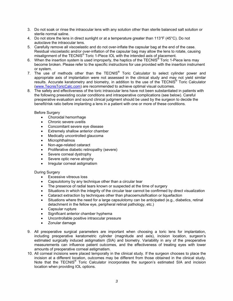
3. Do not soak or rinse the intraocular lens with any solution other than sterile balanced salt solution or sterile normal saline.
4. Do not store the lens in direct sunlight or at a temperature greater than 113°F (45°C). Do not autoclave the intraocular lens.
5. Carefully remove all viscoelastic and do not over-inflate the capsular bag at the end of the case. Residual viscoelastic and/or over-inflation of the capsular bag may allow the lens to rotate, causing misalignment of the TECNIS® Toric 1-Piece IOL with the intended axis of placement.
6. When the insertion system is used improperly, the haptics of the TECNIS® Toric 1-Piece lens may become broken. Please refer to the specific instructions for use provided with the insertion instrument or system.
7. The use of methods other than the TECNIS® Toric Calculator to select cylinder power and appropriate axis of implantation were not assessed in the clinical study and may not yield similar results. Accurate keratometry and biometry, in addition to the use of the TECNIS® Toric Calculator (www.TecnisToricCalc.com) are recommended to achieve optimal visual outcomes.
8. The safety and effectiveness of the toric intraocular lens have not been substantiated in patients with the following preexisting ocular conditions and intraoperative complications (see below). Careful preoperative evaluation and sound clinical judgment should be used by the surgeon to decide the benefit/risk ratio before implanting a lens in a patient with one or more of these conditions. Before Surgery
Choroidal hemorrhage Chronic severe uveitis Concomitant severe eye disease Extremely shallow anterior chamber Medically uncontrolled glaucoma Microphthalmos Non-age-related cataract Proliferative diabetic retinopathy (severe) Severe corneal dystrophy Severe optic nerve atrophy Irregular corneal astigmatism
During Surgery
Excessive vitreous loss Capsulotomy by any technique other than a circular tear The presence of radial tears known or suspected at the time of surgery Situations in which the integrity of the circular tear cannot be confirmed by direct visualization Cataract extraction by techniques other than phacoemulsification or liquefaction Situations where the need for a large capsulotomy can be anticipated (e.g., diabetics, retinal
detachment in the fellow eye, peripheral retinal pathology, etc.) Capsular rupture Significant anterior chamber hyphema Uncontrollable positive intraocular pressure Zonular damage
9. All preoperative surgical parameters are important when choosing a toric lens for implantation,
including preoperative keratometric cylinder (magnitude and axis), incision location, surgeon’s estimated surgically induced astigmatism (SIA) and biometry. Variability in any of the preoperative measurements can influence patient outcomes, and the effectiveness of treating eyes with lower amounts of preoperative corneal astigmatism.
10. All corneal incisions were placed temporally in the clinical study. If the surgeon chooses to place the incision at a different location, outcomes may be different from those obtained in the clinical study. Note that the TECNIS® Toric Calculator incorporates the surgeon’s estimated SIA and incision location when providing IOL options.
4
GENERAL ADVERSE EVENTS FOR IOLS
Potential adverse events during or following cataract surgery with implantation of an IOL may include but are not limited to: Endophthalmitis/intraocular infection Hypopyon Pupillary block Retinal detachment IOL dislocation Persistent corneal stromal edema Persistent cystoid macular edema Secondary surgical intervention (including implant repositioning, removal, or other surgical procedure) Any other adverse event that leads to permanent visual impairment or requires surgical or medical
intervention to prevent permanent visual impairment
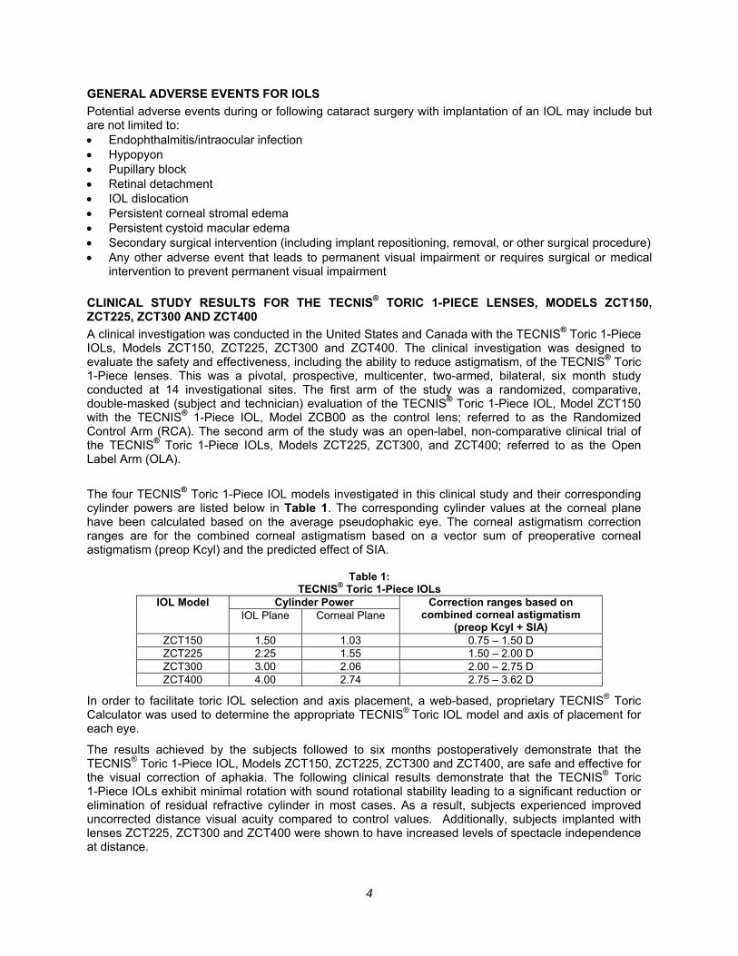
CLINICAL STUDY RESULTS FOR THE TECNIS® TORIC 1-PIECE LENSES, MODELS ZCT150, ZCT225, ZCT300 AND ZCT400
A clinical investigation was conducted in the United States and Canada with the TECNIS® Toric 1-Piece IOLs, Models ZCT150, ZCT225, ZCT300 and ZCT400. The clinical investigation was designed to evaluate the safety and effectiveness, including the ability to reduce astigmatism, of the TECNIS® Toric 1-Piece lenses. This was a pivotal, prospective, multicenter, two-armed, bilateral, six month study conducted at 14 investigational sites. The first arm of the study was a randomized, comparative, double-masked (subject and technician) evaluation of the TECNIS® Toric 1-Piece IOL, Model ZCT150 with the TECNIS® 1-Piece IOL, Model ZCB00 as the control lens; referred to as the Randomized Control Arm (RCA). The second arm of the study was an open-label, non-comparative clinical trial of the TECNIS® Toric 1-Piece IOLs, Models ZCT225, ZCT300, and ZCT400; referred to as the Open Label Arm (OLA).
The four TECNIS® Toric 1-Piece IOL models investigated in this clinical study and their corresponding cylinder powers are listed below in Table 1. The corresponding cylinder values at the corneal plane have been calculated based on the average pseudophakic eye. The corneal astigmatism correction ranges are for the combined corneal astigmatism based on a vector sum of preoperative corneal astigmatism (preop Kcyl) and the predicted effect of SIA.
Table 1: TECNIS® Toric 1-Piece IOLs
IOL Model Cylinder Power Correction ranges based on combined corneal astigmatism
(preop Kcyl + SIA) IOL Plane Corneal Plane
ZCT150 1.50 1.03 0.75 – 1.50 D ZCT225 2.25 1.55 1.50 – 2.00 D ZCT300 3.00 2.06 2.00 – 2.75 D ZCT400 4.00 2.74 2.75 – 3.62 D
In order to facilitate toric IOL selection and axis placement, a web-based, proprietary TECNIS® Toric Calculator was used to determine the appropriate TECNIS® Toric IOL model and axis of placement for each eye.
The results achieved by the subjects followed to six months postoperatively demonstrate that the TECNIS® Toric 1-Piece IOL, Models ZCT150, ZCT225, ZCT300 and ZCT400, are safe and effective for the visual correction of aphakia. The following clinical results demonstrate that the TECNIS® Toric 1-Piece IOLs exhibit minimal rotation with sound rotational stability leading to a significant reduction or elimination of residual refractive cylinder in most cases. As a result, subjects experienced improved uncorrected distance visual acuity compared to control values. Additionally, subjects implanted with lenses ZCT225, ZCT300 and ZCT400 were shown to have increased levels of spectacle independence at distance.

5
TECNIS® TORIC 1-PIECE IOL CLINICAL STUDY PATIENT POPULATION:
A total of 269 subjects were enrolled in the study; 197 were in the RCA and 72 in the OLA. Of the 197 in the RCA, 95 were implanted in the first eye with the control ZCB00 lens and 102 with a ZCT150 toric lens. Of the 72 in the OLA, 17 were implanted with the ZCT225 lens in the first eye and 55 with either ZCT300 or ZCT400. Overall, 174 first eyes were implanted with a TECNIS® Toric 1-Piece IOL. The 6-month study results are presented for all study groups.
The subject population implanted with the ZCT150 lens in the RCA consisted of 53.9% females to 46.1% males, and subjects implanted with the ZCB00 control lens consisted of 57.9% females and 42.1% males. The OLA arm of the study consisted of 55.6% females and 44.4% males. Stratifying by race, the ZCT150 population consisted of 94.1% Caucasian, 3.9% African American, and 2.0% Asian; the ZCB00 control population consisted of 95.8% Caucasian, 3.2% African American and 1.1% Asian; and the OLA group consisted of 94.4% Caucasian, 4.2% African American and 1.4% Asian. The mean ages were 69.9 years for the ZCT150 population, 71.3 years for the ZCB00 control population and 68.8 years for the OLA population.
REDUCTION IN CYLINDER
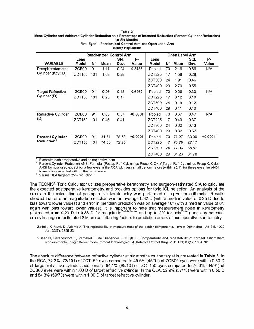
Percent reduction in cylinder was calculated as the ratio of achieved postoperative refractive cylinder to the target refractive cylinder, adjusted for preoperative keratometric cylinder. Specifically, the difference between postoperative refractive cylinder and preoperative keratometric cylinder was divided by the difference between the target refractive cylinder and preoperative keratometric cylinder to calculate the percent reduction in cylinder. The target refractive cylinder is a combination of preoperative keratometric cylinder, SIA from the cataract incision and the toric IOL. The calculation was performed similarly for all eyes; in the RCA, the target refractive cylinder for ZCB00 eyes was calculated as if the control subjects were receiving a ZCT150 IOL. As shown in Table 2, no statistically significant differences were observed in preoperative keratometric cylinder or target refractive cylinder between ZCT150 toric and ZCB00 control eyes in the RCA; however, statistically significant differences were observed for mean refractive cylinder and the mean percent reduction in cylinder in favor of the ZCT150 lens group compared to the ZCB00 control at six months postoperative. Additionally, the mean percent reduction in cylinder for OLA first eyes at six months was statistically significantly higher than the target value of 25%. For all toric first eyes in the RCA and OLA safety populations combined (N=171), the mean percent reduction in cylinder was 75.24 (SD=59.29).
6
Table 2: Mean Cylinder and Achieved Cylinder Reduction as a Percentage of Intended Reduction (Percent Cylinder Reduction)
at Six Months First Eyesa - Randomized Control Arm and Open Label Arm
Safety Population
Randomized Control Arm Open Label Arm
VARIABLE Lens Model Na Mean
Std.Dev.
P-Value
Lens Model Na Mean
Std. Dev.
P-Value
PreopKeratometric Cylinder (Kcyl; D)
ZCB00 91 1.11 0.24 0.3436 Pooled 70 2.16 0.66 N/A
ZCT150 101 1.08 0.28 ZCT225 17 1.58 0.28
ZCT300 24 1.91 0.46
ZCT400 29 2.70 0.55
Target Refractive Cylinder (D)
ZCB00 91 0.26 0.18 0.6267 Pooled 70 0.26 0.30 N/A
ZCT150 101 0.25 0.17 ZCT225 17 0.12 0.10
ZCT300 24 0.19 0.12
ZCT400 29 0.41 0.40
Refractive Cylinder (D)
ZCB00 91 0.85 0.57 <0.0001 Pooled 70 0.67 0.47 N/A
ZCT150 101 0.45 0.41 ZCT225 17 0.49 0.37
ZCT300 24 0.62 0.43
ZCT400 29 0.82 0.52
Percent Cylinder Reductionb
ZCB00 91 31.61 78.73 <0.0001 Pooled 70 76.27 33.09 <0.0001c
ZCT150 101 74.53 72.25 ZCT225 17 73.78 27.17
ZCT300 24 72.03 38.57
ZCT400 29 81.23 31.78 a Eyes with both preoperative and postoperative data b Percent Cylinder Reduction ANSI Formula=(Postop Ref. Cyl. minus Preop K. Cyl.)/(Target Ref. Cyl. minus Preop K. Cyl.);
ANSI formula used except for a few eyes in the RCA with very small denominators (within ±0.1); for these eyes the ANSI formula was used but without the target value.
c Versus OLA target of 25% reduction The TECNIS® Toric Calculator utilizes preoperative keratometry and surgeon-estimated SIA to calculate the expected postoperative keratometry and provides options for toric IOL selection. An analysis of the errors in the calculation of postoperative keratometry was performed using vector arithmetic. Results showed that error in magnitude prediction was on average 0.32 D (with a median value of 0.25 D due to bias toward lower values) and error in meridian prediction was on average 16° (with a median value of 8°, again with bias toward lower values). It is important to note that measurement noise in keratometry (estimated from 0.20 D to 0.83 D for magnitudeZadnik,Visser and up to 20° for axisVisser) and any potential errors in surgeon-estimated SIA are contributing factors to prediction errors of postoperative keratometry.
Zadnik, K, Mutti, D, Adams A. The repeatability of measurement of the ocular components. Invest Ophthalmol Vis Sci. 1992 Jun; 33(7): 2325-33
Visser N, Berendschot T, Verbakel F, de Brabander J, Nuijts R. Comparability and repeatability of corneal astigmatism
measurements using different measurement technologies. J. Cataract Refract Surg. 2012 Oct; 38(1): 1764-70” The absolute difference between refractive cylinder at six months vs. the target is presented in Table 3. In the RCA, 72.3% (73/101) of ZCT150 eyes compared to 49.5% (45/91) of ZCB00 eyes were within 0.50 D of target refractive cylinder; additionally, 94.1% (95/101) of ZCT150 eyes compared to 70.3% (64/91) of ZCB00 eyes were within 1.00 D of target refractive cylinder. In the OLA, 52.9% (37/70) were within 0.50 D and 84.3% (59/70) were within 1.00 D of target refractive cylinder.
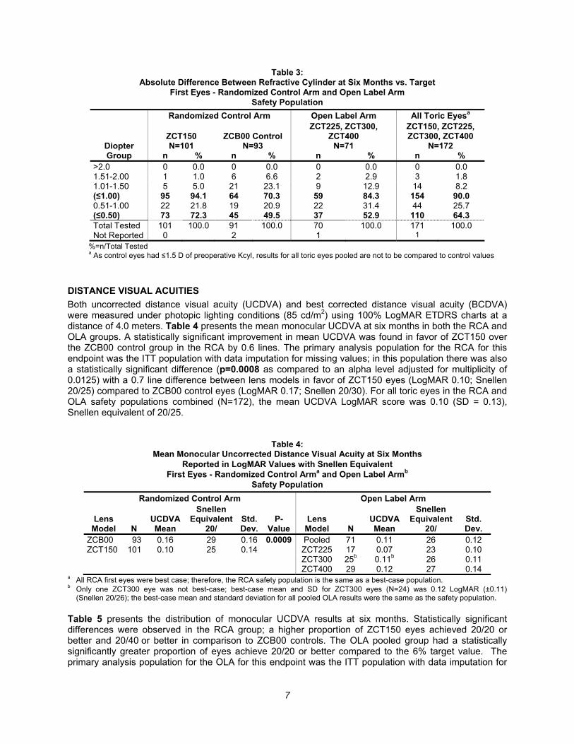
7
Table 3: Absolute Difference Between Refractive Cylinder at Six Months vs. Target
First Eyes - Randomized Control Arm and Open Label Arm Safety Population
Randomized Control Arm Open Label Arm All Toric Eyesa
Diopter Group
ZCT150 N=101
ZCB00 ControlN=93
ZCT225, ZCT300, ZCT400
N=71
ZCT150, ZCT225, ZCT300, ZCT400
N=172 n % n % n % n %
>2.0 0 0.0 0 0.0 0 0.0 0 0.0 1.51-2.00 1 1.0 6 6.6 2 2.9 3 1.8 1.01-1.50 5 5.0 21 23.1 9 12.9 14 8.2 (≤1.00) 95 94.1 64 70.3 59 84.3 154 90.00.51-1.00 22 21.8 19 20.9 22 31.4 44 25.7 (≤0.50) 73 72.3 45 49.5 37 52.9 110 64.3Total Tested 101 100.0 91 100.0 70 100.0 171 100.0 Not Reported 0 2 1 1
%=n/Total Tested a As control eyes had ≤1.5 D of preoperative Kcyl, results for all toric eyes pooled are not to be compared to control values
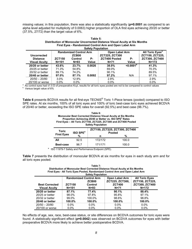
DISTANCE VISUAL ACUITIES
Both uncorrected distance visual acuity (UCDVA) and best corrected distance visual acuity (BCDVA) were measured under photopic lighting conditions (85 cd/m2) using 100% LogMAR ETDRS charts at a distance of 4.0 meters. Table 4 presents the mean monocular UCDVA at six months in both the RCA and OLA groups. A statistically significant improvement in mean UCDVA was found in favor of ZCT150 over the ZCB00 control group in the RCA by 0.6 lines. The primary analysis population for the RCA for this endpoint was the ITT population with data imputation for missing values; in this population there was also a statistically significant difference (p=0.0008 as compared to an alpha level adjusted for multiplicity of 0.0125) with a 0.7 line difference between lens models in favor of ZCT150 eyes (LogMAR 0.10; Snellen 20/25) compared to ZCB00 control eyes (LogMAR 0.17; Snellen 20/30). For all toric eyes in the RCA and OLA safety populations combined (N=172), the mean UCDVA LogMAR score was 0.10 (SD = 0.13), Snellen equivalent of 20/25.
Table 4: Mean Monocular Uncorrected Distance Visual Acuity at Six Months
Reported in LogMAR Values with Snellen Equivalent First Eyes - Randomized Control Arma and Open Label Armb
Safety Population
Randomized Control Arm Open Label Arm
Lens Model N
UCDVA Mean
Snellen Equivalent
20/ Std.Dev.
P-Value
Lens Model N
UCDVA Mean
Snellen Equivalent
20/ Std. Dev.
ZCB00 93 0.16 29 0.16 0.0009 Pooled 71 0.11 26 0.12 ZCT150 101 0.10 25 0.14 ZCT225 17 0.07 23 0.10 ZCT300 25b 0.11b 26 0.11 ZCT400 29 0.12 27 0.14
a All RCA first eyes were best case; therefore, the RCA safety population is the same as a best-case population. b Only one ZCT300 eye was not best-case; best-case mean and SD for ZCT300 eyes (N=24) was 0.12 LogMAR (±0.11)
(Snellen 20/26); the best-case mean and standard deviation for all pooled OLA results were the same as the safety population.
Table 5 presents the distribution of monocular UCDVA results at six months. Statistically significant differences were observed in the RCA group; a higher proportion of ZCT150 eyes achieved 20/20 or better and 20/40 or better in comparison to ZCB00 controls. The OLA pooled group had a statistically significantly greater proportion of eyes achieve 20/20 or better compared to the 6% target value. The primary analysis population for the OLA for this endpoint was the ITT population with data imputation for
8
missing values; in this population, there was also a statistically significantly (p=0.0001 as compared to an alpha level adjusted for multiplicity of 0.0063) higher proportion of OLA first eyes achieving 20/20 or better (37.5%, 27/72) than the target value of 6%.
Table 5: Distribution of Monocular Uncorrected Distance Visual Acuity at Six Months
First Eyes - Randomized Control Arm and Open Label Arm Safety Population
Uncorrected Distance
Visual Acuity
Randomized Control Arm Open Label Arm All Toric Eyesa
ZCT150 N=101
ZCB00Control
N=93 P-
Value
ZCT225, ZCT300ZCT400 Pooled
N=71 P-
Value
ZCT150, ZCT225, ZCT300, ZCT400
N=172 20/20 or better 43.6% 23.7% 0.0026 38.0% <0.0001b 41.3%20/25 or better 71.3% 54.8% 69.0% 70.3% 20/32 or better 89.1% 74.2% 90.1% 89.5% 20/40 or better 97.0% 87.1% 0.0092 97.2% N/A 97.1% 20/50 – 20/80 3.0% 12.9% 2.8% 2.9%
20/100 or worse 0.0% 0.0% 0.0% 0.0% a As control eyes had ≤1.5 D of preoperative Kcyl, results for all toric eyes pooled are not to be compared to control values b Versus target value of 6%
Table 6 presents BCDVA results for all first-eye TECNIS® Toric 1-Piece lenses (pooled) compared to ISO SPE rates. At six months, 100% of all toric eyes and 100% of toric best-case toric eyes achieved BCDVA of 20/40 or better, exceeding the ISO SPE rates for overall (92.5%) and best case (96.7%).
Table 6: Monocular Best Corrected Distance Visual Acuity at Six Months
Proportion Achieving 20/40 or Better vs. ISO SPEa Rates First Eyes – All Toric ZCT150, ZCT225, ZCT300 and ZCT400 Pooled
Safety Population
Toric First Eyes
ISO SPEa %
ZCT150, ZCT225, ZCT300, ZCT400 Pooled
n %All 92.5 172/172 100.0
Best-case 96.7 171/171 100.0 a ISO 11979-7 Safety and Performance Endpoint (SPE).
Table 7 presents the distribution of monocular BCDVA at six months for eyes in each study arm and for all toric eyes pooled.
Table 7: Distribution of Monocular Best Corrected Distance Visual Acuity at Six Months
First Eyes - All Toric Eyes Pooled, Randomized Control Arm and Open Label Arm Safety Population
Best Corrected Visual Acuity
Randomized Control Arm Open Label Arm ZCT225, ZCT300,
ZCT400 N=71
All Toric Eyes ZCT150, ZCT225, ZCT300, ZCT400
N=172 ZCT150 N=101
ZCB00Control
N=93 20/20 or better 87.1% 77.4% 90.1% 88.4% 20/25 or better 98.0% 97.8% 95.8% 97.1% 20/32 or better 99.0% 100.0% 98.6% 98.8% 20/40 or better 100.0% 100.0% 100.0% 100.0% 20/50 – 20/80 0.0% 0.0% 0.0% 0.0% 20/100 or worse 0.0% 0.0% 0.0% 0.0%
No effects of age, sex, race, best-case status, or site differences on BCDVA outcomes for toric eyes were found. A statistically significant effect (p=0.0042) was observed on BCDVA outcomes for eyes with better preoperative BCDVA more likely to achieve better postoperative BCDVA.
9
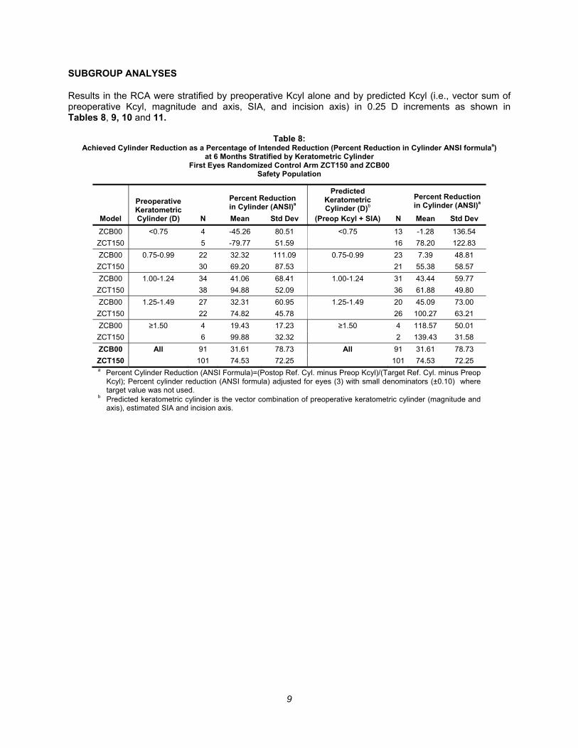
SUBGROUP ANALYSES Results in the RCA were stratified by preoperative Kcyl alone and by predicted Kcyl (i.e., vector sum of preoperative Kcyl, magnitude and axis, SIA, and incision axis) in 0.25 D increments as shown in Tables 8, 9, 10 and 11.
Table 8: Achieved Cylinder Reduction as a Percentage of Intended Reduction (Percent Reduction in Cylinder ANSI formulaa)
at 6 Months Stratified by Keratometric Cylinder First Eyes Randomized Control Arm ZCT150 and ZCB00
Safety Population
Model
Preoperative Keratometric Cylinder (D) N
Percent Reduction in Cylinder (ANSI)a
Predicted Keratometric Cylinder (D)b
(Preop Kcyl + SIA)
N
Percent Reduction in Cylinder (ANSI)a
Mean Std Dev Mean Std Dev
ZCB00 <0.75 4 -45.26 80.51 <0.75 13 -1.28 136.54
ZCT150 5 -79.77 51.59 16 78.20 122.83
ZCB00 0.75-0.99 22 32.32 111.09 0.75-0.99 23 7.39 48.81
ZCT150 30 69.20 87.53 21 55.38 58.57
ZCB00 1.00-1.24 34 41.06 68.41 1.00-1.24 31 43.44 59.77
ZCT150 38 94.88 52.09 36 61.88 49.80
ZCB00 1.25-1.49 27 32.31 60.95 1.25-1.49 20 45.09 73.00
ZCT150 22 74.82 45.78 26 100.27 63.21
ZCB00 ≥1.50 4 19.43 17.23 ≥1.50 4 118.57 50.01
ZCT150 6 99.88 32.32 2 139.43 31.58
ZCB00 All 91 31.61 78.73 All 91 31.61 78.73
ZCT150 101 74.53 72.25 101 74.53 72.25 a Percent Cylinder Reduction (ANSI Formula)=(Postop Ref. Cyl. minus Preop Kcyl)/(Target Ref. Cyl. minus Preop
Kcyl); Percent cylinder reduction (ANSI formula) adjusted for eyes (3) with small denominators (±0.10) where target value was not used.
b Predicted keratometric cylinder is the vector combination of preoperative keratometric cylinder (magnitude and axis), estimated SIA and incision axis.

10
Table 9: Residual Refractive Cylinder at 6 Months Stratified by Keratometric Cylinder
First Eyes Randomized Control Arm ZCT150 and ZCB00 Safety Population
Model
Preoperative Keratometric Cylinder (D) N
Residual Refractive Cylinder (D)
Predicted Keratometric Cylinder (D)a
(Preop Kcyl + SIA) N
Residual Refractive Cylinder (D)
Mean Std Dev Mean Std Dev
ZCB00 <0.75 5 0.85 0.42 <0.75 14 0.77 0.49
ZCT150 5 0.91 0.14 16 0.55 0.43
ZCB00 0.75-0.99 22 0.56 0.50 0.75-0.99 23 1.03 0.51
ZCT150 30 0.50 0.40 21 0.43 0.33
ZCB00 1.00-1.24 34 0.80 0.55 1.00-1.24 31 0.84 0.68
ZCT150 38 0.36 0.36 36 0.48 0.45
ZCB00 1.25-1.49 27 1.09 0.59 1.25-1.49 21 0.84 0.52
ZCT150 22 0.48 0.49 26 0.39 0.43
ZCB00 ≥1.50 5 1.35 0.28 ≥1.50 4 0.43 0.42
ZCT150 6 0.34 0.44 2 0.38 0.18
ZCB00 All 93 0.86 0.57 All 93 0.86 0.57
ZCT150 101 0.45 0.41 101 0.45 0.41 a Predicted keratometric cylinder is the vector combination of preoperative keratometric cylinder (magnitude and axis),
estimated SIA and incision axis.
Table 10: Mean Uncorrected Distance Visual Acuity at Six Months Stratified by Keratometric Cylinder
Reported in LogMAR Values with Snellen Equivalent First Eyes Randomized Control Arm ZCT150 and ZCB00
Safety Population
Model
Preoperative Keratometric Cylinder (D) N
UCDVA Predicted Keratometric Cylinder (D)a
(Preop Kcyl + SIA) N
UCDVA
LogMAR Mean
Snellen Equiv.
Std Dev
LogMAR Mean
Snellen Equiv. Std Dev
ZCB00 <0.75 5 0.04 22 0.19 <0.75 14 0.08 24 0.14 ZCT150 5 0.17 30 0.14 16 0.06 23 0.13 ZCB00 0.75-0.99 22 0.09 25 0.11 0.75-0.99 23 0.22 33 0.15 ZCT150 30 0.08 24 0.11 21 0.15 28 0.17 ZCB00 1.00-1.24 34 0.18 30 0.16 1.00-1.24 31 0.17 30 0.17 ZCT150 38 0.08 24 0.16 36 0.09 25 0.12 ZCB00 1.25-1.49 27 0.20 32 0.16 1.25-1.49 21 0.16 29 0.14 ZCT150 22 0.10 25 0.12 26 0.09 25 0.12 ZCB00 ≥1.50 5 0.26 36 0.13 ≥1.50 4 0.08 24 0.13 ZCT150 6 0.18 30 0.12 2 0.13 27 0.18 ZCB00 All 93 0.16 29 0.16 All 93 0.16 29 0.16 ZCT150 101 0.10 25 0.14 101 0.10 25 0.14 a Predicted keratometric cylinder is the vector combination of preoperative keratometric cylinder (magnitude and axis),
estimated SIA and incision axis.
11
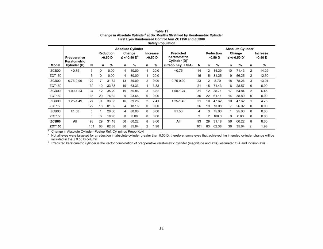
Table 11 Change in Absolute Cylindera at Six Months Stratified by Keratometric Cylinder
First Eyes Randomized Control Arm ZCT150 and ZCB00 Safety Population
Absolute Cylinder Absolute Cylinder
Model
Preoperative Keratometric Cylinder (D)
Reduction >0.50 D
Change ≤ +/-0.50 Db
Increase >0.50 D
Predicted Keratometric Cylinder (D)c
(Preop Kcyl + SIA)
Reduction >0.50 D
Change ≤ +/-0.50 Db
Increase >0.50 D
N n % n % n % N n % n % n %
ZCB00 <0.75 5 0 0.00 4 80.00 1 20.0 <0.75 14 2 14.29 10 71.43 2 14.29
ZCT150 5 0 0.00 4 80.00 1 20.0 16 5 31.25 9 56.25 2 12.50
ZCB00 0.75-0.99 22 7 31.82 13 59.09 2 9.09 0.75-0.99 23 2 8.70 18 78.26 3 13.04
ZCT150 30 10 33.33 19 63.33 1 3.33 21 15 71.43 6 28.57 0 0.00
ZCB00 1.00-1.24 34 12 35.29 19 55.88 3 8.82 1.00-1.24 31 12 38.71 17 54.84 2 6.45
ZCT150 38 29 76.32 9 23.68 0 0.00 36 22 61.11 14 38.89 0 0.00
ZCB00 1.25-1.49 27 9 33.33 16 59.26 2 7.41 1.25-1.49 21 10 47.62 10 47.62 1 4.76
ZCT150 22 18 81.82 4 18.18 0 0.00 26 19 73.08 7 26.92 0 0.00
ZCB00 ≥1.50 5 1 20.00 4 80.00 0 0.00 ≥1.50 4 3 75.00 1 25.00 0 0.00
ZCT150 6 6 100.0 0 0.00 0 0.00 2 2 100.0 0 0.00 0 0.00
ZCB00 All 93 29 31.18 56 60.22 8 8.60 All 93 29 31.18 56 60.22 8 8.60
ZCT150 101 63 62.38 36 35.64 2 1.98 101 63 62.38 36 35.64 2 1.98 a Change in Absolute Cylinder=Postop Ref. Cyl minus Preop Kcyl b Not all eyes were targeted for a reduction in absolute cylinder greater than 0.50 D; therefore, some eyes that achieved the intended cylinder change will be
included in the ± 0.50 D column c Predicted keratometric cylinder is the vector combination of preoperative keratometric cylinder (magnitude and axis), estimated SIA and incision axis.
12
SPECTACLE INDEPENDENCE
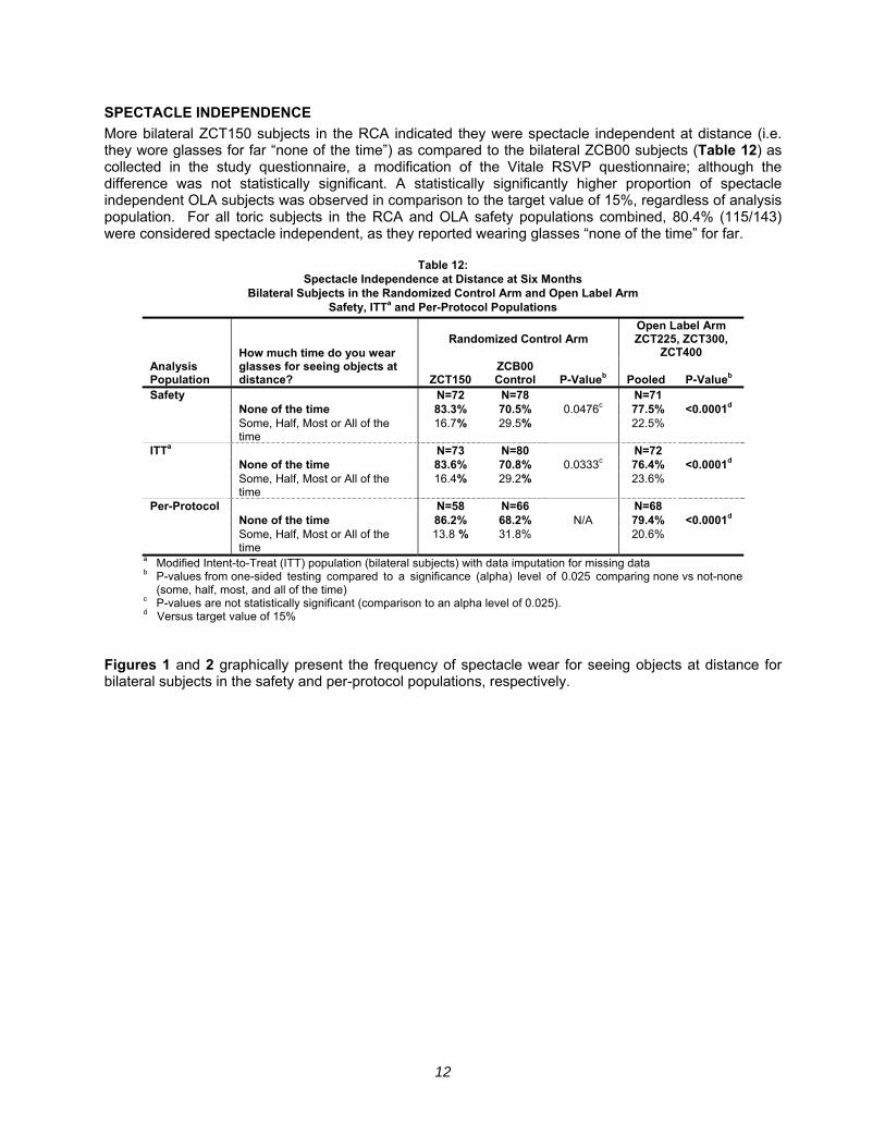
More bilateral ZCT150 subjects in the RCA indicated they were spectacle independent at distance (i.e. they wore glasses for far “none of the time”) as compared to the bilateral ZCB00 subjects (Table 12) as collected in the study questionnaire, a modification of the Vitale RSVP questionnaire; although the difference was not statistically significant. A statistically significantly higher proportion of spectacle independent OLA subjects was observed in comparison to the target value of 15%, regardless of analysis population. For all toric subjects in the RCA and OLA safety populations combined, 80.4% (115/143) were considered spectacle independent, as they reported wearing glasses “none of the time” for far.
Table 12: Spectacle Independence at Distance at Six Months
Bilateral Subjects in the Randomized Control Arm and Open Label Arm Safety, ITTa and Per-Protocol Populations
Analysis Population
How much time do you wear glasses for seeing objects at distance?
Randomized Control Arm Open Label Arm ZCT225, ZCT300,
ZCT400
ZCT150 ZCB00 Control P-Valueb Pooled P-Valueb
Safety N=72 N=78 N=71 None of the time 83.3% 70.5% 0.0476c 77.5% <0.0001d Some, Half, Most or All of the time
16.7% 29.5% 22.5%
ITTa N=73 N=80 N=72 None of the time 83.6% 70.8% 0.0333c 76.4% <0.0001d Some, Half, Most or All of the time
16.4% 29.2% 23.6%
Per-Protocol N=58 N=66 N=68 None of the time 86.2% 68.2% N/A 79.4% <0.0001d Some, Half, Most or All of the time
13.8 % 31.8% 20.6%
a Modified Intent-to-Treat (ITT) population (bilateral subjects) with data imputation for missing data
b P-values from one-sided testing compared to a significance (alpha) level of 0.025 comparing none vs not-none (some, half, most, and all of the time)
c P-values are not statistically significant (comparison to an alpha level of 0.025). d Versus target value of 15%
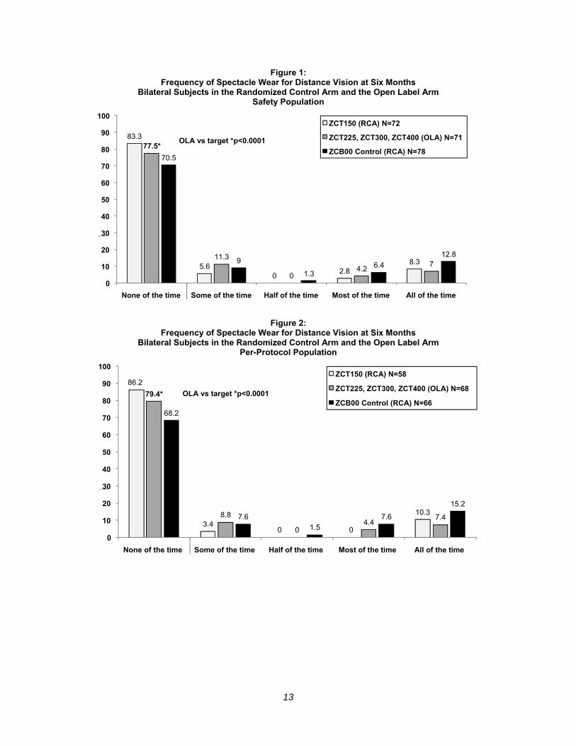
Figures 1 and 2 graphically present the frequency of spectacle wear for seeing objects at distance for bilateral subjects in the safety and per-protocol populations, respectively.
13
Figure 1: Frequency of Spectacle Wear for Distance Vision at Six Months
Bilateral Subjects in the Randomized Control Arm and the Open Label Arm Safety Population
Figure 2:
Frequency of Spectacle Wear for Distance Vision at Six Months Bilateral Subjects in the Randomized Control Arm and the Open Label Arm
Per-Protocol Population
86.2
3.4 0 0
10.3
79.4*
8.8
04.4
7.4
68.2
7.6
1.5
7.6
15.2
0
10
20
30
40
50
60
70
80
90
100
None of the time Some of the time Half of the time Most of the time All of the time
Percent of Subjects
ZCT150 (RCA) N=58
ZCT225, ZCT300, ZCT400 (OLA) N=68
ZCB00 Control (RCA) N=66 OLA vs target *p<0.0001
83.3
5.6 0
2.88.3
77.5*
11.3
04.2
7
70.5
9
1.36.4
12.8
0
10
20
30
40
50
60
70
80
90
100
None of the time Some of the time Half of the time Most of the time All of the time
Percent of Subjects
ZCT150 (RCA) N=72
ZCT225, ZCT300, ZCT400 (OLA) N=71
ZCB00 Control (RCA) N=78
OLA vs target *p<0.0001
14
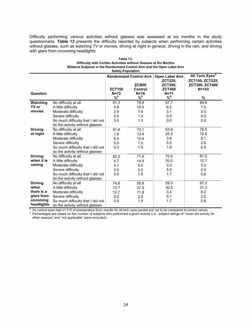
Difficulty performing various activities without glasses was assessed at six months in the study questionnaire. Table 13 presents the difficulty reported by subjects when performing certain activities without glasses, such as watching TV or movies, driving at night in general, driving in the rain, and driving with glare from oncoming headlights.
Table 13: Difficulty with Certain Activities without Glasses at Six Months
Bilateral Subjects in the Randomized Control Arm and the Open Label Arm Safety Population
Question
Randomized Control Arm Open Label Arm All Toric Eyesa
ZCT150 N=72
ZCB00 Control
N=78
ZCT225, ZCT300, ZCT400
N=71
ZCT150, ZCT225, ZCT300, ZCT400
N=143
%b %b %b %Watching TV or movies
No difficulty at all 91.3 78.9 87.7 89.6 A little difficulty 5.8 10.5 9.2 7.5 Moderate difficulty 2.9 7.9 3.1 3.0 Severe difficulty 0.0 1.3 0.0 0.0 So much difficulty that I did not do the activity without glasses
0.0 1.3 0.0 0.0
Driving at night
No difficulty at all 91.9 73.1 63.6 78.6 A little difficulty 1.6 13.4 25.5 12.8 Moderate difficulty 6.5 10.4 3.6 5.1 Severe difficulty 0.0 1.5 5.5 2.6 So much difficulty that I did not do the activity without glasses
0.0 1.5 1.8 0.9
Driving when it is raining
No difficulty at all 92.2 71.6 70.0 81.5 A little difficulty 4.7 14.9 20.0 12.1 Moderate difficulty 3.1 9.0 3.3 3.2 Severe difficulty 0.0 3.0 5.0 2.4 So much difficulty that I did not do the activity without glasses
0.0 1.5 1.7 0.8
Driving when there is a glare from oncoming headlights
No difficulty at all 74.6 55.9 59.3 67.2 A little difficulty 12.7 27.9 30.5 21.3 Moderate difficulty 12.7 11.8 3.4 8.2 Severe difficulty 0.0 2.9 5.1 2.5 So much difficulty that I did not do the activity without glasses
0.0 1.5 1.7 0.8
a As control eyes had ≤1.5 D of preoperative Kcyl, results for all toric eyes pooled are not to be compared to control values b Percentages are based on the number of subjects who performed a given activity (i.e., subject ratings of “never did activity for
other reasons” and “not applicable” were excluded).
15
SUBJECT SATISFACTION
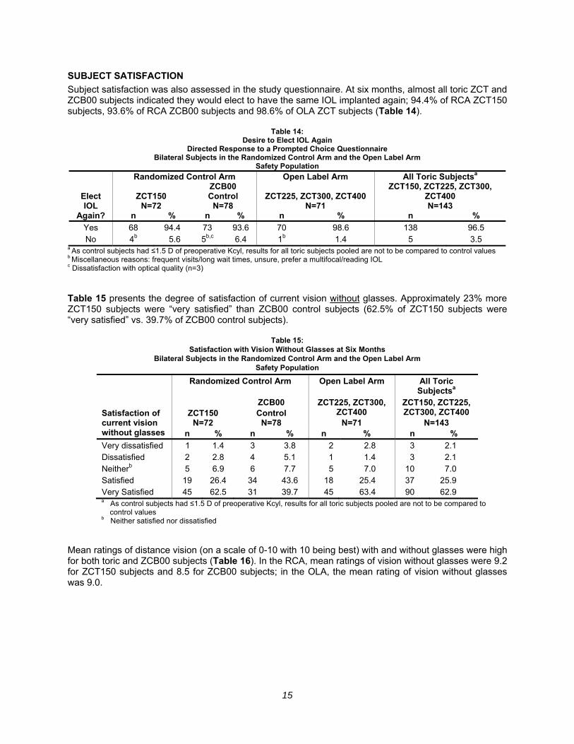
Subject satisfaction was also assessed in the study questionnaire. At six months, almost all toric ZCT and ZCB00 subjects indicated they would elect to have the same IOL implanted again; 94.4% of RCA ZCT150 subjects, 93.6% of RCA ZCB00 subjects and 98.6% of OLA ZCT subjects (Table 14).
Table 14: Desire to Elect IOL Again
Directed Response to a Prompted Choice Questionnaire Bilateral Subjects in the Randomized Control Arm and the Open Label Arm
Safety Population
Elect IOL
Again?
Randomized Control Arm Open Label Arm All Toric Subjectsa
ZCT150 ZCB00Control ZCT225, ZCT300, ZCT400
ZCT150, ZCT225, ZCT300, ZCT400
N=72 N=78 N=71 N=143 n % n % n % n %
Yes 68 94.4 73 93.6 70 98.6 138 96.5 No 4b 5.6 5b,c 6.4 1b 1.4 5 3.5
a As control subjects had ≤1.5 D of preoperative Kcyl, results for all toric subjects pooled are not to be compared to control values b Miscellaneous reasons: frequent visits/long wait times, unsure, prefer a multifocal/reading IOL
c Dissatisfaction with optical quality (n=3)
Table 15 presents the degree of satisfaction of current vision without glasses. Approximately 23% more ZCT150 subjects were “very satisfied” than ZCB00 control subjects (62.5% of ZCT150 subjects were “very satisfied” vs. 39.7% of ZCB00 control subjects).
Table 15: Satisfaction with Vision Without Glasses at Six Months
Bilateral Subjects in the Randomized Control Arm and the Open Label Arm Safety Population
Satisfaction of current vision without glasses
Randomized Control Arm Open Label Arm All Toric Subjectsa
ZCT150 N=72
ZCB00 Control
N=78
ZCT225, ZCT300, ZCT400
N=71
ZCT150, ZCT225, ZCT300, ZCT400
N=143 n % n % n % n %
Very dissatisfied 1 1.4 3 3.8 2 2.8 3 2.1 Dissatisfied 2 2.8 4 5.1 1 1.4 3 2.1 Neitherb 5 6.9 6 7.7 5 7.0 10 7.0 Satisfied 19 26.4 34 43.6 18 25.4 37 25.9 Very Satisfied 45 62.5 31 39.7 45 63.4 90 62.9 a As control subjects had ≤1.5 D of preoperative Kcyl, results for all toric subjects pooled are not to be compared to
control values b Neither satisfied nor dissatisfied
Mean ratings of distance vision (on a scale of 0-10 with 10 being best) with and without glasses were high for both toric and ZCB00 subjects (Table 16). In the RCA, mean ratings of vision without glasses were 9.2 for ZCT150 subjects and 8.5 for ZCB00 subjects; in the OLA, the mean rating of vision without glasses was 9.0.

16
Table 16: Rating of Distance Visiona at Six Months
Bilateral Subjects in the Randomized Control Arm and Open Label Arm Safety Population
Rating of distance vision
Randomized Control Arm
Open Label Arm All Toric Subjectsb
ZCT150
N=72
ZCB00 Control
N=78
ZCT225, ZCT300, ZCT400
N=71
ZCT150, ZCT225, ZCT300, ZCT400
N=143
Rating of distance vision without glasses
N 71 78 70 141 Mean 9.2 8.5 9.0 9.1 SD 1.13 1.78 1.35 1.24
Rating of distance vision with glasses
Nc 15 23 18 33 Mean 9.5 8.5 9.3 9.4 SD 0.74 1.27 0.83 0.78
a On a scale from 0 to 10, where 0 means “completely blind” and 10 means “perfect vision”. b As control subjects had ≤1.5 D of preoperative Kcyl, results for all toric subjects pooled are not to be compared to control
values c Number of subjects who have worn glasses for distance vision in the past month
ROTATIONAL STABILITY
The degree of lens axis rotation between time points was measured using a direct photographic method. Table 17 presents the change in axis rotation between stability time points (one to three months and three to six months) for toric first eyes. The TECNIS® Toric 1-Piece IOLs achieved the ANSI Standard for Toric IOLs, Z80.30 rotational stability requirement (>90% of eyes having ≤5° axis change between consecutive visits approximately three months apart) as ≥93% of toric first eyes had a change in axis of ≤5° between stability visits approximately three months apart.
Table 17: Absolute Difference in Axis Alignment Between Visits
First Eyes - All Toric ZCT150, ZCT225, ZCT300, ZCT400 Pooled Safety Population
a Eyes with photographic axis data at all visits through six months b Eyes with photographic axis data at two or more consecutive visits but not necessarily all visits c Results achieved the ANSI Standard for Toric IOLs, Z80.30 rotational stability requirements (>90% of
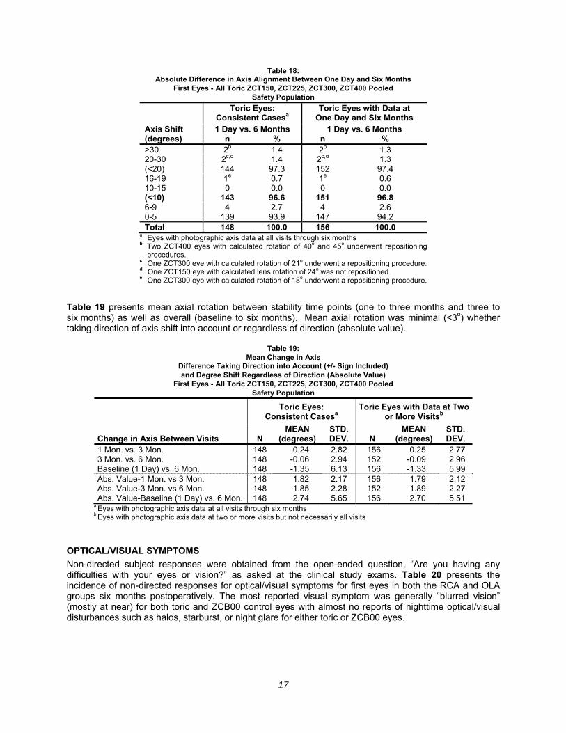
eyes having ≤5° axis change between consecutive visits approximately three months apart) Table 18 presents axis change for toric eyes between baseline (day 1) and six months. Of toric first eyes, 97% had <10° of axis change between baseline and six months.
Toric Eyes: Consistent Casesa
Toric Eyes with Data at Two or More Consecutive Visitsb
Axis Shift (degrees)
1 Month vs. 3 Months
3 Months vs. 6 Months
1 Month vs. 3 Months
3 Months vs. 6 Months
n % n % n % n % >30 0 0.0 0 0.0 0 0.0 0 0.0 16-30 0 0.0 0 0.0 0 0.0 0 0.0 10-15 2 1.4 3 2.0 2 1.3 3 2.0 (<10) 146 98.6 145 98.0 154 98.7 149 98.0 6-9 9 6.1 6 4.1 9 5.8 6 3.9 0-5 137 92.6c 139 93.9c 145 92.9c 143 94.1c Total 148 100.0 148 100.0 156 100.0 152 100.0
17
Table 18: Absolute Difference in Axis Alignment Between One Day and Six Months
First Eyes - All Toric ZCT150, ZCT225, ZCT300, ZCT400 Pooled Safety Population
Toric Eyes: Consistent Casesa
Toric Eyes with Data at One Day and Six Months
Axis Shift 1 Day vs. 6 Months 1 Day vs. 6 Months (degrees) n % n %>30 2b 1.4 2b 1.320-30 2c,d 1.4 2c,d 1.3(<20) 144 97.3 152 97.416-19 1e 0.7 1e 0.610-15 0 0.0 0 0.0(<10) 143 96.6 151 96.86-9 4 2.7 4 2.60-5 139 93.9 147 94.2Total 148 100.0 156 100.0
a Eyes with photographic axis data at all visits through six months b Two ZCT400 eyes with calculated rotation of 40o and 45o underwent repositioning
procedures. c One ZCT300 eye with calculated rotation of 21o underwent a repositioning procedure. d One ZCT150 eye with calculated lens rotation of 24o was not repositioned. e One ZCT300 eye with calculated rotation of 18o underwent a repositioning procedure.
Table 19 presents mean axial rotation between stability time points (one to three months and three to six months) as well as overall (baseline to six months). Mean axial rotation was minimal (<3o) whether taking direction of axis shift into account or regardless of direction (absolute value).
Table 19: Mean Change in Axis
Difference Taking Direction into Account (+/- Sign Included) and Degree Shift Regardless of Direction (Absolute Value)
First Eyes - All Toric ZCT150, ZCT225, ZCT300, ZCT400 Pooled Safety Population
Toric Eyes:
Consistent Casesa Toric Eyes with Data at Two
or More Visitsb
Change in Axis Between Visits N MEAN
(degrees) STD. DEV. N
MEAN (degrees)
STD. DEV.
1 Mon. vs. 3 Mon. 148 0.24 2.82 156 0.25 2.77 3 Mon. vs. 6 Mon. 148 -0.06 2.94 152 -0.09 2.96 Baseline (1 Day) vs. 6 Mon. 148 -1.35 6.13 156 -1.33 5.99 Abs. Value-1 Mon. vs 3 Mon. 148 1.82 2.17 156 1.79 2.12 Abs. Value-3 Mon. vs 6 Mon. 148 1.85 2.28 152 1.89 2.27 Abs. Value-Baseline (1 Day) vs. 6 Mon. 148 2.74 5.65 156 2.70 5.51
a Eyes with photographic axis data at all visits through six months b Eyes with photographic axis data at two or more visits but not necessarily all visits
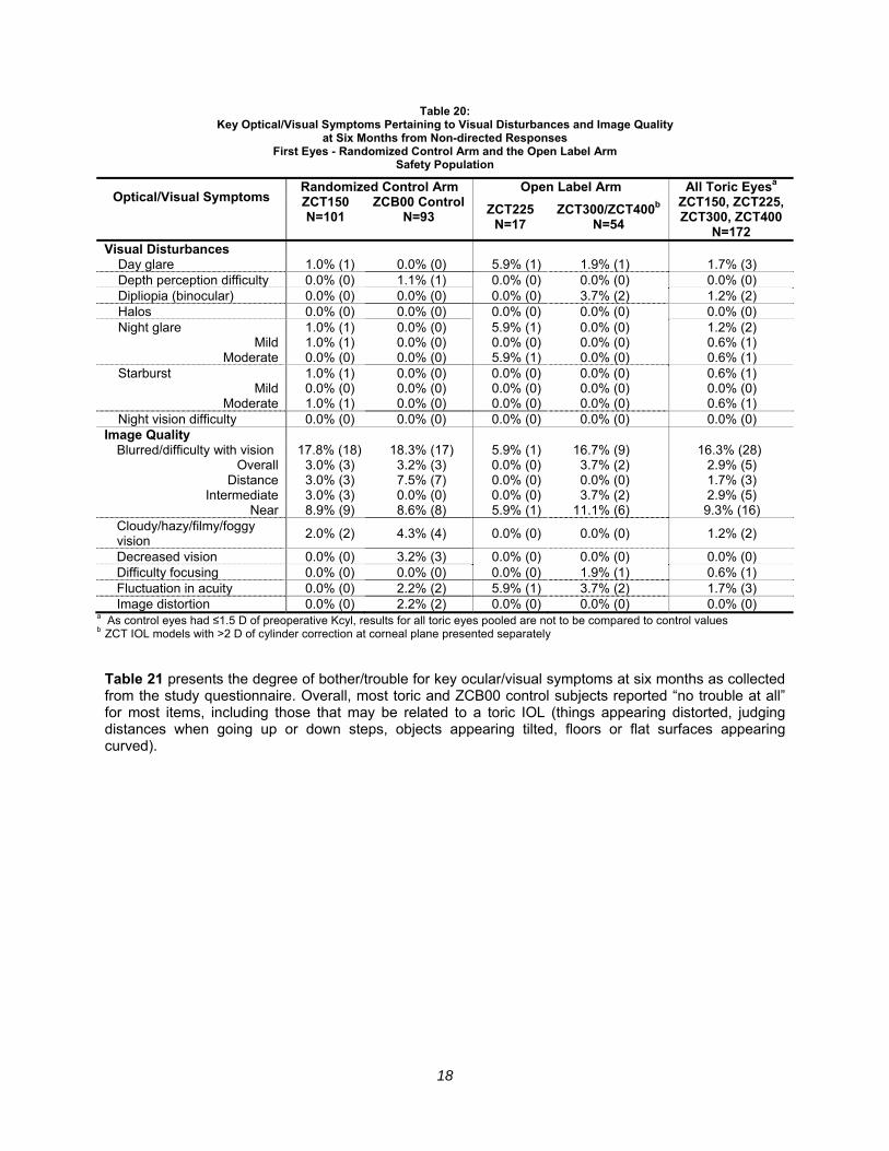
OPTICAL/VISUAL SYMPTOMS
Non-directed subject responses were obtained from the open-ended question, “Are you having any difficulties with your eyes or vision?” as asked at the clinical study exams. Table 20 presents the incidence of non-directed responses for optical/visual symptoms for first eyes in both the RCA and OLA groups six months postoperatively. The most reported visual symptom was generally “blurred vision” (mostly at near) for both toric and ZCB00 control eyes with almost no reports of nighttime optical/visual disturbances such as halos, starburst, or night glare for either toric or ZCB00 eyes.
18
Table 20: Key Optical/Visual Symptoms Pertaining to Visual Disturbances and Image Quality
at Six Months from Non-directed Responses First Eyes - Randomized Control Arm and the Open Label Arm
Safety Population
Optical/Visual Symptoms Randomized Control Arm Open Label Arm All Toric Eyesa
ZCT150 N=101
ZCB00 ControlN=93
ZCT225 N=17
ZCT300/ZCT400b N=54
ZCT150, ZCT225, ZCT300, ZCT400
N=172 Visual Disturbances
Day glare 1.0% (1) 0.0% (0) 5.9% (1) 1.9% (1) 1.7% (3) Depth perception difficulty 0.0% (0) 1.1% (1) 0.0% (0) 0.0% (0) 0.0% (0) Dipliopia (binocular) 0.0% (0) 0.0% (0) 0.0% (0) 3.7% (2) 1.2% (2) Halos 0.0% (0) 0.0% (0) 0.0% (0) 0.0% (0) 0.0% (0) Night glare 1.0% (1) 0.0% (0) 5.9% (1) 0.0% (0) 1.2% (2)
Mild 1.0% (1) 0.0% (0) 0.0% (0) 0.0% (0) 0.6% (1) Moderate 0.0% (0) 0.0% (0) 5.9% (1) 0.0% (0) 0.6% (1)
Starburst 1.0% (1) 0.0% (0) 0.0% (0) 0.0% (0) 0.6% (1) Mild 0.0% (0) 0.0% (0) 0.0% (0) 0.0% (0) 0.0% (0)
Moderate 1.0% (1) 0.0% (0) 0.0% (0) 0.0% (0) 0.6% (1) Night vision difficulty 0.0% (0) 0.0% (0) 0.0% (0) 0.0% (0) 0.0% (0)
Image Quality Blurred/difficulty with vision 17.8% (18) 18.3% (17) 5.9% (1) 16.7% (9) 16.3% (28)
Overall 3.0% (3) 3.2% (3) 0.0% (0) 3.7% (2) 2.9% (5) Distance 3.0% (3) 7.5% (7) 0.0% (0) 0.0% (0) 1.7% (3)
Intermediate 3.0% (3) 0.0% (0) 0.0% (0) 3.7% (2) 2.9% (5) Near 8.9% (9) 8.6% (8) 5.9% (1) 11.1% (6) 9.3% (16)
Cloudy/hazy/filmy/foggy vision
2.0% (2) 4.3% (4) 0.0% (0) 0.0% (0) 1.2% (2)
Decreased vision 0.0% (0) 3.2% (3) 0.0% (0) 0.0% (0) 0.0% (0) Difficulty focusing 0.0% (0) 0.0% (0) 0.0% (0) 1.9% (1) 0.6% (1) Fluctuation in acuity 0.0% (0) 2.2% (2) 5.9% (1) 3.7% (2) 1.7% (3) Image distortion 0.0% (0) 2.2% (2) 0.0% (0) 0.0% (0) 0.0% (0)
a As control eyes had ≤1.5 D of preoperative Kcyl, results for all toric eyes pooled are not to be compared to control values b ZCT IOL models with >2 D of cylinder correction at corneal plane presented separately
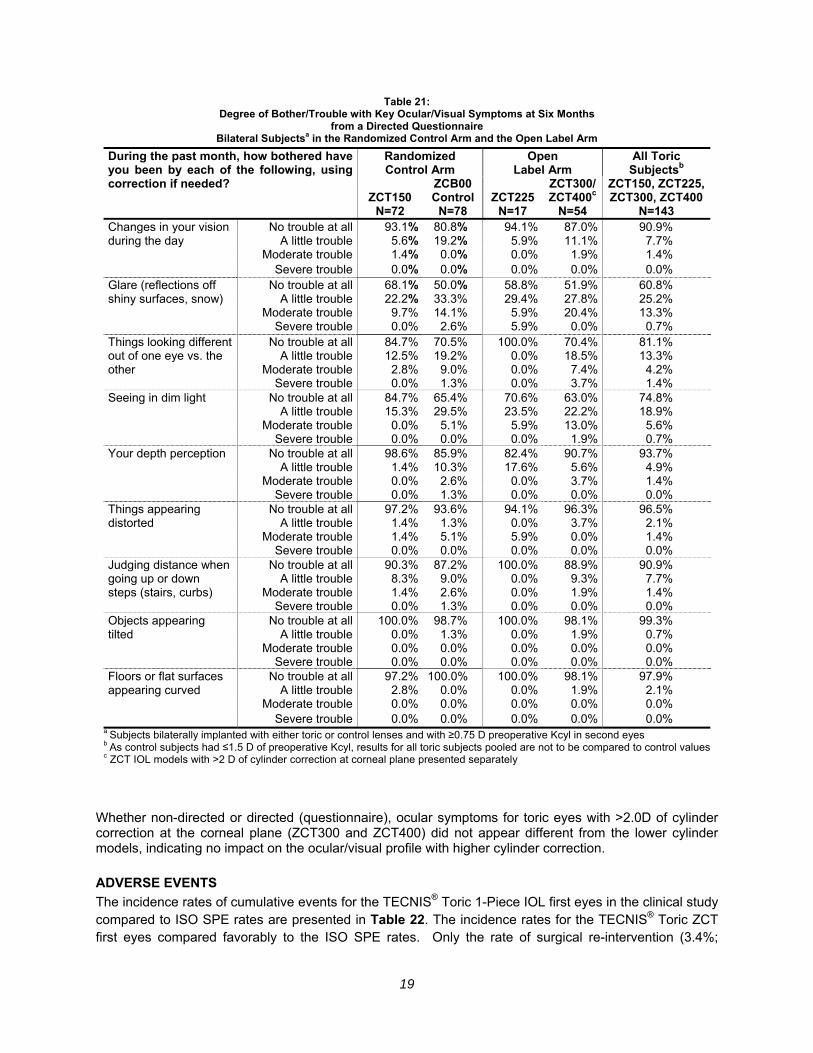
Table 21 presents the degree of bother/trouble for key ocular/visual symptoms at six months as collected from the study questionnaire. Overall, most toric and ZCB00 control subjects reported “no trouble at all” for most items, including those that may be related to a toric IOL (things appearing distorted, judging distances when going up or down steps, objects appearing tilted, floors or flat surfaces appearing curved).
19
Table 21: Degree of Bother/Trouble with Key Ocular/Visual Symptoms at Six Months
from a Directed Questionnaire Bilateral Subjectsa in the Randomized Control Arm and the Open Label Arm
During the past month, how bothered have you been by each of the following, using correction if needed?
Randomized Control Arm
Open Label Arm
All ToricSubjectsb
ZCT150N=72
ZCB00Control
N=78 ZCT225
N=17
ZCT300/ ZCT400c
N=54
ZCT150, ZCT225, ZCT300, ZCT400
N=143 Changes in your vision during the day
No trouble at all 93.1% 80.8% 94.1% 87.0% 90.9% A little trouble 5.6% 19.2% 5.9% 11.1% 7.7%
Moderate trouble 1.4% 0.0% 0.0% 1.9% 1.4% Severe trouble 0.0% 0.0% 0.0% 0.0% 0.0%
Glare (reflections off shiny surfaces, snow)
No trouble at all 68.1% 50.0% 58.8% 51.9% 60.8% A little trouble 22.2% 33.3% 29.4% 27.8% 25.2%
Moderate trouble 9.7% 14.1% 5.9% 20.4% 13.3% Severe trouble 0.0% 2.6% 5.9% 0.0% 0.7%
Things looking different out of one eye vs. the other
No trouble at all 84.7% 70.5% 100.0% 70.4% 81.1% A little trouble 12.5% 19.2% 0.0% 18.5% 13.3%
Moderate trouble 2.8% 9.0% 0.0% 7.4% 4.2% Severe trouble 0.0% 1.3% 0.0% 3.7% 1.4%
Seeing in dim light No trouble at all 84.7% 65.4% 70.6% 63.0% 74.8% A little trouble 15.3% 29.5% 23.5% 22.2% 18.9%
Moderate trouble 0.0% 5.1% 5.9% 13.0% 5.6% Severe trouble 0.0% 0.0% 0.0% 1.9% 0.7%
Your depth perception No trouble at all 98.6% 85.9% 82.4% 90.7% 93.7% A little trouble 1.4% 10.3% 17.6% 5.6% 4.9%
Moderate trouble 0.0% 2.6% 0.0% 3.7% 1.4% Severe trouble 0.0% 1.3% 0.0% 0.0% 0.0%
Things appearing distorted
No trouble at all 97.2% 93.6% 94.1% 96.3% 96.5% A little trouble 1.4% 1.3% 0.0% 3.7% 2.1%
Moderate trouble 1.4% 5.1% 5.9% 0.0% 1.4% Severe trouble 0.0% 0.0% 0.0% 0.0% 0.0%
Judging distance when going up or down steps (stairs, curbs)
No trouble at all 90.3% 87.2% 100.0% 88.9% 90.9% A little trouble 8.3% 9.0% 0.0% 9.3% 7.7%
Moderate trouble 1.4% 2.6% 0.0% 1.9% 1.4% Severe trouble 0.0% 1.3% 0.0% 0.0% 0.0%
Objects appearing tilted
No trouble at all 100.0% 98.7% 100.0% 98.1% 99.3% A little trouble 0.0% 1.3% 0.0% 1.9% 0.7%
Moderate trouble 0.0% 0.0% 0.0% 0.0% 0.0% Severe trouble 0.0% 0.0% 0.0% 0.0% 0.0%
Floors or flat surfaces appearing curved
No trouble at all 97.2% 100.0% 100.0% 98.1% 97.9% A little trouble 2.8% 0.0% 0.0% 1.9% 2.1%
Moderate trouble 0.0% 0.0% 0.0% 0.0% 0.0% Severe trouble 0.0% 0.0% 0.0% 0.0% 0.0%
a Subjects bilaterally implanted with either toric or control lenses and with ≥0.75 D preoperative Kcyl in second eyes
b As control subjects had ≤1.5 D of preoperative Kcyl, results for all toric subjects pooled are not to be compared to control values c ZCT IOL models with >2 D of cylinder correction at corneal plane presented separately
Whether non-directed or directed (questionnaire), ocular symptoms for toric eyes with >2.0D of cylinder correction at the corneal plane (ZCT300 and ZCT400) did not appear different from the lower cylinder models, indicating no impact on the ocular/visual profile with higher cylinder correction.
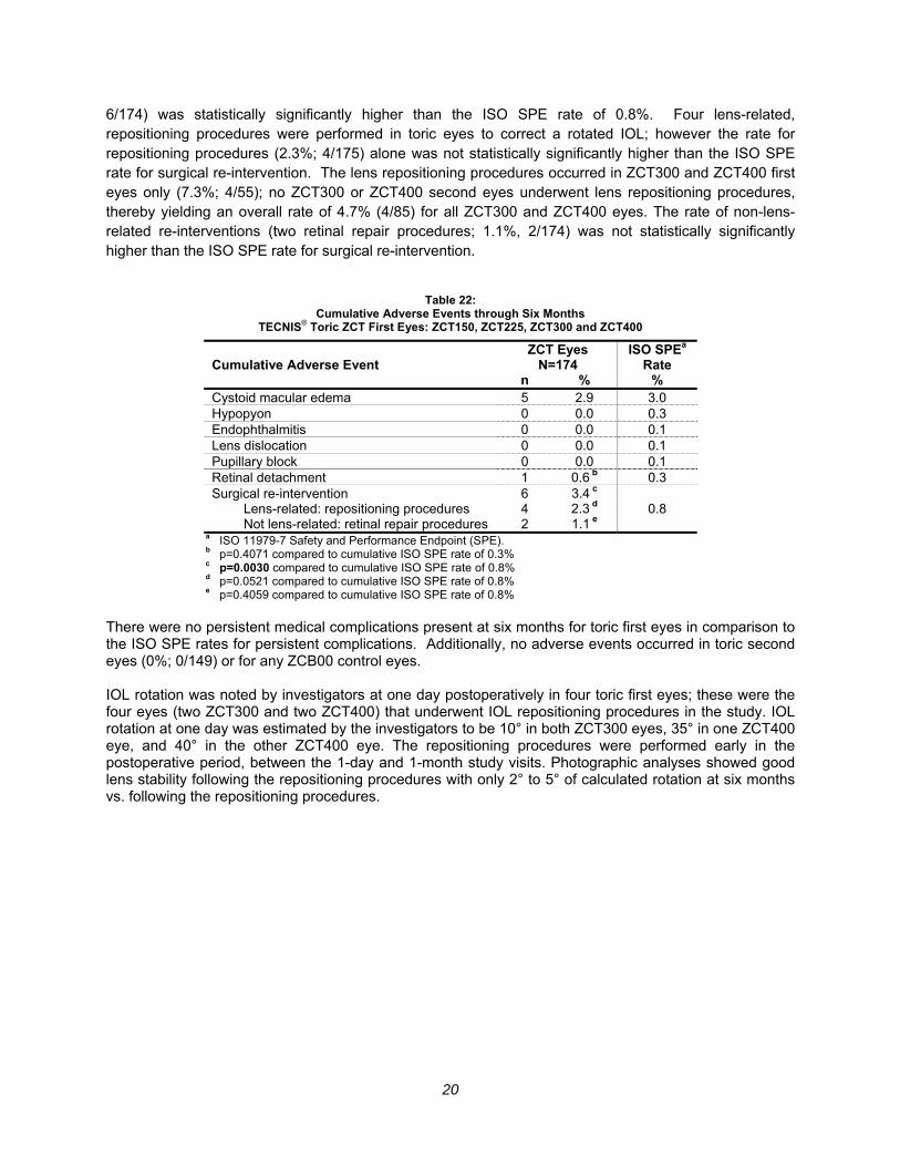
ADVERSE EVENTS
The incidence rates of cumulative events for the TECNIS® Toric 1-Piece IOL first eyes in the clinical study compared to ISO SPE rates are presented in Table 22. The incidence rates for the TECNIS® Toric ZCT first eyes compared favorably to the ISO SPE rates. Only the rate of surgical re-intervention (3.4%;
20
6/174) was statistically significantly higher than the ISO SPE rate of 0.8%. Four lens-related, repositioning procedures were performed in toric eyes to correct a rotated IOL; however the rate for repositioning procedures (2.3%; 4/175) alone was not statistically significantly higher than the ISO SPE rate for surgical re-intervention. The lens repositioning procedures occurred in ZCT300 and ZCT400 first eyes only (7.3%; 4/55); no ZCT300 or ZCT400 second eyes underwent lens repositioning procedures, thereby yielding an overall rate of 4.7% (4/85) for all ZCT300 and ZCT400 eyes. The rate of non-lens-related re-interventions (two retinal repair procedures; 1.1%, 2/174) was not statistically significantly higher than the ISO SPE rate for surgical re-intervention.
Table 22:
Cumulative Adverse Events through Six Months TECNIS® Toric ZCT First Eyes: ZCT150, ZCT225, ZCT300 and ZCT400
Cumulative Adverse Event ZCT Eyes
N=174 ISO SPEa
Rate n % %
Cystoid macular edema 5 2.9 3.0 Hypopyon 0 0.0 0.3 Endophthalmitis 0 0.0 0.1 Lens dislocation 0 0.0 0.1 Pupillary block 0 0.0 0.1 Retinal detachment 1 0.6 b 0.3 Surgical re-intervention 6 3.4 c
0.8 Lens-related: repositioning procedures 4 2.3 d Not lens-related: retinal repair procedures 2 1.1 e
a ISO 11979-7 Safety and Performance Endpoint (SPE). b p=0.4071 compared to cumulative ISO SPE rate of 0.3% c p=0.0030 compared to cumulative ISO SPE rate of 0.8% d p=0.0521 compared to cumulative ISO SPE rate of 0.8% e p=0.4059 compared to cumulative ISO SPE rate of 0.8%
There were no persistent medical complications present at six months for toric first eyes in comparison to the ISO SPE rates for persistent complications. Additionally, no adverse events occurred in toric second eyes (0%; 0/149) or for any ZCB00 control eyes. IOL rotation was noted by investigators at one day postoperatively in four toric first eyes; these were the four eyes (two ZCT300 and two ZCT400) that underwent IOL repositioning procedures in the study. IOL rotation at one day was estimated by the investigators to be 10° in both ZCT300 eyes, 35° in one ZCT400 eye, and 40° in the other ZCT400 eye. The repositioning procedures were performed early in the postoperative period, between the 1-day and 1-month study visits. Photographic analyses showed good lens stability following the repositioning procedures with only 2° to 5° of calculated rotation at six months vs. following the repositioning procedures.
21
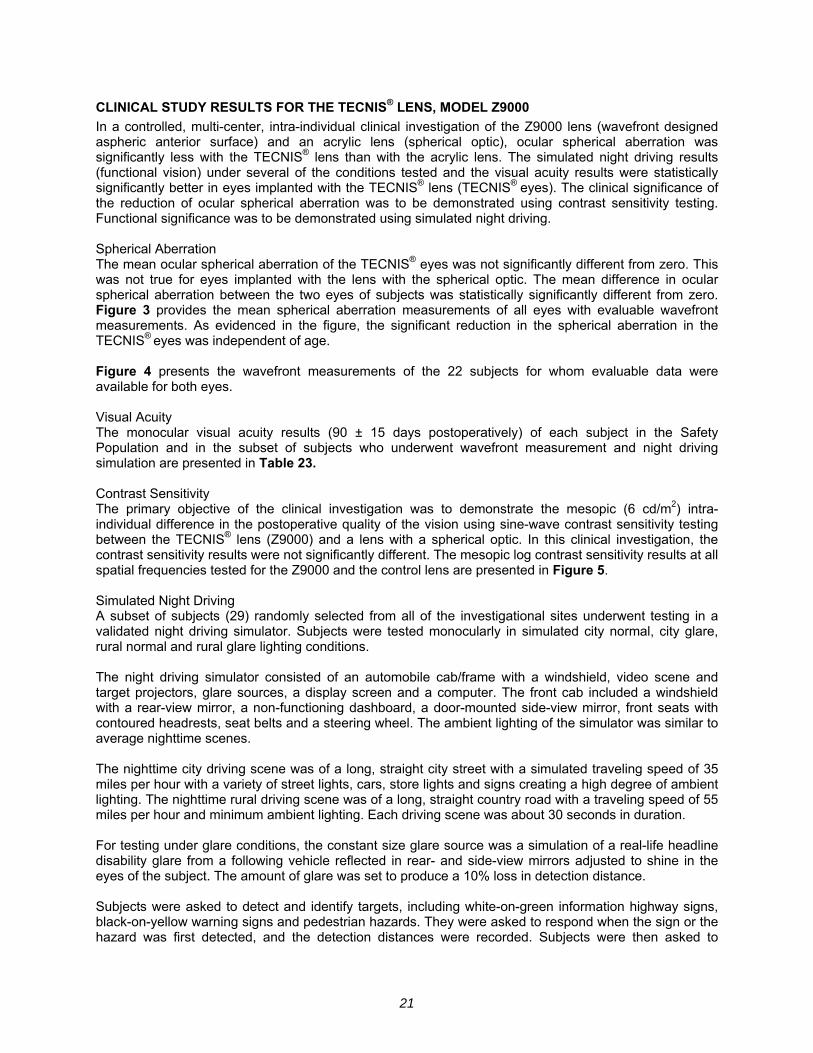
CLINICAL STUDY RESULTS FOR THE TECNIS® LENS, MODEL Z9000
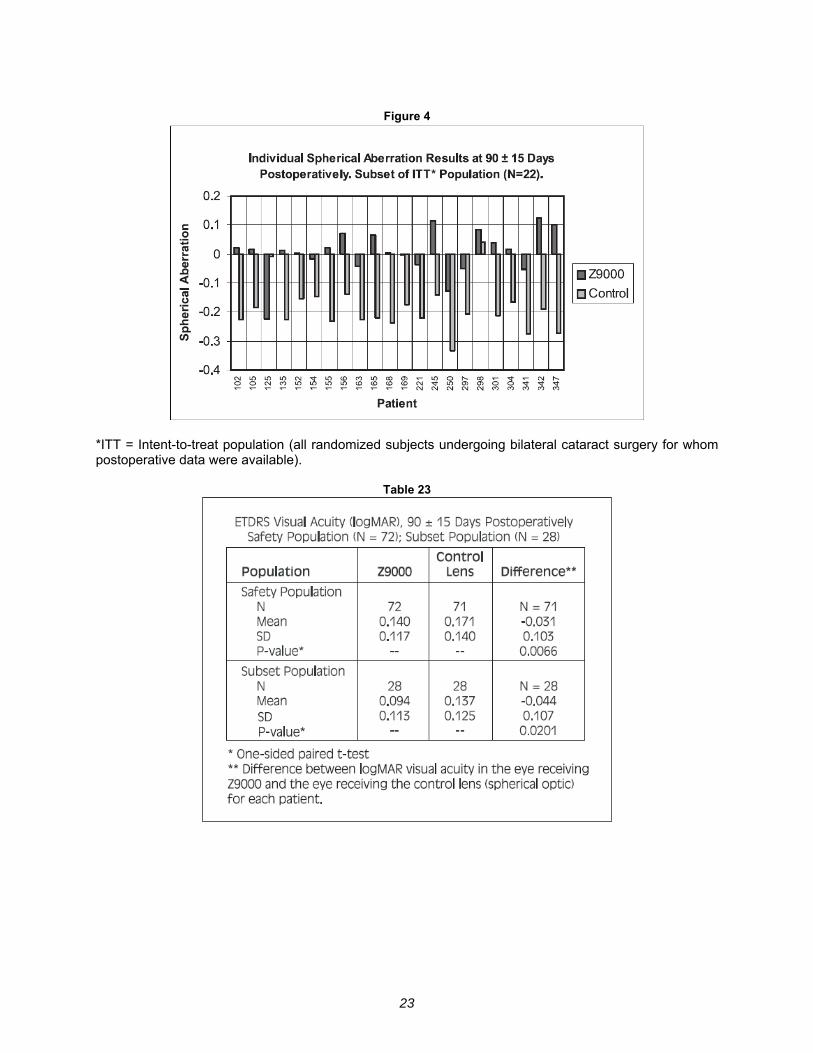
In a controlled, multi-center, intra-individual clinical investigation of the Z9000 lens (wavefront designed aspheric anterior surface) and an acrylic lens (spherical optic), ocular spherical aberration was significantly less with the TECNIS® lens than with the acrylic lens. The simulated night driving results (functional vision) under several of the conditions tested and the visual acuity results were statistically significantly better in eyes implanted with the TECNIS® lens (TECNIS® eyes). The clinical significance of the reduction of ocular spherical aberration was to be demonstrated using contrast sensitivity testing. Functional significance was to be demonstrated using simulated night driving. Spherical Aberration The mean ocular spherical aberration of the TECNIS® eyes was not significantly different from zero. This was not true for eyes implanted with the lens with the spherical optic. The mean difference in ocular spherical aberration between the two eyes of subjects was statistically significantly different from zero. Figure 3 provides the mean spherical aberration measurements of all eyes with evaluable wavefront measurements. As evidenced in the figure, the significant reduction in the spherical aberration in the TECNIS® eyes was independent of age. Figure 4 presents the wavefront measurements of the 22 subjects for whom evaluable data were available for both eyes. Visual Acuity The monocular visual acuity results (90 ± 15 days postoperatively) of each subject in the Safety Population and in the subset of subjects who underwent wavefront measurement and night driving simulation are presented in Table 23. Contrast Sensitivity The primary objective of the clinical investigation was to demonstrate the mesopic (6 cd/m2) intra-individual difference in the postoperative quality of the vision using sine-wave contrast sensitivity testing between the TECNIS® lens (Z9000) and a lens with a spherical optic. In this clinical investigation, the contrast sensitivity results were not significantly different. The mesopic log contrast sensitivity results at all spatial frequencies tested for the Z9000 and the control lens are presented in Figure 5. Simulated Night Driving A subset of subjects (29) randomly selected from all of the investigational sites underwent testing in a validated night driving simulator. Subjects were tested monocularly in simulated city normal, city glare, rural normal and rural glare lighting conditions. The night driving simulator consisted of an automobile cab/frame with a windshield, video scene and target projectors, glare sources, a display screen and a computer. The front cab included a windshield with a rear-view mirror, a non-functioning dashboard, a door-mounted side-view mirror, front seats with contoured headrests, seat belts and a steering wheel. The ambient lighting of the simulator was similar to average nighttime scenes. The nighttime city driving scene was of a long, straight city street with a simulated traveling speed of 35 miles per hour with a variety of street lights, cars, store lights and signs creating a high degree of ambient lighting. The nighttime rural driving scene was of a long, straight country road with a traveling speed of 55 miles per hour and minimum ambient lighting. Each driving scene was about 30 seconds in duration. For testing under glare conditions, the constant size glare source was a simulation of a real-life headline disability glare from a following vehicle reflected in rear- and side-view mirrors adjusted to shine in the eyes of the subject. The amount of glare was set to produce a 10% loss in detection distance. Subjects were asked to detect and identify targets, including white-on-green information highway signs, black-on-yellow warning signs and pedestrian hazards. They were asked to respond when the sign or the hazard was first detected, and the detection distances were recorded. Subjects were then asked to

22
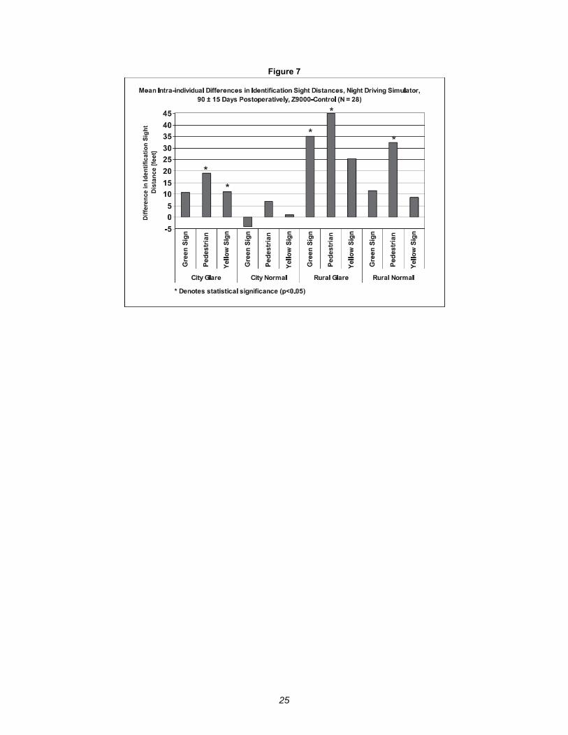
respond when the sign or hazard could first be identified, i.e., what did the sign say, what direction was the pedestrian walking, and the identification distances were recorded. The subject responses for each target set and visibility condition were averaged. Figures 6 and 7 present the average difference between the detection and identification distances with testing of the Z9000 eye and the detection and identification distances with testing of the spherical optic lens of each subject (the mean of the intra-individual differences). The Z9000 eyes performed functionally better than the control eyes in 21 of the 24 conditions tested. This means the Z9000 lens improves both detection and identification distances across the driving scenes (city and rural) and visibility conditions (with/without glare) compared to the control lens. Z9000 eyes performed statistically significantly better than the control eyes in 9 of the test conditions. The greatest advantage of the Z9000 lens is for increased detection and identification of the pedestrian hazard under rural visibility conditions with and without glare. Under these conditions, the increased visibility distance at 55 miles per hour provides for an average of about 0.5 seconds more time to perception and reaction time is functionally significant in increasing the time to take evasive action, time to stop or effect of impact. These findings suggest there is likely to be a meaningful safety benefit to elderly drivers with TECNIS® lenses, and to the drivers and pedestrians with whom they share the road. The results of this performance/functional test demonstrate that the TECNIS® lens improved functional vision, which in turn may improve patient safety for other life situations under low visibility conditions.
Figure 3
23
Figure 4
*ITT = Intent-to-treat population (all randomized subjects undergoing bilateral cataract surgery for whom postoperative data were available).
Table 23

24
Figure 5
Figure 6
25
Figure 7
26
CLINICAL STUDY RESULTS FOR THE SENSAR® 1-PIECE LENS, MODEL AAB00:
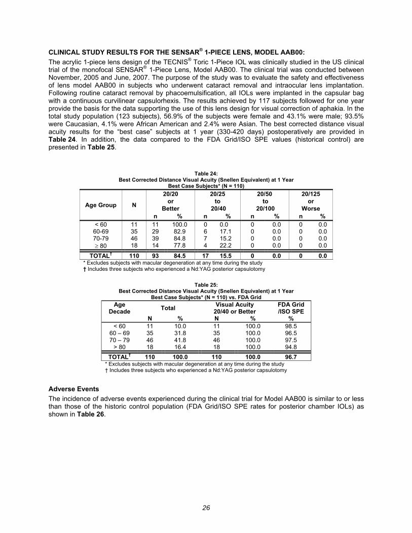
The acrylic 1-piece lens design of the TECNIS® Toric 1-Piece IOL was clinically studied in the US clinical trial of the monofocal SENSAR® 1-Piece Lens, Model AAB00. The clinical trial was conducted between November, 2005 and June, 2007. The purpose of the study was to evaluate the safety and effectiveness of lens model AAB00 in subjects who underwent cataract removal and intraocular lens implantation. Following routine cataract removal by phacoemulsification, all IOLs were implanted in the capsular bag with a continuous curvilinear capsulorhexis. The results achieved by 117 subjects followed for one year provide the basis for the data supporting the use of this lens design for visual correction of aphakia. In the total study population (123 subjects), 56.9% of the subjects were female and 43.1% were male; 93.5% were Caucasian, 4.1% were African American and 2.4% were Asian. The best corrected distance visual acuity results for the “best case” subjects at 1 year (330-420 days) postoperatively are provided in Table 24. In addition, the data compared to the FDA Grid/ISO SPE values (historical control) are presented in Table 25.
Table 24:
Best Corrected Distance Visual Acuity (Snellen Equivalent) at 1 Year Best Case Subjects* (N = 110)
Age Group N
20/20 or
Better
20/25 to
20/40
20/50 to
20/100
20/125 or
Worse n % n % n % n %
< 60 11 11 100.0 0 0.0 0 0.0 0 0.0 60-69 35 29 82.9 6 17.1 0 0.0 0 0.0 70-79 46 39 84.8 7 15.2 0 0.0 0 0.0 80 18 14 77.8 4 22.2 0 0.0 0 0.0
TOTAL† 110 93 84.5 17 15.5 0 0.0 0 0.0 * Excludes subjects with macular degeneration at any time during the study † Includes three subjects who experienced a Nd:YAG posterior capsulotomy
Table 25:
Best Corrected Distance Visual Acuity (Snellen Equivalent) at 1 Year Best Case Subjects* (N = 110) vs. FDA Grid
Age Decade
Total Visual Acuity
20/40 or Better FDA Grid /ISO SPE
N % N % % < 60 11 10.0 11 100.0 98.5
60 – 69 35 31.8 35 100.0 96.5 70 – 79 46 41.8 46 100.0 97.5
> 80 18 16.4 18 100.0 94.8
TOTAL† 110 100.0 110 100.0 96.7 * Excludes subjects with macular degeneration at any time during the study † Includes three subjects who experienced a Nd:YAG posterior capsulotomy
Adverse Events
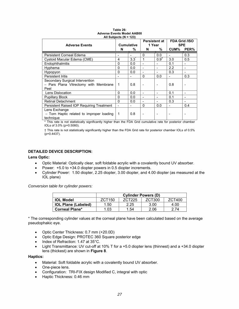
The incidence of adverse events experienced during the clinical trial for Model AAB00 is similar to or less than those of the historic control population (FDA Grid/ISO SPE rates for posterior chamber IOLs) as shown in Table 26.
27
Table 26: Adverse Events Model AAB00
All Subjects (N = 123)
Adverse Events Cumulative Persistent at
1 Year FDA Grid /ISO
SPE N % N % CUM% PER%
Persistent Corneal Edema - - 0 0.0 - 0.3 Cystoid Macular Edema (CME) 4 3.3* 1 0.9† 3.0 0.5 Endophthalmitis 0 0.0 - - 0.1 - Hyphema 0 0.0 - - 2.2 - Hypopyon 0 0.0 - - 0.3 - Persistent Iritis - - 0 0.0 - 0.3 Secondary Surgical Intervention – Pars Plana Vitrectomy with Membrane Peel
1 0.8 - - 0.8 -
Lens Dislocation 0 0.0 - - 0.1 - Pupillary Block 0 0.0 - - 0.1 - Retinal Detachment 0 0.0 - - 0.3 - Persistent Raised IOP Requiring Treatment - - 0 0.0 - 0.4 Lens Exchange – Torn Haptic related to improper loading technique
1 0.8 - - - -
* This rate is not statistically significantly higher than the FDA Grid cumulative rate for posterior chamber IOLs of 3.0% (p=0.5060).
† This rate is not statistically significantly higher than the FDA Grid rate for posterior chamber IOLs of 0.5% (p=0.4437).
DETAILED DEVICE DESCRIPTION:
Lens Optic:
Optic Material: Optically clear, soft foldable acrylic with a covalently bound UV absorber. Power: +5.0 to +34.0 diopter powers in 0.5 diopter increments. Cylinder Power: 1.50 diopter, 2.25 diopter, 3.00 diopter, and 4.00 diopter (as measured at the
IOL plane) Conversion table for cylinder powers:
Cylinder Powers (D) IOL Model ZCT150 ZCT225 ZCT300 ZCT400 IOL Plane (Labeled) 1.50 2.25 3.00 4.00 Corneal Plane* 1.03 1.54 2.06 2.74
* The corresponding cylinder values at the corneal plane have been calculated based on the average pseudophakic eye.
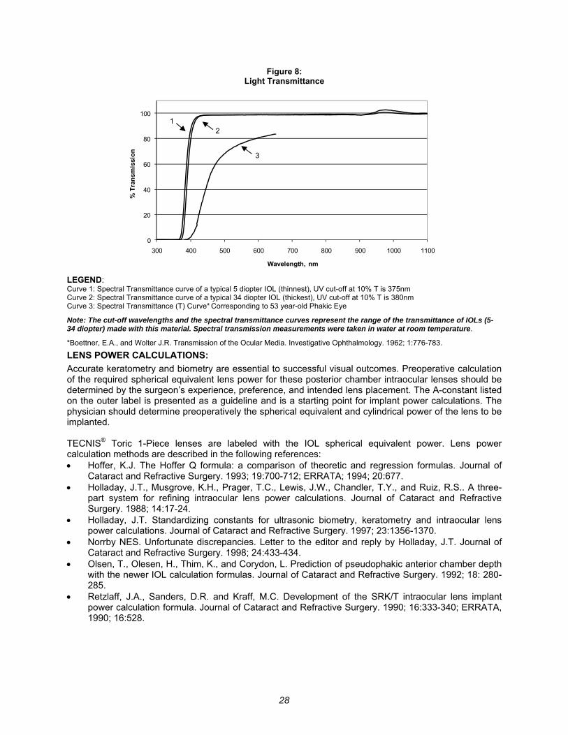
Optic Center Thickness: 0.7 mm (+20.0D) Optic Edge Design: PROTEC 360 Square posterior edge Index of Refraction: 1.47 at 35°C. Light Transmittance: UV cut-off at 10% T for a +5.0 diopter lens (thinnest) and a +34.0 diopter
lens (thickest) are shown in Figure 8.
Haptics:
Material: Soft foldable acrylic with a covalently bound UV absorber. One-piece lens. Configuration: TRI-FIX design Modified C, integral with optic Haptic Thickness: 0.46 mm
28
Figure 8: Light Transmittance
LEGEND: Curve 1: Spectral Transmittance curve of a typical 5 diopter IOL (thinnest), UV cut-off at 10% T is 375nm Curve 2: Spectral Transmittance curve of a typical 34 diopter IOL (thickest), UV cut-off at 10% T is 380nm Curve 3: Spectral Transmittance (T) Curve* Corresponding to 53 year-old Phakic Eye
Note: The cut-off wavelengths and the spectral transmittance curves represent the range of the transmittance of IOLs (5-34 diopter) made with this material. Spectral transmission measurements were taken in water at room temperature.
*Boettner, E.A., and Wolter J.R. Transmission of the Ocular Media. Investigative Ophthalmology. 1962; 1:776-783.
LENS POWER CALCULATIONS:
Accurate keratometry and biometry are essential to successful visual outcomes. Preoperative calculation of the required spherical equivalent lens power for these posterior chamber intraocular lenses should be determined by the surgeon’s experience, preference, and intended lens placement. The A-constant listed on the outer label is presented as a guideline and is a starting point for implant power calculations. The physician should determine preoperatively the spherical equivalent and cylindrical power of the lens to be implanted. TECNIS® Toric 1-Piece lenses are labeled with the IOL spherical equivalent power. Lens power calculation methods are described in the following references: Hoffer, K.J. The Hoffer Q formula: a comparison of theoretic and regression formulas. Journal of
Cataract and Refractive Surgery. 1993; 19:700-712; ERRATA; 1994; 20:677. Holladay, J.T., Musgrove, K.H., Prager, T.C., Lewis, J.W., Chandler, T.Y., and Ruiz, R.S.. A three-
part system for refining intraocular lens power calculations. Journal of Cataract and Refractive Surgery. 1988; 14:17-24.
Holladay, J.T. Standardizing constants for ultrasonic biometry, keratometry and intraocular lens power calculations. Journal of Cataract and Refractive Surgery. 1997; 23:1356-1370.
Norrby NES. Unfortunate discrepancies. Letter to the editor and reply by Holladay, J.T. Journal of Cataract and Refractive Surgery. 1998; 24:433-434.
Olsen, T., Olesen, H., Thim, K., and Corydon, L. Prediction of pseudophakic anterior chamber depth with the newer IOL calculation formulas. Journal of Cataract and Refractive Surgery. 1992; 18: 280-285.
Retzlaff, J.A., Sanders, D.R. and Kraff, M.C. Development of the SRK/T intraocular lens implant power calculation formula. Journal of Cataract and Refractive Surgery. 1990; 16:333-340; ERRATA, 1990; 16:528.
0
20
40
60
80
100
300 400 500 600 700 800 900 1000 1100
% T
ran
smis
sio
n
Wavelength, nm
12
3
29
SELECTION AND PLACEMENT OF THE TECNIS® TORIC 1-PIECE IOL:
The astigmatism to be corrected should be determined from keratometry and biometry data rather than refractive data since the presence of lenticular astigmatism in the crystalline lens to be removed may influence results. The size and location of the surgical incision may affect the amount of postoperative corneal astigmatism as well as the respective axis. In order to facilitate IOL selection and axis placement, AMO provides a web-based proprietary tool, the TECNIS® Toric Calculator (www.TecnisToricCalc.com) for the surgeon. The corneal astigmatism to be corrected at the time of surgery is calculated by the TECNIS® Toric Calculator using vector summation of the preoperative corneal astigmatism and the expected surgically induced astigmatism. The cylinder IOL power calculation is based on the Holladay 1 formula (Holladay JT, Musgrove KH, Prager TC, Lewis JW, Chandler TY, and Ruiz RS. A three-part system for refining intraocular lens power calculations. Journal of Cataract and Refractive Surgery. 1988; 14:17-24). This yields an individual calculation instead of using a fixed ratio based on average ocular parameters. For optimal toric IOL calculations, it is recommended that surgeons customize their surgically induced corneal astigmatism values based upon individual surgical technique and past results. An example of this calculation can be found within the following reference (Holladay JT, Cravy TV, Koch DD. “Calculating the surgically induced refractive change following ocular surgery”, J Cataract Refract Surg. 1992;18:429-43) Preoperative keratometry and biometry data, incision location, spherical equivalent IOL power, and the surgeon’s estimated surgically induced corneal astigmatism are used as inputs for the TECNIS® Toric Calculator. These inputs are used to determine the axis of placement in the eye and the predicted residual refractive astigmatism for up to three different TECNIS® Toric 1-Piece IOL models. In eyes with low levels of corneal astigmatism, the predicted residual refractive astigmatism for implantation of a TECNIS® 1-Piece lens, Model ZCB00, will be displayed for evaluation by the surgeon to determine the clinically meaningful benefit of implanting a toric IOL. For optimal results, the surgeon must ensure the correct placement and orientation of the lens within the capsular bag. The anterior surface of the IOL is marked with indentations (four at opposite sides) at the haptic/optic junction that identify the flat meridian of the TECNIS® Toric 1-Piece optic. These indentations, or axis marks, form an imaginary line representing the plus cylinder axis (note: IOL cylinder steep meridian is 90° away). The TECNIS® Toric 1-Piece IOL cylinder axis marks should be aligned with the post-incision steep corneal meridian (intended axis of placement). Prior to surgery the operative eye should be marked in the following manner: With the patient sitting upright, precisely mark the twelve o’clock and/or the six o’clock position with a T marker, a surgical skin marker, or a marking pencil indicated for ophthalmic use. Using these marks as reference points, an axis marker can be used immediately prior to or during surgery to mark the axis of lens placement following the use of the web-based TECNIS® Toric Calculator, www.TecnisToricCalc.com to determine the optimal axis of placement. After the lens is inserted, precisely align the axis marking indentations on the TECNIS® Toric 1-Piece IOL with the marked axis of lens placement. Carefully remove all viscoelastic from the capsular bag. This may be accomplished by manipulating the IOL optic with the I/A tip and using standard irrigation/aspiration techniques to remove all viscoelastic from the eye. Special care should be taken to ensure proper positioning of the TECNIS® Toric 1-Piece IOL at the intended axis following viscoelastic removal and/or inflation of the capsular bag at the end of the surgical case. Residual viscoelastic and/or over-inflation of the bag may allow the lens to rotate, causing misalignment of the TECNIS® Toric 1-Piece IOL with the intended axis of placement. Misalignment of the axis of the lens with the intended axis of placement may compromise its astigmatic correction. Such misalignment can result from inaccurate keratometry or marking of the cornea, inaccurate placement of the TECNIS® Toric 1-Piece IOL axis during surgery, an unanticipated surgically induced change in the cornea, or physical rotation of the TECNIS® Toric 1-Piece IOL after implantation. In order to minimize this effect, the surgeon should be careful to ensure that preoperative keratometry and biometry is accurate and that the IOL is properly oriented prior to the end or surgery.
30
DIRECTIONS FOR USE:
1. Prior to implanting, examine the lens package for IOL type, power, proper configuration and expiration date.
2. Open the peel pouches and remove the lens in a sterile environment. Verify the dioptric power and cylinder power of the lens.
3. Examine the lens thoroughly to ensure particles have not become attached to it, and examine the lens’ optical surfaces for other defects.
4. If desired, the lens may be soaked or rinsed in sterile balanced salt solution until ready for implantation.
5. AMO recommends using The UNFOLDER® Platinum 1 Series Implantation System (the 1MTEC30 Cartridge and the DK7796 inserter). Alternate validated insertion systems that can be used to insert the TECNIS® Toric 1-Piece lens include the UNFOLDER® EMERALD-AR Series Implantation System (with the 1CART30 Cartridge), the ONE SERIES Ultra Insertion System (the 1VPR30 Cartridge and the DK7786 or DK7791 inserters) or any other AMO-qualified insertion system. Only insertion instruments that have been validated and approved for use with this lens should be used. Please refer to the directions for use of the insertion instrument or system for additional information.
6. Carefully remove all viscoelastic from the capsular bag and align the lens with the intended axis of placement.
Factors to consider in deciding whether to implant a toric lens: effectiveness of implanting a toric lens in reducing postoperative astigmatism is affected by many factors, including the following:
The degree of mismatch between the postoperative magnitude of corneal astigmatism and effective IOL power in the corneal plane.
Misalignment between the intended axial position and final IOL axial orientation. Error in prediction of the postoperative corneal cylinder axis and power. Error in prediction of cylinder
axis is greatest for lower levels of preoperative corneal astigmatism. Manufacturing variation in power and axis markings can influence intended correction. Based on the
tolerances set in the ANSI standard Z80.30, cylinder power variation may cause the intended correction at the corneal plane to vary by up to ±0.34 D, and cylinder axis tolerance may reduce intended correction by up to 16%.
CAUTION:
Do not use the lens if the package has been damaged. The sterility of the lens may have been compromised.
PATIENT CARD:
An implant identification card, to be supplied to the patient, is included in the package. The patient should be instructed to keep the card as a permanent record of his/her implant and to show the card to any eye care practitioner he/she may see in the future.
REPORTING:
Adverse events and/or potentially sight-threatening complications that may reasonably be regarded as lens-related and that were not previously expected in nature, severity or rate of occurrence must be reported to AMO. This information is being requested from all surgeons in order to document potential long-term effects of intraocular lens implantation, especially in younger patients. Physicians are required to report these events in order to aid in identifying emerging or potential problems with posterior chamber lenses. These problems may be related to a specific lot of lenses or may be indicative of long-term problems associated with these lenses or with intraocular lenses in general.
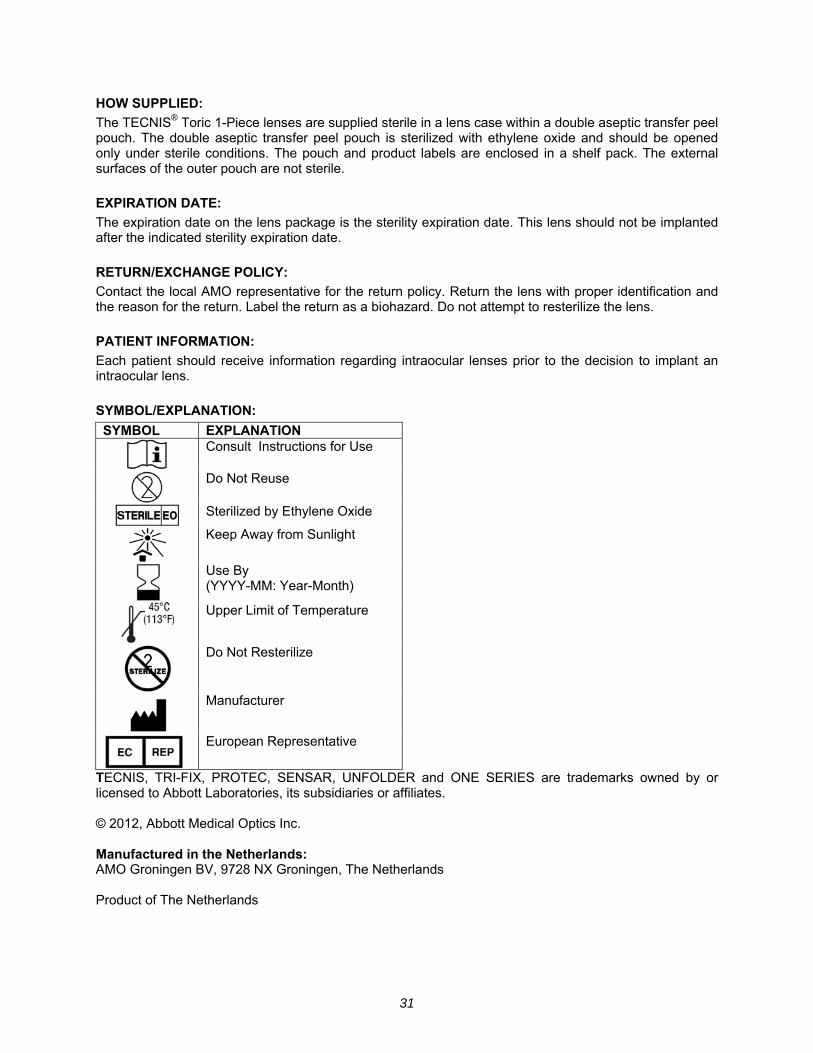
31
HOW SUPPLIED:
The TECNIS® Toric 1-Piece lenses are supplied sterile in a lens case within a double aseptic transfer peel pouch. The double aseptic transfer peel pouch is sterilized with ethylene oxide and should be opened only under sterile conditions. The pouch and product labels are enclosed in a shelf pack. The external surfaces of the outer pouch are not sterile.
EXPIRATION DATE:
The expiration date on the lens package is the sterility expiration date. This lens should not be implanted after the indicated sterility expiration date.
RETURN/EXCHANGE POLICY:
Contact the local AMO representative for the return policy. Return the lens with proper identification and the reason for the return. Label the return as a biohazard. Do not attempt to resterilize the lens.
PATIENT INFORMATION:
Each patient should receive information regarding intraocular lenses prior to the decision to implant an intraocular lens.
SYMBOL/EXPLANATION:
SYMBOL EXPLANATION
Consult Instructions for Use
Do Not Reuse
Sterilized by Ethylene Oxide
Keep Away from Sunlight
Use By (YYYY-MM: Year-Month)
Upper Limit of Temperature
Do Not Resterilize
Manufacturer
European Representative
TECNIS, TRI-FIX, PROTEC, SENSAR, UNFOLDER and ONE SERIES are trademarks owned by or licensed to Abbott Laboratories, its subsidiaries or affiliates. © 2012, Abbott Medical Optics Inc. Manufactured in the Netherlands: AMO Groningen BV, 9728 NX Groningen, The Netherlands Product of The Netherlands

32
Abbott Medical Optics Inc. 1700 E. St. Andrew Place Santa Ana, CA 92705 USA www.amo-inc.com AMO Ireland Block B Liffey Valley Office Campus Quarryvale, Co. Dublin, Ireland