The Special Senses - · PDF fileOr you can google: Anatomy and Physiology online quiz.
40
Copyright 2009, John Wiley & Sons, Inc. The Special Senses
Transcript of The Special Senses - · PDF fileOr you can google: Anatomy and Physiology online quiz.
Copyright 2009, John Wiley & Sons, Inc.
The Special Senses
Videos
Bozeman senses: https://youtu.be/TAzTFgPSPiU
How hearing works: https://youtu.be/flIAxGsV1q0
How does the ear work: https://youtu.be/qgdqp-oPb1Q
Khan, smell: https://youtu.be/5-McqAO8_Qw
Khan, taste: https://youtu.be/-vp1X7_u3KU
TedEd, smell: https://youtu.be/snJnO6OpjCs
Vsauce, why two nostrils: https://youtu.be/eiAx2kqmUpQ
Handwritten, the eye: https://youtu.be/MJxxFwVu1OM
Its okay to be smart, how many smells?: https://youtu.be/-yEtBps-BnI
Is your red the same as mine? https://youtu.be/evQsOFQju08
How the eye works: https://youtu.be/RE1MvRmWg7I
TedEd how we see color: https://youtu.be/l8_fZPHasdo
Ted Ed, Why we cry: https://youtu.be/keMF8YzQoRM
Copyright 2009, John Wiley & Sons, Inc.

Comparison of General and Special Senses
General Senses
Include somatic sensations (heat,
pain).
Sensors are scattered throughout
the body.
Simple structures.
Special Senses
Include smell, taste, vision, hearing
and equilibrium.
Concentrated in specific locations
in the head.
Complex.
Copyright 2009, John Wiley & Sons, Inc.

Copyright 2009, John Wiley & Sons, Inc.
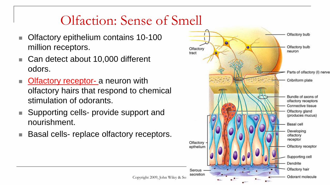
Olfaction: Sense of Smell Olfactory epithelium contains 10-100
million receptors.
Can detect about 10,000 different
odors.
Olfactory receptor- a neuron with
olfactory hairs that respond to chemical
stimulation of odorants.
Supporting cells- provide support and
nourishment.
Basal cells- replace olfactory receptors.
Copyright 2009, John Wiley & Sons, Inc.
Olfaction: Sense of Smell Olfactory epithelium contains 10-100
million receptors.
Can detect about 10,000 different
odors.
Olfactory receptor- a neuron with
olfactory hairs that respond to chemical
stimulation of odorants.
Supporting cells- provide support and
nourishment.
Basal cells- replace olfactory receptors.

Copyright 2009, John Wiley & Sons, Inc.
Olfaction: Sense of Smell Odorant binds to the receptor of an
olfactory hair→
nerve impulse through olfactory nerves
(using Na+ channels)→
olfactory bulbs→
olfactory tract→
primary olfactory area of the cerebral
cortex.

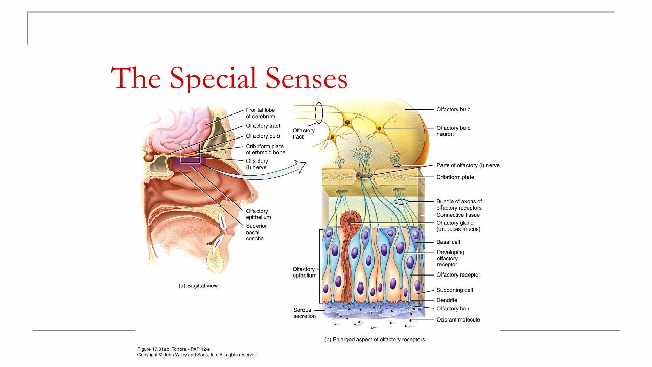
Olfactory Epithelium and
Olfactory Receptors
continued…
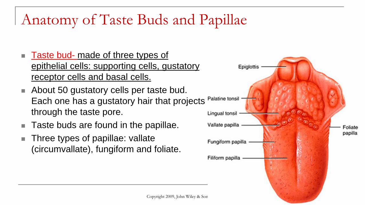
Anatomy of Taste Buds and Papillae
Taste bud- made of three types of
epithelial cells: supporting cells, gustatory
receptor cells and basal cells.
About 50 gustatory cells per taste bud.
Each one has a gustatory hair that projects
through the taste pore.
Taste buds are found in the papillae.
Three types of papillae: vallate
(circumvallate), fungiform and foliate.
Copyright 2009, John Wiley & Sons, Inc.
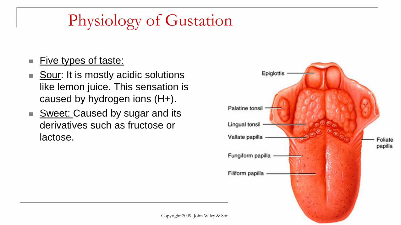
Physiology of Gustation
Five types of taste:
Sour: It is mostly acidic solutions
like lemon juice. This sensation is
caused by hydrogen ions (H+).
Sweet: Caused by sugar and its
derivatives such as fructose or
lactose.
Copyright 2009, John Wiley & Sons, Inc.

Physiology of Gustation
Bitter: There are a wide range of
sensory cells that respond to bitter
substances. From an evolutionary
standpoint, this can be explained by
the many different bitter species of
plants, some of which were
poisonous.
Salty: Food containing table salt.
Which consists of sodium and
chloride.
Umami: ????
Copyright 2009, John Wiley & Sons, Inc.
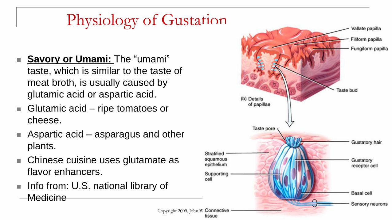
Physiology of Gustation
Savory or Umami: The “umami”
taste, which is similar to the taste of
meat broth, is usually caused by
glutamic acid or aspartic acid.
Glutamic acid – ripe tomatoes or
cheese.
Aspartic acid – asparagus and other
plants.
Chinese cuisine uses glutamate as
flavor enhancers.
Info from: U.S. national library of
Medicine
Copyright 2009, John Wiley & Sons, Inc.

Gustatory Pathway

Vision or Sight
Visible light:
400-700 nm.
Copyright 2009, John Wiley & Sons, Inc.
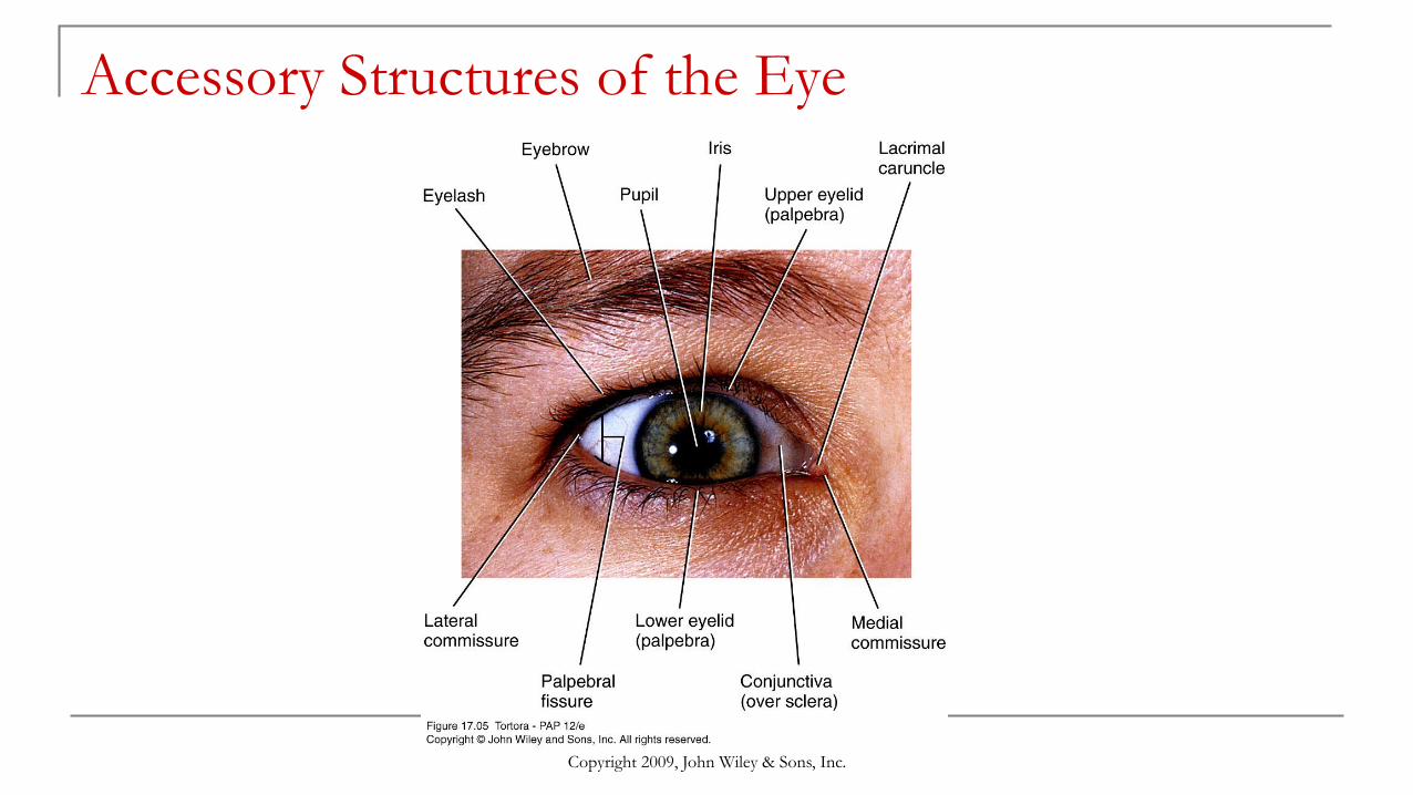
Accessory Structures of the Eye
Copyright 2009, John Wiley & Sons, Inc.

Accessory Structures of the Eye

Anatomy of the Eyeball
Copyright 2009, John Wiley & Sons, Inc.
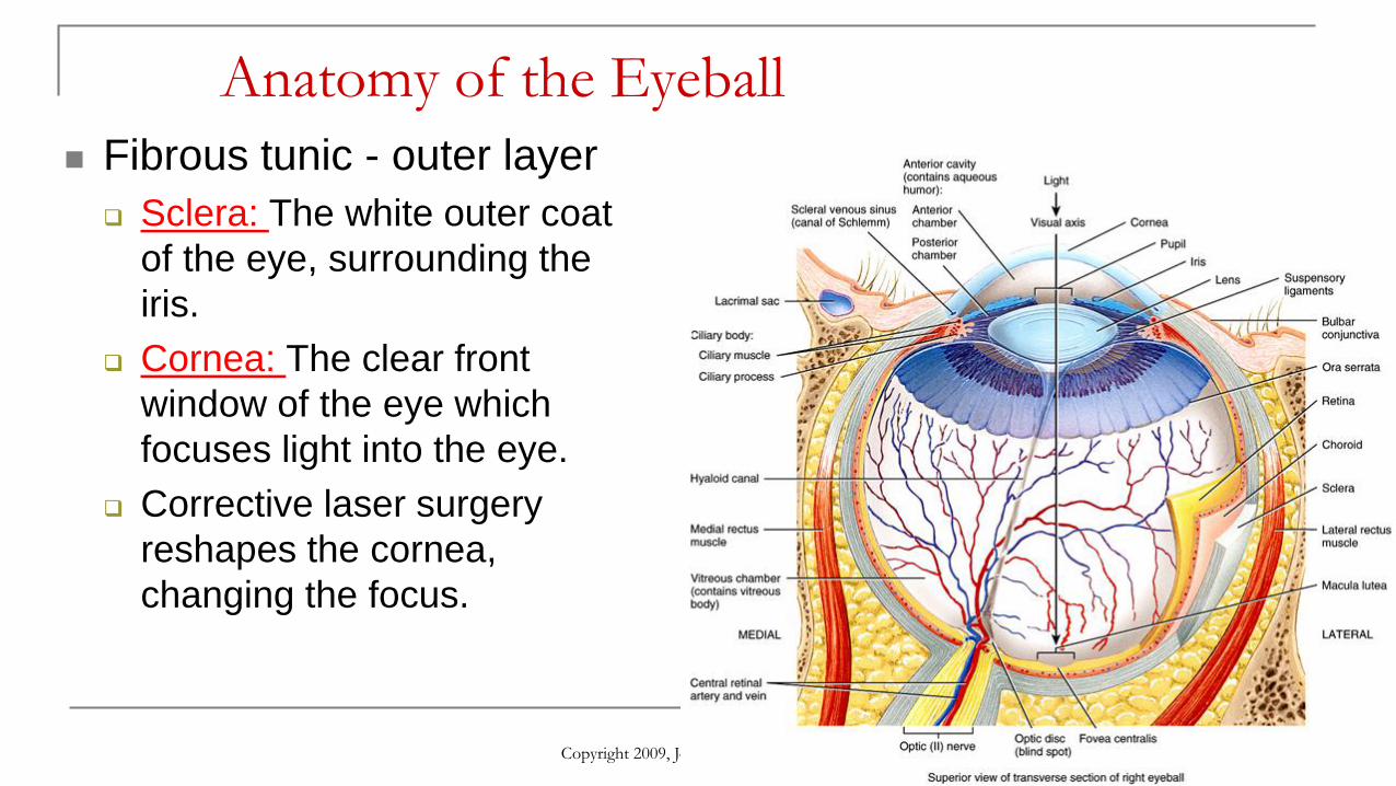
Fibrous tunic - outer layer
Sclera: The white outer coat
of the eye, surrounding the
iris.
Cornea: The clear front
window of the eye which
focuses light into the eye.
Corrective laser surgery
reshapes the cornea,
changing the focus.
Anatomy of the Eyeball
Copyright 2009, John Wiley & Sons, Inc.
Fibrous tunic - outer layer
Sclera: The white outer coat
of the eye, surrounding the
iris.
Cornea: The clear front
window of the eye which
focuses light into the eye.
Corrective laser surgery
reshapes the cornea,
changing the focus.
Anatomy of the Eyeball
Copyright 2009, John Wiley & Sons, Inc.
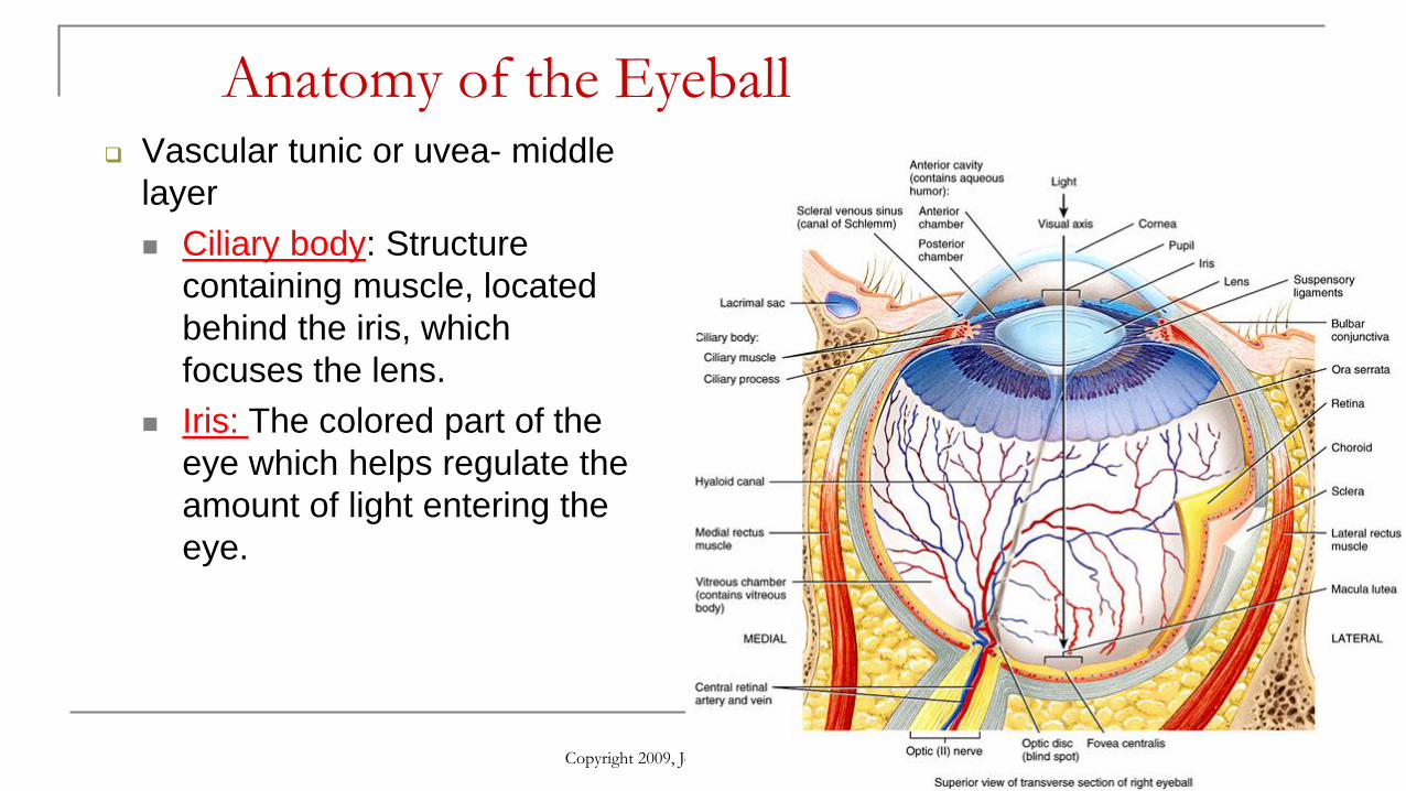
Vascular tunic or uvea- middle
layer
Ciliary body: Structure
containing muscle, located
behind the iris, which
focuses the lens.
Iris: The colored part of the
eye which helps regulate the
amount of light entering the
eye.
Anatomy of the Eyeball
Copyright 2009, John Wiley & Sons, Inc.
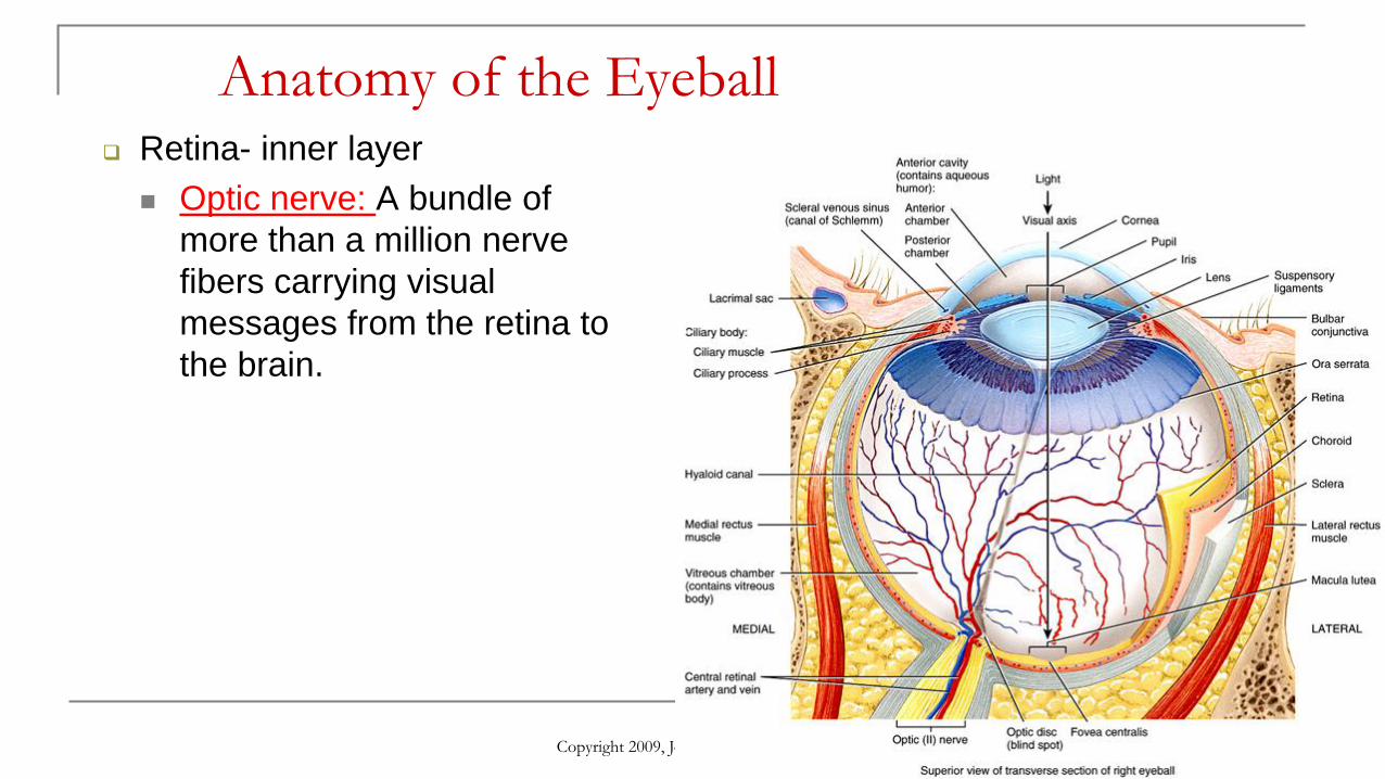
Retina- inner layer
Optic nerve: A bundle of
more than a million nerve
fibers carrying visual
messages from the retina to
the brain.
Anatomy of the Eyeball
Copyright 2009, John Wiley & Sons, Inc.
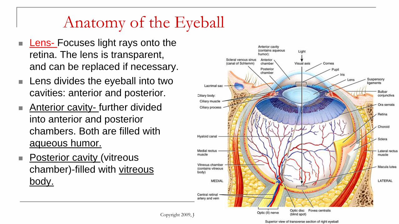
Lens- Focuses light rays onto the
retina. The lens is transparent,
and can be replaced if necessary.
Lens divides the eyeball into two
cavities: anterior and posterior.
Anterior cavity- further divided
into anterior and posterior
chambers. Both are filled with
aqueous humor.
Posterior cavity (vitreous
chamber)-filled with vitreous
body.

Anatomy of the Eyeball
Copyright 2009, John Wiley & Sons, Inc.
Pupil is an opening in the center
of the iris.
Contraction of the circular
muscles of the iris causes
constriction of the pupil.
Contraction of the radial muscles
causes dilation of the pupil.
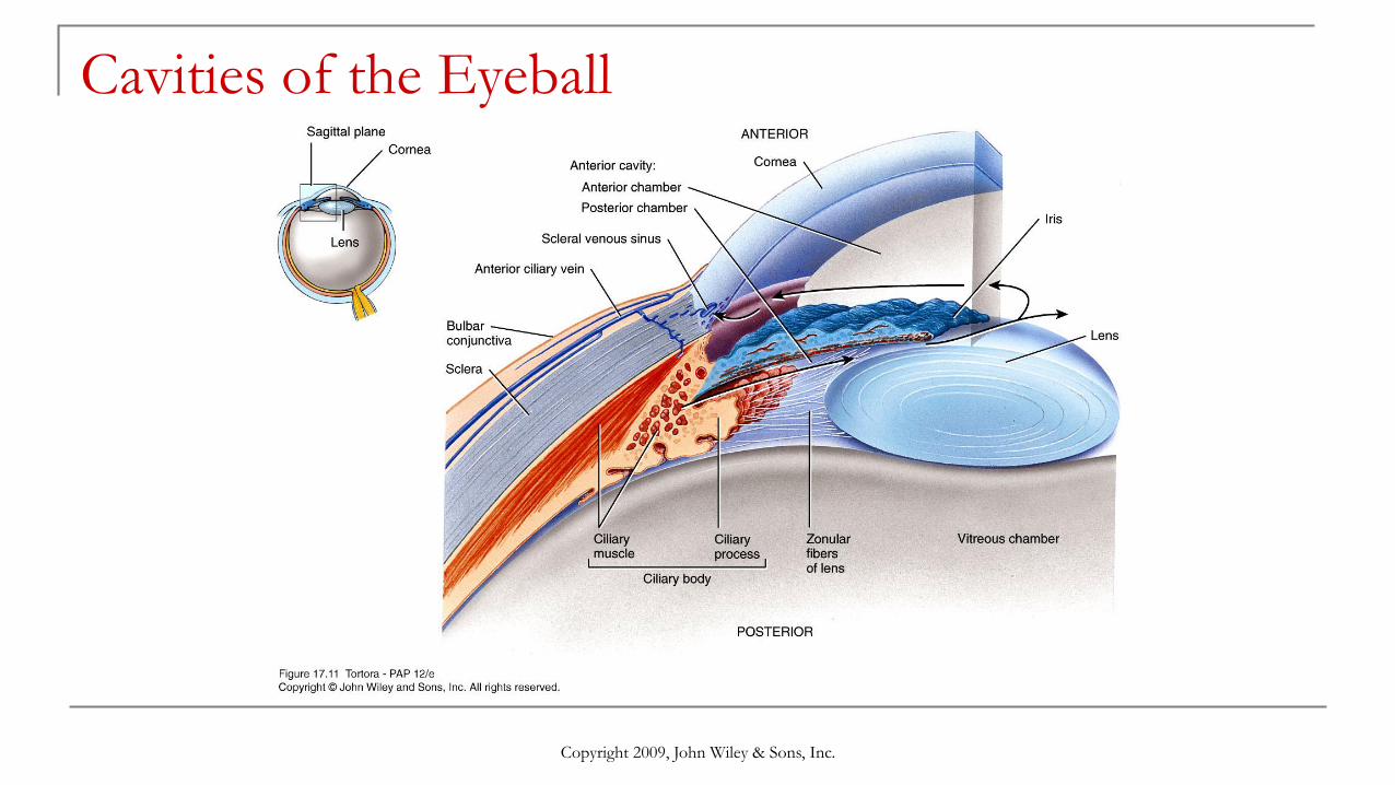
Cavities of the Eyeball
Copyright 2009, John Wiley & Sons, Inc.

The Lacrimal Apparatus
Copyright 2009, John Wiley & Sons, Inc.

Structure of Rod and Cone Photoreceptors
Rods: involved in non-colored and reduced light vision.
Cones: involved in color and acute vision.
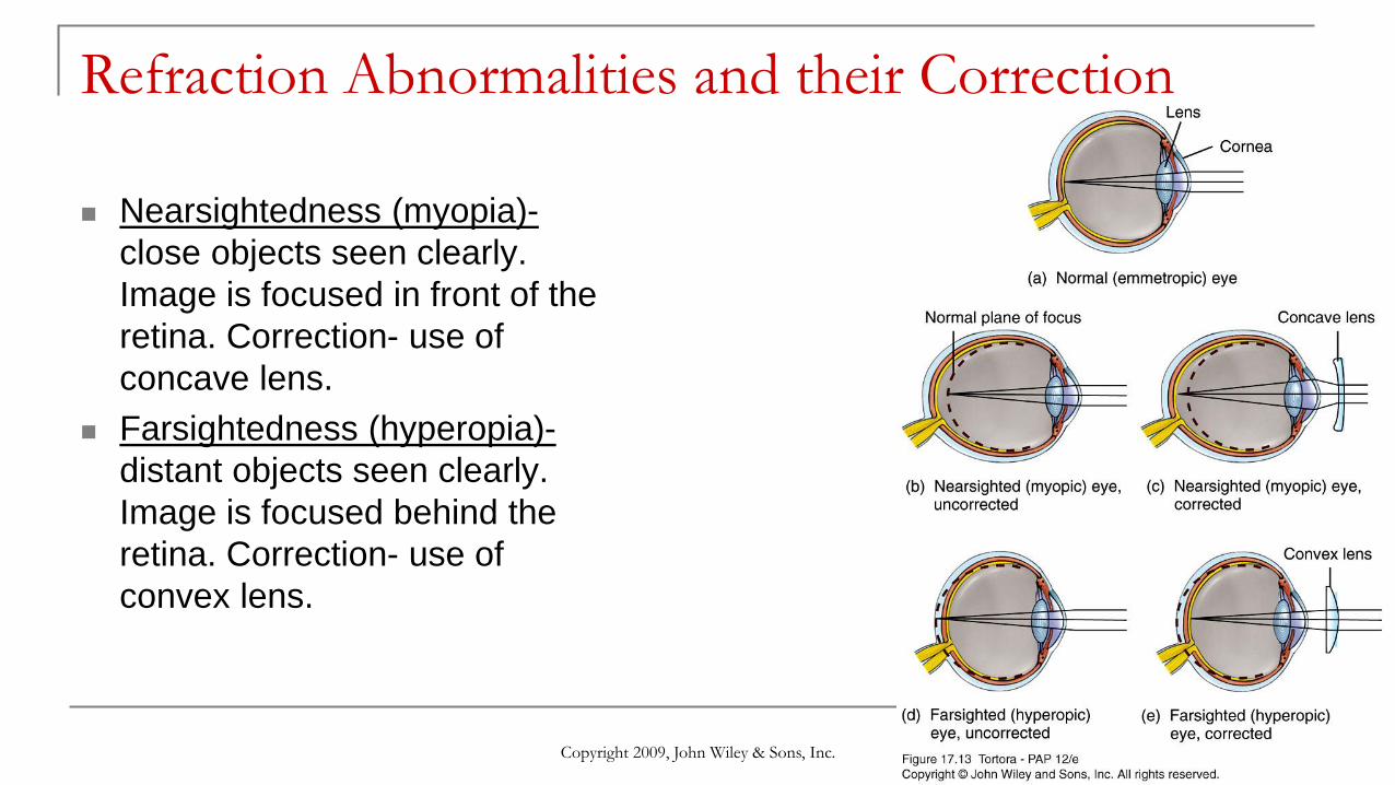
Refraction Abnormalities and their Correction
Nearsightedness (myopia)-
close objects seen clearly.
Image is focused in front of the
retina. Correction- use of
concave lens.
Farsightedness (hyperopia)-
distant objects seen clearly.
Image is focused behind the
retina. Correction- use of
convex lens.
Copyright 2009, John Wiley & Sons, Inc.
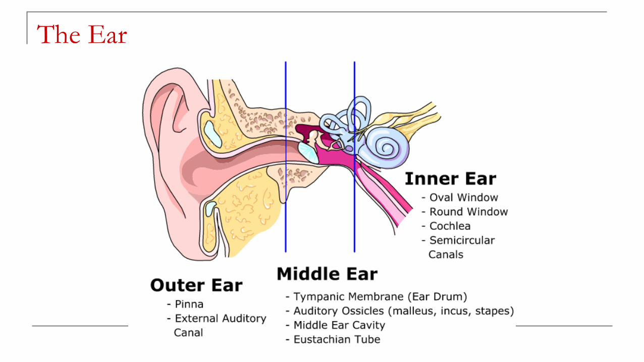
The Ear

Anatomy of the Ear
Copyright 2009, John Wiley & Sons, Inc.

Anatomy of the Ear
External (outer) ear
Auricle or pinna: the outer part of
the ear, serves to "catch" the sound
waves.
Tympanic membrane: commonly
called the eardrum. It is positioned
between the ear canal and
the middle ear.
Copyright 2009, John Wiley & Sons, Inc.
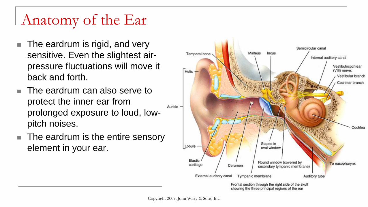
Anatomy of the Ear
The eardrum is rigid, and very
sensitive. Even the slightest air-
pressure fluctuations will move it
back and forth.
The eardrum can also serve to
protect the inner ear from
prolonged exposure to loud, low-
pitch noises.
The eardrum is the entire sensory
element in your ear.
Copyright 2009, John Wiley & Sons, Inc.

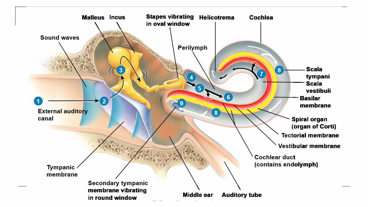
Anatomy of the Ear
Middle ear
Malleus: When the
eardrum vibrates, it moves
the malleus from side to
side like a lever.
Incus: The malleus is
connected to the incus,
which is attached to the
stapes.
Stapes: The other end of
the stapes rests against
the cochlea, through
the oval window.

Anatomy of the Ear
Middle ear
Auditory (eustachian)
tube: Creates pressure
balance.
How hearing works:
http://www.youtube.com/w
atch?v=flIAxGsV1q0

Copyright 2009, John Wiley & Sons, Inc.
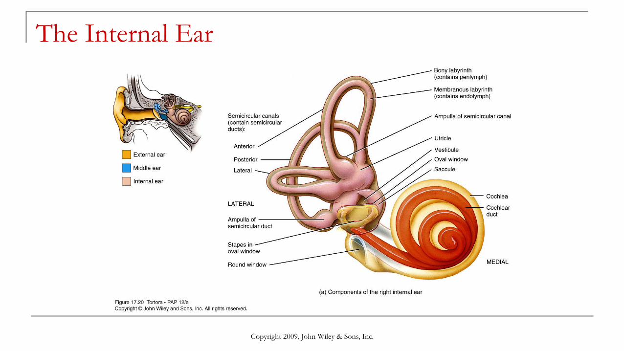
The Internal Ear
Copyright 2009, John Wiley & Sons, Inc.

The Internal Ear
Semicircular canals: sense
head rotations and balance.
Cochlea: contains the spiral
organ of Corti, the receptor
for hearing.
Organ of Corti: consists of
tiny hair cells that translate
the fluid vibration of sounds
into electrical impulses that
are carried to the brain.
Copyright 2009, John Wiley & Sons, Inc.

Physiology of Equilibrium
Two types of equilibrium:
Static - maintenance of the
body position relative to the
force of gravity.
Dynamic - maintenance of
body position (mainly head) in
response to rotational
acceleration and deceleration.

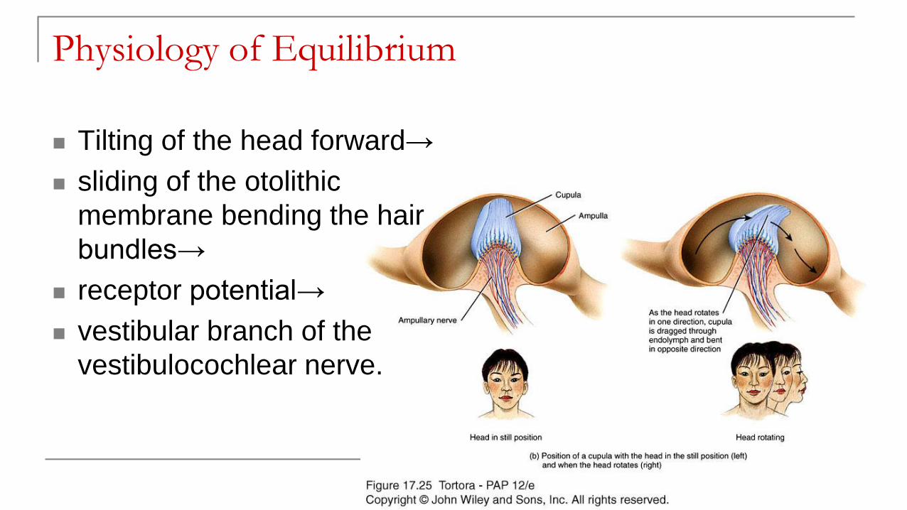
Physiology of Equilibrium
Tilting of the head forward→
sliding of the otolithic
membrane bending the hair
bundles→
receptor potential→
vestibular branch of the
vestibulocochlear nerve.
Physiology of Equilibrium
Tilting of the head forward→
sliding of the otolithic
membrane bending the hair
bundles→
receptor potential→
vestibular branch of the
vestibulocochlear nerve.

Test Review
The online quiz is here:
http://highered.mheducation.com/sites/0072351136/student_view
0/chapter15/chapter_quiz.html
Or you can google: Anatomy and Physiology online quiz.
Make sure you are on Chapter 15: The Special Senses
Questions to focus on: 24, 25, 26, 28, 31, 35, 41, 44, 45, 46, 58,
63, 64, 65, 66, 67, 68, 69, 70, 78, 80, 85, 86, 87, 88.
Copyright 2009, John Wiley & Sons, Inc.

The Senses Flowchart
Create a flowchart that explains
how each sense receives input and
how it relays that information to the
brain.
Properly use or label the key terms
in the diagrams or in the
explanations.
Each flowchart should include at
least four steps with each step
being at least one sentence.
Olfactory pathway: Olfactory tract, olfactory
nerve, olfactory epithelium, olfactory cilia,
olfactory bulb, and cribriform plate.
Gustatory pathway: Taste bud, taste pore,
gustatory cell, gustatory cortex, cranial
nerve fibers, and gustatory hair.
Visual pathway: Lens, pupil, iris, cornea,
retina, sclera, optic nerve, optic tract,
visual cortex, rods, and cones.
Auditory pathway: Auditory canal,
Tympanic membrane, Malleus, Incus,
Stapes, Cochlea, Organ of Corti, auditory
nerve, and auditory cortex.
Copyright 2009, John Wiley & Sons, Inc.