The Role of Ultrasound in Objectives the Trauma Patient Role of Ultrasound in the Trauma Patient...
13
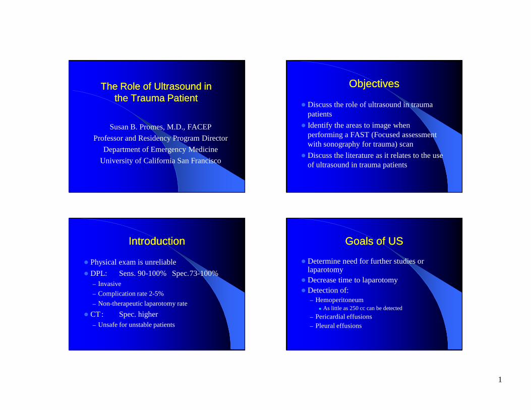
1 The Role of Ultrasound in The Role of Ultrasound in the Trauma Patient the Trauma Patient Susan B. Promes, M.D., FACEP Professor and Residency Program Director Department of Emergency Medicine University of California San Francisco Objectives Objectives Discuss the role of ultrasound in trauma patients Identify the areas to image when performing a FAST (Focused assessment with sonography for trauma) scan Discuss the literature as it relates to the use of ultrasound in trauma patients Introduction Introduction Physical exam is unreliable DPL: Sens. 90-100% Spec.73-100% – Invasive – Complication rate 2-5% – Non-therapeutic laparotomy rate CT : Spec. higher – Unsafe for unstable patients Goals of US Goals of US Determine need for further studies or laparotomy Decrease time to laparotomy Detection of: – Hemoperitoneum As little as 250 cc can be detected – Pericardial effusions – Pleural effusions
Transcript of The Role of Ultrasound in Objectives the Trauma Patient Role of Ultrasound in the Trauma Patient...
1
The Role of Ultrasound in The Role of Ultrasound in the Trauma Patientthe Trauma Patient
Susan B. Promes, M.D., FACEP
Professor and Residency Program Director
Department of Emergency Medicine
University of California San Francisco
ObjectivesObjectives
� Discuss the role of ultrasound in trauma patients
� Identify the areas to image when performing a FAST (Focused assessment with sonography for trauma) scan
� Discuss the literature as it relates to the use of ultrasound in trauma patients
IntroductionIntroduction
� Physical exam is unreliable
� DPL: Sens. 90-100% Spec.73-100%– Invasive
– Complication rate 2-5%
– Non-therapeutic laparotomy rate
� CT: Spec. higher– Unsafe for unstable patients
Goals of USGoals of US
� Determine need for further studies or laparotomy� Decrease time to laparotomy� Detection of:
– Hemoperitoneum� As little as 250 cc can be detected
– Pericardial effusions– Pleural effusions
2
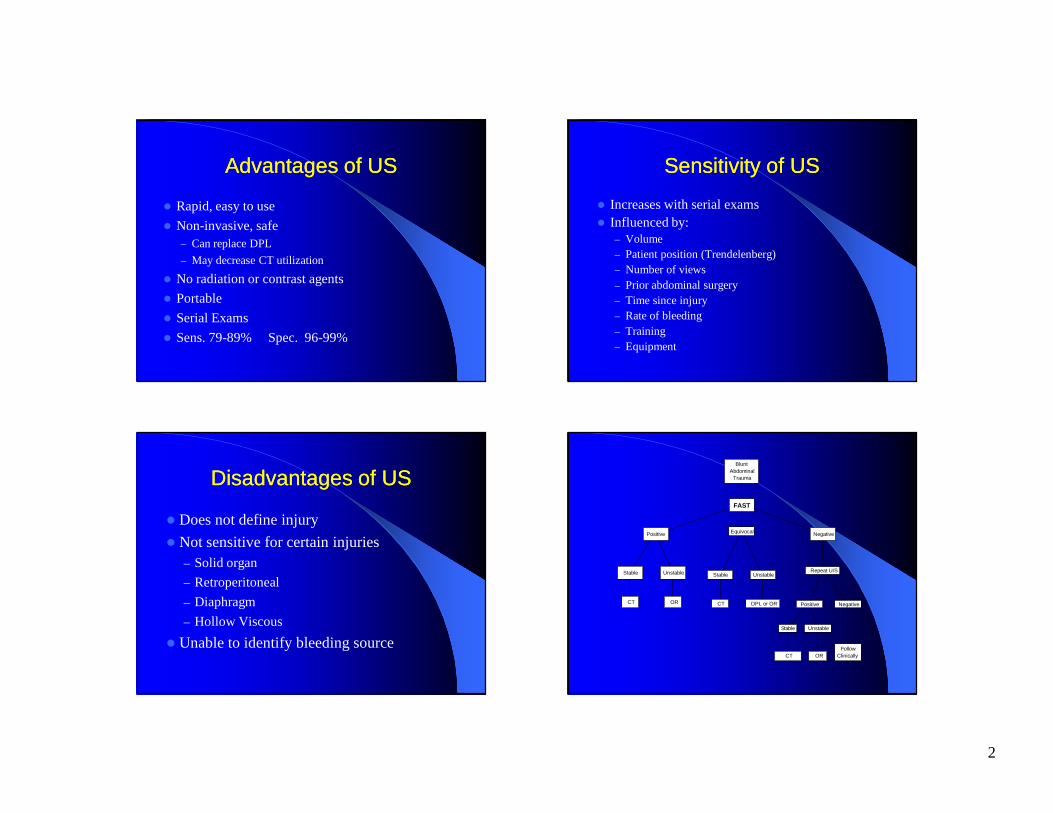
Advantages of USAdvantages of US
� Rapid, easy to use
� Non-invasive, safe– Can replace DPL
– May decrease CT utilization
� No radiation or contrast agents
� Portable
� Serial Exams
� Sens. 79-89% Spec. 96-99%
Sensitivity of USSensitivity of US
� Increases with serial exams� Influenced by:
– Volume– Patient position (Trendelenberg)– Number of views– Prior abdominal surgery– Time since injury– Rate of bleeding– Training– Equipment
Disadvantages of USDisadvantages of US
� Does not define injury
� Not sensitive for certain injuries– Solid organ
– Retroperitoneal
– Diaphragm
– Hollow Viscous
� Unable to identify bleeding source
BluntAbdominal
Trauma
FAST
Equivocal NegativePositive
Stable Unstable
CT OR
Stable Unstable
CT DPL or OR
Repeat U/S
Positive Negative
FollowClinically
Stable Unstable
CT OR
3
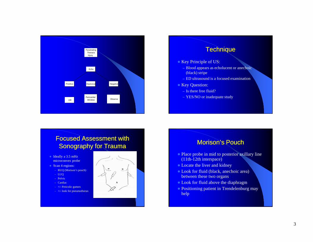
Penetrating ThoracicInjury
Echo
Positive Equivocal Negative
ORPericardial
Window Observe
TechniqueTechnique
� Key Principle of US: – Blood appears as echolucent or anechoic
(black) stripe
– ED ultrasound is a focused examination
� Key Question:– Is there free fluid?
– YES/NO or inadequate study
Focused Assessment with Focused Assessment with Sonography for TraumaSonography for Trauma
� Ideally a 3.5 mHz microconvex probe
� Scan 4 regions:– RUQ (Morison’s pouch)
– LUQ
– Pelvis
– Cardiac
– +/- Pericolic gutters
– +/- look for pneumothorax
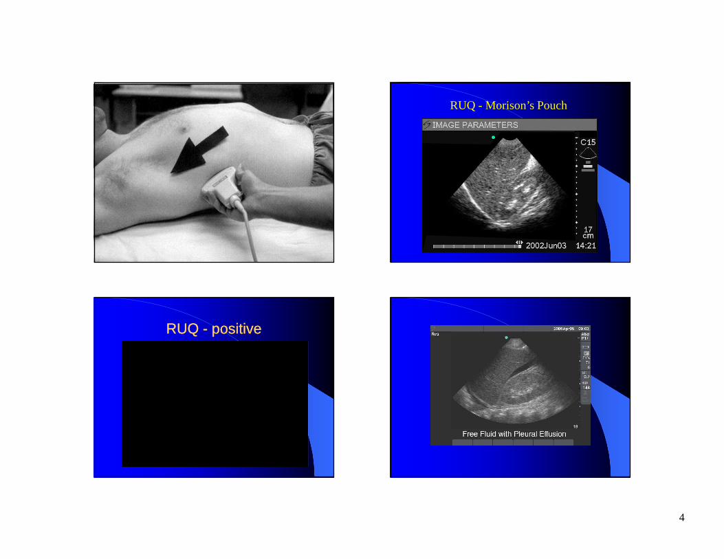
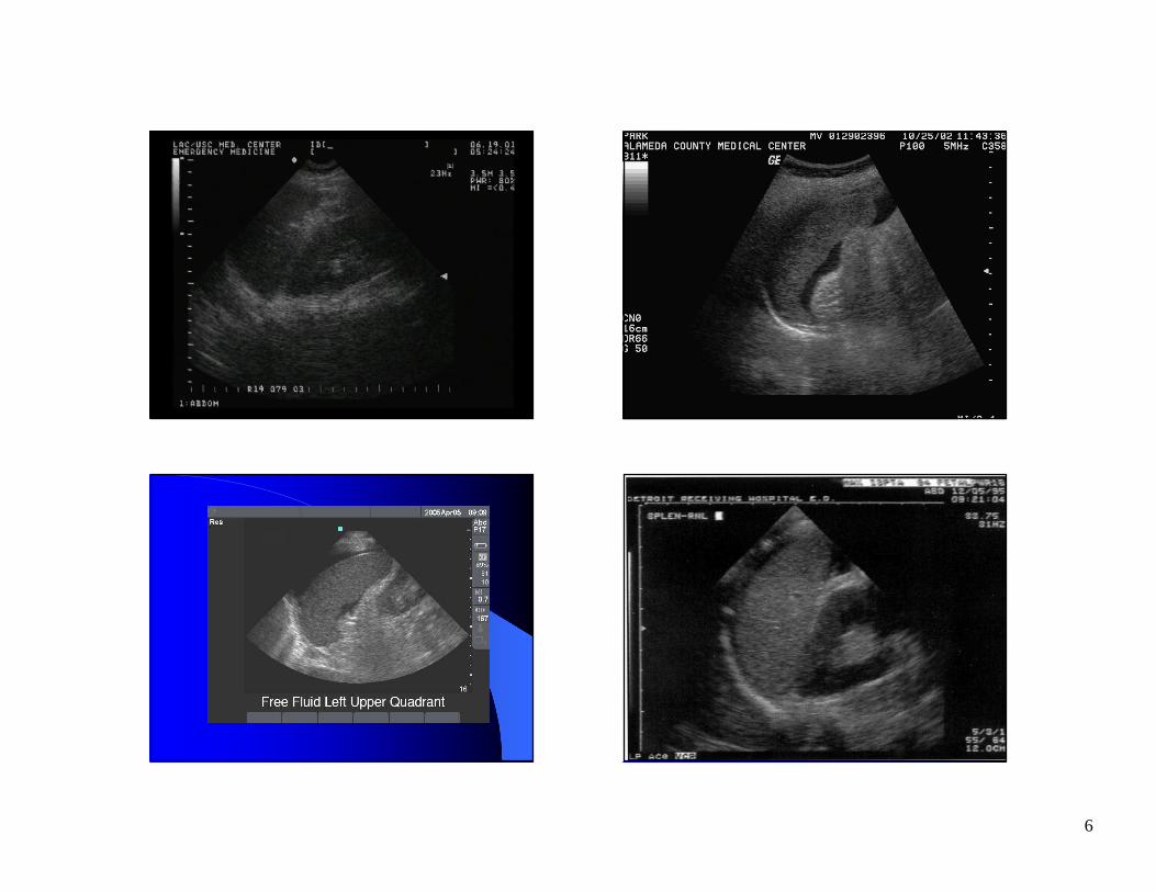
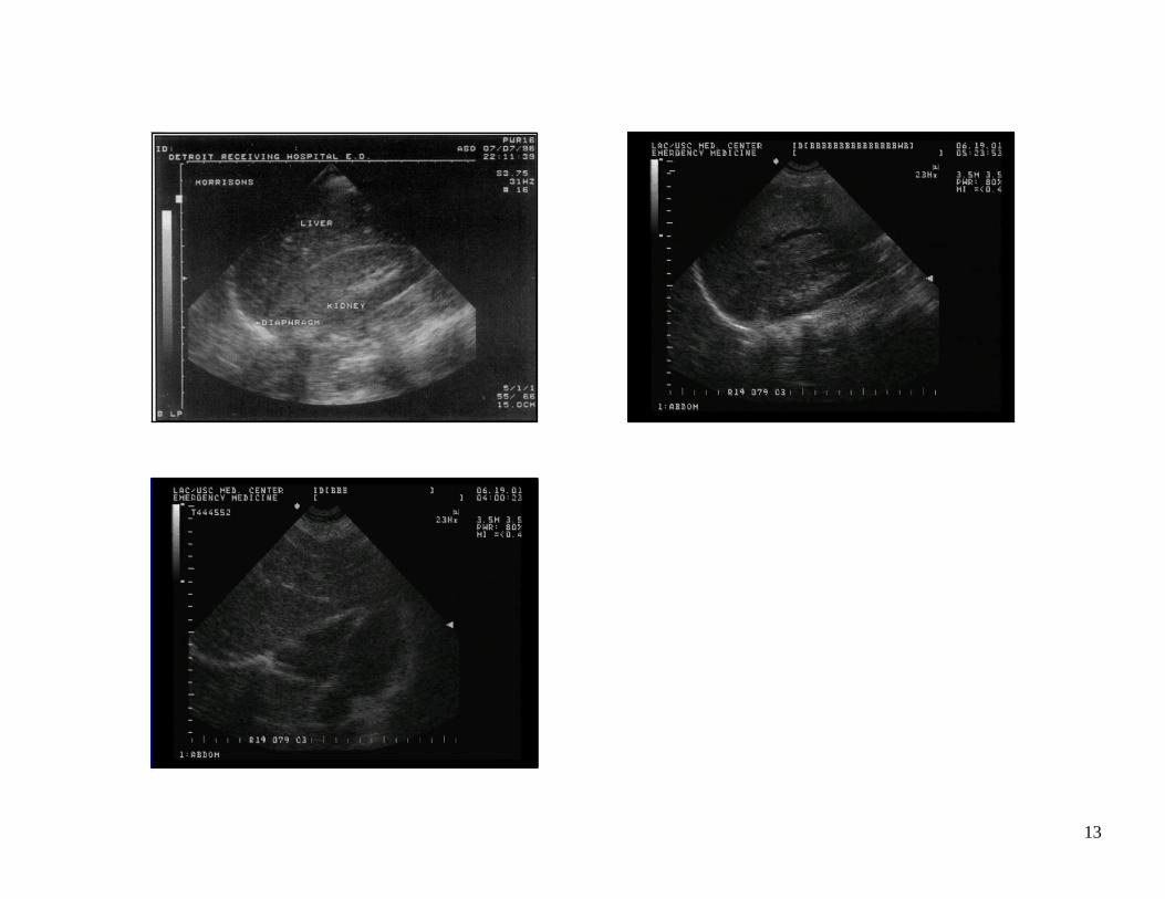
Morison’s PouchMorison’s Pouch
� Place probe in mid to posterior axillary line (11th-12th interspace)� Locate the liver and kidney� Look for fluid (black, anechoic area)
between these two organs� Look for fluid above the diaphragm� Positioning patient in Trendelenburg may
help
4
RUQRUQRUQ - Morison’s Pouch
RUQ RUQ -- positivepositive
5
Morison’s pouch Morison’s pouch -- pospos
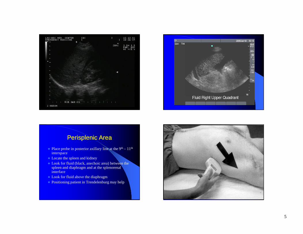
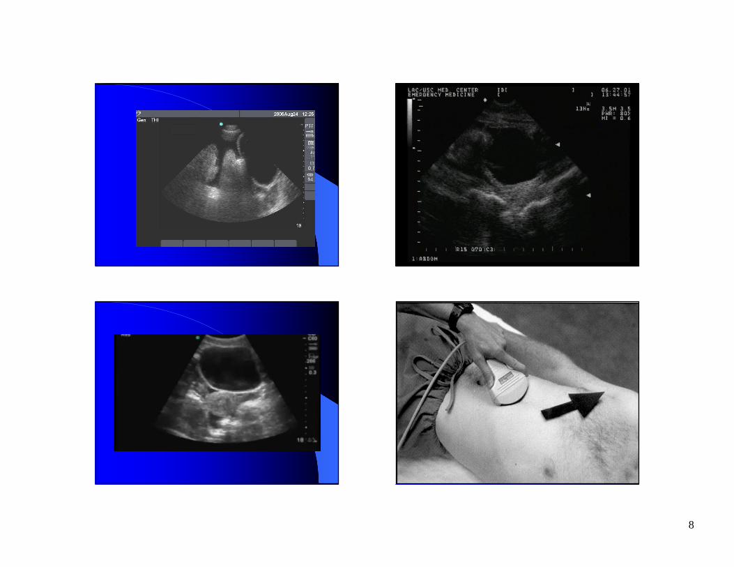
Perisplenic AreaPerisplenic Area
� Place probe in posterior axillary line at the 9th – 11th
interspace� Locate the spleen and kidney� Look for fluid (black, anechoic area) between the
spleen and diaphragm and at the splenorenal interface� Look for fluid above the diaphragm� Positioning patient in Trendelenburg may help
LUQLUQ
6
Perisplenic Perisplenic –– nlnl
7
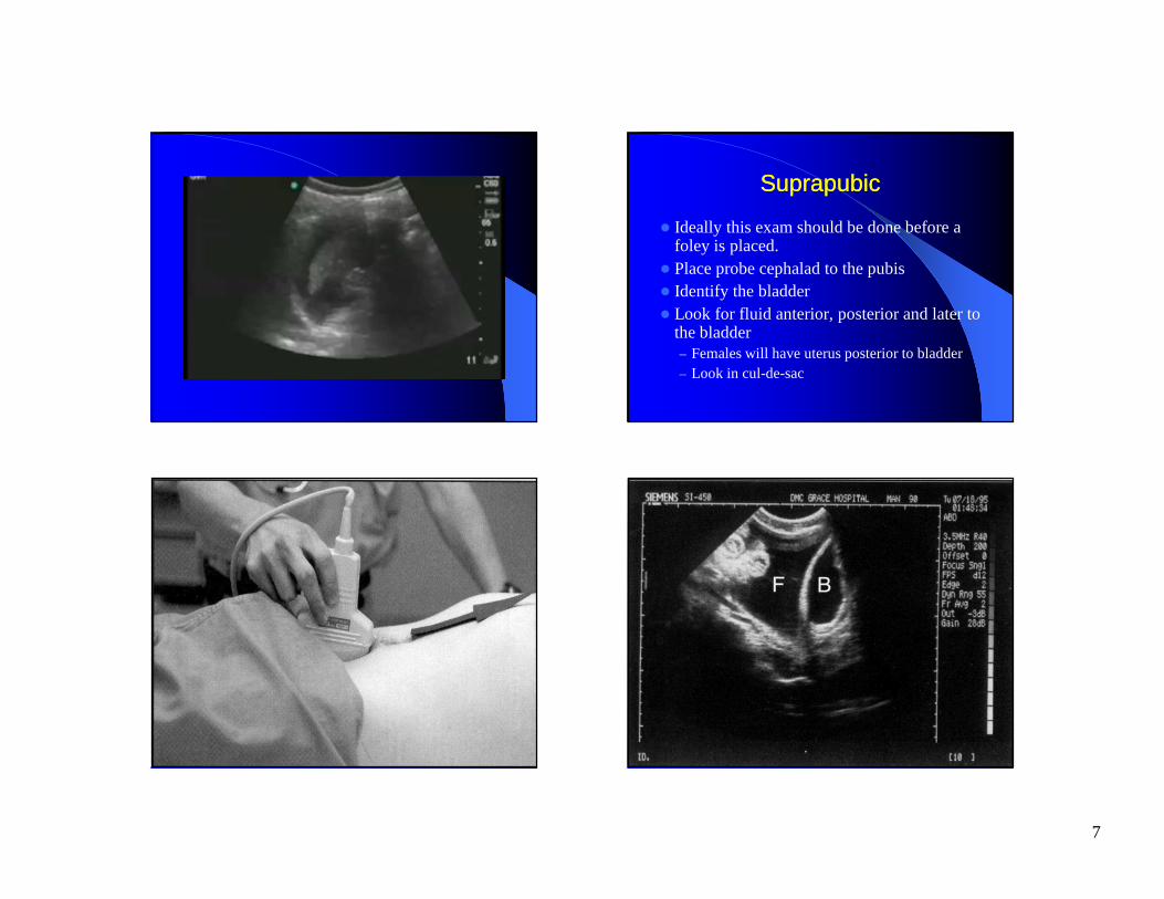
SuprapubicSuprapubic
� Ideally this exam should be done before a foley is placed.� Place probe cephalad to the pubis� Identify the bladder� Look for fluid anterior, posterior and later to
the bladder– Females will have uterus posterior to bladder– Look in cul-de-sac
8

9
PericardiumPericardium
� Place probe in subcostal region below xiphoid angled toward left shoulder
� Look for fluid surrounding the (white, hyperechoic) myocardium
10
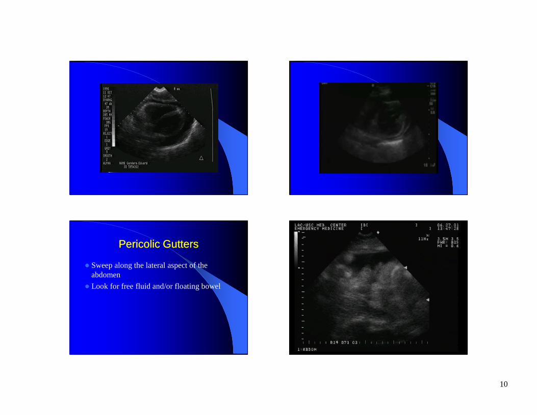
Pericolic GuttersPericolic Gutters
� Sweep along the lateral aspect of the abdomen
� Look for free fluid and/or floating bowel
Pericolic gutter Pericolic gutter –– pospos
11
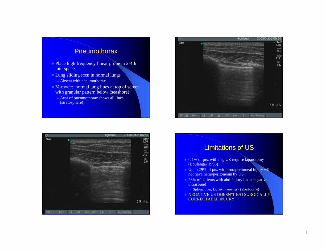
PneumothoraxPneumothorax
� Place high frequency linear probe in 2-4th interspace� Lung sliding seen in normal lungs
– Absent with pneumothorax
�M-mode: normal lung lines at top of screen with granular pattern below (seashore)– Area of pneumothoraz shows all lines
(stratosphere)
Limitations of USLimitations of US
� < 1% of pts. with neg US require laparotomy (Boulanger 1996)� Up to 29% of pts. with intraperitoneal injury will
not have hemoperitoneum by US� 26% of patients with abd. injury had a negative
ultrasound– Spleen, liver, kidney, mesentery (Sherbourne)
� NEGATIVE US DOESN’T R/O SURGICALLY CORRECTABLE INJURY
12
PitfallsPitfalls
� Failure to do multiple view exam
� Failure to consider other etiologies of free intraperitoneal fluid
� Failure to do serial exams
� Over reliance on ultrasonography
�Misinterpretation of US
TrainingTraining
� SAEM 1994– 40 hrs of instruction and 150 exams for EM US
� ACEP 2001– Minimum 8 hrs of instruction and 25-50
proctored exams for Trauma US
– Refer to ACEP web page and call for ACEP Resource Document
Go Ultrasound!Go Ultrasound!
13
RUQ RUQ -- nlnl Morison’s pouch Morison’s pouch --nlnl