Emergency Ultrasound in Trauma
31
description
Emergency Ultrasound in Trauma. Fahad Khan, MD St. Luke’s/Roosevelt Hospital Center Columbia University, New York City April 24, 2009. E -FAST. Focused Assessment with Sonograghy for Trauma Cardiac RUQ LUQ Pelvis Extended Lung bases for pleural fluid - PowerPoint PPT Presentation
Transcript of Emergency Ultrasound in Trauma

Emergency Emergency Ultrasound in TraumaUltrasound in Trauma
Fahad Khan, MDFahad Khan, MDSt. Luke’s/Roosevelt Hospital CenterSt. Luke’s/Roosevelt Hospital CenterColumbia University, New York CityColumbia University, New York City
April 24, 2009April 24, 2009


EE-FAST-FAST Focused Assessment with Focused Assessment with
Sonograghy for TraumaSonograghy for Trauma CardiacCardiac RUQRUQ LUQLUQ PelvisPelvis
ExtendedExtended Lung bases for pleural fluidLung bases for pleural fluid Anterior lung apices for pneumothoraxAnterior lung apices for pneumothorax

IndicationsIndications
Blunt thoraco-abdominal trauma Blunt thoraco-abdominal trauma Unexplained hypotensionUnexplained hypotension Trauma in pregnancyTrauma in pregnancy

Key QuestionsKey Questions
Is there FREE FLUID present?Is there FREE FLUID present? In the pericardial spaceIn the pericardial space In the peritoneal cavityIn the peritoneal cavity In the pleural spaceIn the pleural space
Is there a PNEUMOTHORAX?Is there a PNEUMOTHORAX?

AdvantagesAdvantages
RapidRapid ReproducibleReproducible Non-invasiveNon-invasive PortablePortable No radiation or contrastNo radiation or contrast

DisadvantagesDisadvantages
Difficult to distinguishDifficult to distinguish Type of fluidType of fluid Solid organ injurySolid organ injury
Cannot evaluate retroperitoneumCannot evaluate retroperitoneum Difficult in the obese patientDifficult in the obese patient

AlgorithmAlgorithm
Blunt Thoraco-abdominal Trauma
Hemodynamically Stable Hemodynamically Unstable
Peritoneal Signs
LaparotomyUltrasound
Free Fluid/Organ Injury
Laparotomy CT Scan
UltrasoundFree Fluid/Organ Injury
LaparotomyRepeat U/S
CT Scan

TechniqueTechnique

Intraperitoneal Fluid Intraperitoneal Fluid FlowFlow

TechniqueTechnique
Low frequency Low frequency probeprobe
2.5 – 5.0 MHz2.5 – 5.0 MHz Tissue penetrationTissue penetration

Sub-xiphoidSub-xiphoid

Pericardial FluidPericardial Fluid

Pericardial EffusionPericardial Effusion

Hepato-renal RecessHepato-renal Recess
Trendelenburg positionTrendelenburg position Anterior axillary lineAnterior axillary line

Hepato-renal FluidHepato-renal Fluid
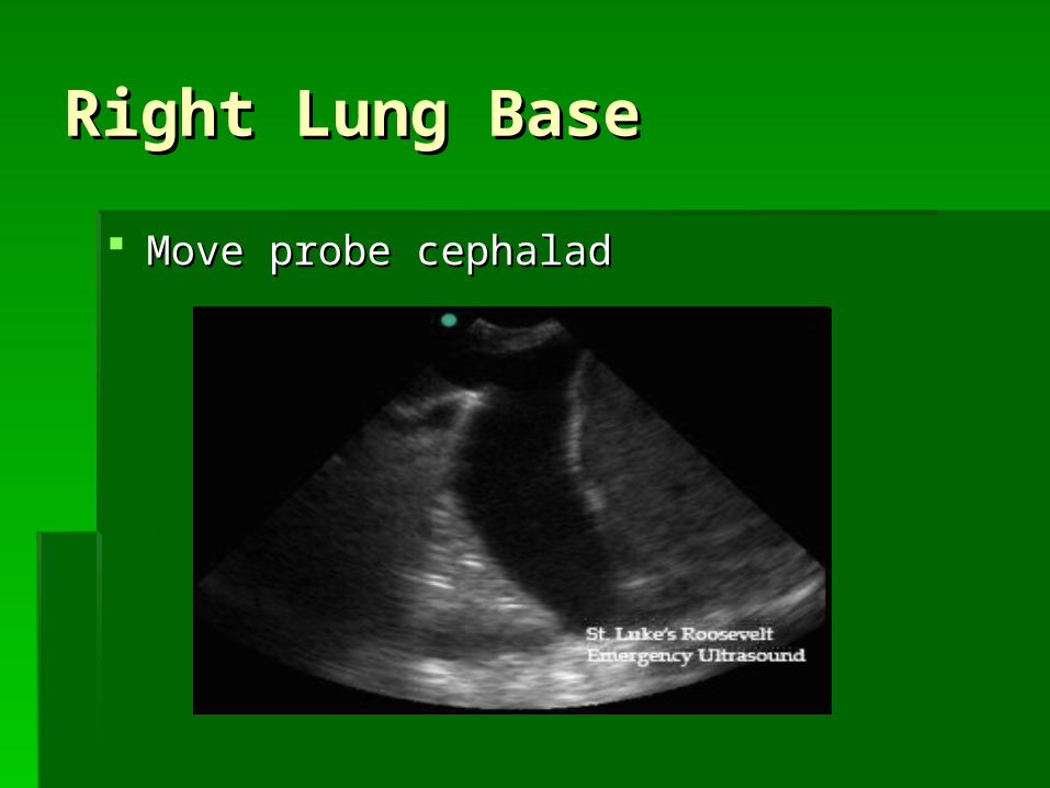
Right Lung BaseRight Lung Base
Move probe cephaladMove probe cephalad

Spleno-renal RecessSpleno-renal Recess

Spleno-renal FluidSpleno-renal Fluid

Left Lung BaseLeft Lung Base
Move probe cephaladMove probe cephalad

PelvisPelvis

Pelvic Free FluidPelvic Free Fluid

TechniqueTechnique

Lung Scanning for Lung Scanning for PneumothoraxPneumothorax
““Bat” SignBat” Sign Comet tailsComet tails

Normal LungNormal Lung

PneumothoraxPneumothorax

PitfallsPitfalls
Scan all quadrantsScan all quadrants Repeating scansRepeating scans Inferior polesInferior poles Solid organ injuriesSolid organ injuries FatFat RetroperitoneumRetroperitoneum

After a short training program, physicians After a short training program, physicians can use FAST in early assessment of trauma can use FAST in early assessment of trauma patients with sufficient specificity to patients with sufficient specificity to expedite decision making. expedite decision making.

Increased physician ultrasound experience is Increased physician ultrasound experience is associated with increased physician accuracy associated with increased physician accuracy in FAST examinations.in FAST examinations.
This can directly lead to a reduction in the use This can directly lead to a reduction in the use of CT scans, and ultimately, medical costs.of CT scans, and ultimately, medical costs.

Questions?Questions?