The Role of Norepinephrine and Serotonin in ADHD · 1 The Role of Norepinephrine and Serotonin in...
31
1 The Role of Norepinephrine and Serotonin in ADHD Robert D. Oades PhD University Clinic for Child and Adolescent Psychiatry, Virchowstr. 184, 45147 Essen, Germany. In, “Attention Deficit Hyperactivity Disorder: From Genes to Animal Models to Patients” Eds. D. Gozal and D. L. Molfese : Humana Press (2005) The final publication is available at www.springerlink.com (http://www.springer.com/medicine/psychiatry/book/978-1-58829- 312-1?changeHeader) CONTENTS: 1. Introduction .......... 1 2. Biochemistry .......... 2 3. CNS Pathways .......... 3 3.1 NE .......... 3 3.2 5-HT .......... 4 4. Interactions between monoamines .......... 5 4.1 5-HT – NE .......... 5 4.2 5-HT – DA .......... 5 4.3 NE – DA .......... 6 5. Development .......... 7 5.1 NE .......... 7 5.2 5-HT .......... 8 6. Evidence for monoamine contributions to ADHD -- Genetic studies .......... 8 6.1 NE .......... 8 6.2 5-HT .......... 9 7. Methods .......... 10 8. Animal Models .......... 12 8.1 Rodent .......... 12 8.2 Primate .......... 14 9. Evidence for monoamine contributions to ADHD -- NE and 5-HT activity .......... 13 9.1 Evidence from group comparisons .......... 16 9.2 Evidence from pharmacological treatments .......... 18 10. General .......... 23 1. Introduction: The first actor in this chapter is norepinephrine (NE). NE belongs to the chemical group of the catecholamines and is also known outside the Americas as noradrenaline. The second actor is serotonin, an indole-amine that is better described chemically as 5-hydroxy-tryptamine (5-HT). Together with the catecholamines dopamine (DA) and epinephrine (adrenaline) they are known as the monoamines. These monoamines have an agent role in transmission between neurons – often in the synapse between neurons and their elements in apposition, sometimes between release and receptor sites that are further apart. Then the role is more reminiscent of hormonal communication. Both roles are subsumed as ‘neurotransmission’. These transmitters are located in well-characterized, similar neural pathways throughout the vertebrates. This chapter is essentially concerned with the role of NE and 5-HT in the central nervous system (CNS) and how characteristics of 5-HT and NE transmission could contribute to the principle features of ADHD. This review starts with the basic aspects of monoamine biochemistry and neurochemical anatomy and proceeds over mechanisms of function (animal work) to investigations of their role in the neuropsychology, and nosology thought to underlie ADHD. However, throughout these considerations it should not be overlooked that
Transcript of The Role of Norepinephrine and Serotonin in ADHD · 1 The Role of Norepinephrine and Serotonin in...
1
The Role of Norepinephrine and Serotonin in ADHD
Robert D. Oades PhD
University Clinic for Child and Adolescent Psychiatry, Virchowstr. 184, 45147 Essen, Germany.
In, “Attention Deficit Hyperactivity Disorder: From Genes to Animal Models to Patients”
Eds. D. Gozal and D. L. Molfese : Humana Press (2005)
The final publication is available at www.springerlink.com (http://www.springer.com/medicine/psychiatry/book/978-1-58829-
312-1?changeHeader)
CONTENTS:
1. Introduction .......... 1
2. Biochemistry .......... 2
3. CNS Pathways .......... 3
3.1 NE .......... 3
3.2 5-HT .......... 4
4. Interactions between monoamines .......... 5
4.1 5-HT – NE .......... 5
4.2 5-HT – DA .......... 5
4.3 NE – DA .......... 6
5. Development .......... 7
5.1 NE .......... 7
5.2 5-HT .......... 8
6. Evidence for monoamine contributions to ADHD -- Genetic studies .......... 8
6.1 NE .......... 8
6.2 5-HT .......... 9
7. Methods .......... 10
8. Animal Models .......... 12
8.1 Rodent .......... 12
8.2 Primate .......... 14
9. Evidence for monoamine contributions to ADHD -- NE and 5-HT activity .......... 13
9.1 Evidence from group comparisons .......... 16
9.2 Evidence from pharmacological treatments .......... 18
10. General .......... 23
1. Introduction:
The first actor in this chapter is
norepinephrine (NE). NE belongs to the
chemical group of the catecholamines and is
also known outside the Americas as
noradrenaline. The second actor is serotonin, an
indole-amine that is better described chemically
as 5-hydroxy-tryptamine (5-HT). Together with
the catecholamines dopamine (DA) and
epinephrine (adrenaline) they are known as the
monoamines. These monoamines have an agent
role in transmission between neurons – often in
the synapse between neurons and their
elements in apposition, sometimes between
release and receptor sites that are further apart.
Then the role is more reminiscent of hormonal
communication. Both roles are subsumed as
‘neurotransmission’. These transmitters are
located in well-characterized, similar neural
pathways throughout the vertebrates.
This chapter is essentially concerned with the
role of NE and 5-HT in the central nervous
system (CNS) and how characteristics of 5-HT
and NE transmission could contribute to the
principle features of ADHD. This review starts
with the basic aspects of monoamine
biochemistry and neurochemical anatomy and
proceeds over mechanisms of function (animal
work) to investigations of their role in the
neuropsychology, and nosology thought to
underlie ADHD. However, throughout these
considerations it should not be overlooked that

2
both 5-HT and NE pathways are widely
distributed peripherally with functions
additional to those considered here1. Further it
is also important to bear in mind in the ensuing
discussion of NE and 5-HT function that many of
the effects simply attributed to the activity of
one or the other monoamine, are through
multiple interactions, additionally dependent on
another monoamine.
2. Biochemistry:
5-HT and NE synthesis depends on the
availability of the amino-acids, tryptophan and
phenylalanine, respectively. Tryptophan is
hydroxylated in the rate-limiting step by
tryptophan hydroxylase to the precursor 5-
hydroxytryptophan (5-HTP) prior to conversion
to 5-HT by decarboxylation. For NE synthesis,
phenylalanine is hydroxylated to tyrosine prior
to the rate-limiting hydroxylation to L-DOPA
(Fig. 1). Decarboxylation then produces DA that
can be dehydroxylated to NE. Many studies
examining the effects of enhancing or depleting
NE make use of the crucial role of tyrosine
hydroxylase (TOH) and dopamine beta-
hydroxylase (DBH). Studies of 5-HT depletion
often use diets free of tryptophan for examining
the effect of reducing 5-HT activity. Thus it is not
surprising that dietary effects on the availability
of factors needed for transmitter synthesis has
been part of the agenda in some ADHD studies.
Breakdown (catabolism) occurs following
post-synaptic uptake of the neurotransmitter,
when the transmitter remains unused in the
synapse, or after pre-synaptic re-uptake when
not stored in vesicles. In detail the NE and 5-HT
catabolic pathways can differ. Several enzymes
are involved in both. But primary is the
oxidation process (monoamine oxidase, MAO).
For 5-HT this leads to 5-hydroxy-indoleacetic
acid (5-HIAA: Fig. 2); for NE there are many
intermediates resulting from the activities of
several enzymes.
1 For example, 5-HT has a prominent role in pulmonary and renal blood flow, as well as the enteric autonomic system (smooth muscle contraction): NE, released from post-ganglionic sympathetic neurons, also actively modulates vasoconstriction/dilation, especially heart and smooth muscle function (also the uterus, intestine, bronchi and iris). In addition NE modulates insulin secretion and several metabolic activities: (note also that NE is the precursor to epinephrine synthesis in the adrenal medulla).
Three trends emerge from metabolic studies
that help the interpretation of clinical results.
First, the primary products of stimulated central
NE synthesis are mostly 3-methoxy- and
dihydroxy-phenyl-glycol (MHPG, DHPG), while
extra-neuronal products also include
metanephrine and normetanephrine (MN,
NMN: 1). As these latter metabolites along with
vanillomandelic acid (VMA) do not cross the
blood brain barrier peripheral measures of
these metabolites likely reflect peripheral
sources. Secondly these metabolites (e.g. NMN,
VMA) often measured peripherally, can be
excreted partially, after further metabolism, as
homovanillic acid (HVA). This leads to some
confusion over identifying the relative roles of
NE and DA activity. Thirdly, NE and 5-HT are the
preferred substrates for MAO type A, while
tyramine, tryptamine and DA are the preferred
substrates of MAO type B: but, the separation of
function between these two isoenzymes is not
tight. (e.g. selective inhibitors of both MAO-A
[clorgyline] and MAO-B [selegiline] can reduce
5-HT catabolism.)
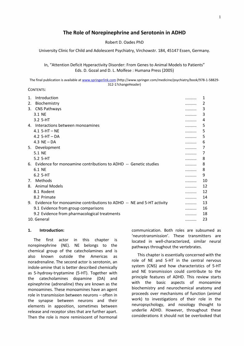
3. CNS Pathways:
3.1 NE
In the 1950s pioneer work demonstrated NE
to be a chemical transmitter and to have its cells
of origin in the brain stem (2, 3). The locus
ceruleus (LC: A6) is located in the dorso-lateral
pontine tegmentum just lateral to the fourth
ventricle (4, 5: Fig. 3). It and the nearby A5, A7
nuclei (subcoeruleus) give rise to NE fibers
innervating the forebrain (dorsal noradrenergic
bundle), diencephalon, cerebellum and local
brainstem nuclei. Some fibers also descend in
the spinal cord (6). A more ventral bundle with
fibers from the Nucleus tractus solitarius (A2)
also innervates the diencephalon and a number
of sub-cortical limbic regions (7). The LC in man
is about 15 mm long and in adults contains
some 40-60 thousand NE-containing cells. Of
interest for animal models, there is much
similarity between the LC in humans and that of
the rat – even if the latter contains only 3% of
the number of neurons in the human LC. Other
transmitting agents such as neuropeptide Y,
galanin and GABA may also be colocalized in
these neurons.

3
Figure 1
NE Metabolism: Biochemical pathways showing the synthesis and breakdown of NE.
Figure 2
5-HT Metabolism: Biochemical pathways showing the synthesis and breakdown of 5-HT
To understand the function of the NE system
it is important to appreciate that there is much
dendrite branching locally within the LC and
axonal branching between widely separate
areas innervated by the same neuron (8). If one
considers the vast areas of cortex innervated it
may be that as few as 5% of transmitter
containing varicosities are located in
conventional synapses (9). Most of the
transmitter released has effect at a distance
from the end of the axon. The densest input is
to the laminae III and IV (review: 10). Alpha-1
and alpha-2 receptor types that can be pre- or
post-synaptically located are distributed more
across the superficial laminae, while beta sites
may be found in most cortical laminae. (α2a
have a primarily frontal, α2b a more thalamic
and α2c a brainstem distribution).
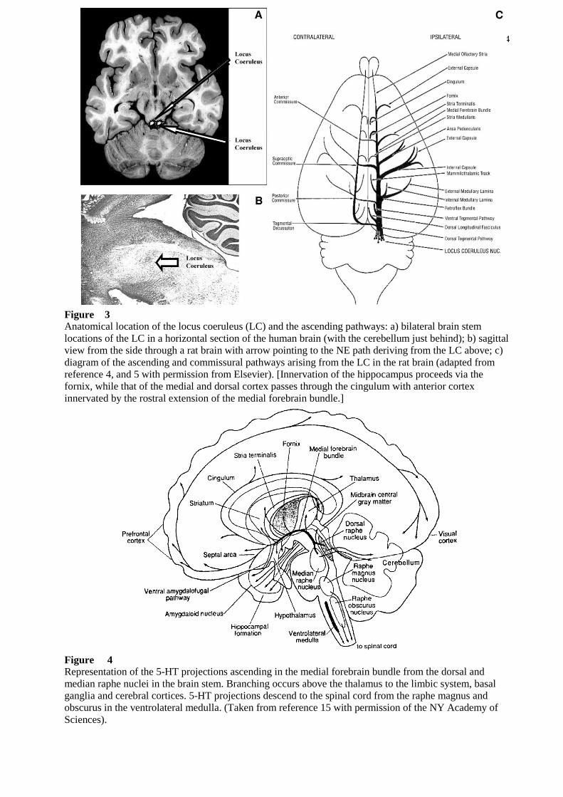
3.2 5-HT
5-HT was first demonstrated in the CNS of
cats and dogs about 50 years ago (11, 12). The
development of fluorescence histochemistry 10
years later led to the description of the basic
components of the 5-HT projection system (13).
In succeeding decades the development of
antibodies, of immunohistochemical (13) and
immunocytochemical methods led to the
current understanding of the cell body origins
and their heterogeneous termination patterns
(14). For 5-HT there are 9 cell groups (B1-B9).
B1-B5 are small cell groups located in the
midline from the mid-pons to the caudal
medulla (Fig. 4). They project locally and down
the dorsal and ventral horns of the spinal cord.
More significant for the current discussion are
B6 and B7 (the dorsal raphe nuclei) that lie
along the floor of the fourth ventricle near the
LC, and ventrally the B8 group (the median
raphe on the borders of the pons and midbrain.
4
Figure 3 Anatomical location of the locus coeruleus (LC) and the ascending pathways: a) bilateral brain stem locations of the LC in a horizontal section of the human brain (with the cerebellum just behind); b) sagittal view from the side through a rat brain with arrow pointing to the NE path deriving from the LC above; c) diagram of the ascending and commissural pathways arising from the LC in the rat brain (adapted from reference 4, and 5 with permission from Elsevier). [Innervation of the hippocampus proceeds via the fornix, while that of the medial and dorsal cortex passes through the cingulum with anterior cortex innervated by the rostral extension of the medial forebrain bundle.]
Figure 4 Representation of the 5-HT projections ascending in the medial forebrain bundle from the dorsal and median raphe nuclei in the brain stem. Branching occurs above the thalamus to the limbic system, basal ganglia and cerebral cortices. 5-HT projections descend to the spinal cord from the raphe magnus and obscurus in the ventrolateral medulla. (Taken from reference 15 with permission of the NY Academy of Sciences).
5
The dorsal raphe is the larger group but along
with the median raphe, both contain neurons
using other transmitters (e.g. DA: 15).
There is a fairly broad overlap for the
forebrain innervation from these two nuclei.
The emphasis is on the neostriatum and frontal
lobe for the dorsal raphe (with a decreasing
gradient over the more caudal cortical regions),
while the median raphe projects more to
diencephalic and limbic structures. Output from
the median raphe relays not just to the
hippocampus, but extends to the cingulate and
fairly evenly through the parietal and
neighbouring cortices. The sensory and motor
cortices show a mixed pattern, some with much
5-HT innervation (e.g. auditory and
somatosensory cortex) some with less (e.g.
motor cortex). Some areas receive high and low
patches of input (visual cortex). There are
morphologically two quite different forms of
innervation, although their functional relevance
remains obscure. The one with fine axons and
small varicosities (inclusions), is found
throughout cortical terminal regions, and is
largely of dorsal raphe origin. The other is
coarser with a large beaded form, is more
sparsely distributed (mostly fronto-parietal and
hippocampal regions) and mostly of median
raphe origin (reviews: 15-17). 5-HT1a binding
sites are found as autoreceptors as well as
postsynaptically on cholinergic neurons, and
those using amino-acid transmission. It is
noteworthy that 5-HT2a sites are frequently
found on DA and NE neurons (see reviews of the
widespread distribution of the 5-HT1 and 5-HT2
classes of binding site in 18 and 19).
4. Interactions between monoamines:
4.1 5-HT – NE interactions
Many central effects of monoamines are
modified by activity in pathways releasing other
monoamines. Indeed, some of the autonomic
effects of 5-HT of central origin are exerted via
5-HT2a receptors on processes of the NE
networks arising in the N. tractus solitarius (20).
Interactions between the brainstem nuclei work
both ways. NE can facilitate 5-HT release (e.g.
via alpha-1 binding sites: 21, 22), while 5-HT can
reduce NE activity (23, 24). This latter effect can
occur in the brainstem via 5-HT1a sites
potentiating local NE inhibitory feedback (25).
However, in the cortices NE usually inhibits 5-HT
release (via alpha-2 receptors: 26), while 5-HT
can facilitate or reduce NE release (5-HT2a
[heteroceptor] or 5-HT2c binding sites
[autoreceptors] depending on their pre-
/postsynaptic loci: 27, 28).
4.2 5-HT - DA interactions
Many of the central effects of 5-HT arise via
modulation of activity in DA paths. Often the
levels of DA and 5-HT metabolites in samples of
cerebrospinal fluid (CSF) drawn from healthy
subjects are highly inter-correlated (29). Indeed,
in ADHD children high levels of 5-HIAA and HVA
decreased together in those responding to
psychostimulant treatment (30). Thus it is not
surprising to learn that increases of
amphetamine-induced locomotion (31) and the
associated induced release of DA (32) are
modulated by 5-HT at 5-HT2a receptors: both
effects are suppressed by 5-HT2a antagonists.
Other ADHD-like features modeled in animals
show DA/5-HT interactions. Shifts of attention
and stimulus-reward learning, facilitated by
methylphenidate, are impaired by reduced 5-HT
synthesis (33). A separate psychostimulant
action on reinforcement - the amphetamine-
induced enhancement of response for
conditioned reward - is suppressed by 5-HT
stimulation (at mesolimbic 5-HT1b sites: 34).
Reverse influences of DA on 5HT activity
should not be overlooked. Neonatal damage to
DA systems leads to large increases of 5-HT in
the basal ganglia and cerebellum, though not in
the cortex (35). There are potential
consequences of such interactions in terms of
treatment. Impulsivity in ADHD has a basis in
the responsiveness of 5-HT neurons (36: 8.1
below) and the stimulation by 5-HT2 agonists of
premature responses in rats performing a
choice task can be brought under control with
DA antagonists (37).
A number of receptor sites underlie these
mechanisms. Currently the 5-HT2a /2c are
among those that are better understood. 5-
HT2a sites are often located on neurons with
projections ascending from the ventral
tegmental area (38) and modulate active DA
transmission, while 5-HT2c sites affect tonic DA
outflow (39). Agonism at these two sites
6
suppresses, while antagonism stimulates DA
outflow. This action is better documented for
mesocortical sites with 5-HT2a, and for
mesolimbic sites with 5-HT2c sites (40-42).
Effects of the 5-HT1 receptor classes on DA
release are less well understood2 (26, 43).
4.3 NE - DA interactions
NE activity modulates the stimulation by
amphetamine of DA release (46). But the
mechanisms seem to differ between subcortical
and cortical areas. In mesolimbic regions NE
alpha-1 sites are needed for amphetamine to
raise DA levels and elicit locomotion (e.g. 1b-
knockout mice: 47). Alpha-2 agonists decrease
mesolimbic DA levels, while alpha-2 antagonists
are without effect (48). Mesolimbic DA release
is also influenced by NE at beta-sites (49). But, in
cortical regions alpha-1 sites can interfere with
DA D1 function (50) and blocking alpha-2 sites
can raise DA levels like DA D2 antagonists (51:
cf. 9.2).
In cortical regions the interactions are
complicated by an extra mechanism that has
consequences for understanding ADHD
treatment. Considerable extrasynaptic levels of
DA are likely to interact with the numerous
extrasynaptic DA receptors. But, this DA can also
be taken up and cleared by NE transporters (52).
So it is not surprising that chronic imipramine
blockade of these sites leads to a down
regulation of D1 sites (53). Clearance of DA by
both DA and NE transporters has been
confirmed (54). But, further, a comparison of
NE-innervated cortices with those receiving
more or less DA innervation has shown that in
both cases NE and DA levels can be reduced by
alpha-2 agonists (e.g., clonidine) and increased
by alpha-2 antagonists (e.g. idazoxan; 55). This
demonstrates the co-release of DA from NE
transporters. Thus, uptake and release of DA
was recorded at NE uptake sites in the cortices
(but not the basal ganglia). Inhibition of NE
transporters influences both mesocortical NE-
and DA- dependent function.
2 Differences between reports likely reflect separate site-specific presynaptic roles on newly synthesized versus basal DA levels that in turn may vary between brain regions. For example, 5-HT1a sites are mostly presynaptic in the brainstem, but postsynaptic in many projection areas. Thus, the presence of 5-HT1a sites on dendrites in the VTA suggests a disinhibitory role (44), while 5-HT1b mesolimbic sites facilitate DA release (45).
5. Development
5.1 NE
Catecholamine synthesis in the brainstem is
in place in the middle of the second month of
gestation. This matures up to around 13 weeks
in parallel with the development of the
ascending pathways (medial forebrain bundle)
that penetrate the cortical plate at this time
(56). Animal studies suggest the development
lags behind that for DA at first, but overtakes it
later (57).
Rodent and primate studies suggest that
basal and stress-induced NE activity soars
prepubertally, but falls back in adolescence,
whereby changes in those reared away from
their mother are less marked (58-60). Cortical
alpha-2 receptors are evident before alpha-1
sites, but the latter expand postnatally while the
alpha-2 concentration levels off. In puberty
alpha-1 levels fall more than alpha-2
concentrations (61). Efficient control of NE
function is mirrored by transporter mechanisms
that also decline through puberty but rise again
somewhat on attaining adulthood (62, 63).
These developmental changes are reflected in
24h urine collections in human subjects (64).
Compared to 8-12y-old children, in groups of
younger and older teenagers NE levels fell by ca.
40% and its metabolite (MHPG) by two thirds
(implying a halving of turnover activity). Yet by
20 years of age levels of both substances had
again increased by a third.
It is not clear if there are gender differences
in the development of the NE system. In
contrast in the DA system a more marked
overproduction of D2- and D1-like receptors
between birth and puberty is reported for
males. Indeed, in rodents mesolimbic D1
binding appears to remain elevated in males
(65)3.
5.2 5HT
Reports on the 5-HT system in animals show
that the fine-axon system develops steadily
from birth, with the fibers gradually
concentrating in the first three layers of the
cortex. The larger more beaded neurons
3 This difference may be further exaggerated by a leftward bias in males compared to a rightward bias of D1 binding in females. However with maturation there is a decrease in the asymmetry in terms of DA and its metabolism (75).

7
develop later, but they also innervate the first
three cortical layers and are forming pericellular
innervation arrays by adolescence (66). 5-HT
turnover remains relatively steady early in
development while DA activity is rapidly
increasing. However 5-HT activity, sensitive to
stressors, may be depressed for example by
rearing in isolation (67, 68). CSF measures taken
from premature neonates to 6 month-old
infants broadly confirm a large increase of DA
metabolism while 5-HT turnover remains steady
(69). Across this age-range the HVA/5-HIAA ratio
doubled. This should not disguise, of course,
that there is a large continuing prepubertal
development of the 5-HT innervation of limbic
and cortical areas in terms of binding sites and
activity. However, the pace is moderate by
comparison with the DA system (60, 70).
Human studies (platelet binding,
postmortem reports) suggest that from the age
of 10y, certainly from adolescence, 5-HT
turnover and binding for 5-HT2a and transporter
sites decrease markedly (71-73). Indeed an
associated down-regulation of 5-HT2a sites has
been monitored electrophysiologically (74).
Concordant with this a drop of 50% or more was
noted for 5-HT and its metabolite in urinary
measures between 8-12 and 14-17 y-olds (64).
This resulted in a halving of turnover rates that
only partially recovered in young adults. In
summary, the cortical innervation by 5-HT
neurons is basically in place by birth, hyper-
innervation is evident during childhood and this
is cut back over puberty and adolescence.
Details of the timing and localization of spurts
and pauses are notable for numerous examples
that are not in phase with DA developments.
This provides many sensitive moments when
environmental influences could disturb the
balance of DA/5-HT interactions with largely
unknown consequences.
6. Evidence for a monoaminergic
contribution to ADHD -- Genetic studies
6.1 NE
Ten years on from Hechtman’s review (76)
studies are only starting to get under way to test
her argument that genetic influences on NE will
inform on ADHD. Genetic studies of features
important to NE transmission and relevant to
the ADHD condition have been few. They have
concentrated on the alpha-2a site for which NE
has high affinity (where increased binding has
been related to stress and frontal lobe cognition
[77, 78]) and the re-uptake site, which if blocked
(like the alpha-2a site) will lead to a decrease of
neuronal firing (79). Metabolic enzymes (DBH,
COMT and MAO: Fig. 1) have also received some
attention. MAO activity, relevant for the
breakdown of all the monoamines, has been
related (inversely) to the expression of
personality features thought to be relevant for
groups or subgroups of ADHD subjects (e.g.
impulsiveness, aggression and sensation-
seeking: see discussion in 80).
A study using a so-called ‘line-item’ approach
to the alpha-2a receptor (approximately the
inverse of more conventional studies with single
base-pair polymorphisms) found an allele
associated with clusters of symptoms relevant
to ADHD along with oppositional and conduct
disorders (81). In contrast to this, another allele
they examined related to anxiety and schizoid
features. Studies focusing on this receptor seem
promising. In contrast to the situation with DA,
first reports on several polymorphisms relating
to the NE transporter (NET1) have drawn a
blank (82, 83). There is no evidence as yet that
the NE transporter is relevant to the heritability
of the ADHD phenotype.
Catechol-o-methyl transferase (COMT)
activity is relevant to both DA and NE
metabolism (Fig. 1). There is a low activity allele
with methionine substitutions that is reported
to be preferentially transferred in male Han-
Chinese with ADHD, while the high activity form
with valine substitutions was more common in
the females (84). But while there is support for
the transmission of the valine form in Israeli
triads (85), in view of negative results from
three other countries, the situation remains
controversial.
Several polymorphisms have figured in
studies of the genetic transmission of DBH (also
for TOH), but there is little evidence for
preferential transmission in ADHD (but see 86)
and none for linkage (87). Consideration of MAO
heritability also seems irrelevant to questions
concerning the role of NE and 5-HT in ADHD.
Associations were reported from a case control
study of ADHD with comorbid externalizing
problems (88) but earlier reports of
8
relationships to novelty-seeking have not been
replicated (89).
6.2 5-HT
Little is known in relation to mental health
about the genetic bases of the 22 or so subtypes
of 5-HT binding sites currently known. Most
studies have concentrated on
a) variants of the 5-HT1 class of receptors
(especially 5-HT1b),
b) the 5-HT2 class (because of an association
with DA release and motor activity (45), and an
association of 5-HT2a blockade with reduced
impulsivity in animals [90] and ‘harm
avoidance’4 in healthy adults [91]), and
c) the transporter (5-HTT). For the latter
there are some features (alleles) that are
transmitted and associated with a risk for ADHD
(92, 93). Compared with a long form of the allele
there is a short form with less efficient
transcription efficiency and diminished 5-HT
uptake.
Temperament contributes strongly to the
normal response to novelty. The challenge of
novel stimuli, as in the form of a stranger,
naturally can lead to anxiety in the very young.
This is important as temperamental or
internalizing coping responses characterize
ADHD children with very different comorbid
problems. It is therefore of some interest that
more anxiety was recorded to strangers in
infants homozygous for the short form of the 5-
HTT-linked promoter region length
polymorphism (5HTT LPR), but less anxiety was
observed in those with genotypes including one
or more copies of the long form (94). Auerbach
et al (95) also reported that infants homozygous
for the short form were less easily distressed,
and tended to be more withdrawn needing a
longer latency to smile. Yet, it may emerge that
the absence of the short form characterizes
vulnerability for a heritable form of ADHD (96),
for if it is associated with higher thresholds for
provoking anxiety, it may coincide with the ease
of risk-taking evident in many ADHD subjects.
One awaits the results from prospective infant
studies with interest.
4 Harm avoidance is one of three personality dimensions on the Cloninger scales. The other two dimensions, novelty seeking and reward dependence, were not related to 5-HT2a binding in this study.
With respect to 5-HT1b receptors, a recent
report on 115 ADHD families using the
transmission disequilibrium test for a particular
polymorphism (G861C) showed a tendency for
parental transmission of this allele, and in
particular for paternal transmission to the child
that was affected (97, 98). Quist et al. (99) had
already pointed out a linkage disequilibrium of
the 5-HT2a receptor (polymorphism His 452Tyr
allele) with ADHD in these families indicating a
preferential transmission of the 452Tyr allele to
the affected offspring. While this was not
confirmed by Hawi et al. (97), data from a
symptomatic adult group also suggest that the
gene for 5-HT2a sites played a role in the ADHD
pathology recorded (100). Clearly one is still too
close to the onset of such studies to be able to
draw firm conclusions.
7. Methods:
Invasive methods for measuring transmitter
activity in the CNS in vivo are available in
animals (e.g. dialysis probes, electrochemistry)
and adult persons (e.g. PET studies of ligand
binding) but are not justified from an ethical
standpoint in children. Measures must be
conducted peripherally. There are 3 possible
points of access along the route of elimination
of excess monoamines and their metabolic
products. These are the cerebrospinal fluid
(CSF), the blood (including plasma and platelets)
and the urine. Opinions differ widely on the
extent to which these peripheral measures can
reflect CNS function. Somatic sources of 5HT are
particularly high. As there is no reason to
suspect that in otherwise somatically healthy
ADHD children that central systems are
differentially impaired with respect to
peripheral systems, crude indicators may be
sought in the comparison of baseline measures
between groups. The effects of challenges with
monoaminergic drugs or environmental
conditions on biochemical measures represent a
good method for testing the functionality of NE
and 5-HT pathways.
The extracerebral release of transmitters
does not interfere with CNS transmission as
there is a blood brain barrier with a powerful
pump that transports them from brain to blood.
What can cross the blood-brain barrier out of
the brain and influence concentrations
measured peripherally? Basically all the
monoamines can pass with varying degrees of

9
ease passively or actively out of CNS tissue
(review, 101), although as acid metabolites do
not equilibrate across the blood-brain
membranes, they are sensitive to active
transport mechanisms (101). These mechanisms
of active clearance may contribute to
differences reported between blood or plasma
and CSF measures. (Regions where the blood
brain barrier does not so function include the
circumventricular and sub-fornical organs, the
choroid plexus and area postrema of the
medulla.) However, measures derived from
venous blood and urine, often reflect challenges
to the system, at least at a qualitative level.
Peripheral and central monoamine activities are
often correlated: if the correlations are not
good, they are still strong enough to be relevant
to the study of behavior (103).
Some limits and influences on the study of
monoamine activity from peripheral sources
should also be recognized. In most cases
changes in a peripheral catchment cannot not
be attributed to over or under activity in any
particular part of the CNS5. Further, it should
not be overlooked that just as the processes of
synthesis, release and uptake of transmitters
change with age, so do the characteristics of the
blood-brain barrier (104). These are poorly
documented. The integrity of the blood brain
membranes may receive insult from illness and
their properties may be influenced by drug
treatment. For example, it has been suggested
that neuroleptic treatment can increase
permeability (105).
An alternative approach is with the use of
models that represent the specific feature of
interest rather than the whole system. Relevant
choices here include selection of the platelet
fraction from blood to examine receptor
function: thus, the binding characteristics of
platelet 5-HT transporters model precisely those
of the central transporter (106). A rather
different type of model involves study of a
particular breed of animal whose CNS
5 Usually blood samples for plasma or platelet analyses are collected from the arm. However, a series of studies compared veno-arterial gradients form the left/right jugular, hepatosplanchnic, forearm and cardiac vessels and showed that it is possible to separate the contributions from various somatic organs, as well as cortical vs. sub-cortical contributions (e.g. 101, 107-109).
responsivity resembles in certain ways that of
children with ADHD (next section).
8. Animal models:
8.1 Rodent
Two widely cited models come to mind. The
one proposes to ‘model’ hyperactivity with
chemical lesion of DA pathways with 6-
hydroxydopamine (usually using desipramine to
protect NE terminals). The other compares
some symptom dimensions shown by
spontaneous hypertensive rats (SHR) in
comparison to their genetic controls, the
Wistar-Kyoto strain (WKY). In this second
example, while largely peripheral NE systems
contribute to the dominant feature of
hypertension, the changes do not leave central
NE systems unaffected. Further, 5-HT systems
are also partly involved in the control of blood
pressure6. The strength of the ‘lesion-model’ lies
in the reliable stimulation of increased
locomotion. However there is an overriding
weakness. While the lesion renders the system
hypofunctional in one sense, DA receptors
become supersensitive to DA stimulation to
produce the activity. This form of DA
hyperactivity is not the basis for motor
hyperactivity in ADHD subjects where there is
much evidence for a (relatively) hypo-DA
function. Nonetheless, as both psychostimulants
and agents acting on other monoaminergic
systems can calm ADHD patients (section 10), it
is important that not only methylphenidate
antagonizes hyperlocomotion in lesioned rats,
but antagonists of 5-HT and NE transporters also
reduce the locomotion elicited from lesioned
rats (110). Indeed, the 5-HT modulation is not
limited to the transporter and DA D2
mechanisms. 5-HT2 antagonists (e.g. ritanserin)
also prevent D1 stimulation of hyperlocomotion
arising from a lesion-induced supersensitive
neostriatum (111). Clearly this most
dopaminergic of symptoms, motor activity, can
also be modulated by activity of the other
monoamines, one way in psychopathology and
in another way perhaps with successful
treatment.
6 Regulation of blood-pressure by the Nucleus of the Tractus Solitarius is upregulated by increased 5-HT turnover in the SHR (118): hypertension may reflect an increase of sensitivity to stimulation of 5-HT2 receptors (119, 120).
10
What features pertinent to ADHD does the
SHR model that may also be influenced by NE
and 5-HT? The SHR explores more (112), though
activity can be context dependent (113),
reminiscent of situational rather than pervasive
hyperkinetic children. SHRs may learn Hebb-
mazes, active-avoidance tasks and multiple
reversals faster than controls (114, 115), yet this
sometimes reflects poor WKY performance
(113). Sometimes the SHR has difficulty with
passive avoidance, water-mazes extinctions,
longer-term working memory and delayed
response learning (e.g. temporarily withholding
response for gratification: 116, 117). To a
degree these difficulties, especially the last one,
do mirror some of the features of ADHD.
Unfortunately neither quantitative
relationships of NE and 5-HT activity to SHR
behavioral function nor their responses to
pharmacological challenge have been much
studied. A few reports suggest that NE and 5-HT
systems function differently, but even here the
locus of control is poorly understood. Basal
release of NE in slices of prefrontal cortex does
not differ between SHR and WKY rats (121). The
vesicular stores are not depleted. But
brainstem, cortical (122) and even CSF levels
(123) of NE are higher than normal. These levels
are managed better after treatment with alpha-
2 agonists that specifically reduce NE release
(121, see guanfacine in section 9.1). Thus,
autoreceptor-mediated control of NE release
seems to be poorly regulated in the prefrontal
cortex of SHRs (124) even though synaptosomal
NE uptake is also reported to be higher in SHRs
vs. WKY controls (125). What about the 5-HT
system? An analysis of amines and metabolites
in the prefrontal cortex and parts of the
brainstem containing the LC and raphe (122)
showed a significant decrease of 5-HT turnover
in the brain stem (and a non-significantly lower
turnover in the cortex). While this may simply
reflect the bases for hypertension, one should
recognise the influence this could have on
mesocortical NE and DA activity (section 4).
Further, considering the difficulty that SHRs
(and ADHD children) have in withholding
response on interval schedules, data consistent
with the SHR neurochemistry just described
comes from a study of blockade of NE and 5-HT
uptake on differential responding at low rates of
response (a 72 sec schedule:126). This study
reported that a range of NE uptake inhibitors
enhanced, while a range of 5-HT uptake
inhibitors impaired the efficiency of withholding
responses appropriate to the delays of the
schedule. One may conclude that the rodent
model provides evidence for the “potential” for
NE and 5-HT control of higher (dys)functions
relevant to ADHD.
In looking to the future, it is appropriate to
introduce a potentially useful model based on a
new genetic variant of mouse, the Coloboma
strain. Hyperactivity in this animal appears to
result from a reduction of SNAP-25, a protein
that regulates presynaptic exocytotic
catecholamine release (127). Unexpectedly,
while DA utilization is low, calcium-dependent
NE concentrations are high. Also unexpected is
that use of a neurotoxin specific to NE terminals
(DSP-4) not only reduces NE but also
hyperactivity. This suggests a link between NE
transmission and motor activity, and prompts
the search for other potentially relevant mouse
models that are suitable to study with genetic
knockout techniques. One concerns neurexin
proteins involved in exocytotic mechanisms and
in the binding to postsynaptic neuroligins (128).
This promotes the coupling of impulse-related
transmitter release to efficient post-synaptic
docking. Arguably this mechanism in its
(in)efficiency could make an important
contribution to aspects of the ADHD condition.
Lastly, in the absence of an established
model of developmental processes leading to
ADHD, a brief mention is made of the potential
for further study of the role of perinatal
anoxia/hypoxia. The model involves placing rat
pups in a nitrogen atmosphere for about 25
minutes. After 3-9 weeks DA and 5-HT
metabolism is unusually high in the
hippocampus and neostriatum (129), a feature
that leads animals to make many errors on tests
of sustained attention (130). The stages of a
rat’s development are difficult to equate with
that of a child, but considering that major
(differential) changes of 5-HT activity were
noted during development (section 5) closer
study could prove valuable. Another effect of
anoxia is to alter CNS and peripheral levels of
neuropeptide Y (NPY: 131). NPY is commonly
localized in NE neurons, and raised NPY levels
have been reported in many ADHD children
(132), as would be expected from raised NE
11
levels. Work with the SHR shows increased NPY
binding, that NPY enhances the effects of alpha-
2 receptor agonism (e.g. vasoconstriction) and
that while NPY administration decreases motor
activity in normotensive animals, it increases it
in the SHR (133, 134). Clearly there are several
leads in the developmental hypoxia model and
the SHR that should be followed up.
8.2 Primate
Recent reviews on the contribution of
transmitter systems to ADHD give prominence
to NE alongside DA, to the neglect of 5-HT and
other candidates (135). These views are
predicated on the undisputed role of impaired
frontal activity in ADHD performance where
delayed reinforcement (136), response
inhibition, error- (137) and change-detection
were studied (138). But the weight of the
argument lies on a series of studies
demonstrating that stimulation of NE activity in
monkeys, when catecholamines are depleted,
enhances working memory (WM) task
performance: too little impairs, facilitated by
alpha-2 stimulation; too much impairs,
reflecting alpha-1 stimulation (where the low
affinity of alpha-1 sites for NE means that they
are active at high NE concentrations: 77, 139).
Yet, the evidence for WM dysfunction rather
than impairments of other executive functions
in ADHD remains equivocal. A few studies have
reported impairments of digit/arithmetic (140,
141) and visuo-spatial span (142-144). But the
impairments are often small (c.1 standard
deviation, 145), more of a problem for those
with comorbid reading/learning difficulties (146)
or are found only where the task loads on
attentional capacity (147). Indeed, many of the
differences disappear after covarying for IQ
(148) and with increasing age (149-151). It is
doubtful if impaired WM performance is a
salient part of the neuropsychological profile of
ADHD (152) or contributes significantly to other
executive functions such as planning (153, 154).
It is therefore important to define the role of
NE in tasks pertinent to ADHD. NE activity
relates to vigilance, signal-detection abilities and
attention-related processes. NE activity can alter
(tune) the signal to noise ratio improving
attention to relevant stimulation (review 10,
155). A series of studies has shown that
fluctuations of neuronal discharge in the LC of
monkeys correlate with performance on a
continuous performance test (CPT) of sustained
attention (156). These authors have shown that
while phasic LC firing is associated with good
performance, elevated tonic discharge rates are
associated with errors of commission,
decreased sensitivity (d-prime), increased
criterion levels for stimulus identification (beta
decreased). The latter situation was improved
by clonidine. Although clonidine does not seem
to help ADHD children on the CPT (the sedative
action seems to dominate), guanfacine can
improve performance (157). Nonetheless, while
the monkey-model provides some insight as to
what could be happening in ADHD, it is not
surprising that this complex relationship is not
mirrored in a simple relationship between
MHPG and CPT performance. Neither urinary
nor plasma nor CSF levels of MHPG were related
to CPT errors of omission or commission (158-
160). However, the latter study (160) did
mention a trend for a negative relationship
between the HVA/MHPG ratio with d-prime.
This suggests there is a potentially important
imbalance between the two main
catecholamine actors in ADHD in the
determination of ‘currently’ relevant
stimulation. The question remains open
whether action at the alpha-2 receptor is the
best way to ‘tune’ the NE role in tuning in ADHD
cognition.
9. Evidence for monoamine contributions
to ADHD -- NE and 5-HT activity
9.1 Evidence from group comparisons
Does the metabolism of NE and 5-HT differ
between children with ADHD and those without
a psychiatric or medical diagnosis? The question
is based on the assumptions that
a) pathological-developmental factors
affecting transmitters in the body will affect
peripheral and central metabolism similarly, and
b) transmitter metabolism underlies the
expression of the behavioral and cognitive
measures typical of ADHD.
To a degree both assumptions are equivocal.
The main limit to interpretation of the answer
(apart from the caveat over the sample’s
source) lies with the knowledge that there are
many other factors involved in the efficient
coupling of nervous activity to the appropriate
post-synaptic response that have not been
12
studied, and may not necessarily influence the
metabolic parameters as currently measured.
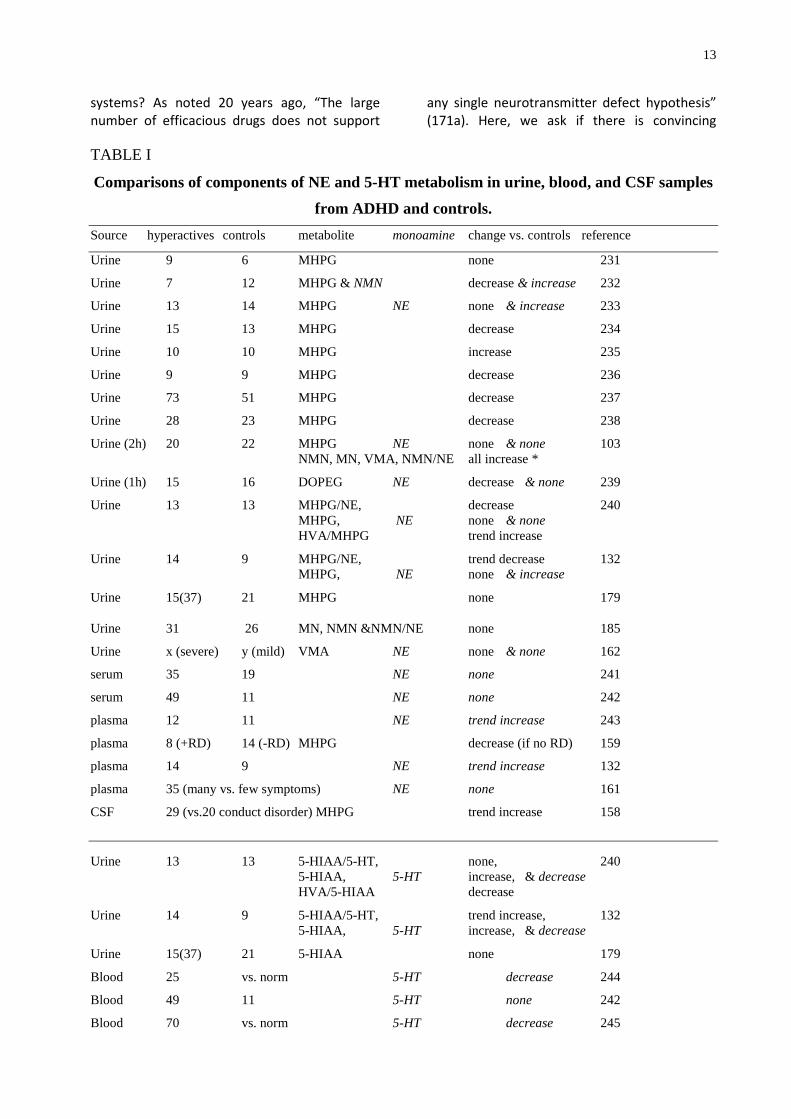
Analyses of CSF, blood compartments and
urine (Table 1 below) indicate that in the ADHD
condition MHPG levels (NE metabolite) are
usually lower than normal: less clearly NE levels
may be increased. Overall this suggests a
decreased turnover. There is a hint that other
catabolic pathways may be differentially
affected (cf. NMN levels). The severity of the
core symptoms does not influence the results
(161, 162). But, over the 4-5 years from pre- to
post-puberty when a number of symptoms
regress, MHPG levels have been noted to
increase or normalize (163). Further, some
studies that deliberately contrasted sub-groups
find that several comorbidities (independent of
their nature) appear to counteract the
metabolic decrease: e.g. in those with a reading
disorder (159), and in 15 subjects with high
levels of anxiety (not in Table: 103).
The results for the 5-HT system are more
limited reflecting in part the methodological
issues (section 7). However, if one brings the
separate findings together, there is an indication
of an increase of 5-HT turnover largely reflecting
decreases in 5-HT levels (Table 1). Nonetheless,
as with NE, it must be recognized that there will
be sub-groups, however defined, for whom the
effects associated with the core symptoms will
be masked by other features. One such example
is shown by the contrast between ADHD boys
brought up in families with or without alcoholic
fathers (164). Those with this experience
showed a larger cortisol response to a challenge
dose of fenfluramine than those without an
alcoholic father. This was interpreted as
reflecting increased 5-HT receptor sensitivity.
Another example of the influences of
comorbidity on 5-HT activity concerns
impulsivity. Impulsive aggression (oppositional
behaviors; 30, 165) has been associated with
low plasma and CSF 5-HIAA and synaptic
availability of 5-HT. This contrasts with the
generalization noted in the preceding
paragraph. Intriguingly, Oades et al. (36)
compared the binding characteristics of the
platelet 5-HT transporter with clinical ratings
(impulsivity/ distractibility, externalizing/
aggression) and the (in)ability to withhold
responses on the Stop-Signal task (cognitive
impulsivity). Decreased affinity correlated with
poor response inhibition (cognitive
impulsiveness) but not clinical ratings, even
though the cognitive and clinical indices of
impulsivity were related. In contrast aggressive
behavior related to increased 5-HT transporter
affinity7. (See section 6.2: genetic control of 5-
HT availability by the transporter [HTTLPR]).
Cognitive impulsivity might be expected to
reveal itself on the CPT test of sustained
attention in the form of an increased rate of
false alarms. However, as yet, both high (blood:
166) and low levels of 5-HT (tryptophan
depletion: 167) have been related to more
errors of commission. But, d-prime, reflecting
target sensitivity, was reported to decrease as
the excretion of the 5-HT metabolite increased
(160), which supports interpretations of the
platelet study, above.
Lastly another indication that there may be 2
ADHD sub-groups differing in the sensitivity of
the 5-HT system comes from neurophysiological
study of the augmenting-reducing response
using event-related potentials. The N1/P2
component may increase (augment) or decrease
(reduce) in response to increases of salience
(loudness of sounds). An augmenting response
is a feature of sensation-seeking (168), and
ADHD subjects who respond to amphetamine
(169). Increasing stimulus intensity-dependence
relates to decreasing 5-HT activity (and vice
versa, cf. effects of alcohol and lithium: 170).
Among ADHD subjects who do not respond to
amphetamine, a reducing response to auditory
stimuli is typical (169). It remains unclear how
closely coupled 5-HT activity is with the
augmenting-reducing phenomenon. But, it
would be worthwhile combining biochemical
measures in ADHD subjects with/without the
conduct problems that are influenced by 5-HT
activity with this paradigm
9.2 Evidence from pharmacological
treatments:
The question addressed here is whether
there is evidence that treatments that exert a
good effect on the ADHD condition also exert a
minor or major effect by way of the NE or 5-HT
7 Reductions of binding site affinity should normally be off-set by increased receptor capacity. If this does not occur then more 5-HT remains available in the synapse.
13
systems? As noted 20 years ago, “The large
number of efficacious drugs does not support
any single neurotransmitter defect hypothesis”
(171a). Here, we ask if there is convincing
TABLE I
Comparisons of components of NE and 5-HT metabolism in urine, blood, and CSF samples
from ADHD and controls.
Source hyperactives controls metabolite monoamine change vs. controls reference
Urine 9 6 MHPG none 231
Urine 7 12 MHPG & NMN decrease & increase 232
Urine 13 14 MHPG NE none & increase 233
Urine 15 13 MHPG decrease 234
Urine 10 10 MHPG increase 235
Urine 9 9 MHPG decrease 236
Urine 73 51 MHPG decrease 237
Urine 28 23 MHPG decrease 238
Urine (2h) 20 22 MHPG NE none & none 103 NMN, MN, VMA, NMN/NE all increase *
Urine (1h) 15 16 DOPEG NE decrease & none 239
Urine 13 13 MHPG/NE, decrease 240 MHPG, NE none & none HVA/MHPG trend increase
Urine 14 9 MHPG/NE, trend decrease 132 MHPG, NE none & increase
Urine 15(37) 21 MHPG none 179
Urine 31 26 MN, NMN &NMN/NE none 185
Urine x (severe) y (mild) VMA NE none & none 162
serum 35 19 NE none 241
serum 49 11 NE none 242
plasma 12 11 NE trend increase 243
plasma 8 (+RD) 14 (-RD) MHPG decrease (if no RD) 159
plasma 14 9 NE trend increase 132
plasma 35 (many vs. few symptoms) NE none 161
CSF 29 (vs.20 conduct disorder) MHPG trend increase 158
Urine 13 13 5-HIAA/5-HT, none, 240 5-HIAA, 5-HT increase, & decrease HVA/5-HIAA decrease
Urine 14 9 5-HIAA/5-HT, trend increase, 132 5-HIAA, 5-HT increase, & decrease
Urine 15(37) 21 5-HIAA none 179
Blood 25 vs. norm 5-HT decrease 244
Blood 49 11 5-HT none 242
Blood 70 vs. norm 5-HT decrease 245
14
Serum 11 11 5-HT decrease 246
Platelet 17 75 5-HIAA none 241
Platelet 55 38 5-HT none 247
plasma 35 (many vs. few symptoms) 5-HT decrease 161
CSF 24 6 5-HIAA none 248
CSF 6 16 5-HIAA none 249
CSF 29 (vs.20 conduct disorder) 5-HIAA none 158
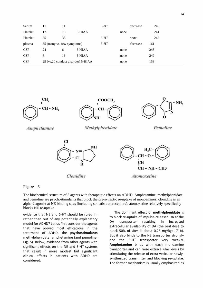
Figure 5 The biochemical structure of 5 agents with therapeutic effects on ADHD. Amphetamine, methylphenidate and pemoline are psychostimulants that block the pre-synaptic re-uptake of monoamines: clonidine is an alpha-2 agonist at NE binding sites (including somatic autoreceptors): atomoxetine relatively specifically blocks NE re-uptake
evidence that NE and 5-HT should be ruled in,
rather than out of any potentially explanatory
model for ADHD? Let us first consider the agents
that have proved most efficacious in the
treatment of ADHD, the psychostimulants
methylphenidate, amphetamine (and pemoline:
Fig. 5). Below, evidence from other agents with
significant effects on the NE and 5-HT systems
that result in more modest but significant
clinical effects in patients with ADHD are
considered.
The dominant effect of methylphenidate is
to block re-uptake of impulse-released DA at the
DA transporter resulting in increased
extracellular availability of DA (the oral dose to
block 50% of sites is about 0.25 mg/kg: 171b).
But it also binds to the NE transporter strongly
and the 5-HT transporter very weakly.
Amphetamine binds with each monoamine
transporter and can raise extracellular levels by
stimulating the release of extra-vesicular newly-
synthesized transmitter and blocking re-uptake.
The former mechanism is usually emphasized as
15
treatments that block catecholamine synthesis
inhibit the effects of amphetamine more than
methylphenidate. (Caveat: the mechanism of
stimulating the transporter to release
transmitter, or block the re-uptake varies with
dose, and specific data vary with measures
made in vitro or in vivo: 172). A modest degree
of MAO inhibition has also been reported.
Pemoline (caveat: liver toxicity) will not be
further discussed: its effects are specific to the
release and uptake of DA (173).
In preclinical studies in rodents,
methylphenidate (0.75-3.0 mg/kg, iv) does not
increase motor activity or mesolimbic levels of
DA, but it does increase extracellular levels of
NE (e.g. in the limbic system: 174). Similar doses
of amphetamine (sc) increase limbic and frontal
levels of NE to a greater extent (and release DA:
172). While higher doses (e.g. 20 mg/kg) of
methylphenidate still release NE they do not
increase levels of 5-HT. Nonetheless such
pharmacological doses have been reported to
enhance 5-HT metabolite levels in fronto-striatal
regions (175). In contrast 2.5/3.0 mg/kg
amphetamine can raise 5-HT levels 3-fold and
increase its metabolism (e.g. neostriatum: 176).
Sub-chronic amphetamine treatment has been
reported to sensitize brainstem 5-HT1a, but not
5-HT2a sites (177).
Do the biochemical responses to the
psychostimulants reflect expectations from the
preclinical results? First, care must be taken
with the interpretation of results as the
variability between reports, whether from
different or the same authors can be marked for
measures taken from the CSF, plasma or urine.
Secondly, HVA levels, as noted above can reflect
peripheral NE metabolism8, also tend to
decrease/normalize after methylphenidate
treatment, whether or not the patients
responded clinically (urine, 179; CSF, 30). Both
studies noted that although 5-HT metabolism
was not necessarily high, levels tended to
decrease with treatment following corrections
of high levels DA metabolism and symptom
improvement..
The only clear result for NE, 5-HT and their
metabolites is that urinary MHPG levels
decrease after amphetamine (7/7 studies) but
8 Peripheral NA metabolites were reported to be high in the ADHD urinary samples (178).
not after methylphenidate treatment (3/3
studies: Table 14.1 in 180). VMA levels were
also reduced after amphetamine in 3/3 studies.
For other metabolites increases and decreases
have been reported and no clear pattern
emerges. It is surprising that unequivocal
changes of NE levels are not usually recorded
after methylphenidate treatment. At first sight it
is enigmatic that the frequently reported low
turnover for NE in ADHD patients should be
further lowered in those who respond to
psychostimulant treatment (181). A possible
explanation derives from electrophysiological
recordings in primates (182). A parallel is drawn
between an overly tonic firing mode for the LC
during poor CPT performance and the sustained
attention problems in ADHD. Low activity
facilitates interactions with many stimuli rather
than focused attention. Stimulants decrease the
tonic activity and facilitate a transition to a
phasic firing mode. This counteracts the ‘hypo-
arousal’ in the system. The coupling of
information transfer is improved, even though
the overall NE turnover rate decreases further.
Raising the issue of arousal encourages
mention of the biochemical support for the
concept of hypo-arousal in ADHD from
measures of adrenaline and phenylethylamine
(PEA). Adrenaline levels tend to be low in urine
samples from ADHD children and the adrenergic
(and cortisol) response to stress is reduced (183-
1859). Adrenaline levels rise with
methylphenidate or amphetamine treatment
(186-188). This is consistent with the simple
concept of low levels of arousal becoming
partially normalized by stimulant treatment.
PEA is a naturally occurring amphetamine-like
derivative that results from decarboxylation of
phenylalanine, a precursor to normal
catecholamine synthesis (Fig.1). Levels are
frequently found to be raised in a range of
psychiatric, excited conditions (e.g. acute
schizophrenia, bipolar disorder, some obsessive
compulsive and psychopathic conditions) but
reduced in depression (189-192). They are lower
9 However, in highly anxious children, usually with internalizing problems, urinary adrenalin levels can indeed be high with respect to patients without prominent anxiety (180). Slightly higher levels of plasma adrenaline reported in ADHD children (132) may likewise have reflected the cognitive testing that occurred around the same time.
16
in ADHD, even if PEA levels are not significantly
correlated with symptom severity itself (179,
193). Psychostimulant treatments raise PEA
levels (194, 195). PEA levels may reflect
endogenous homeostatic mechanisms for
promoting catecholamine activity (e.g., like
amphetamine, PEA increases CSF levels of NE
and DA in non-human primates [196]). In
summary, while both psychostimulants lead to
an increase of extracellular catecholamines,
they differ a) on the mechanism at the
transporter, b) on its relation to impulse flow
and c) at clinically relevant doses only
amphetamine significantly influences 5-HT
activity; yet it is clear that specific effects of
methylphenidate (and atomoxetine) at the NE
transporter can bring about significant changes
in the activity of both catecholamines, especially
in mesocortical regions.
The relatively recent introduction of
atomoxetine, a selective NE transport inhibitor,
as an efficacious form of ADHD treatment
merits attention; however, independent studies
of the nature of the improvement and
biochemical effects remain sparse. In rodents it
raises mesocortical NE and DA levels three-fold.
Like methylphenidate it is without influence on
the 5-HT system, but in contrast it is without
effect on nigro-striatal or mesolimbic
catecholamines (197). [Note that
methylphenidate also raises mesocortical NE
and DA levels to a similar degree.] The focus of
attention on the mechanisms underlying its
efficacy returns to the role of cortical NE
transporters on the availability of both
catecholamines (section 4.3). Atomoxetine
improves each of the diagnostically important
symptom clusters (inattention, impulsivity and
activity: 198), but results of more specific tests
of attentional abilities or of cognitive impulsivity
remain unclear.
A range of well-known antidepressants can
also positively influence ADHD symptoms (e.g.
MAO inhibitors, desipramine, review 199). In 7
trials desipramine (DMI), known for its blockade
of NE uptake, is reported to modestly improve
hyperactivity, impulsivity, distractibility and
some limited aspects of learning (paired
associates) and recall (match to familiar figures:
200, 201). Yet it has no apparent effect on the
CPT measure of sustained attention (201, 202).
Cardiac side-effects discourage the use of DMI,
but, as with other “helpful” treatments, DMI can
decrease NE excretion, along with its central
and peripheral metabolites (203). DMI may not
so much alter basal levels of NE but increase
those arising from stimulus-coupled release of
NE, a parallel to methylphenidate’s action (10).
Unfortunately there is little information on
dose-dependent biochemical effects or
correlations with the reported behavioral
improvements.
Less well-documented are effects of DMI on
the 5-HT system. This surprises as tertiary anti-
depressants like imipramine with an effect on
the 5-HT transporter exert modest
improvements like the secondary anti-
depressants (e.g. DMI: 204). Recently Overtoom
and colleagues (205) reported on a left-right
discrimination test in ADHD children treated
with either DMI, methylphenidate, L-dopa or
placebo. The discrimination became a stop-
signal test with a no-go tone rapidly following
some of the discriminanda. Methylphenidate
treatment speeded reaction times and
decreased omissions and discrimination errors.
That L-dopa (promoting post-synaptic DA levels)
had no effect does not show that DA had no
effect, as the synaptic mechanisms differ from
the other agents investigated. But it promotes
speculation that methylphenidate was at least
in part influencing the NE system. More
intriguing still is that inhibition on the stop-task
improved only after DMI treatment. Fortunately
the authors recorded prolactin responses to
treatment. These decreased as expected after
the two “DA” treatments, but increased after
DMI. The supposition that this was a 5-HT effect
was confirmed by their finding that serum 5-
HIAA levels decreased. This seems to confirm
the proposition (section 9.1) that changes in the
5-HT system may relate to cognitive impulsivity,
while other attentional effects may reflect NE /
catecholaminergic activity.
Clonidine is not a treatment of first choice.
This reflects its side effects (blood-pressure,
sedation, dizziness) and that its efficacy is
largely restricted to oppositional problems (e.g.
aggression10
, frustration tolerance, co-
10 Relevant to the study of NE‘s role in comorbid aspects of ADHD is that beta-NE blockers also yield positive results in treating problems related to aggression, despite potential cardiac problems with this form of treatment (214).

17
operation: 206). However, some improvements
in hyperactivity and impulsivity have been
reported (meta-analysis, 207), especially when
co-administered with methylphenidate (208,
209). Further, performance on some specific
tests of frontal executive functions can be
enhanced (210), and response speed and errors
on tests of sustained attention improved with
clonidine treatment (211). Two reports (212,
213) confirmed the inhibitory effects of
clonidine expected from preclinical studies by
showing that MHPG levels decreased in ADHD
children and young adults. (The literature on
hypertension also shows falling NE
concentrations.) It is therefore of interest to look
at clonidine’s agonist activity at alpha-2 NE
receptors.
A direct action at alpha sites was assumed to
underlie the enhanced growth hormone
response to a pharmacological challenge with
clonidine (213). While an increased receptor
sensitivity may be consistent with simple
interpretations of clonidine’s inhibitory
influence, and perhaps that of guanfacine, the
implication from platelet alpha-2 receptor
binding is different (215). This group used the
platelet model of binding to predict stimulant
response. They found a generally low level of
binding: ADHD children with relatively normal
binding responded to treatment, and those with
low levels were non-responders. However,
other interpretations of clonidine’s action are
possible, and some expectations can be
generated from animal studies. Using systemic
doses in the range of 0.1-1.0 mg/kg (and local
treatments), clonidine not only reduces NE
release (in the brainstem and cortices) but
reduces brainstem and cortical 5-HT release (21,
216). These studies show that alpha-1 and
alpha-2 sites exert opposite facilitatory and
inhibitory influences on 5-HT release. Further, of
interest for the interpretation of the roles of
mesocortical DA and NE (section 4.3), the
stimulation of increases of cortical DA and NE
(e.g. by clozapine treatment) can be prevented
by quite moderate doses of clonidine (0.015
mg/kg: 217).
In summary, clinical and preclinical work with
clonidine show a) limited but significant
improvements in some areas of function
relevant to ADHD; b) reduced 5-HT release could
underlie the modulation by clonidine of
aggressive and impulsive behavior (15); c) a
mechanism for reduced NE release to incur
reduced DA release, which could have both
helpful (hyperactivity) and less helpful
consequences (appreciation of reinforcement).
In a similar vein, lowering NE release may
enhance sustained attention performance (211),
and it may raise the degree to which alpha-2
rather than alpha-1 receptors (with a lower NE
affinity) might assist cortical function (e.g.
working memory and related executive
functions, 218). Nonetheless hard evidence for
binding differences in ADHD children is lacking,
and treatments aimed at the alpha-2 receptor
could be counter productive in the appropriate
control of responses to stress.
Two of three open trials of Guanfacine, an
agonist at alpha-2 NE sites, found a modest
improvement of ratings of attention and
impulsivity, with one demonstrating fewer
errors on the CPT (157, 219, 220). Controlled
trials (vs. amphetamine) in adult ADHD patients
showed comparable reductions of symptoms
and even an improvement of the Stroop color-
word naming, so often impaired in childhood
ADHD (221). In children with ADHD and
comorbid tics teacher ratings improved in half
the patients, who also performed a CPT more
accurately (222). Thus, a modest degree of
success for Arnsten’s alpha-2 NE hypothesis
(139) appears to be realized, although with a
certain risk of lethargy, bradycardia and
hypotension the agent should perhaps be held
in reserve for psychostimulant non-responders.
Despite indications that some treatments
may achieve therapeutic effects (e.g.
impulsivity) by an action on 5-HT systems, direct
attempts using agents with unequivocal effects
on 5-HT metabolism have been largely without
success (e.g. the precursor amino acid
tryptophan, [223] fenfluramine that facilitates
5-HT release, [186]; an agonist at 5-HT1a sites,
buspirone [review: 224]). It is sobering and
important to note that while a particular agent
may reduce symptoms and alter monoaminergic
metabolism, it is not known that the metabolic
changes are related to the psychopathological
changes. The report of Donnelly and colleagues
(186) is salutary. Fenfluramine treatment (0.6-
2.0 mg/d) had no significant therapeutic effect
on ADHD boys aged 6-12y. However, urinary NE,
MHPG, VMA and epinephrine all decreased
18
significantly, as did plasma MHPG and platelet
5-HT levels. Yet, for those with impulsive
aggression, and delinquency there is a clear
relationship with low 5-HT activity, be it
expressed as reduced platelet binding of
imipramine (e.g. 225), plasma 5-HIAA (165) or
prolactin response to fenfluramine challenge
(226).
10. General:
Early proposals that NE could have a causal
role in ADHD and hyperkinetic behavior in
particular (227) were based on the effect of
amphetamine to reduce NE activity during
arousal. Now there is a widespread belief that
children with ADHD are under- rather than over-
aroused: yet there is an increasing consensus
that NE function has something to do with the
symptoms (204). Evidence in this chapter shows
that NE activity undoubtedly modulates
attentional mechanisms both directly (tuning
signal to noise ratios) and indirectly (via the
control of mesocorticolimbic DA release). NE
may influence other relevant behaviors
depending on their dependence on cognitive
mechanisms (e.g. environmental stimulation
facilitating hyperkinesis) and the nature of the
mechanisms underlying comorbid conditions.
Crucial mechanisms include the control of
catecholamine availability in the cortex (via the
transporter) and phasic firing modes in the LC.
Both of these should be targets for treatment.
Common to a consideration of the relative
role of NE and 5-HT in ADHD is the increasing
appreciation of a crucial role for the transporter
in determining the availability of monoamines.
Thus, cortical NE transporters can release DA
(55), the DA transporter is regulated by a variety
of substrates including 5-HT (228) and the NE
transporter (cf. knockout mice) modulates the
perception of reinforcement (229). This latter
finding has implications for understanding the
aversion to accepting delays between response
and reinforcement gradients associated with the
SHR and with ADHD subjects (230).
5-HT mechanisms are also relevant to the
expression of features of ADHD by direct
(transporter mediated reuptake mechanisms)
and indirect mechanisms (modulation of DA
activity, especially in the initiation of behavioral
responses). These have been under-researched
in view of more clearly established relations of
5-HT activity to the expression of externalizing
responses more frequent in comorbid
conditions. Now it is appreciated that 5-HT
activity has a role in information processing
(modulating gain) and cognitive impulsivity. The
appreciation of these roles and the interactions
of the three monoamines should make it easier
to taylor treatment to the particular individual
(im)balance of the pattern of cognitive,
motivational and motor bases to be found in a
given patient.
References:
1. Moleman P, Tulen JHM, Blankestijn PJ, Man in
t'Veld A, Boomsma F. Urinary excretion of
catecholamines and their metabolites in relation
to circulating catecholamines: six-hour infusion
of epinephrine and norepinephrine in healthy
volunteers. Arch Gen Psychiatry 1992; 49: 568-
572.
2. Carlsson A, Falck B, Hillarp NA. Cellular
localization of brain monoamines. Acta Physiol
Scand 56 suppl 1962: 1-28.
3. Vogt M. The concentration of sympathin in
different parts of the central nervous system
under normal conditions and after
administration of drugs. J Physiol (London)
1954;123:451-481.
4. Anderson CD, Pasquier DA, Forbes WB,
Morgane PJ. Locus coeruleus-to-dorsal raphe
input examined by electrophysiological and
morphological methods. Brain Res Bull 1977; 2:
209-221.
5. Jones BE, Moore RY. Ascending projections
of the locus coeruleus in the rat. II.
Autoradiographic study. Brain Res 1977; 127:
23-53.
6. Loughlin SE, Foote SL, Bloom FE. Efferent
projections of nucleus locus coeruleus:
topographic organization of cells of origin
demonstrated by three-dimensional
reconstruction. Neurosci 1986; 18: 291-306
7. Delfs JM, Zhu JP, Druhan JP, Aston-Jones GS.
Origin of noradrenergic afferents to the shell
subregion of the nucleus accumbens:
anterograde and retrograde tract-tracing
studies in the rat. Brain Res 1998; 806: 127-140.
8. Chan-Palay V, Asan E. Quantitation of
catecholamine neurons in the locus coeruleus in
human brains of normal young and older adults
19
and in depression. J Comp Neurol 1989; 287:
357-372.
9. Descarries L, Watkins KC, Lapierre Y.
Noradrenergic axon terminals in the cerebral
cortex of rat. III. Topometric ultrastructural
analysis. Brain Res 1977; 133: 197-222.
10. Berridge CW, Waterhouse BD. The locus
coeruleus - noradrenergic system: modulation
of behavioral state and state-dependent
cognitive processes. Brain Res Rev 2003; 42: 33-
84.
11. Amin AH, Crawford TBB, Gaddum JH. The
distribution of substance P and 5-hydroxy-
tryptamine in the central nervous system of the
dog. J Physiol 1954; 126: 596-618.
12. Twarog BM, Page ICH. Serotonin content of
some mammalian tissues and urine and method
for its determination. Am J Physiol 1953; 175:
479-485.
13. Dahlström A, Fuxe K. Evidence for the
existence of monoamine containing neurons in
the central nervous system. I. Demonstration of
monoamines in cell bodies of brain neurons.
Acta Physiol Scand 1964; 62 (suppl 232): 1-55.
13. Steinbusch HWM. Distribution of serotonin-
immunoreactivity in the central nervous system
of the rat: cell bodies and terminals. Neurosci
1981; 6: 557-618.
14. Kosofsky BE, Molliver ME. The serotonergic
innervation of the cerebral cortex: Different
classes of axon terminals arise from the dorsal
and median raphe nuclei. Synapse 1987; 1: 153-
168.
15. Törk I. Anatomy of the serotonergic system.
Ann NY Acad Sci 1990; 600: 9-34.
16. Hornung J-P, Fritschy J-M, Törk I. Distribution
of subsets of serotoninergic axons in the
cerebral cortex of the Marmoset. J Comp Neurol
1990; 297: 165-181.
17. Molliver ME. Serotonergic neuronal systems:
what their anatomic organization tells us about
function. J Clin Psychopharmacol 1987; 7: 3S-
23S
18. De Haes JI, Bosker FJ, Van Waarde A, et al. 5-
HT1A receptor imaging in the human brain:
Effect of tryptophan depletion and infusion on
[18F] MPPF binding. Synapse 2002; 46: 108-115.
19. Wright DE, Seroogy KB, Lundgren KH, Davis
BM, Jennes L. Comparative localization of
serotonin1A, 1C and 2 receptor subtype mRNAs in
rat brain. J Comp Neurol 1995; 351: 357-373.
20. Nosjean A, Hamon M, Darmon M. 5-HT2A
receptors are expressed by catecholaminergic
neurons in the rat nucleus tractus solitarii.
NeuroReport 2003; 13: 2365-2369.
21. Bortolozzi A, Artigas F. Control of 5-
hydroxytryptamine release in the dorsal raphe
nucleus by the noradrenergic system in rat
brain. Role of alpha-adrenoceptors.
Neuropsychopharmacol 2003; 28: 421-434.
22. Pudovkina OL, Cremers TI, Westerink BHC.
The interaction between the locus coeruleus
and dorsal raphe nucleus studied with dual-
probe microdialysis. Eur J Pharmacol 2002; 445:
37-42.
23. Birthelmer A, Ehret A, Amtage F, et al.
Neurotransmitter release and its presynaptic
modulation in the rat hippocampus after
selective damage to cholinergic or/and
serotonergic afferents. Brain Res Bull 2002; 59:
371-381.
24. Szabo ST, Blier P. Effects of serotonin (5-
hydroxytryptamine, 5-HT) reuptake inhibition
plus 5-HT (2A) receptor antagonism on the firing
activity of norepinephrine neurons. J Pharmacol
Exp Ther 2002; 302: 983-991.
25. Ruiz-Ortega JA, Ugedo L. Activation of 5-
HT1A receptors potentiates the clonidine
inhibitory effect in the locus coeruleus. Eur J
Pharmacol 1997; 333: 159-162.
26. Gobert A, Rivet J-M, Audinot V, Newman-
Tancredi A, Cistarelli L, Millan MJ. Simultaneous
quantification of serotonin, dopamine and
noradrenaline levels in single frontal cortex
dialysates of freely-moving rats reveals a
complex pattern of reciprocal auto- and
heteroceptor-mediated control of release.
Neurosci 1998; 84: 413-429.
27. Gobert A, Millan MJ. Serotonin 5-HT 2a
receptor activation enhances dialysate levels of
dopamine and noradrenaline, but not 5-HT, in
the frontal cortex of freely-moving rats.
Neuropharmacol 1999; 38: 315-318.
28. Millan MJ, Dekeyne A, Gobert A. Serotonin
(5HT2c) receptors tonically inhibit dopamine
(DA) and noradrenaline (NA), but not 5-HT,

20
release in the frontal cortex in vivo.
Neuropharmacol 1998; 37: 953-955.
29. Geracioti TD, Keck PE, Ekhator NN, et al.
Continuous covariability of dopamine and
serotonin metabolites in human cerebrospinal
fluid. Biol Psychiatry 1998; 44: 228-233.
30. Castellanos FX, Elia J, Kruesi MJP, et al.
Cerebrospinal fluid homovanillic acid predicts
behavioral response to stimulants in 45 boys
with attention deficit/hyperactivity disorder.
Neuropsychopharmacol 1996; 14: 125-137.
31. O'Neill MF, Heron-Maxwell CL, Shaw G. 5-
HT2 receptor antagonism reduces hyperactivity
induced by amphetamine, cocaine and MK-801
but not D-1 agonist c-APB. Pharmacol Biochem
Behav 1999; 63: 237-244.
32. Porras G, Di Matteo V, Fracasso C, et al. 5-HT
(2A) and 5-HT (2C/2B) receptor subtypes
modulate dopamine release induced in vivo by
amphetamine and morphine in both the rat
nucleus accumbens and striatum.
Neuropsychopharmacol 2002;26:311-324.
33. Rogers RD, Blackshaw AJ, Middleton HC, et
al. Tryptophan depletion impairs stimulus
reward learning while methylphenidate disrupts
attentional control in healthy young adults:
implications for the monoaminergic basis of
impulsive behaviour. Psychopharmacol 1999;
146: 482-491.
34. Fletcher PJ, Korth KM. Activation of 5-HT1B
in the nucleus accumbens reduces
amphetamine induced enhancement of
responding for conditioned reward.
Psychopharmacol 1999; 142: 165-174.
35. Luthman J, Fredeiksson A, Sundström E,
Jonsson G, Archer T. Selective lesion of central
dopamine or noradrenaline neuron systems in
the neonatal rat: motor behavior and
monoamine alterations at adult stage. Behav
Brain Res 1989; 33: 267-277.
36. Oades RD, Slusarek M, Velling S, Bondy B.
Serotonin platelet-transporter measures in
childhood attention-deficit/hyperactivity
disorder (ADHD): clinical versus experimental
measures of impulsivity. World J Biol Psychiat
2002; 3: 96-100.
37. Koskinen T, Sirviö J. Studies on the
involvement of the dopaminergic system in the
5-HT2 agonist DOI-induced premature
responding in a five-choice serial reaction time
task. Brain Res Bull 2001; 54: 65-75.
38. Nocjar C, Roth BL, Pehek EA Localization of 5-
HT (2A) receptors on dopamine cells in sub
nuclei of the midbrain A10 cell group.
Neuroscience 2002; 111: 163-176.
39. Lucas G, Spampinato U. Role of striatal
serotonin2A and serotonin2C receptor subtypes
in the control of the in vivo dopamine outflow in
the rat striatum. J Neurochem 2000; 74: 693-
701.
40. Di Giovanni G, Di Matteo V, Di Mascio M,
Esposito E. Preferential modulation of
mesolimbic vs. nigrostriatal dopaminergic
function by serotonin2c/2b receptor agonists: a
combined in vivo electrophysiological and
microdialysis study. Synapse 2000; 35: 53-61.
41. Di Matteo V, Cacchio M, Di Giulio C, Esposito
E. Role of serotonin (2C) receptors in the control
of brain dopaminergic function. Pharmacol
Biochem Behav 2002; 71: 727-734.
42. Hutson PH, Barton CL, Jay M, et al. Activation
of mesolimbic dopamine function by
phencyclidine is enhanced by 5-HT2C/2B
receptor antagonists: neurochemical and
behavioural studies. Neuropharmacol 2000; 39:
2318-2328.
43. Kuroki T, Dai J, Meltzer HY, Ichikawa J. R(+)-8-
OH-DPAT, a selective 5-HT1A receptor agonist,
attenuated amphetamine-induced dopamine
synthesis in rat striatum, but not nucleus
accumbens or medial prefrontal cortex. Brain
Res 2000; 872: 204-207.
44. Doherty MD, Pickel VM. Targeting of
serotonin 1a receptors to dopaminergic neurons
within the parabrachial subdivision of the
ventral tegmental area in rat brain. J Comp
Neurol (2001); 433: 490-500.
45. Yan QS, Yan SE. Activation of 5.HT1B/(1D)
receptors in the mesolimbic dopamine system:
increases dopamine release from the nucleus
accumbens. Eur J Pharmacol 2001; 418: 55-64.
46. Pan WHT, Sung JC, Fuh SMR. Locally
application of amphetamine into the ventral
tegmental area enhances dopamine release in
the nucleus accumbens and the medial
prefrontal cortex through noradrenergic
neurotransmission. J Pharmacol Exp Ther 1996;
21
278: 725-731.
47. Auclair A, Cotecchia S, Glowinski J, Tassin J-P.
D-amphetamine fails to increase extracellular
dopamine levels in mice lacking alpha 1b-
adrenergic receptors: relationship between
functional and non-functional dopamine
release. J Neurosci 2002; 22: 9150-9154.
48. Yavich L, Lappalainen R, Sirviö J, Haapalinna
A, MacDonald E. 2-Adrenergic control of
dopamine overflow and metabolism in mouse
striatum. Eur J Pharmacol 1997; 339: 113-119.
49. Tuinstra T, Cools AR. High and low responders
to novelty: effects of adrenergic agents on the
regulation of accumbal dopamine under
challenged and non-challenged conditions.
Neurosci 2000; 99: 55-64
50. Gioanni Y, Thierry A-M, Glowinski J, Tassin J-
P. 1-adrenergic, D1 and D2 receptors
interactions in the prefrontal cortex:
implications for the modality of action of
different types of neuroleptics. Synapse 1998;
30: 362-370.
51. Hertel P, Fagerquist MV, Svensson TH.
Enhanced cortical dopamine output and
antipsychotic-like effects of raclopride by 2
adrenoceptor blockade. Science 1999; 286: 105-
107.
52. Carboni E, Tanda GL, Frau R, Di Chiara G.
Blockade of the noradrenaline carrier increases
extracellular dopamine concentrations in the
prefrontal cortex: evidence that dopamine is
taken up in vivo by noradrenergic terminals. J
Neurochem 1990; 55: 1067-1070
53. De Montis G, Devoto P, Gessa GL, et al.
Central dopaminergic transmission is selectively
increased in the limbic system of rats chronically
exposed to antidepressants. Eur J Pharmacol
1990; 180: 31-35.
54. Wayment HK, Schenk JO, Sorg BA.
Characterization of extracellular dopamine
clearance in the medial prefrontal cortex: role of
monoamine uptake and monoamine oxidase
inhibition. J Neurosci 2001; 21: 35-44.
55. Devoto P, Flore G, Pani L, Gessa GL. Evidence
for co-release of noradrenaline and dopamine
from noradrenergic neurons in the cerebral
cortex. Mol Psychiatry 2001; 6: 657-664.
56. Zevcevic N, Verney C. Development of the
catecholamine neurons in human embryos and
fetuses, with special emphasis on the
innervation of the cerebral cortex. J comp
Neurol 1995; 351: 509-535.
57. Tomasini R, Kema IP, Muskiet FAJ, Neiborg G,
Staal MJ, Go KG. Catecholaminergic
development of fetal ventral mesencephalon:
characterization by high performance liquid
chromatography with electrochemical detection
and immunohistochemistry. Exp Neurol 1997;
145: 434-441.
58. Choi S-J, Kellogg CK. Adolescent development
influences functional responsiveness of
noradrenergic projections to the hypothalamus
in male rats. Dev Brain Res 1996; 94: 144-151.
59. Clarke AS, Hedeker DR, Ebert MH, Schmidt
DE, McKinney WT, Kraemer GW. Rearing
experience and biogenic amine activity in infant
rhesus monkeys. Biol Psychiatry 1996; 40: 338-
352.
60. Miguez JM, Aldegrunde M, Paz-Valinas L,
Recio J, Sanchez-Barcelo E. Selective changes in
the contents of noradrenaline, dopamine and
serotonin in rat brain areas during aging. J Neur
Transm 2000; 106: 1089-1098
61. Hartley EJ, Seeman P. Development of
receptors for dopamine and noradrenaline in rat
brain. Eur J Pharmacol 1983;91:391-397.
62. Moll GH, Mehnert C, Wicker M, et al. Age-
associated changes in the densities of the
presynaptic monoamine transporters in
different regions of the rat brain from early
juvenile life to late adulthood. Dev Brain Res
2000; 119: 251-257.
63. Roux JC, Mamet J, Perrin J, et al.
Neurochemical development of the brainstem
catecholaminergic cell groups in rat. J Neur
Transm 2003; 110: 51-65.
64. Oades RD, Röpcke B, Schepker R. A test of
conditioned blocking and its development in
childhood and adolescence: relationship to
personality and monoamine metabolism. Dev
Neuropsychol 1996; 12: 207-230.
65. Andersen SL, Teicher MH. Sex differences in
dopamine receptors and their relevance to
ADHD. Neurosci Biobehav Rev 2000; 24: 137-
141.
22
66. Vu DH, Törk I. Differential development of
the dual serotonergic fiber system in the
cerebral cortex of the cat. J Comp Neurol 1992;
317: 156-174.
67. Jones GH, Hernandez TD, Kendall DA,
Marsden CA, Robbins TW. Dopaminergic and
serotonergic function following isolation rearing
in rats: study of behavioural responses and
postmortem and in vivo neurochemistry.
Pharmacol Biochem Behav 1992; 43: 17-35.
68. Santana C, Rodriguez M, Alfonso D, Arevalo
R. Dopaminergic neuron development in rats:
biochemical study from prenatal life to
adulthood. Brain Res Bull 1992; 29: 7-13.
69. Silverstein FS, Donn S, Buchanan K, Johnston
MV. Concentrations of homovanillic acid and 5-
hydroxyindoleacetic acid in cerebrospinal fluid
from human infants in the perinatal period. J
Neurochem 1984; 43: 1769-1772.
70. Murrin LC, Gibbens DL, Ferrer JR. Ontogeny
of dopamine, serotonin and spirodecanone
receptors in rat forebrain – an autoradiographic
study. Dev Brain Res 1985; 23: 91-109.
71. Konradi C, Kornhuber J, Sofic E, Heckers S,
Riederer P, Beckmann H. Variations of
monoamines and their metabolites in the
human brain putamen. Brain Res 1992; 579:
285-290.
72. Retz W, Kornhuber J, Riederer P.
Neurotransmission and the ontogeny of human
brain. J Neur Transm 1996; 103: 403-419.
73. Sigurdh J, Spigset O, Allard P, Mjörndahl T,
Hägglof B. Binding of [3H]lysergic acid
diethylamide to serotonin 5-HT2A receptors and
of [3H]paroxetine to serotonin uptake sites in
platelets from healthy children, adolescents and
adults. Neuropsychobiol 1999; 40: 183-187.
74. Zhang ZW. Serotonin induces tonic firing in
layer V pyramidal neurons of rat prefrontal
cortex during postnatal development. J Neurosci
2003; 23: 3373-3384.
75. Rodriguez M, Martin L, Santana C. Ontogenic
development of brain asymmetry in
dopaminergic neurons. Brain Res Bull 1994; 33:
163-171.
76. Hechtman L. Genetic and neurobiological
aspects of attention deficit hyperactive disorder:
a review. J Psychiat Neurosci 1994; 19: 193-201.
77. Arnsten AFT. Catecholamine regulation of
the prefrontal cortex. J Psychopharmacol 1997;
1: 151-162.
78. Flugge G, van Kampen M, Meyer H, Fuchs E,
Alpha2A and alpha2C-adrenoceptor regulation
in the brain: alpha2A changes persist after
chronic stress. Eur J Neurosci 2003;17:917-928.
79. Linner L, Arborelius L, Nomikos GG,
Bertilsson L, Svensson TH. Locus coeruleus
neuronal activity and noradrenaline availability
in the frontal cortex of rats chronically treated
with imipramine: effect of α2-adrenoceptor
blockade. Biol Psychiatry 1999; 46: 776-774.
80. Fahlke C, Garpenstrand H, Oreland L, Suomi
SJ, Higley JD. Platelet monoamine oxidase
activity in nonhuman primate model of type 2
excessive alcohol consumption. Am J Psychiat
2002; 159: 2107-2109.
81. Comings DE, Gonzalez NS, Cheng LS-C,
MacMurray J. A “line-item” approach to the
identification of genes involved in polygenic
behavioral disorders: the adrenergic α2A
(ADRA2A) gene. Am J Med Genet
2003;118B:110-114.
82. Barr CL, Kroft J, Feng Y, et al. The
norepinephrine transporter gene and attention
deficit hyperactivity disorder. Am J Genet 2002;
114: 255-259.
83. McEvoy B, Hawi Z, Fitzgerald M, Gill M. No
evidence of linkage or association between
norepinephrine transporter (NET) gene
polymorphisms and ADHD in the Irish
population. Am J Med Genet 2002; 114: 656-
666.
84. Qian Q, Wang Y, Zhou R, et al. Family based
and case control association studies of catechol-
o-methyltransferase in attention deficit
hyperactivity disorder suggest genetic sexual
dimorphism. Am J Med Genet 2003; 118B: 103-
109.
85. Eisenberg J, Mei-Tal G, Steinberg A, et al.
Haplotype relative risk study of catechol-O-
methyl-transferase (COMT) and attention deficit
hyperactivity disorder (ADHD): association of
the high-enzyme activity Val allele with ADHD
impulsive-hyperactive phenotype. Am J Med
Genet 1999; 88: 497-502.
86. Kirley A, Hawi Z, Daly G, et al. Dopaminergic
system genes in ADHD: toward a biological
23
hypothesis. Neuropsychopharmacol 2002; 27:
607-619.
87. Wigg K, Zai G, Schachar R, Tannock R, et al.
Attention deficit hyperactivity disorder and the
gene for dopamine beta hydroxylase. Am J
Psychiatry 2002; 159: 1046-1048.
88. Lawson DC, Turie D, Langley K, et al.
Association analysis of monoamine oxidase A
and attention deficit hyperactivity disorder. J
Med Genet 2003; 116B: 84-89.
89. Garpenstrand H, Ekblom J, Hallman J,
Oreland L. Platelet monoamine oxidase activity
in relation to alleles of dopamine D4 and
tyrosine hydroxylase genes. Acta Psychiat Scand
1997; 96: 295-300.
90. Dalley JW, Theobald DE, Eagle DM, Passetti F,
Robbins TW. Deficits in impulse control
associated with tonically-elevated serotonergic
function in rat prefrontal cortex.
Neuropsychopharmacol 2002; 26: 716-728.
91. Moresco FM, Dieci M, Vita A, In vivo
serotonin 5HT(2A) receptor binding and
personality traits in healthy subjects: a positron
emission tomography study. NeuroImage 2002;
17: 1470-1478.
92. Kent L, Doerry U, Hary E, et al. Evidence that
variation at the serotonin transporter gene
influences susceptibility to attention deficit
hyperactivity disorder (ADHD): analysis and
pooled analysis. Mol Psychiatry 2002; 7: 908-
912.
93. Smeraldi E, Zanardi R, Benedetti F, Di Bella D,
Perez J, Catalano M. Polymorphism within the
promoter of the serotonin transporter gene and
antidepressant efficacy of fluvoxamine. Mol
Psychiatry 1998; 3: 508-511.
94. Lakatos K, Nemoda Z, Birkas E, et al.
Association of D4 dopamine receptors gene and
serotonin transporter promoter polymorphisms
with infants’ response to novelty. Mol
Psychiatry 2003; 8: 90-97.
95. Auerbach JG, Faroy M, Ebstein R. The
association of the dopamine D4 receptor gene
/DRD4) and the serotonin transporter promoter
gene (5-HTTLPR) with temperament in 12-
month-old infants. J Child Psychol Psychiat 2001;
42: 777-783.
96. Zoroglu SS, Erdal ME, Alasehirli B, et al.
Significance of the serotonin transporter gene 5-
HTTLPR and variable number tandem repeat
polymorphism in attention deficit hyperactivity
disorder. Neuropsychobiology 2002; 45: 176-
181.
97. Hawi Z, Dring M, Kirley A, et al. Serotonergic
system and attention deficit hyperactivity
disorder (ADHD): a potential susceptibility locus
at the 5-HT1B receptor gene in 273 nuclear
families from a multicentre sample. Mol
Psychiatry 2002; 7: 718-725.
98. Quist JF, Barr CL, Schachar R, et al. The
serotonin 5-HT1B receptor gene and attention
deficit hyperactivity disorder. Mol Psychiatry
2003; 8: 98-102.
99. Quist JF, Barr CL, Schachar R, et al. Evidence
for the serotonin HTR2A receptor gene as a
susceptibility factor in attention deficit
hyperactivity disorder (ADHD). Mol Psychiatry
2000; 5: 537-541.
100. Levitan RD, Masellis M, Basile VS, et al.
Polymorphism of the serotonin-2A receptor
gene (HTR2A) associated with childhood
attention deficit hyperactivity disorder (ADHD)
in adult women with seasonal affective disorder.
J Affect Disord 2002; 71: 229-233.
101. Lambert GW, Horne M, Kalff V, et al.
Central nervous noradrenergic and
dopaminergic turnover in response to acute
neuroleptic challenge. Life Sci 1995; 56: 1545-
1555.
102. Potter WZ, Manji HK. Are monoamine
metabolites in cerebrospinal fluid worth
measuring? Arch Gen Psychiatry 1993; 50: 653-
656.
103. Plizka SR, Maas JW, Javors MA, Rogeness
GA, Baker J. Urinary catecholamines in
attention-deficit hyperactivity disorder with and
without comorbid anxiety. J Am Acad Child
Adolesc Psychiatry 1994; 33: 1165-1173.
104. Tohgi H, Takahashi S, Abe T. The effect of
age on concentrations of monoamines, amino
acids and their related substances in the
cerebrospinal fluid. J Neur Transm (PD section)
1993; 5: 215-226.
105. Müller N, Empl M, Riedel M, Schwarz M,
Ackenheil M. Neuroleptic treatment increases
soluble IL-2 receptors and decreases soluble IL-6
24
receptors. Eur Arch Psychiat Clin Neurosci 1997;
247: 308-313.
106. Cheetham SC, Viggers JA, Slater NA, Heal
DJ, Buckett WR. [3H] Paroxetine binding in rat
frontal cortex strongly correlates with [3H] 5HT
uptake: effect of administration of various
antidepressant treatments. Neuropharmacol
1993; 32: 737-743.
107. Lambert GW, Cox HS, Horne M, et al.
Direct determination of homovanilic acid
release from the human brain, an indicator of
central dopaminergic activity. Life Sci 1991; 49:
1061-1072.
108. Lambert GW, Eisenhofer G, Jennings GL,
Esler MD. Regional homovanillic acid production
in humans. Life Sci 1993; 53: 63-75.
109. Lambert GW, Ferrier C, Kaye DM, et al.
Monoaminergic neuronal activity in subcortical
brain regions in essential hypertension. Blood
Press 1994; 3: 55-66.
110. Davids E,, Zhang K, Kula NS, Tarazi FI,
Baldessarini RJ. Effects of norepinephrine and
serotonin transporter inhibitors on hyperactivity
induced by neonatal 6-hydroxydopamine
lesioning in the rat. J Exp Pharmacol Ther 2002;
301: 1097-1102.
111. Bishop C, Kamdar DP, Walker PD. Intra-
striatal serotonin 5-HT2 receptors mediate
dopamine D1-inducd hyperlocomotion in 6-
hydrosxydopamine-lesioned rats. Synapse 2003;
50: 164-170.
112. Sagvolden T, Metzger MA, Schiorbeck HK,
Rugland A-L, Spinnangr I, Sagvolden G. The
spontaneously hypertensive rat (SHR) as an
animal model of childhood hyperactivity
(ADHD): changed reactivity to reinforcers and to
psychomotor stimulants. Behav Neur Biol 1992;
58: 103-112.
113. Rogers LJ, Sink MS, Hambley JW.
Exploration, fear and maze learning in
spontaneously hypertensive and normotensive
rats. Behav Neur Biol 1988; 49: 222-233.
114. Lukaszewska I, Niewiadomska G. The
difference in learning abilities between
hypertensive (SHR) and Wistar normotensive
rats are cue dependent. Neurobiol Learn
Memory 1995; 63: 43-53.
115. Nakamura-Palacios EM, Caldas CK, Fiorini
A, Chaga KN, Vasquez EC. Deficits of spatial
learning and working memory in spontaneously
hypertensive rats. Behav Brain Res 1996; 74:
217-221.
116. Gattu M, Pauly JR, Boss K.L, Summers JB,
Buccafusco JJ. Cognitive impairment in
spontaneously hypertensive rats: role of central
nicotinic receptors. Part I Brain Res
1997;771:89-103.
117. Sagvolden T, Pettersen MB, Larsen MC.
Spontaneously hypertensive rats (SHR) as a
putative animal model of childhood
hyperkinesis. Physiol Behav 1993; 5 4: 1047-
1955.
118. Dev BR, Philip L. Extracellular catechol
and indole turnover in the nucleus of the
solitary tract of spontaneously hypertensive and
Wistar-Kyoto normotensive rats in response to
drug-induced changes in arterial blood pressure.
Brain Res Bull 1996; 40: 111-116.
119. Bagdy G, Szemeredi K, Listwak SJ, Keiser
HR, Goldstein DS. Plasma catecholamine, renin
activity and ACTH responses to the serotonin
agonist DOI in juvenile spontaneously
hypertensive rats. Life Sci 1993; 53: 1573-1582.
120. Tsukamoto K, Sved AF, Ito S, Komatsu K,
Kanmatsuse K. Enhanced serotonin-mediated
responses in the nucleus tractus solitarius of
spontaneously hypertensive rats. Brain Res
2000; 863: 1-8.
121. Russell V, Allie S, Wiggins T. Increased
noradrenergic activity in prefrontal cortex slices
of an animal model for attention-deficit
hyperactivity disorder – the spontaneously
hypertensive rat. Behav Brain Res 2001; 117: 69-
74.
122. De Villiers AS, Russell VA, Sagvolden T,
Searson A, Jffer A, Taljaard JF. ά2-adrenoceptor
mediated inhibition of [3H]dopamine release
from nucleus accumbens slices and monoamine
levels in a rat model for attention-deficit
hyperactivity disorder. Neurochem Res 1995;
20: 427-433.
123. Togashi H, Matsumoto M, Yoshioka M,
Hirokami M, Minami M, Saito H. Neurochemical
profiles in cerebrospinal fluid of stroke-prone
spontaneously hypertensive rats. Neurosci Lett
1994; 166: 117-120.
25
124. Russell V. Hypodopaminergic and
hypernoradrenergic activity in prefrontal cortex
slices of an animal model for attention-deficit
hyperactivity disorder – the spontaneously
hypertensive rat. Behav Brain Res 2002; 130:
191-196.
125. Davids E, Zhang K, Tarazi FI, Baldessarini
RJ. Animal models of attention-deficit
hyperactivity disorder. Brain Res Rev 2003; 42:
1-21.
126. Dekeyne A, Gobert A, Auclair A, Girardon
S, Millan MJ. Differential modulation of
efficiency in a food-rewarded differential
reinforcement of low rate 72-s schedule in rats
by norepinephrine and serotonin re-uptake
inhibitors. Psychopharmacol 2002; 162: 156-
167.
127. Jones MD, Hess EJ. Norepinephrine
regulates locomotor hyperactivity in the mouse
mutant Coloboma. Pharmacol Biochem Behav
2003; 75: 209-216.
128. Missler M, Zhang W, Rohlmann A, et al. α-
Neurexins couple Ca2+
channels to synaptic
vesicle exocytosis. Nature 2003; 423: 939-948.
129. Dell’Anna ME, Luthman J, Lindqvust E,
Olson L. Development of monoamine systems
after neonatal anoxia in rats. Brain Res Bull
1993; 32: 159-170.
130. Puumala T, Ruotsalainen S, Jäkälä P,
Koivisto E, Riekkinen P, Sirviö J. Behavioral and
pharmacological studies on the validation of a
new animal model of attention deficit
hyperactivity disorder. Neurobiol Learn Memory
1996; 66: 198-211.
131. Poncet L, Denoroy L, Dalmaz Y, Pequignot
JM, Jouvet M. Alteration in central and
peripheral substance P and neuropeptide Y like
immunoreactivity after chronic hypoxia in the
rat. Brain Res 1996; 733: 64-72.
132. Oades RD, Daniels R, Rascher W. Plasma
neuropeptide Y levels, monoamine metabolism,
electrolyte excretion and drinking behavior in
children with attention-deficit hyperactivity
disorder (ADHD). Psychiatry Res 1998; 80; 177-
186.
133. Dumont Y, Martel JC, Fournier A, St-Pierre
S, Quirion R. Neuropeptide Y and neuropeptide
Y receptor subtypes in brain and peripheral
tissue. Prog Neurobiol 1992; 38: 125-167.
134. Lehmann J. Neuropeptide Y: an overview.
Drug Dev Res 1990; 19: 329-351.
135. Castellanos FX. The psychobiology of
attention-deficit/hyperactivity disorder. In:
Quay HC, Cohen TP, eds. Handbook of disruptive
behavior disorders. New York: Kluwer
Academic/Plenum Publishers, 1999:179-198
136. Ernst M, Kimes AS, London ED, et al.
Neural substrates of decision making in adults
with attention deficit hyperactivity disorder. Am
J Psychiat 2003; 160: 1061-1070.
137. Rubia K, Smith AB, Brammer MJ, Taylor E.
Right inferior prefrontal cortex mediates
response inhibition while mesial prefrontal
cortex is responsible for error detection.
NeuroImage 2003; 20 351-358.
138. Oades RD, Dittmann-Balcar A, Schepker R,
Eggers C. Auditory event-related potentials and
mismatch negativity in healthy children and
those with attention-deficit- or Tourette-like
symptoms. Biol Psychol 1996; 43: 163-185.
139. Arnsten AFT. Dopaminergic and
noradrenergic influences on cognitive functions
mediated by prefrontal cortex. In: Solanto MV,
Arnsten AFT, Castellanos FY, eds. Stimulant
drugs and ADHD: basic and clinical
neuroscience. Oxford: Oxford University Press,
2001: 185-208.
140. Kalff AC, Hendriksen JGM, Kroes M, et al.
Neurocognitive performance of 5- and 6-year
old children who met criteria for attention
deficit/hyperactivity disorder at 18 months
follow-up: results from a prospective population
study. J Abnorm Child Psychol 2002; 30: 589-
598.
141. Stevens J, Quittner AL, Zuckerman JB,
Moore S. Behavioral inhibition, self-regulation of
motivation, and working memory in children
with attention deficit hyperactivity disorder. Dev
Neuropsychol 2002; 21: 117-139.
142. Barnett R, Maruff P, Vance A, et al.
Abnormal executive function in attention deficit
hyperactivity disorder: the effect of stimulant
medication and age on spatial working memory.
Psychol Med 2001; 31: 1107-1115.
143. Karetekin C, Asarnow RF. Working
memory in childhood-onset schizophrenia and
attention-deficit/hyperactivity disorder. Psychiat
Res 1998; 80: 165-176.
26
144. Tripp G, Ryan J, Peace K.
Neuropsychological functioning in children with
DSM-IV combined type attention deficit
hyperactivity disorder. Aust NZ J Psychiat 2003;
36: 771-779.
145. Muir-Broaddus J, Rosenstein LD, Medina
DE, Soderberg C. Neuropsychological test
performance of children with ADHD relative to
test norms and parent behavioral ratings. Arch
Clin Neuropsychol 2002; 17: 671-689.
146. Roodenrys S, Koloski N, Grainger J.
Working memory function in attention deficit
hyperactivity disordered and reading disabled
children. Br J Dev Psychol 2001; 19: 325-337.
147. Oie M, Sundet K, Rund BR. Contrasts in
memory functions between adolescents with
schizophrenia or ADHD. Neuropsychologia 1999;
37: 1351-1358.
148. Kuntsi J, Oosterlaan J, Stevenson J.
Psychological mechanisms in hyperactivity; I
Response inhibition deficit, working memory
impairment, delay aversion, or something else?
J Child Psychol Psychiat 2001; 42: 199-210.
149. Barkley RA, Edwards G, Laneri M, Fletcher
K. Executive functioning, temporal discounting
and sense of time in adolescents with attention
deficit hyperactivity disorder (ADHD) and
oppositional defiance disorder (ODD). J Abnorm
Child Psychol 2001; 29: 541-556.
150. Shallice T, Marzocchi GM, Coser S, Del
Salvio M, Meuter RF, Rumiati RI. Executive
function profile of children with attention deficit
hyperactivity disorder. Dev Neuropsychol 2002;
21: 43-71.
151. Siegel LS, Ryan EB. The development of
working memory in normally achieving and
subtypes of learning disabled children. Child Dev
1989; 60: 973-980.
152. Sergeant JA, Geurts H, Oosterlaan J. How
specific is a deficit of executive function for
attention-deficit/hyperactivity disorder? Behav
Brain Res 2002; 130: 3-28.
153. Sonuga-Barke EJS, Dalen L, Daley D,
Remington B. Are planning, working memory,
and inhibition associated with individual
differences in preschool ADHD symptoms? Dev
Neuropsychol 2002; 21: 255-272
154. Welsh MC, Satterlee-Cartnell T, Stine M.
Towers of Hanoi and London: contribution of
working memory and inhibition to performance
Brain Cogn 1999; 41: 231-242.
155. Oades RD. The role of noradrenaline in
tuning and dopamine in switching between
signals in the CNS. Neurosci Biobehav Rev 1985;
9: 261-283.
156. Usher M, Cohen JD, Servan-Schreiber D,
Rajkowski J, Aston-Jones GS. The role of the
locus coeruleus in the regulation of cognitive
performance. Science 1999; 283: 549-554.
157. Chappell PB, Riddle MA, Scahill L, et al.
Guanfacine treatment of comorbid attention-
deficit hyperactivity disorder and Tourette’s
syndrome: preliminary clinical experience. J Am
Acad Child Adolesc Psychiatry 1995; 34: 1140-
1146.
158. Castellanos FX, Elia J, Kruesi MJP, et al.
Cerebrospinal fluid monoamine metabolites in
boys with attention-deficit hyperactivity
disorder. Psychiatry Res 1994; 52: 305-316.
159. Halperin JM, Newcorn JH, Koda VH, Pick L,
McKay K.E, Knott P. Noradrenergic mechanisms
in ADHD children with and without reading
disabilities: a replication and extension. J Am
Acad Child Adolesc Psychiatry 1997; 36: 1688-
1697.
160. Oades RD. Differential measures of
‘sustained attention’ in children with attention-
deficit/ hyperactivity or tic disorders: relations
to monoamine metabolism. Psychiatry Res
2000; 93: 165-178.
161. Spivak B, Vered Y, Yoran-Hegesh R,
Averbuch E, Mester R, Graf E. Circulatory levels
of catecholamines, serotonin and lipids in
attention deficit hyperactivity disorder. Acta
Psychiatr Scand 1999; 99: 300-304.
162. Uzbekov MG, Misionzhnik EY. Changes in
urinary monoamine excretion in hyperkinetic
children. Hum Psychopharmacol 2003 18: 493-
497.
163. Pick LH, Halperin JM, Schwartz ST,
Newcorn JH. A longitudinal study of
neurobiological mechanisms in boys with
attention-deficit hyperactivity disorder:
preliminary findings. Biol Psychiatry 1999; 45:
371-373.
164. Schulz KP, McKay KE, Newcorn JH, Sharma
27
V, Gabriel S, Halperin JM. Serotonin function
and risk for alcoholism in boys with attention-
deficit hyperactivity disorder. Neuropsycho-
pharmacol 1998; 18: 10-17.
165. van Goozen SHM, Matthys W, Cohen-
Kettenis PT, Westenberg H, van Engeland H.
Plasma monoamine metabolites and aggression:
two studies of normal and oppositional defiant
disorder children. Eur Neuropsychopharmacol
1999; 9: 141-147.
166. Cook EH, Stein MA, Ellison T, Unis AS,
Leventhal BL. Attention deficit hyperactivity
disorder and whole blood serotonin levels:
effects of comorbidity. Psychiatry Res 1995; 57:
13-20.
167. Walderhaug E, Lunde H, Nordvik JE,
Landro NI, Refsum H, Magnusson A. Lowering of
serotonin by rapid tryptophan depletion
increases impulsiveness in normal individuals.
Psychopharmacol 2002; 164: 385-391
168. Juckel G, Schmidt LG, Rommelspacher H,
Hegerl U. The tridimensional personality
questionnaire and the intensity dependence of
auditory evoked dipole source activity. Biol
Psychiatry 1995; 37: 311-317.
169. Buchsbaum MS, Wender PH. Averaged
evoked responses in normal and minimally brain
dysfunctioned children treated with
amphetamine. A preliminary report. Arch Gen
Psychiatry 1973; 29: 764-770.
170. Hegerl U, Juckel G. Auditory evoked
dipole source activity: indicator of central
serotonergic dysfunction in psychiatric patients?
Pharmacopsychiatry 1994; 27 75-78.
171a. Zametkin AJ, Rapoport JL. Neurobiology
of attention deficit disorder with hyperactivity:
where have we come in 50 years? J Am Acad
Child Adolesc Psychiat 1987; 26: 676-686.
171b Swanson J, Volkow ND. Pharmacokinetic
and pharmacodynamic properties of
methylphenidate in humans. In: Solanto MV,
Arnsten AFT, Castellanos FY, eds. Stimulant
drugs and ADHD: basic and clinical
neuroscience. Oxford: Oxford University Press,
2001: 259-282.
172. Kuczenski R, Segal D. Regional
norepinephrine response to amphetamine using
dialysis: comparison with caudate dopamine.
Synapse 1992; 11: 164-169.
173. Patrick KS, Markowitz JS. Pharmacology of
methylphenidate, amphetamine enantiomers
and pemoline in attention-deficit hyperactivity
disorder. Hum Psychopharmacol 1997; 12: 527-
546.
174. Kuczenski R, Segal D. Exposure of
adolescent rats to oral methylphenidate:
preferential effects on extracellular
norepinephrine and absence of sensitization
and cross-sensitization to methamphetamine. J
Neurosci 2002; 22: 7264-7271.
175. Kuczenski R, Segal DS, Leith NJ, Applegate
CD. Effects of amphetamine, methylphenidate
and apomorphine on regional brain serotonin
and 5-hydroxyindole acetic acid.
Psychophamacol 1987; 93: 329-335.
176. Kuczenski R, Segal D. Effects of
methylphenidate on extracellular dopamine,
serotonin and norepinephrine: comparison with
amphetamine. J Neurochem 1997; 68: 2032-
2037.
177. Bonhomme N, Cador M, Stinus L, Le Moal
M, Spampinato U. Short and long-term changes
in dopamine and serotonin receptor binding
sites in amphetamine-sensitized rats: a
quantitative autoradiographic study. Brain Res
1995; 675: 215-223.
178. Plizka SR, McCracken JT, Maas JW.
Catecholamines in attention-deficit
hyperactivity disorder: current perspectives. J
Am Acad Child Adolesc Psychiatry 1996; 35: 264-
272.
179. Kusaga A, Yamashita Y, Koeda T, et al.
Increased urine phenylethylamine after
methylphenidate treatment in children with
ADHD. Ann Neurol 2002; 52: 371-374.
180. Plizka SR. Comparing the effects of
stimulant and non-stimulant agents on
catecholamine function: implications for
theories of ADHD. In: Solanto MV, Arnsten AFM,
Castellanos FX, ed. Stimulant drugs and ADHD:
Basic and clinical neuroscience, Oxford: OUP,
2001, 332-352.
181. Oades RD. Attention deficit disorder and
hyperkinetic syndrome: biological perspectives.
In: Sagvolden T, Archer T, eds. Attention Deficit
Disorder: Clinical and Basic Research, Hillsdale,
NJ: Lawrence Erlbaum, 1989, 353-368.
28
182. Aston-Jones G, Rajkowski J, Cohen J. Role
of locus coeruleus in attention and behavioral
flexibility. Biol Psychiat 1999; 46: 1309-1320.
183. Hanna GL, Ornitz EM, Hariharan M.
Urinary epinephrine excretion during
intelligence testing in attention-deficit
hyperactivity disorder and normal boys. Biol
Psychiatry 1996a; 40: 553-555.
184. King JA, Barkley RA, Barrett S. Attention-
deficit hyperactivity disorder and the stress
response. Biol Psychiatry 1998; 44: 72-74.
185. Konrad K, Gauggel S, Schurek J.
Catecholamine functioning in children with
traumatic brain injuries and children with
attention-deficit/hyperactivity disorder. Cogn
Brain Res 2003; 16: 425-433.
186. Donnelly M, Zametkin AJ, Rapoport JL, et
al. Treatment of childhood hyperactivity with
desipramine: plasma drug concentration,
cardiovascular effects, plasma and urinary
catecholamine levels, and clinical response. Clin
Pharmacol Ther 1986; 39: 72-81.
187. Elia J, Borcherding BG, Potter WZ,
Mefford IN, Rapoport JL, Keysor CS. Stimulant
drug treatment of hyperactivity: biochemical
correlates. Clin Pharmacol Ther 1990; 48: 57-66.
188. McCracken JT, Hinshaw SP, Henker B,
Whalen CK, Zupran B. Urinary catecholamine
differences in ADHD versus normal children and
effects of methylphenidate. Society for Research
in Child and Adolescent Psychopathology, Los
Angeles, 1990 (cit. Plizka 2001).
189. Boulton AA. Phenylethylaminergic
modulation of catecholaminergic neuro-
transmission. Prog Neuropsychopharmacol Biol
Psychiatry 1991; 15: 139-156.
190. De Groot CM, Bornstein RA, Baker GB.
Obsessive-compulsive symptom clusters and
urinary amine correlates in Tourette syndrome.
J Nerv Ment Dis 1995; 183: 224-230.
191. Karoum F, Linnoila M, Potter WZ, Chuang
LW, Goodwin FK, Wyatt RJ. Fluctuating high
urinary phenylethylamine excretion rates in
some bipolar affective disorder patients.
Psychiatry Res 1982; 6: 215-222.
192. O’Reilly R, Davis BA, Durden DA, Thorpe L,
Machnee H, Boulton AA. Plasma
phenylethylamine in schizophrenic patients. Biol
Psychiatry 1991; 30: 145-150.
193. Baker GB, Bornstein RA, Rouget AC,
Ashton AC, Van Muyden JC, Coutts RT.
Phenylethylaminergic mechanisms in attention
deficit disorder. Biol Psychiatry 1991; 29: 15-22.
194. Chuang LW, Karoum F, Wyatt RJ. Different
effects of behaviorally equipotent doses of
amphetamine and methamphetamine on brain
biogenic amines: specific increase of
phenylethylamine by amphetamine. Eur J
Pharmacol 1982; 81: 385-392.
195. Zametkin AJ, Karoum F, Linnoila M, et al.
Stimulants, urinary catecholamines and
indoleamines in hyperactivity: a comparison of
methylphenidate and amphetamine. Arch Gen
Psychiat 1985; 42: 251-255.
196. Perlow MJ, Chieuh CC, Lake CR, Wyatt RJ.
Increased dopamine and norepinephrine
concentrations in primate CSF following
amphetamine and phenylethylamine
administration. Brain Res 1980;186: 469-473.
197. Bymaster FP, Katner JS, Nelson DL, et al.
Atomoxetine increases extracellular levels of
norepinephrine and dopamine in prefrontal
cortex of the rat: a potential mechanism for
efficacy in attention deficit/hyperactivity
disorder. Neuropsychopharmacol 2002; 27: 699-
711.
198. Michelson D, Adler L, Spencer T, et al.
Atomoxetine in adults with ADHD: two
randomized placebo-controlled studies. Biol
Psychiatry 2003; 53: 211-220.
199. Spencer T, Biederman J, Wilens TE,
Harding M, ‚Donnell D, Griffin S.
Pharmacotherapy of attention-deficit
hyperactivity disorder across the life cycle. J Am
Acad Child Adolesc Psychiatry 1996; 35: 409-
432.
200. Garfinkel BD, Wender PH, Sloman L,
O'Neill I. Tricyclic antidepressant and
methylphenidate treatment of attention deficit
disorder in children. J Am Acad Child Adolesc
Psychiat 1983; 22: 343-348.
201. Rapport MD, Carlson GA, Kelly KL, Pataki
C. Methylphenidate and desipramine in
hospitalized children: I. Separate and combined
effects on cognitive function. J Am Acad Child
Adolesc Psychiatry 1993; 32: 333-342.
29
202. Gualtieri CT, Keenan PA, Chandler M
Clinical and neuropsychological effects of
desipramine in children with attention deficit
disorder. J Clin Psychopharmacol 1991; 11: 155-
159.
203. Donnelly M, Rapoport JL, Potter WZ, et al.
Fenfluramine and dextroamphetamine
treatment of childhood hyperactivity. Clinical
and biochemical findings. Arch Gen Psychiatry
1989; 46: 205-212.
204. Biederman J, Spencer T. Attention-
deficit/hyperactivity disorder (ADHD) as a
noradrenergic disorder. Biol Psychiatry 1999;
46: 1234-1242.
205. Overtoom CCE, Verbaten MN, Kemner C,
et al. Effects of methylphenidate, desipramine
and L-dopa on attention and inhibition in
children with attention deficit hyperactivity
disorder. Behav Brain Res 2003; 145: 7-15.
206. Simeon JV, Wiggins DM.
Pharmacotherapy of attention-deficit-
hyperactivity disorder. Canad J Psychiatry 1993;
38: 443-448.
207. Connor DF, Fletcher KE, Swanson JM. A
meta-analysis of clonidine for symptoms of
attention-deficit disorder. J Am Acad Child
Adolesc Psychiatry 1999; 38: 1551-1559.
208. Connor DF, Barkley RA, Davis HT. A pilot
study of methylphenidate, clonidine, or a
combination in ADHD comorbid with aggressive
oppositional defiant or conduct disorder. Clin
Pediatr 2000; 39; 15-25.
209. Hazell PL, Stuart JE. A randomized control
trial of clonidine added to psychostimulant
medication for hyperactive and aggressive
children. J Am Acad Child Adolesc Psychiat 2003;
42: 886-894.
210. Berridge CW, Arnsten AFT, Foote SL.
Noradrenergic modulation of cognitive function:
clinical implications of anatomical,
electrophysiological and behavioural studies in
animal models. Psychol Med 1993; 23: 557-564.
211. Van der Meere J, Gunning B, Stemerdink
N, The effect of methylphenidate and clonidine
on response inhibition and state regulation in
children with ADHD. J Child Psychol Psychiatry
1999; 40: 291-298.
212. Halliday RA, Callaway E, Lannon R. The
effects of clonidine and yohimbine on human
information processing. Psychopharmacol 1989;
99: 563-566.
213. Hunt RD, Cohen DJ, Anderson G, Clark L.
Possible change in noradrenergic receptor
sensitivity following methylphenidate
treatment: growth hormone and MHPG
response to clonidine challenge in children with
attention deficit disorder and hyperactivity. Life
Sci 1984; 35: 885-897.
214. Campbell M, Cueva JE.
Psychopharmacology in child and adolescent
psychiatry: a review of the past seven years.
Part 1. J Am Acad Child Adolesc Psychiatry 1995;
34: 124-1132.
215. Shekim WO, Bylund DB, Hodges K, Glaser
R, Ray-Prenger C, Oetting G. Platelet alpha 2-
adrenergic receptor binding and the effects of d-
amphetamine in boys with attention deficit
hyperactivity disorder. Neuropsychobiol 1994;
29: 120-124.
216. Pudovkina OL, Cremers TI, Westerink
BHC. Regulation of the release of serotonin in
the dorsal raphe nucleus by alpha1 and alpha2
adrenoceptors. Synapse 2003; 50: 77-82.
217. Devoto P, Flore G, Vacca G, et al. Co-
release of noradrenaline and dopamine from
noradrenergic neurons in the cerebral cortex
induced by clozapine, the prototype atypical
antipsychotic. Psychopharmacol 2003; 167: 78-
84.
218. Franowicz JS, Arnsten AFT. Actions of
alpha-2 noradrenergic agonists on spatial
working memory and blood pressure in rhesus
monkeys appear to be mediated by the same
receptor subtype. Psychopharmacol 2002; 162:
304-312.
219. Horrigan JP, Barnhill LJ. Guanfacine for
the treatment of attention deficit hyperactivity
disorder in boys. J Child Adolesc
Psychopharmacol 1995; 5: 215-223.
220. Hunt RD, Arnsten AFT, Asbell MD. An
open trial of guanfacine in the treatment of
attention deficit hyperactivity disorder. J Am
Acad Child Adolesc Psychiat 1995; 34: 50-54.
221. Taylor FB, Russo J. Comparing guanfacine
and dextroamphetamine for the treatment of
30
adult attention-deficit/hyperactivity disorder. J
Clin Psychopharmacol 2001; 21: 223-228.
222. Scahill L, Chappell PB, Kim YS, et al. A
placebo-controlled study of guanfacine in the
treatment of children with tic disorders and
attention deficit hyperactivity disorder. Am J
Psychiatry 2001; 158: 1067-1074.
223. Nemzer ED, Arnold LE, Vololato NA,
McConnell H. Amino acid supplementation as
therapy for attention deficit disorder. J Am Acad
Child Adolesc Psychiatry 1986; 25: 509-513.
224. Popper CW. Pharmacologic alternatives to
psychostimulants for the treatment of
attention-deficit/ hyperactivity disorder. Child
Adolesc Psychiatry Clin N Am 2000; 9: 605-646.
225. Birmaher B, Stanley M, Greenhill L,
Twomey J, Gavrilescu A, Rabinovich H. Platelet
imipramine binding in children and adolescents
with impulsive behavior. J Am Acad Child
Adolesc Psychiatry 1990: 29: 914-918.
226. Halperin JM, Schulz, KP, McKay KE,
Sharma V, Newcorn JH. Familial correlates of
central serotonergic function in children with
disruptive behavior disorders. Psychiatry Res
2003; 119: 205-216.
227. Kornetsky C. Psychoactive drugs in the
immature organism. Psychopharmacol 1970; 17:
105-136.
228. Gulley JM, Zahniser NR. Rapid regulation
of dopamine transporter function by substrates,
blockers and presynaptic ligands. Eur J
Pharmacol 2003; 479: 139-152.
229. Rocha B. Stimulant and reinforcement
effects of cocaine in monoamine transporter
knockout mice. Eur J Pharmacol 2003; 479: 107-
115.
230. Sagvolden T, Johansen EB, Aase H, Russell
VA. A Dynamic Developmental Theory of
Attention-Deficit/Hyperactivity Disorder (ADHD)
Predominantly Hyperactive/Impulsive and
Combined Subtypes. Behav Brain Sci 2004 in
press.
231. Wender PH, Epstein RS, Kopin IJ, Gordon
EK. Urinary monoamine metabolites in children
with minimal brain dysfunction. Am J Psychiat
1971; 127: 1411-1415.
232. Shekim WO, Dekirmenjian H, Chapel JL.
Urinary catecholamine metabolites in
hyperactive boys treated with d-amphetamine.
Am J Psychiatry 1977; 134: 1276-1279.
233. Rapoport JL, Mikkelsen EJ, Ebert MH,
Brown GL, Weise VL, Kopin IJ. Urinary
catecholamine and amphetamine excretion in
hyperactive and normal boys. J Nerv Ment Dis
1978; 66: 731-737.
234. Shekim WO, Dekirmenjian H, Chapel JL.
Urinary MHPG in minimal brain dysfunction and
its modification by d-amphetamine. Am J
Psychiatry 1979; 136: 667-671.
235. Khan AU, Dekirmenjian H. Urinary
excretion of catecholamine metabolites in
hyperkinetic child syndrome. Am J Psychiatry
1981; 138: 108-112
236. Shekim WO, Javaid J, Dans JM, Bylund
DBN. Urinary MHPG and HVA excretion in boys
with attention deficit disorder and hyperactivity
treated with d-amphetamine. Biol Psychiatry
1983; 18: 707-714.
237. Shen Y-C, Wang Y-F. The effect of
methylphenidate on urinary catecholamine
excretion in hyperactivity: a partial replication.
Biol Psychiatry 1988; 350-356.
238. Shekim WO Sinclair E, Glaser RD, Horwitz
E, Javaid J, Bylund DB. Norepinephrine and
dopamine metabolites and educational
variables in boys with attention deficit disorder
and hyperactivity. J Child Neurol 1987; 2: 50-56
239. Hanna GL, Ornitz EM, Hariharan M.
Urinary catecholamine excretion and behavioral
differences in ADHD and normal boys. J Child
Adolesc Psychopharmacol 1996b; 6: 63-73.
240. Oades RD, Müller BW. The development
of conditioned blocking and monoamine
metabolism in children with attention-deficit-
hyperactivity disorder or complex tics and
healthy controls: an exploratory analysis. Behav
Brain Res 1997; 88: 95-102.
241. Rapoport JL, Quinn PO, Scribanic N,
Murphy DL. Platelet serotonin of hyperactive
school age boys. Br J Psychiatry 1974; 125: 138-
140.
242. Ferguson HB, Pappas BA, Trites RL, Peters
DA, Taub H. Plasma free and total tryptophan,
blood serotonin, and the hyperactivity
syndrome: no evidence for the serotonin
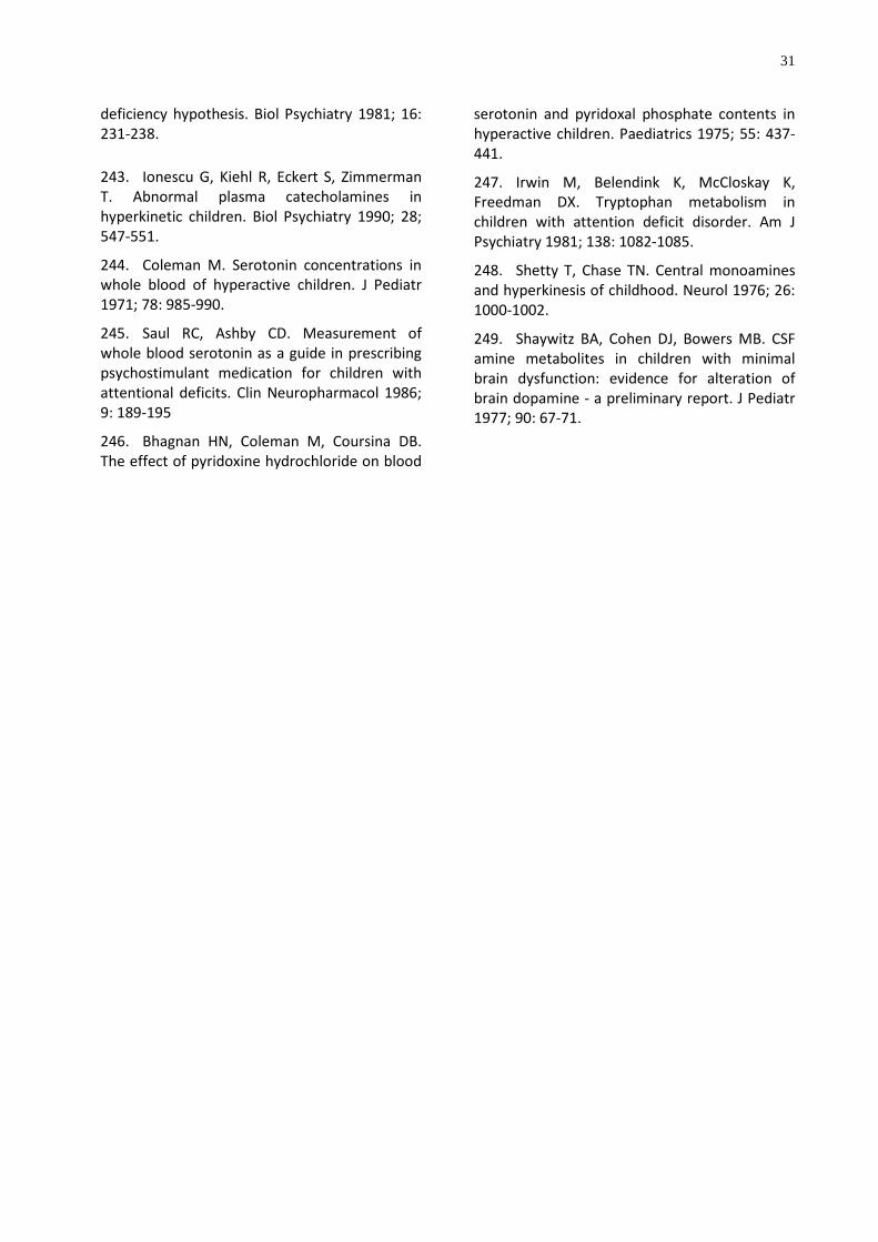
31
deficiency hypothesis. Biol Psychiatry 1981; 16:
231-238.
243. Ionescu G, Kiehl R, Eckert S, Zimmerman
T. Abnormal plasma catecholamines in
hyperkinetic children. Biol Psychiatry 1990; 28;
547-551.
244. Coleman M. Serotonin concentrations in
whole blood of hyperactive children. J Pediatr
1971; 78: 985-990.
245. Saul RC, Ashby CD. Measurement of
whole blood serotonin as a guide in prescribing
psychostimulant medication for children with
attentional deficits. Clin Neuropharmacol 1986;
9: 189-195
246. Bhagnan HN, Coleman M, Coursina DB.
The effect of pyridoxine hydrochloride on blood
serotonin and pyridoxal phosphate contents in
hyperactive children. Paediatrics 1975; 55: 437-
441.
247. Irwin M, Belendink K, McCloskay K,
Freedman DX. Tryptophan metabolism in
children with attention deficit disorder. Am J
Psychiatry 1981; 138: 1082-1085.
248. Shetty T, Chase TN. Central monoamines
and hyperkinesis of childhood. Neurol 1976; 26:
1000-1002.
249. Shaywitz BA, Cohen DJ, Bowers MB. CSF
amine metabolites in children with minimal
brain dysfunction: evidence for alteration of
brain dopamine - a preliminary report. J Pediatr
1977; 90: 67-71.


![Selective serotonin reuptake inhibitors [SSRIs] and ... SSRIs SNRIs prevention... · Selective serotonin reuptake inhibitors (SSRIs) and serotonin-norepinephrine ... and tension-type](https://static.fdocuments.in/doc/165x107/5ce01be988c99399558de41a/selective-serotonin-reuptake-inhibitors-ssris-and-ssris-snris-prevention.jpg)