The Role of Laboratory in the Management of Hemophilia · The Role of Laboratory in the Management...
48
The Role of Laboratory in the Management of Hemophilia Rahajuningsih D. Setiabudy Departemen Patologi Klinik FKUI-RSCM Jakarta Seminar Pediatric Laboratory 29 Juli 2017
Transcript of The Role of Laboratory in the Management of Hemophilia · The Role of Laboratory in the Management...
The Role of Laboratory
in the Management of
Hemophilia
Rahajuningsih D. Setiabudy
Departemen Patologi Klinik
FKUI-RSCM
JakartaSeminar Pediatric Laboratory 29 Juli 2017
Hemophilia
▪ The most frequent hereditary coagulation disorders
▪ Frequency 1 in 10 000 births
▪ Not influenced by ethnicity, geographic, socio economic
▪ Hemophilia A is caused by deficiency of F VIII and hemophilia B by deficiency of F IX
▪ X linked recessive male are more affected, female as carrier


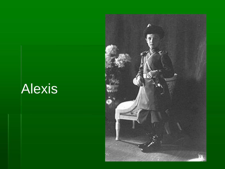
Alexis
Hemophilia in Indonesia
▪ Based on the population of Indonesia around 200 millions it is estimated that the number of PWH in Indonesia 20 000
▪ In facts only around 1800 PWH have been registered
▪ Reasons: under diagnosed due to many PWH died before being diagnosed or moderate and mild hemophilia in the society have not been detected
Clinical manifestation
▪ Easy bruising in early childhood
▪ Spontaneous bleeding into joint and soft tissue
▪ Excessive bleeding following trauma or surgery
▪ Patients with mild hemophilia may not have excessive bleeding unless they have trauma or surgery
▪ Delayed bleeding
Factor VIII and F IX are not required for platelet plug formation
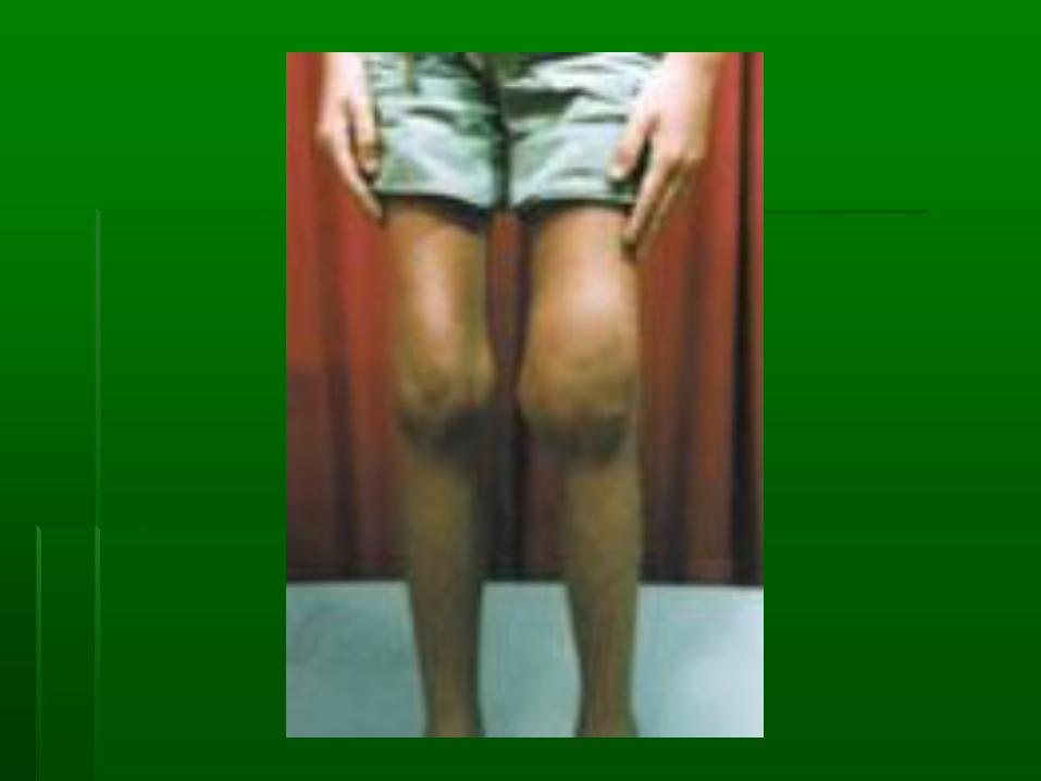
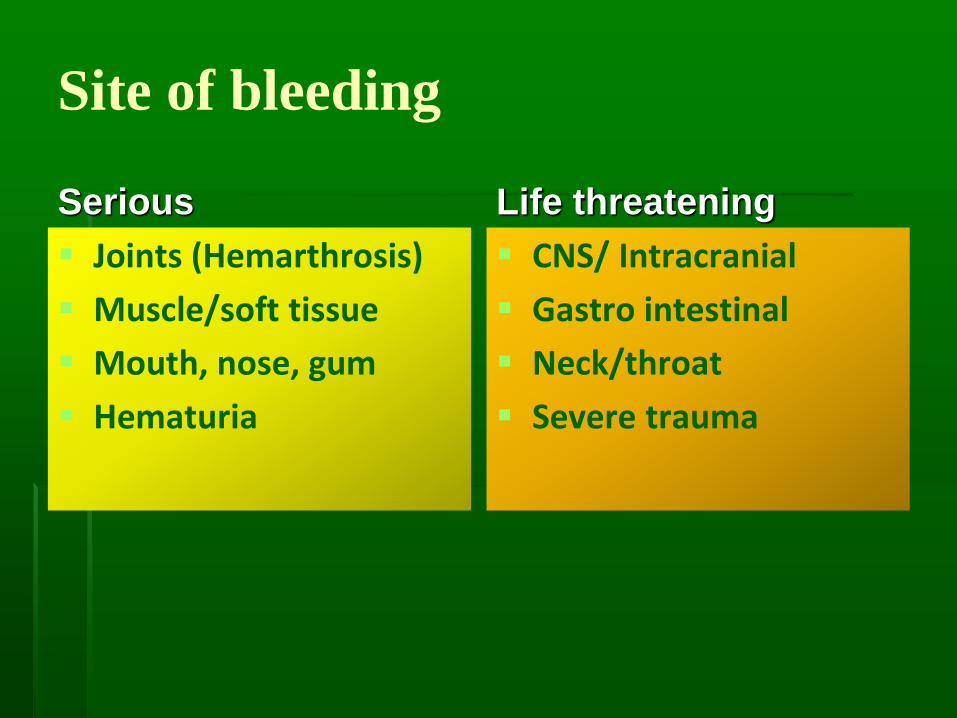
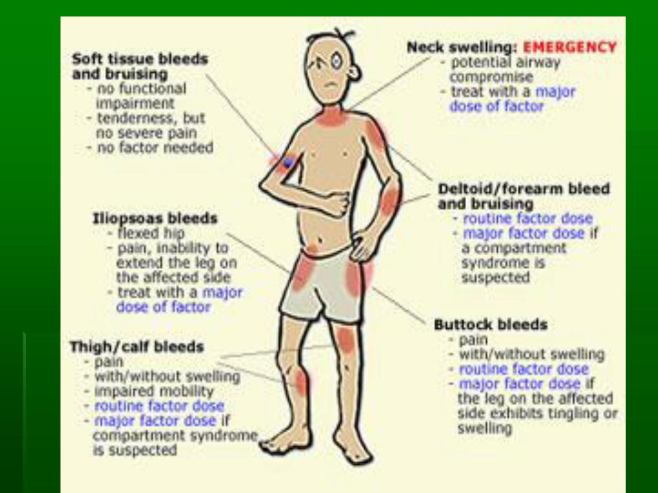
Site of bleeding
Serious
▪ Joints (Hemarthrosis)
▪ Muscle/soft tissue
▪ Mouth, nose, gum
▪ Hematuria
Life threatening
▪ CNS/ Intracranial
▪ Gastro intestinal
▪ Neck/throat
▪ Severe trauma

Severity of bleeding in hemophilia
Severity Clotting factor level% activity (IU/mL)
Bleedingepisodes
Severe 1% (<0.001) Spontaneous bleeding predominantly in joints and muscles
Moderate 1% – 5% (0.01 -0.05) Occasional spontaneous bleeding. Severe bleedingafter trauma or surgery
Mild 5% – 40%(0.05- 0.40) Severe bleeding after major trauma or surgery
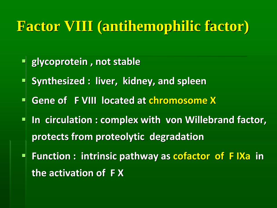
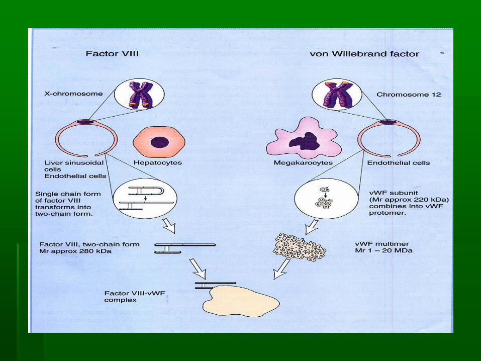
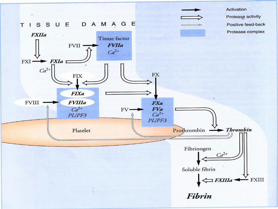
Factor VIII (antihemophilic factor)
▪ glycoprotein , not stable
▪ Synthesized : liver, kidney, and spleen
▪ Gene of F VIII located at chromosome X
▪ In circulation : complex with von Willebrand factor,
protects from proteolytic degradation
▪ Function : intrinsic pathway as cofactor of F IXa in
the activation of F X
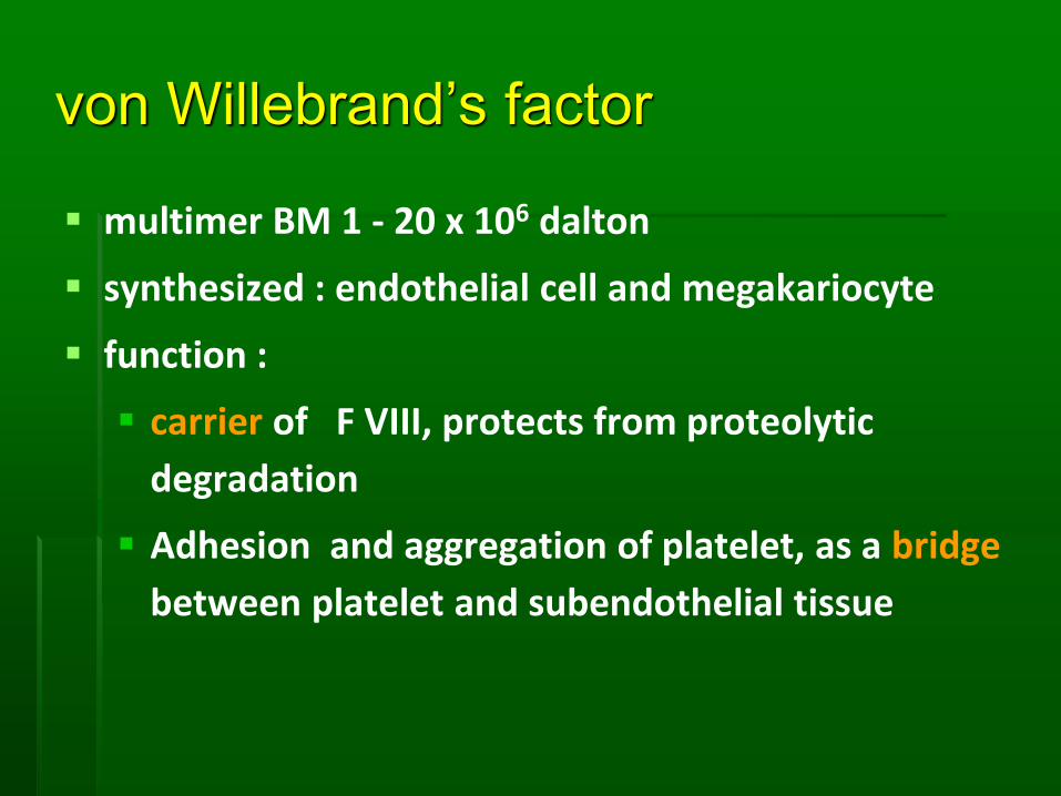
von Willebrand’s factor
▪ multimer BM 1 - 20 x 106 dalton
▪ synthesized : endothelial cell and megakariocyte
▪ function :
▪ carrier of F VIII, protects from proteolytic
degradation
▪ Adhesion and aggregation of platelet, as a bridge
between platelet and subendothelial tissue
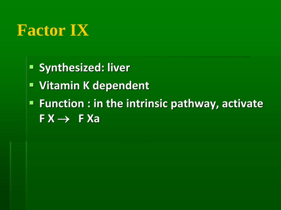
Factor IX
▪ Synthesized: liver
▪ Vitamin K dependent
▪ Function : in the intrinsic pathway, activate F X F Xa
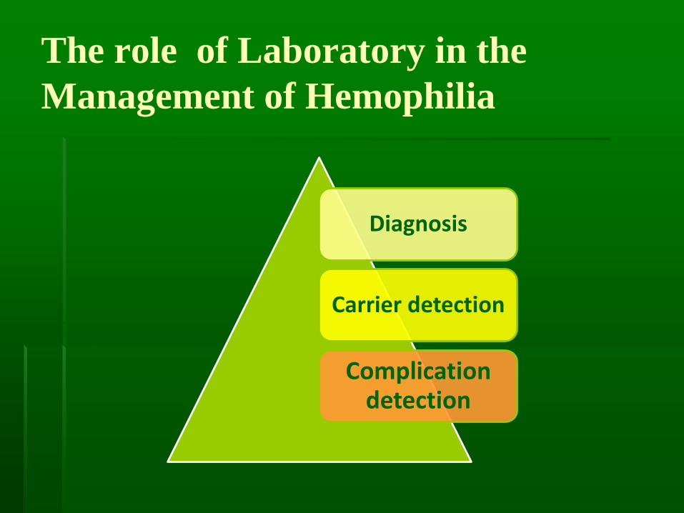
The role of Laboratory in the
Management of Hemophilia
Diagnosis
Carrier detection
Complication detection
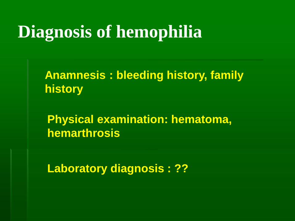
Diagnosis of hemophilia
Anamnesis : bleeding history, family
history
Physical examination: hematoma,
hemarthrosis
Laboratory diagnosis : ??
Laboratory diagnosis of hemophilia
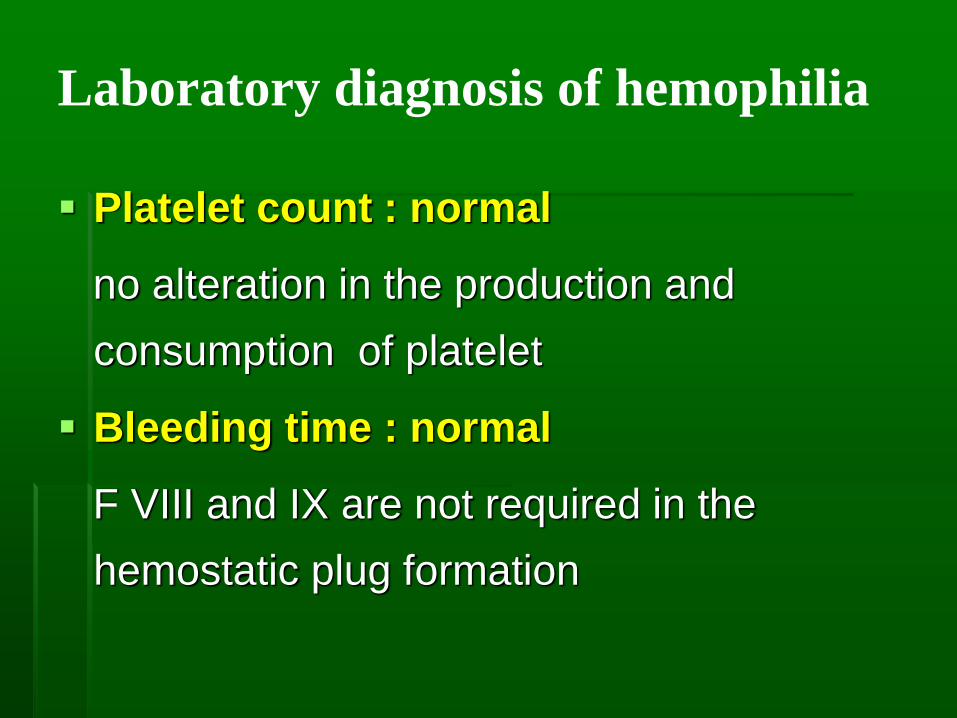
▪ Platelet count : normal
no alteration in the production and
consumption of platelet
▪ Bleeding time : normal
F VIII and IX are not required in the
hemostatic plug formation
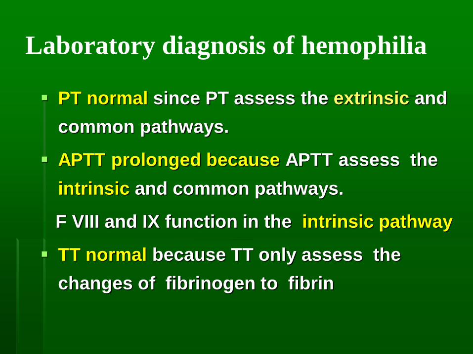
Laboratory diagnosis of hemophilia
▪ PT normal since PT assess the extrinsic and
common pathways.
▪ APTT prolonged because APTT assess the
intrinsic and common pathways.
F VIII and IX function in the intrinsic pathway
▪ TT normal because TT only assess the
changes of fibrinogen to fibrin
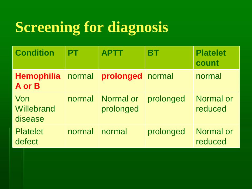
Screening for diagnosis
Condition PT APTT BT Platelet
count
Hemophilia
A or B
normal prolonged normal normal
Von
Willebrand
disease
normal Normal or
prolonged
prolonged Normal or
reduced
Platelet
defect
normal normal prolonged Normal or
reduced
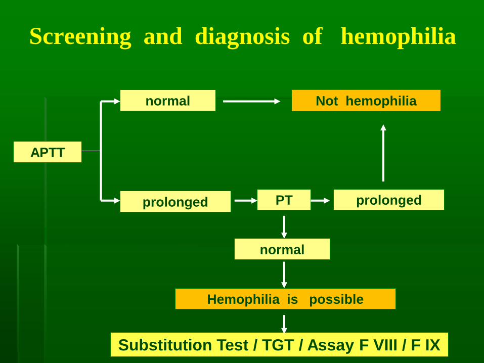
Screening and diagnosis of hemophilia
APTT
prolonged
normal Not hemophilia
PT prolonged
normal
Hemophilia is possible
Substitution Test / TGT / Assay F VIII / F IX
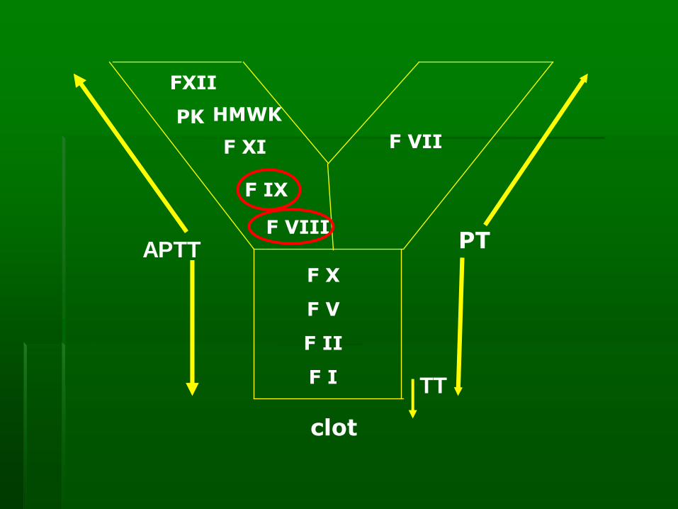
F VII
FXII
PK HMWK
F XI
F IX
F VIII
F X
F V
F II
F I
PT
clot
APTT
TT
Diagnosis of Hemophilia and
DD/ hemophilia A or B
▪ Thromboplastin Generation Time
▪ APTT substitution test
Based on the difference between F VIII and F IX
properties.
F VIII is consumed during coagulation process
F VIII is absent in serum.
F IX is vitamin K dependent factors, is adsorbed
by Ba(SO4) or Al (OH)3 F IX is absent in adsorbed
plasma
▪ Factor VIII/IX Assay
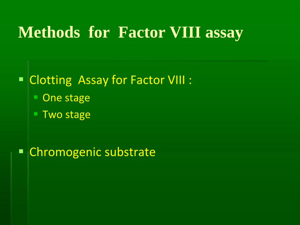
Methods for Factor VIII assay
▪ Clotting Assay for Factor VIII :
▪ One stage
▪ Two stage
▪ Chromogenic substrate
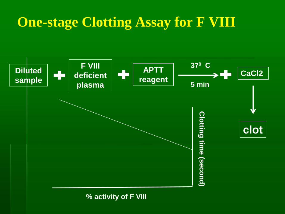
One-stage Clotting Assay for F VIII
Diluted
sample
F VIII
deficient
plasma
APTT
reagent
370 C
clot
5 min
% activity of F VIII
Clo
tting
time
(se
co
nd
)
CaCl2

Two-stage Clotting Assay for Factor VIII
Adsorbed
patient
plasma or
standard
1st stage
F X, activated F IX,
Phospholipid, Ca,
F V in excess
2nd
stage
Conversion F X to activated F X
Clot formation
Source of prothrombin
and fibrinogenClot
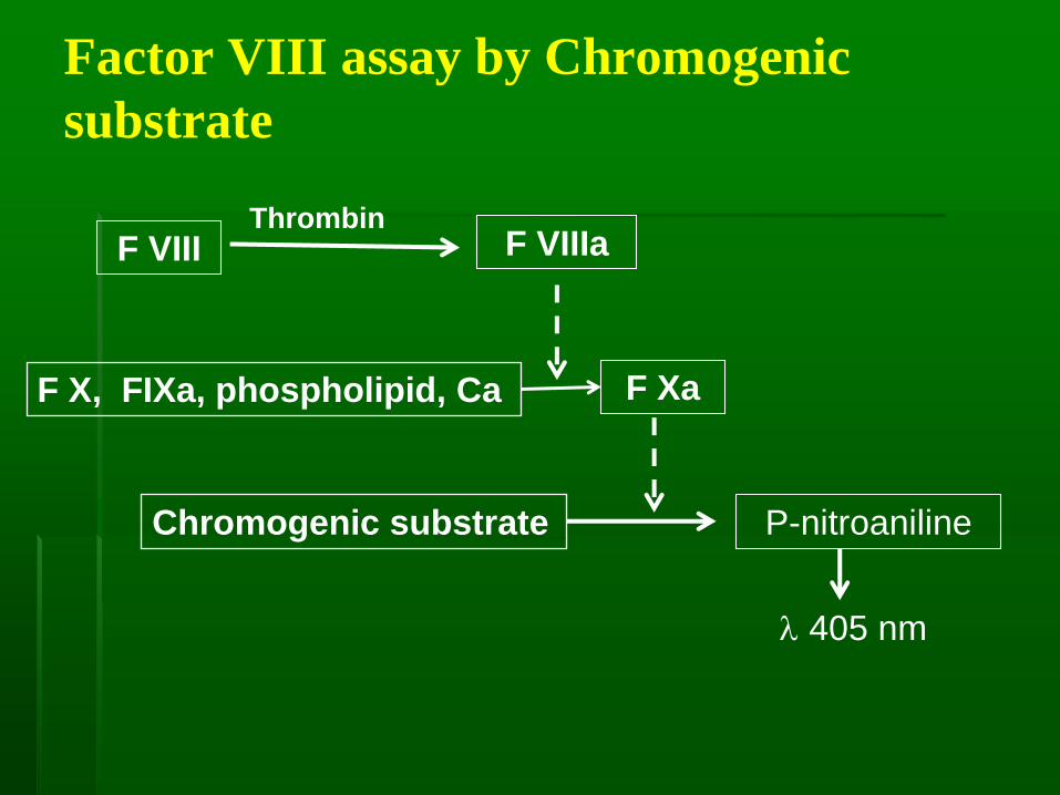
Factor VIII assay by Chromogenic
substrate
F VIIIThrombin
F VIIIa
F X, FIXa, phospholipid, Ca F Xa
Chromogenic substrate P-nitroaniline
405 nm
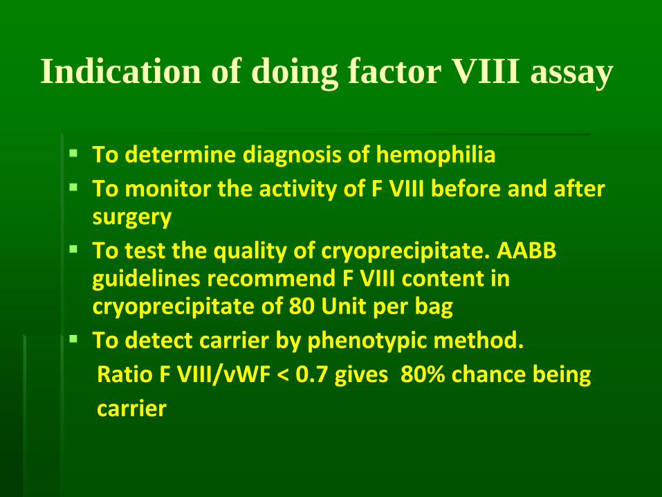
Indication of doing factor VIII assay
▪ To determine diagnosis of hemophilia
▪ To monitor the activity of F VIII before and after surgery
▪ To test the quality of cryoprecipitate. AABB guidelines recommend F VIII content in cryoprecipitate of 80 Unit per bag
▪ To detect carrier by phenotypic method.
Ratio F VIII/vWF < 0.7 gives 80% chance being
carrier
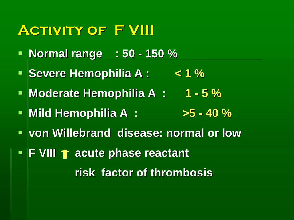
Activity of F VIII
▪ Normal range : 50 - 150 %
▪ Severe Hemophilia A : < 1 %
▪ Moderate Hemophilia A : 1 - 5 %
▪ Mild Hemophilia A : >5 - 40 %
▪ von Willebrand disease: normal or low
▪ F VIII acute phase reactant
risk factor of thrombosis

Activity of F IX
▪ Normal range : 50 - 150 %
▪ Severe Hemophilia B : < 1 %
▪ Moderate Hemophilia B : 1 - 5 %
▪ Mild Hemophilia B : >5 - 40 %
Low :
deficiency of vit. K
Vitamin K antagonist
liver disease
bile obstruction
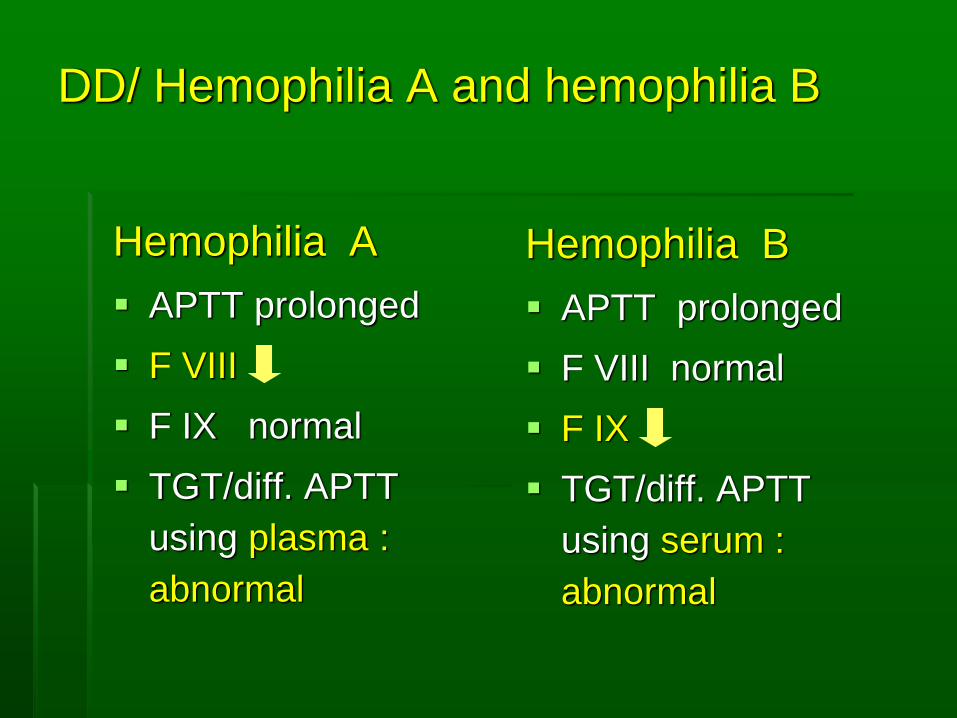
DD/ Hemophilia A and hemophilia B
Hemophilia A
▪ APTT prolonged
▪ F VIII
▪ F IX normal
▪ TGT/diff. APTT
using plasma :
abnormal
Hemophilia B
▪ APTT prolonged
▪ F VIII normal
▪ F IX
▪ TGT/diff. APTT
using serum :
abnormal

DD/ Hemofilia A dan von Willebrand’s d.
Hemofilia A
▪ F VIII
▪ Bleeding time : N
▪ vWF level : N
▪ vWF : Ristosetin
cofactor : N
Von Willebrand’s d.
▪ F VIII N /
▪ Bleeding time
prolonged
▪ vWF level
▪ vWF : Ristosetin
cofactor
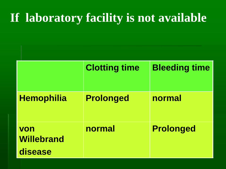
If laboratory facility is not available
Clotting time Bleeding time
Hemophilia Prolonged normal
von
Willebrand
disease
normal Prolonged
Carrier detection
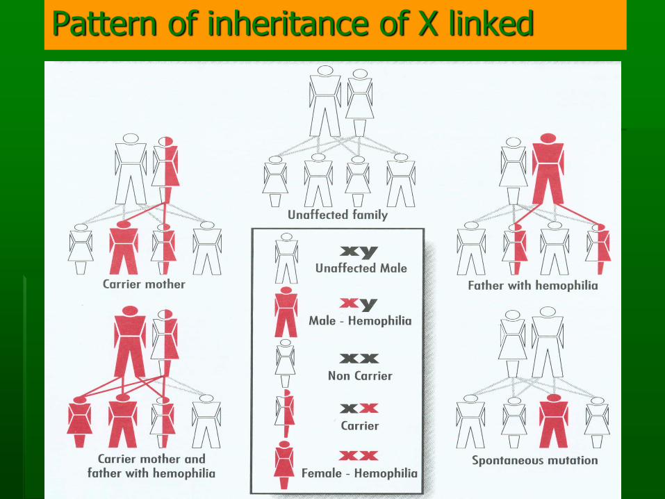
Pattern of inheritance of X linked
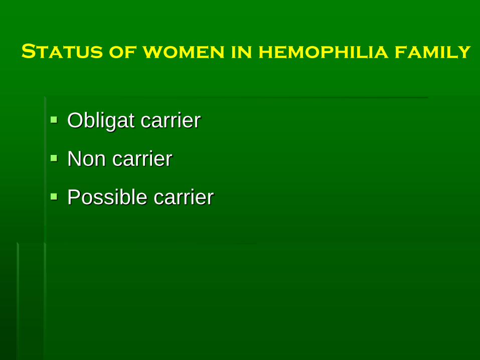
Status of women in hemophilia family
▪ Obligat carrier
▪ Non carrier
▪ Possible carrier
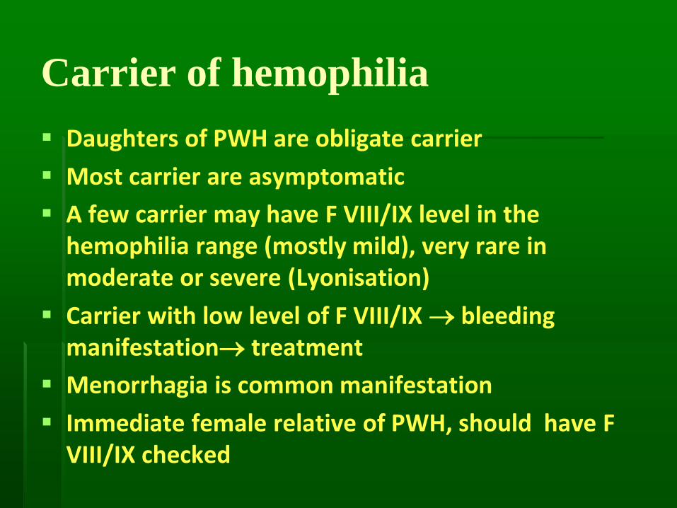
Carrier of hemophilia
▪ Daughters of PWH are obligate carrier
▪ Most carrier are asymptomatic
▪ A few carrier may have F VIII/IX level in the hemophilia range (mostly mild), very rare in moderate or severe (Lyonisation)
▪ Carrier with low level of F VIII/IX bleeding manifestation treatment
▪ Menorrhagia is common manifestation
▪ Immediate female relative of PWH, should have F VIII/IX checked
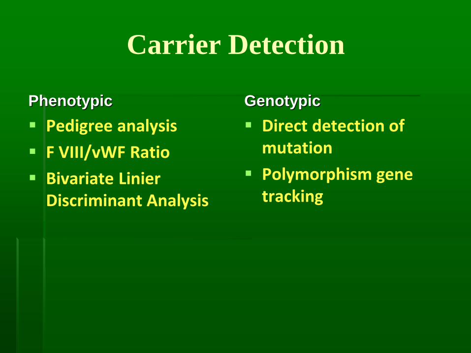
Carrier Detection
Phenotypic
▪ Pedigree analysis
▪ F VIII/vWF Ratio
▪ Bivariate Linier Discriminant Analysis
Genotypic
▪ Direct detection of mutation
▪ Polymorphism gene tracking

Bivariate Linear Discriminant Analysis
Input data from possible carrier
a = age in year
b = blood group group O = 0 Non O = 1
g = vWF Ag in IU/mL
d = VIII in IU/mL
p = genetic probability of carriership
Input data from non carrier reference group
mx = mean of Ln of vWF:Ag level in IU/mL
my = mean of Ln F VIII activity in IU/mL
Calculate for possible carrier
X = ln(g) – mx
Y= ln (d) – my
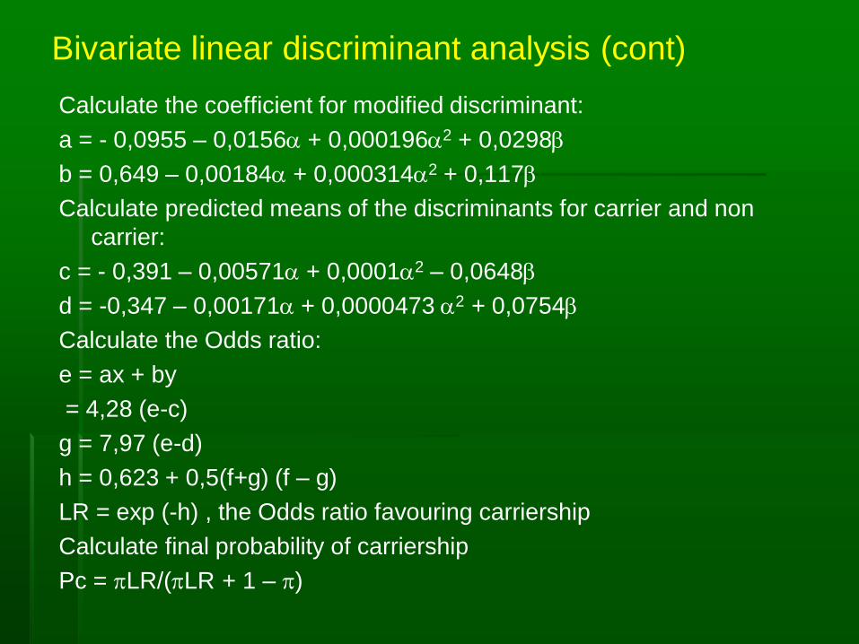
Bivariate linear discriminant analysis (cont)
Calculate the coefficient for modified discriminant:
a = - 0,0955 – 0,0156a + 0,000196a2 + 0,0298b
b = 0,649 – 0,00184a + 0,000314a2 + 0,117b
Calculate predicted means of the discriminants for carrier and non
carrier:
c = - 0,391 – 0,00571a + 0,0001a2 – 0,0648b
d = -0,347 – 0,00171a + 0,0000473 a2 + 0,0754b
Calculate the Odds ratio:
e = ax + by
= 4,28 (e-c)
g = 7,97 (e-d)
h = 0,623 + 0,5(f+g) (f – g)
LR = exp (-h) , the Odds ratio favouring carriership
Calculate final probability of carriership
Pc = pLR/(pLR + 1 – p)
Complication Detection
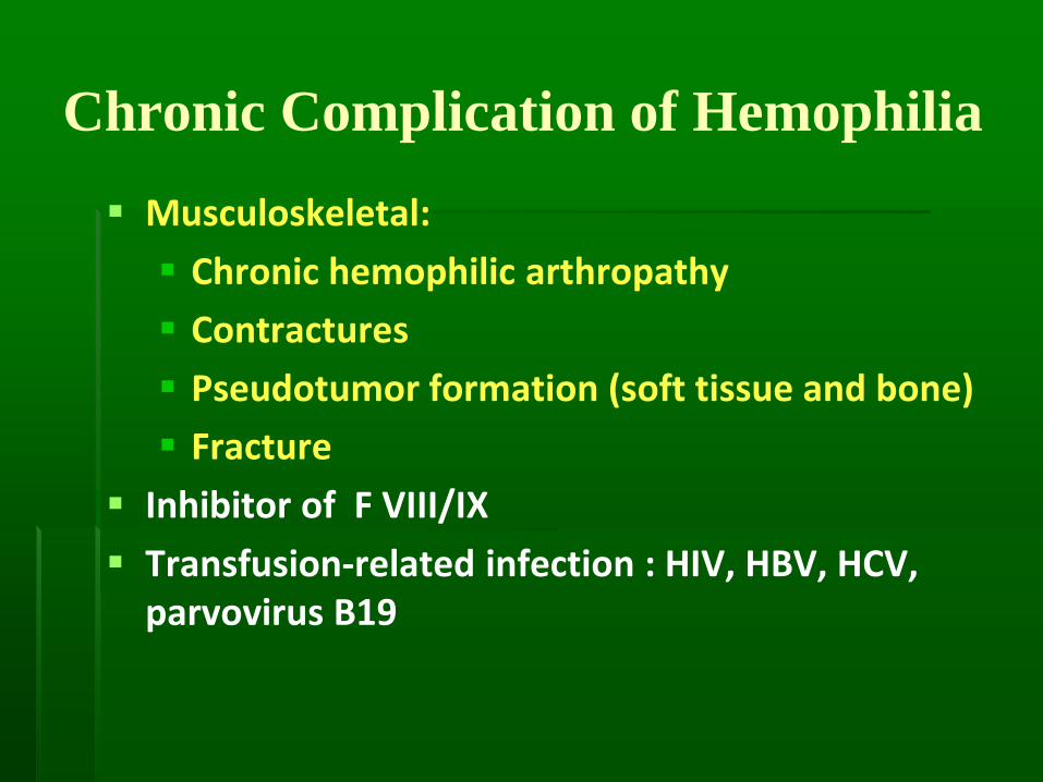
Chronic Complication of Hemophilia
▪ Musculoskeletal:
▪ Chronic hemophilic arthropathy
▪ Contractures
▪ Pseudotumor formation (soft tissue and bone)
▪ Fracture
▪ Inhibitor of F VIII/IX
▪ Transfusion-related infection : HIV, HBV, HCV, parvovirus B19
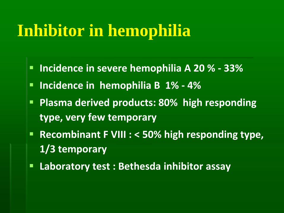
Inhibitor in hemophilia
▪ Incidence in severe hemophilia A 20 % - 33%
▪ Incidence in hemophilia B 1% - 4%
▪ Plasma derived products: 80% high responding
type, very few temporary
▪ Recombinant F VIII : < 50% high responding type,
1/3 temporary
▪ Laboratory test : Bethesda inhibitor assay

Inhibitor F VIII
Patient Plasma + Pool normal
plasmaF VIII def.plasma + pool
normal plasma
Incubate for 2 hours
Perform F VIII assay Perform F VIII assay
Determine residual F VIII
Convert to Bethesda unit
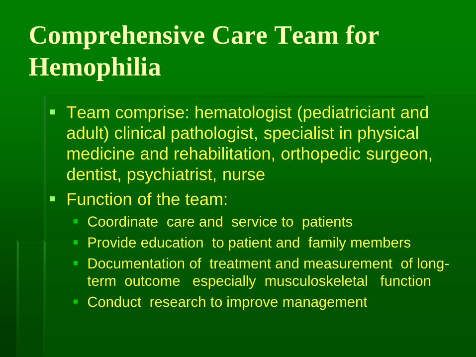
Comprehensive Care Team for
Hemophilia
▪ Team comprise: hematologist (pediatriciant and
adult) clinical pathologist, specialist in physical
medicine and rehabilitation, orthopedic surgeon,
dentist, psychiatrist, nurse
▪ Function of the team:
▪ Coordinate care and service to patients
▪ Provide education to patient and family members
▪ Documentation of treatment and measurement of long-
term outcome especially musculoskeletal function
▪ Conduct research to improve management

Management for Hemophilia in
Indonesia
▪ Since 1997 Comprehensive Care Team for
Hemophilia was established in Dr. Cipto
Mangunkusumo Hospital, Jakarta
▪ Meeting to solve the problem in the patient
management, donation from WFH
▪ Surgery: TKR, implant in hip surgery
▪ We also have Indonesian Hemophilia Society:
PWH, family, doctors, nurse, social workers.

Thank you