The Role of Gap Junctions in Cellular...
132
The Role of Gap Junctions in Cellular Migration by Bado Hewa DeFranco Bachelor’s of Science, Biology and Philosophy, Benedictine University 1996 Master’s of Science, Biology, University of Illinois, 2002 Submitted to the Graduate Faculty of School of Medicine: Department of Cell Biology and Physiology in partial fulfillment of the requirements for the degree of Doctor of Philosophy University of Pittsburgh 2009
-
Upload
phamnguyet -
Category
Documents
-
view
215 -
download
0
Transcript of The Role of Gap Junctions in Cellular...

The Role of Gap Junctions in Cellular Migration
by
Bado Hewa DeFranco
Bachelor’s of Science, Biology and Philosophy, Benedictine University 1996
Master’s of Science, Biology, University of Illinois, 2002
Submitted to the Graduate Faculty of
School of Medicine: Department of Cell Biology and Physiology in partial fulfillment
of the requirements for the degree of
Doctor of Philosophy
University of Pittsburgh
2009

ii
UNIVERSITY OF PITTSBURGH
School of Medicine: Department of Cell Biology and Physiology
This dissertation was presented
by
Bado Hewa DeFranco
It was defended on
September 3, 2009
and approved by
Peter Drain, PhD, Cell Biology and Physiology
David J. Hackam, MD/PhD, Cell Biology and Physiology
Carl Lagenaur, PhD, Neurology
Nirmala Sundar Raj, PhD, Cell Biology and Physiology
Allan Zhao, PhD, Cell Biology and Physiology
Dissertation Advisor: Sandra Murray, PhD, Cell Biology and Physiology

iii
Copyright © by Bado Hewa DeFranco
2009

iv
The body of work presented here focuses on characterizing the role that gap junction intercellular channels play in regulating cellular migration. Cell migration is a ubiquitous process that is required for embryonic development and for maintaining the integrity of tissues and organs. Yet, the status of gap junction channels, with regard to structure and function, in migrating cells is not completely understood. We hypothesized that, “Gap junction channels, as mediators of intercellular communication, play a role in cell migration,” and have investigated and characterized gap junctions in migrating cells. Accordingly, the aims of this dissertation were: (i) to characterize gap junctions and their function during migration, (ii) to determine the effect of altering the status of gap junction expression on cell migration and to (iii) characterize the mechanism of gap junction internalization in migrating cells.
With combined molecular and imaging approaches we have demonstrated that in cells migrating as a sheet, gap junction plaque structures are retained on the plasma membrane surface; that gap junction communication is continuous in migrating cells and that interrupting this communication with connexin 43 specific mimetic peptides reduces migration. We have established a system in a human adrenal cortical cell line (SW13) wherein we significantly reduce gap junction protein expression with siRNA and show that cellular migration is inhibited. We have also demonstrated that gap junction plaque size and orientation are modified during migration. We also discovered that sometimes, migrating cells will spontaneously detach from one another at cellular processes and that during this event the gap junction plaques are internalized. Analysis of gap junction plaque internalization in migrating cells revealed that clathrin and several adaptor proteins associate with surface gap junctions and cytoplasmic annular gap junction structures and possibly regulate the unique mechanism of gap junction plaque removal from the plasma membrane. Within the field of gap junction research this work expands our understanding of gap junctions in living cells as dynamic structures that may play a key role in coordinating the migration of entire cell populations.
The Role of Gap Junctions in Cellular Migration
Bado Hewa DeFranco, MS.
University of Pittsburgh, 2009

v
TABLE OF CONTENTS
ACKNOWLEDGEMENTS ....................................................................................................... XI
1.0 INTRODUCTION ........................................................................................................ 1
1.1 GAP JUNCTION CHANNELS .......................................................................... 2
1.2 GAP JUNCTION CHANNEL ASSEMBLY ..................................................... 5
1.3 GAP JUNCTION PLAQUE FORMATION ..................................................... 7
1.4 GAP JUNCTION CHANNEL GATING ........................................................... 9
1.5 GAP JUNCTION REMOVAL ......................................................................... 10
1.6 CELLULAR MIGRATION .............................................................................. 11
1.7 THE ROLE OF GAP JUNCTIONS IN CELL MIGRATION ...................... 14
1.8 GAP JUNCTIONS AND MIGRATION DURING EMBRYONIC
DEVELOPMENT ............................................................................................................... 17
1.9 GAP JUNCTION EXPRESSION AND METASTATIC CELLULAR
MIGRATION ...................................................................................................................... 18
1.10 THE ROLE OF GAP JUNCTIONS IN CELL MIGRATION DURING THE
INFLAMMATORY IMMUNE RESPONSE ................................................................... 19
1.11 GAP JUNCTIONS AND WOUND HEALING .............................................. 20
1.12 SUMMARY ........................................................................................................ 21
2.0 METHODS ................................................................................................................. 24

vi
2.1 CELL CULTURES ............................................................................................ 24
2.2 TRANSFECTION.............................................................................................. 25
2.3 IMMUNOCYTOCHEMISTRY ....................................................................... 26
2.4 WESTERN BLOT ............................................................................................. 27
2.5 MIGRATION ANALYSIS ................................................................................ 28
2.6 TIME-LAPSE IMAGING................................................................................. 29
2.7 FUNCTIONAL GAP JUNCTION COMMUNICATION ASSAYS ............. 30
2.7.1 Lucifer yellow functional dye-coupling assay ............................................. 30
2.7.2 Fluorescence Recovery after Photobleaching (FRAP) ............................... 30
2.8 TRANSMISSION ELECTRON MICROSCOPY .......................................... 32
2.9 QUANTUM DOT ............................................................................................... 32
2.10 MIMETIC PEPTIDES ...................................................................................... 33
2.11 CONNEXIN 43 SIRNA KNOCKDOWN ........................................................ 33
2.12 STATISTICAL ANALYSIS ............................................................................. 34
3.0 MIGRATING CELLS RETAIN GAP JUNCTION PLAQUE STRUCTURE
AND FUNCTION........................................................................................................................ 35
3.1 ABSTRACT........................................................................................................ 35
3.2 INTRODUCTION ............................................................................................. 36
3.3 RESULTS ........................................................................................................... 37
3.3.1 Characterization of Cell Migration .............................................................. 37
3.3.2 Monitoring Cx43-GFP in Transfected Cells ............................................... 38
3.3.3 Characterization of Gap Junction Function ............................................... 39
3.4 DISCUSSION ..................................................................................................... 51

vii
4.0 ALTERED CONNEXIN 43 EXPRESSION AND GAP JUNCTION
COMMUNICATION REDUCS CELL MIGRATION ........................................................... 55
4.1 ABSTRACT........................................................................................................ 55
4.2 INTRODUCTION ............................................................................................. 56
4.3 RESULTS ........................................................................................................... 57
4.3.1 Migration in Cells with Varied Amounts of Connexin 43 Expression ..... 57
4.3.2 Altered Gap Junctions and Cell Migration ................................................. 58
4.3.3 Altered Gap Junction Communication and Cell Migration ...................... 59
4.3.4 Increasing Gap Junctions and Cellular Migration ..................................... 59
4.4 DISCUSSION ..................................................................................................... 72
5.0 THE MECHANISM OF GAP JUNCTION CHANNEL INTERNALIZATION 77
5.1 ABSTRACT........................................................................................................ 77
5.2 INTRODUCTION ............................................................................................. 78
5.3 RESULTS ........................................................................................................... 80
5.3.1 Gap Junction Dynamics during Process Detachment ................................ 80
5.3.2 Immunocytochemistry Analysis of Gap Junction Internalization ............ 81
5.3.3 Transmission Electron Microscopy Analysis of Gap Junction
Internalization ............................................................................................................ 82
5.4 DISCUSSION ..................................................................................................... 90
6.0 DISCUSSION ............................................................................................................. 94
6.1 FUTURE STUDIES ........................................................................................... 97
6.2 SUMMARY ........................................................................................................ 99
APPENDIX A ............................................................................................................................ 102

viii
APPENDIX B ............................................................................................................................ 104
BIBLIOGRAPHY ..................................................................................................................... 105

ix
LIST OF FIGURES
Figure 1 Model of Gap Junction Formation and Turnover ............................................................. 3
Figure 2. Model of Cell-Cell Communication ................................................................................ 6
Figure 3. The steps of cell migration. ........................................................................................... 13
Figure 4. Migration and Gap Junctions ......................................................................................... 16
Figure 5. Characterization of SW-13 Migration ........................................................................... 41
Figure 6. Connexin 43 gap junctions in SW-13 cells. .................................................................. 42
Figure 7. Fluoresecence microscopy of SW-13 cells. ................................................................... 43
Figure 8. The average number of gap junction plaques per cell. .................................................. 44
Figure 9. Average Plaque Size ...................................................................................................... 45
Figure 10. Cx43-GFP expression in cells. .................................................................................... 46
Figure 11. Gap Junction Dynamics during SW-13 Migration. ..................................................... 47
Figure 12. Functional coupling during migration ......................................................................... 48
Figure 13. Fluorescence Recovery after Photobleaching (FRAP) in SW-13 cells. ...................... 49
Figure 14. Gap junction communication during cell migration. ................................................... 50
Figure 15. Characterization of SBAC cell migration. ................................................................... 61
Figure 16. Characterization of Y-1 cell Migration. ...................................................................... 63
Figure 17. Connexin 43 Knockdown and Gap Junction Expression. ........................................... 64

x
Figure 18. Functional coupling in connexin 43 reduced cells. ..................................................... 65
Figure 19. Fluorescent dye recovery following photobleaching. ................................................. 66
Figure 20. Comparison of percent change in cell migration. ........................................................ 67
Figure 21. Functional dye coupling assessed in cells with gap junction inhibition ...................... 68
Figure 22. Comparison of migration in cells with reduced communication ................................. 69
Figure 23. Migration in cells with increased gap junctions .......................................................... 71
Figure 24. Gap Junction internalization occurs during cell process detachment. ......................... 83
Figure 25. Colocalization of connexin 43 gap junctions and clathrin .......................................... 84
Figure 26. Subcellular localization of gap junctions and the clathrin adaptor protein AP-2 ........ 85
Figure 27. Subcellular localization of connexin 43 gap junctions and Dab2 ............................... 86
Figure 28. Subcellular colocalization of gap junctions and Epsin ................................................ 87
Figure 29. Subcellular colocalization of gap junctions and clathrin assessed with transmission
electron microscopy ...................................................................................................................... 88
Figure 30. Quantum dot transmission immunoelectron microscopy of SW-13 cells. .................. 89
Figure 31. Model of clathrin mediated gap junction plaque endoexocytosis. .............................. 93
Figure 32. Gap junctions and intercellular communication are retained as cells migrate .......... 101

xi
ACKNOWLEDGEMENTS
I would like to first thank God who has sustained me and is the source of my faith. To my
advisor, Dr. Sandra A. Murray, thank you for the opportunity to explore and do exciting research
in your lab, as well for sharing your wisdom and knowledge with me throughout my graduate
career. To my committee members: Dr. Peter Drain (chairperson), Dr. David Hackam, Dr. Carl
Lagenaur, Dr. Nirmala SundarRaj and Dr. Allan Zhao, your knowledge and support are greatly
appreciated. I would like to Beth M. Nickel for sharing her knowledge and talent, you are truly
valued. To the Cell Biology and Physiology department staff members, past and present: Rene
Giles, Carol Staley, Michelle Dobransky, Susan Conway and Cindy Pokora; and to the graduate
office administrators: Cindy Duffy, Veronica Cardamone and Sandra Honick thank you all for
your patience and support over the years. I would like to thank the pioneers of gap junction
research whose efforts from the mid 1960s through the mid 1980s paved the way for the next
generation of gap junction scientists. I would like to thank my family and friends who listened
and were a true source of encouragement and help throughout this process. To my parents,
William and Glenavere Bigham, for being wonderful parents and believing in me. Finally, to my
husband Bryan, a sincere thanks for your constant support, enthusiasm, understanding and most
of all your continuous love.

1
1.0 INTRODUCTION
The fundamental survival of multicellular organisms requires that tissues respond to
specific signals as integrated systems (Peracchia and Girsch, 1985). Much of this cooperation is
attributed to intercellular gap junction communication channels that provide for the mutual
exchange of electrical and chemical information between adjacent cells (Subak-Sharpe et al.,
1969; Lo and Gilula, 1979; McNutt and Weinstein 1970). The majority of cells that comprise
the tissues of our bodies are able to communicate directly with one another via gap junction
channels (Kumar, 1999). A number of these cell populations are also capable of migration, an
event that is thought to rely on the ability of individual cells, in a group, to communicate with
one another (Pepper and Meda, 1992; Matic et al., 1997; Nadarajah et al., 1997; Saitoh et al.,
1997, Civitelli et al., 1998, Winterhager et al., 1999; Ashton et al., 1999; Cina et al., 2007;
Leaphart et al., 2007). According to the current migration model as proposed by the Horwitz
group, the coordination of events for a single cell requires converging intracellular signaling
pathways that regulate cytoskeletal turnover along with contraction/retraction processes (Horwitz
and Parsons, 1999; Ridley et al., 2003, Horwitz and Webb, 2003; Vincente-Mazanares et al.,
2005). Integrating these responses such that an entire population migrates in a regulated and
coordinated manner, that is biologically relevant, would likely require direct cell-cell
communication via gap junction intercellular channels. Based on this model it would be
assumed that gap junctions would either be retained as cells migrate, or that they are

2
disassembled and reassembled in migrating populations such that information could be
transmitted between cells. Yet, issues regarding the status of gap junction communication
channels in cell populations that migrate in a cluster or sheet-like manner have not been
addressed. The hypothesis, “gap junctions and intercellular communication play a pivotal role
in migration,” was tested. Accordingly, the aims of this dissertation are: (i) to characterize gap
junctions and their function in migrating cells, (ii) to determine the effect of altering the status of
gap junction expression on cell migration and to (iii) characterize the mechanism of gap junction
internalization in migrating cells.
1.1 GAP JUNCTION CHANNELS
To better understand intercellular communication between migrating cells it is necessary to
discuss gap junction channels in detail. Gap junction channels are specialized membrane pores
that allow mutual and direct exchange of electrical and chemical information between adjacent
cells in tissues and organs (Loewenstein, 1966; Gilula et al., 1971; Sheridan, 1971). Each
channel is comprised of 12 connexin monomers (Hand and Gobel, 1972). Connexins are four-
pass transmembrane proteins that contain two extracellular loops and cytoplasmically localized
N- and C- termini with amino acid sequences that are targeted for phosphorylation and other
regulatory enzymes (Milks et al., 1988; Musil et al., 1990; Yeager and Gilula, 1992; Torok et al.,
1997) (Fig. 1).
There are twenty-one connexin proteins that have been identified in mammals which vary
by their molecular weight (e.g. Cx32, Cx43, Cx50 etc.) (Kumar and Gilula, 1996 rev. Yeager and
Harris, 2007 rev.). Their expression is tissue specific with most cells expressing more than a

3
Figure 1 Model of Gap Junction Formation and Turnover
Monomeric connexin proteins (shown in the lower right-hand) oligomerize to form hemichannels/connexons.
Following assembly in the Golgi the connexons are inserted into the plasma membrane. Gap junction
channels are formed when connexons of neighboring cells come together or ‘dock’ and form an aqueous pore
that spans across the two nanometers of extracellular space that now separates the plasma membranes. Gap
junction channels aggregate to form ‘plaques’ and are hereafter capable of intercellular communication. An
entire or portion of a plaque is internalized form an annular gap junction which will ultimately be targeted
for degradation.
C-terminus
Cytoplasm
ECM
N-terminus
Connexin (Cx)
ECM
Cytoplasm
Gap Junction Channel Pore
Annular Gap Junction
Golgi complex
Gap Junction Plaque
Cell 1
Cell 2
Cell 1 2 nm
Connexon (Hemichannel)
Cell 2

4
single connexin isotype (Lagree et al., 2003). Most cell types initially express the Cx43 isoform
however; following development hepatocytes, for example, express only Cx32 and Cx26
whereas cardiomyocytes express only Cx43 and Cx26 isotypes (Paul, 1986; Zhang and
Nicholson, 1989; Beyer et al., 1987; Dermietzel et al., 1989). Gap junctions are formed when
connexins are initially assembled into cylindrical, hexameric connexons (hemichannels) (Musil
and Goodenough, 1993) (Fig. 1). It is the characteristics of the individual connexin isotypes that
contributes to the unique specificity of the connexon pore (e.g. size, charge and permeability)
(Oyamada et al., 2005).
Connexons are either: homomeric (same connexin monomers) or heteromeric (different
connexin monomers) and gap junctions may be homocellular (both cells contribute connexons
that are identical) or heterocellular (both cells contribute connexons that are different) in nature
(Diez et al., 1999; Elfgang et al., 1995; Sosinsky, 1995; Larsen, 1977). A single connexon
loaded onto the plasma membrane of a cell will “dock” via non-covalent interaction with its
hemichannel counterpart on the apposing cell membrane and form a single dodecameric gap
junction channel (Foote et al, 1998; Perkins et al, 1998; Yeager et al, 1998) (Fig. 1). The newly
formed intercellular channel contains a hydrophilic central pore and spans across a 2-3
nanometer “gap” that separates the apposing plasma membranes, hence the term “gap junction”
(Revel and Karnovsky, 1967; Goodenough and Revel, 1971) (Fig. 1). Once on the plasma
membrane, gap junction channels form aggregates or ‘plaques’ which are composed of hundreds
or thousands of individual channels and extend several micrometers in length (Goodenough and
Revel, 1970, McNutt and Weinstein, 1970) (Fig. 1). Gap junction channels are typically only
thought to function in cell-cell communication once aggregated into plaques, and the central pore
is virtually leak proof and can accommodate a range of transiting molecules including ions,
5
secondary messengers, amino acids, nucleic acids etc. (Simpson et al., 1977; Lowenstein, 1978;
Saez et al., 1989; Kumar and Gilula, 1996 rev.) (Fig. 1). Incidentally, it has been reported that
connexons/hemichannels may also play a role in molecular transport (Jiang et al., 2007; Krysko
et al., 2005; Bennett et al., 2003). The average turnover for connexin proteins ranges between 1-
5 hours, depending on the cell type (Musil et al., 2005; Fallon and Goodenough, 1981). Factors
that regulate gap junction function, trafficking or dynamics are likely to alter gap junction
channels and thereby affect physiological processes such as cellular migration. The key events
associated with the regulation of gap junction function including: channel assembly, plaque
assembly, channel gating and the removal of the gap junction plaque from the cell surface, will
be discussed in distinct sections.
1.2 GAP JUNCTION CHANNEL ASSEMBLY
The assembly of gap junction channels is a multi-step process, and like many integral
proteins is highly regulated and begins in the biosynthetic pathway (Musil and Goodenough,
1993; Hurtley and Helenius, 1988). Translation of mRNA and subsequent membrane insertion
of connexin monomers occurs in the endoplasmic reticulum (ER) (Falk et al., 1994). The
formation and transport of connexons through the biosynthetic pathway is differentially
regulated and is thought to be based on the cell type (Musil and Goodenough, 1993; Koval et al.,
1997). The oligomerization step, in which six connexins are bound together to form a single

6
Figure 2. Model of Cell-Cell Communication
(A) Small molecules (>1 Kd: e.g. ions, second messengers) transit through gap junction intercellular
communication channels that have formed an aggregate or ‘plaque’ between cell 1 and cell 2. (B)
Transmission Electron Microscopy of a gap junction plaque.
Cell 1
Cell 2
Cell 1
Cell 2 2 nm
Pore
B
IP3 Ca2+ NO- cAMP
A

7
connexon/hemichannel, may take place in the ER or the Golgi apparatus depending on the type
of cell or connexin (Diez et al., 1999; Koval et al., 1997; Kumar et al., 1995; Musil and
Goodenough, 1993; Musil and Goodenough, 1991). The functional association of
phosphorylation with connexins during assembly, has yet to be determined, though it is thought
that phosphorylation may suffice for connexon trafficking through the Golgi or export to the
plasma membrane (Solan and Lampe, 2004). Once assembled, some connexons are thought to
follow the membrane trafficking route through the Golgi, trans-Golgi Network and on to the
plasma membrane where they are inserted (VanSlyke and Musil, 2000; Falk et al., 1997; Wang
et al., 1995) (Fig. 1).
1.3 GAP JUNCTION PLAQUE FORMATION
Gap junction plaques are specialized membrane domains wherein clusters of gap junction
channels function in intercellular communication (Fig. 1). In an important work from the Falk
group, it was demonstrated that plaque formation occurs via the lateral movement of hundreds or
thousands of individual channels on the plasma membrane into a single cluster (Lauf et al.,
2002). The actual regulation of channel aggregation into plaques has been demonstrated to
coincide with physiological processes such as parturition and therefore, may involve hormones
and downstream signaling mechanisms (Garfield, 1980; Risek et al; 1990; Risek et al., 1995).
For example, gap junction formation was increased in smooth muscle (myometrium) cells
following the exposure of uterine tissue to exogenous estrogen and prostaglandins (Garfield et al,
1980). The prostaglandin PGE in particular has been shown to increase intracellular cAMP
levels which are thought to increase gap junction clustering into plaques on the plasma

8
membrane (Wang and Rose, 1995; Brueggemeier et al., 2003; Faucheux et al, 2002).
Interestingly, prostaglandins have also been demonstrated to increase estrogen biosynthesis; the
activation downstream targets of the estrogen receptor includes cAMP and protein kinases (e.g.
PKA) which have been demonstrated to phosphorylate the connexin monomers of gap junction
channels and may be involved in plaque formation (Garfield et al., 1980; Driggers and Segars,
2002). Gap junction clustering is a regulated event that correlates with physiological processes
and formation of a distinct plaque compartment may also be regulated.
The creation of a plaque domain is thought to involve the plasma membrane lipids
components as well as tight junction proteins (Abney et al., 1987; Singh et al., 2005).
Biophysical analyses reveal that the interactions ‘holding’ the plaques together may involve
electrostatic, contact repulsion and lipid mediated forces (Abney et al., 1987). The creation of
this membrane compartment is thought to involve the PDZ domains of tight junction proteins
ZO-1 and 2 and their interaction with the C-terminus of connexin 43 (Singh et al., 2005). It is
thought that ZO-1 and 2 may function by stabilizing the gap junction plaques on the membrane
(Singh et al., 2005). In addition it is has been demonstrated that the α and β catenin proteins may
also play a role in mediating the interaction between ZO-1 and Cx43 during gap junction plaque
assembly specifically, in cardiomyocytes (Wu et al., 2003). Once the plaques are formed, it is
thought that connexons are continuously added to the periphery of existing gap junctions
(Gaietta et al., 2002; Lauf et al., 2002). Once it is part of the plaque, it is thought that the gap
junction channel is capable of opening and closing (gating).
9
1.4 GAP JUNCTION CHANNEL GATING
Gap junction communication consists of the mutual transit of small molecules including ions,
secondary messengers, amino acids and nucleic acids via intercellular channels that can be gated
in response to several stimuli including voltage change, intracellular pH, hormones and connexin
phophorylation (Solan and Lampe, 2004; Simpson et al., 1977; Lowenstein, 1978). Gap junction
proteins are actually targets of a variety of protein kinases that have been reported to affect the
opened and closed status of the channel pore including: protein kinase C, serine/threonine and
tyrosine kinases (Cruciani and Mikalsen, 2002 rev). In fact, the down regulation of gap junction
communication, i.e. open status, has been correlated with increased connexin phosphorylation
(Solan et al., 2003). Several connexin proteins, including Cx32, Cx26 and Cx43 have been
characterized as likely phosphorylation targets of protein kinase C (PKC) activity (Takeda et al.,
1987; Mazzoleni et al., 1996). The phorbol ester (TPA) induced activation of PKC has been
associated, in several cell types, with the inhibition of gap junction intercellular communication
(GJIC) of fluorescent dyes and single channel conductances (Kwak et al., 1995; Mazzoleni et al.,
1996; Rivedal and Liethe, 2005). Growth factors such as the epidermal growth factor (EGF)
bind and activate specific membrane receptors and set into motion the activation of mitogen
activated phosphorylating kinases (MAPK) intracellular signaling cascades that have been
directly associated with reduced or increased gap junction communication, depending on the cell
type (Rivedal et al., 1996; Lau et al., 1992). Disruption of gap junction communication and
phosphorylation of Cx43 serine residues were reported in cells treated with the epidermal growth
factor (Lau et al., 1992; Kanemitsu and Lau, 1993). Reduced gap junction communication was
also reported along with the tyrosine phosphorylation of Cx43 src homology domains (SH2 and
SH3) in v-src transformed cells (Azarnia et al., 1988; Kanemitsu et al., 1997). Alternatively, a

10
MAP kinase dependent signaling pathway is thought to be the cause of increased gap junction
communication observed in human kidney epithelial cells, following the addition of epidermal
growth factor (Rivedal et al., 1996). Under hypoxic conditions, PKA mediated targeting of
connexin 43 has been shown to affect the open status of gap junction channel and thereby
promote electrical coupling of cardiac myocytes (Matsumura et al., 2006). The regulation of gap
junction communication may affect the transit of factors necessary for maintaining normal cell
physiology and it is thought that the regulation of communication may also precede or coincide
with gap junction removal.
1.5 GAP JUNCTION REMOVAL
It is thought that the removal of plaques from the cell surface may be a way of regulating
intercellular communication; and thereby affecting physiological processes (Merk et al., 1973;
Albertini et al., 1974; Dewey and Barr, 1974; Perissel et al., 1976; Larsen et al., 1979; Murray et
al., 2004). The removal of gap junction channels is thought to occur from the center of the
plaque and consists of the internalization of gap junction channel plaques into one of the two
cells for the formation of a double membrane cytoplasmic vesicle referred to as annular gap
junctions (Gaietta et al., 2002) (Fig. 1). Annular gap junctions are vesicular structures that are
formed when gap junction plaques are removed from the plasma membrane by being internalized
into the cytoplasm of one the two contacting cells (Murray et al., 1981). Annular vesicles have
been characterized with light and transmission electron microscopy in several cell types
including, rabbit granulosa, human adrenal cortical adenocarcinoma and HeLa cells (Larsen et
al., 1979; Murray et al., 1981; Piehl et al., 2007). The Larsen group initially demonstrated the

11
association between annular gap junctions and bristle coats at the transmission electron
microscopy level (Larsen et al., 1979). This bristle coat was similar in appearance to the coat
that surrounds clathrin coated pits and vesicles. This has lead to the hypothesis that clathrin may
facilitate gap junction plaque internalization. Further support of this hypothesis was provided by
immunocytochemical studies in which Cx43-GFP was expressed in HeLa cells. It was suggested
that clathrin colocalized with Cx43-GFP positive annular gap junction vesicles, but this could not
be confirmed at the light microscopic level (Piehl et al., 2007). The possibility that clathrin
could be involved in gap junction plaque removal is supported with the finding of several
putative clathrin adaptor protein (AP2) binding sites (YXXΦ) within the C-terminal of the
connexin 43 sequence. However, a direct association between clathrin and/or mediators of
clathrin driven internalization of gap junction plaques and annular gap junctions has yet to be
demonstrated. Furthermore, clathrin mediated endocytosis of integral membrane proteins has
been implicated in cellular migration (Kamiguchi and Lemmon, 2000; Rappoport and Simon,
2003). To better understand the role of gap junctions in cell migration, there is a need to
demonstrate whether there is an association between clathrin and gap junction structures and
assess the nature of the bristle coat and internalized annular gap junction vesicles.
1.6 CELLULAR MIGRATION
To understand the possible role of gap junction intercellular channels in migratory cell
populations, it is useful to begin with a discussion of the physiology of migration. Cell migration
is an evolutionarily conserved mechanism that underlies numerous physiological processes
including; embryonic development, functioning of unicellular and multicellular organisms,
12
maintenance of injured tissues, the inflammatory immune response, angiogenesis and metastasis
(Horwitz and Parsons, 1999; Kurosaka and Kashina, 2008). The cell types that participate in
migration vary, however the process is virtually consistent for each. The stimulus for cell
motility begins with external signals that are converted into intracellular gradients which
establish the cellular polarity necessary to promote migration (Horwitz and Webb, 2003;
Bretscher, 1996 rev.). Protrusion at the leading edge of the plasma membrane is directed by
signaling mediators that organize actin-myosin polymerization into protrusive lamellipodia or
filipodia (Vincente-Manzanares et al., 2005, Jaffe and Hall, 2005) (Fig. 3). Stabilization of the
protruding membrane involves the establishment of large and dynamic focal adhesion protein
complexes which relay signals between the cytoskeleton and extracellular matrix (ECM) (Ridley
et al., 2003; Horwitz and Webb, 2003) (Fig. 3). Contraction and forward motion arise from the
focal adhesion complexes via integrins which serve as traction sites and transmit information
about the physical status of the ECM into the cell and thereby alter the assembly and disassembly
of focal adhesions (Ridley et al., 2003; Vincente- Manzanares et al., 2005) (Fig. 3). In response
to the extracellular signal that is transduced through the integrins, actin polymerization produces
a myosin force at the leading edge and propels the cell body forward (Horwitz and Webb, 2003).
In the final and rate-limiting step, the rear release of adhesion complexes occurs, allowing the
forward progression of migrating cells (Fig. 3). This sequence of events is highly conserved, and
perturbations affecting any one of the aforementioned steps may lead to developmental or
physiological deficiencies.

13
Figure 3. The steps of cell migration.
Protrusion of the leading edge of the plasma membrane (step 1). The leading edge is stabilized
with dynamic focal adhesions that relay signals from the external environment to the cell (step
2). Contraction arises from integrins which are part of the focal adhesion complex and serve as
traction sites to the ECM (step 3). Rear release of focal adhesions from the trailing edge allows
forward progression of the cell (step 4).

14
1.7 THE ROLE OF GAP JUNCTIONS IN CELL MIGRATION
By regulating the passage of small molecules, gap junction channels have been suggested to play
a pivotal role in orderly and effective cell migration, which includes the regulation of proper
direction and rate of cell movement (Fushiki et al. 2003; Huang et al. 1998; Kwak et al. 2001; Lo
1999; Richards et al. 2004; Tate et al. 2006; Xu et al. 2006; Xu et al. 2001). For example, the
trajectory of glioma cell migration in a two-dimensional culture appears to be dependent on cell-
cell communication through gap junction channels (Aubert et al. 2006). Enhanced gap junction
communication in neural crest cell populations is associated with an increased rate of cell
migration (Huang et al. 1998) and inhibition of gap junctions has the opposite effect, in reducing
the rate of cell migration (Ashton et al. 1999; Huang et al. 1998; Lo et al. 1999). The Menezes
group, in particular, have shown that inhibition of gap junction communication reduced
migration of striatal subventricular zone cells to the olfactory bulb (Menezes et al. 2002; Marins
et al., 2009) which would suggest the need for gap junction communication during coordinated
cell movement. The communication of molecules as cells migrate though has not yet been
previously demonstrated. It has however been demonstrated that cell motility was reduced when
cells were treated to decrease communication (Pepper et al., 1989). In contrast, it has been
suggested that the process of uncoupling may actually trigger migration in some cell types
(Batten and Haar, 1979). Based upon these observation it is likely that gap junctions play a
pivotal role in cell migration.
The proper coordination of migratory cell clusters poses a distinct set of
challenges and numerous migration studies have shown that mutations affecting gap junction-
mediated intercellular communication are associated with migration defects which results in
aberrant development (Huang et al, 1998; Reaume et al., 1995; Ruangvoravat and Lo, 1991).

15
These defects alter normal physiology and include heart malformations (visceroatrial
heterotaxia) (Britz-Cunningham., 1995) and oculodentodigital dysplasia with a phenotype that
includes limb syndactyly, craniofacial and skeletal anomalies (Paznekas et al., 2003). It is
suggested, based on such studies, that gap junction channels may facilitate the coordination of
cells for migration, and there is a need for greater understanding of gap junction function and
behavior in migratory cell populations particularly since the coordinated movement of cell
populations appears to be essential for development, wound healing, and other physiological
responses and intercellular communication via gap junctions may be required for the mediation
of coordinated cellular migration. Characterizing the status of gap junctions and the capacity for
communication in migrating cell populations may also further our understanding of the
mechanisms that underlie physiological processes that require migration.

16
Figure 4. Migration and Gap Junctions
(A) Cells grow to confluency and form a monolayer in culture and a micropipette tip is used to create a
scratch down the center of the monolayer and stimulate cells to migrate. (B) Cells migrate in a sheet-like
formation toward the leading edge and maintain intercellular gap junction channels which may aid in
coordinating migration of a group of cells. In order for sheet migration to proceed, the leading cell releases its
rear focal adhesions while the plasma membrane of the trailing cell protrudes and establishes focal adhesions
that stabilize the cell.
Wounded Cell Monolayer
Gap Junction Plaque
Leading Edge
Micropipette Tip
Leading Cell
Trailing Cells
Cell Migration
Focal Adhesion

17
1.8 GAP JUNCTIONS AND MIGRATION DURING EMBRYONIC DEVELOPMENT
It has been suggested that gap junctions play a pivotal role establishing body plan during
embryogenesis and mutations affecting connexin proteins may hinder proper development
(Khalimi and Lo, 1988). Connexin 43 (Cx43) is the primary isoform that is expressed in all
germ cell types, and mutations or knock-out models affecting Cx43 gap junction expression or
function have been shown to affect migrating primordial cells. Mouse embryonic neural crest
cells typically express high levels of Cx43 transcripts and are capable of direct cell-cell
communication and sheet-like migration, however connexin 43 null (Cx43-/-) mice exhibit open
cranial and conotruncal heart malformations due to neural crest cells that fail to migrate to their
proper developmental targets ((Ruangvoravat and Lo, 1992; Lo et al., 1997; Lecanda et al.,
2000; Reaume et al., 1995). Epithelial cells, isolated from Drosophila, fail to migrate during
development when the connexin homologue that is expressed in invertebrates, innexin 2, is
mutated (Bauer et al., 2002, Phelan, 2005 review). Pioneering work from the Lo group showed
that neural crest cells isolated from Cx43 over expressing mouse embryos exhibit increased
migration rates and directionality while the speed and directionality are significantly reduced in
migrating primordial germ cells isolated from Cx43 mouse embryonic knock-outs (Huang et al.,
1998; Francis and Lo, 2006). Furthermore, when Cx43 levels were altered in mouse models and
cardiac neural crest cells (CNCs) were analyzed to determine the effects on polarized cell
migration and directional movement: the speed and directionality of migration were both reduced
in Cx43 knock-out cells, whereas both speed and directionality were increased in cells that over
expressed Cx43 (Xu and Lo, 2006). Multiple mutations of the Cx43 gene affecting regulation of
gap junction mediated communication were associated with heart malformations in patients with
congenital heart defects (visceroatrial heterotaxia) (Britz-Cunningham., 1995). Gap junction

18
mediated communication and the speed of migration were significantly reduced in cellular
explants isolated from Cx43 hemizygotes and knockout mice (Li et al., 2002). It has been
demonstrated with the aid of both molecular and genetic analyses that a missense substitution in
the Cx43 gene results in primordial cells that fail to establish left/right asymmetry during
development (Gebbia et al., 1997). Gap junctions thus may play an important role in cell
migration during the establishing the tissue architecture of the body and may also aid in
preserving tissue structures.
1.9 GAP JUNCTION EXPRESSION AND METASTATIC CELLULAR MIGRATION
Connexin expression is reported to have tumor suppressive properties that may prevent the
uncontroled proliferation and migration of cancer cells (Mehta et al., 1986; Shao et al., 2005).
For instance, the overexpression of Cx26 in the MCF-7 breast cancer cell line was reported to
significantly reduce migration (Momiyam et al., 2003). The migration of MDA-MB-231 breast
cancer cells and underlying human vascular endothelial cells (HUVECs) were also reduced when
both Cx26 and Cx43 were exogenously expressed in the cell lines (McLachlan et al., 2006). In a
similar study, MDA-MB-435 breast tumor cells that expressed either functional or non-
functional forms of Cx26, exhibited significantly slower the rates of cell migration, and reduced
cell invasion (Kalra et al., 2006). Furthermore, the exogenous expression of Cx43 inhibited
breast cancer cell migration into a bone microenvironment (MacLachlan et al., 2007). Brain
derived glioma cells that exhibited greater gap junction intercellular communication and
expressed more surface Cx43 had slower migration and proliferation rates than cells with
decreased Cx43 expression and gap junction communication (McDonough et al., 1999).

19
Conversely, connexin 43 expression in gliomas enhanced the formation of homo- and
heterocellular type gap junctions and is thought to facilitate the invasion into co-cultures of
astrocytes, brain slice cultures and parenchymal tissue in nude mouse brain (Oliviera et al.,
2005). Similarly, gap junction deficient mammary epithelial tumor cells that expressed
exogenous Cx43 were able to form heterocellular gap junctions with human endothelial cells and
exhibited enhanced diapedesis (Pollmann et al., 2005). Thus, it appears that the lack of gap
junction communication may facilitate the out of control proliferation and enhanced metastatic
potential of cancerous cells; and that the same cancerous cells may in turn, be able to use gap
junctions to facilitate invasion and subsequent transformation of normal tissues.
1.10 THE ROLE OF GAP JUNCTIONS IN CELL MIGRATION DURING THE
INFLAMMATORY IMMUNE RESPONSE
Migration that occurs during the immune response involves the cooperation of one or more cell
types, and may require gap junction mediated communication. Early transmission electron
microscopy studies revealed the presence pentilaminar intercellular junctions, or gap junctions,
between vascular endothelial cells and migratory lymphocytes (Campbell., 1983). Similarly,
intercellular communication was detected between endothelial cell layers that formed
heterocellular-typic gap junctions with trans-endothelial migrating lymphocytes (Orvieto-Orda et
al., 2002). Both enterocyte migration and gap junction communication were inhibited by the
release of nitric oxide from macrophages during the inflammatory immune response (Anand et
al., 2008). The migration of enterocytes was also significantly inhibited when gap junction
communication was interrupted in cells that were treated with the proinflammatory cytokine,

20
interferon γ (Leaphart et al., 2007). Alternatively, leukocytes and neutrophils treated with Cx43
inhibiting mimetic peptides displayed enhanced transmigration through endothelial cells (Zahler
et al., 2003). Generally speaking, it is reasonable to assert that gap junction communication
allows for the necessary cooperation between multiple cell types that takes place during the
immune response and may also facilitate the reestablishment of tissues following trauma.
1.11 GAP JUNCTIONS AND WOUND HEALING
Cell migration is critical for wound repair in a variety of tissues, and the role of gap junction
mediated communication has been hypothesized to influence this process. For example,
increased gap junction plaque numbers were observed in epithelial and myofibroblast cell
populations during wound healing (Gabbiani et al., 1978). Furthermore, increased Cx43 protein
and mRNA levels were associated with sustained cell-cell communication during wound healing
(Larson and Haudenschild, 1988; Pepper et al., 1989; Pepper et al., 1992). Conversely, gap
junction communication was not detected during wound healing in migrating cells that were
isolated from the imaginal wing disc of adult Drosophila (Bryant and Fraser, 1988).
Additionally, cell migration rates were increased in mouse fibroblasts that lacked the capacity for
gap junction intercellular communication (Lin et al., 2006). Interestingly, regulated increases
and decreases in the spatio-temporal expression of specific connexin monomers in-situ are also
thought to affect wound healing (Goliger and Paul, 1995; Richards et al., 2004). For example, in
wounded epidermis Cx26 was up-regulated in cells proximal to the wound, but was down-
regulated in cells located at the wound edge; in contrast, Cx31.1 and Cx43 were down-regulated
in cells both peripheral to and at the wounded edge (Goliger and Paul, 1995). In human foreskin

21
keratinocytes, migration was coupled with an initial increase in Cx43 immunostaining and gap
junction intercellular communication (GJIC) at the wound edge followed by a 24 hour period of
restriction of Cx43 expression to the basal epidermal layer and reduced cell-cell communication
(Richards et al., 2004). Thus, while it appears that gap junction expression and communication
during wound healing may be cell type specific, determining the status and behavior of gap
junctions in specific cell types will greatly increase our understanding of the role that gap
junctions play in migrating cells.
1.12 SUMMARY
Cell migration is a ubiquitous event that is necessary for embryonic development and
maintaining the integrity of tissues and organs (Locascio and Nieto, 2001; Kurosaka and
Kashina, 2007). Integrating extra and intra- cellular responses such that an entire population
migrates in a regulated and coordinated manner, which is also biologically relevant, has been
hypothesized to require direct cell-cell communication via gap junction intercellular channels.
Gap junction channels facilitate the direct exchange of electrical and chemical information
between adjacent cells in tissues and organs (Loewenstein, 1966; Gilula et al., 1971; Sheridan,
1971). It is thought that factors that regulate gap junction function or dynamics are likely to alter
gap junction channels and thereby affect physiological processes such as cellular migration.
Cell migration underlies numerous physiological processes including; embryonic
development, the inflammatory immune response, metastasis and wound healing, (Horwitz and
Parsons, 1999; Kurosaka and Kashina, 2008). The proper coordination of migratory cells poses
a distinct set of challenges and numerous migration studies have shown that mutations affecting

22
gap junction mediated intercellular communication are associated with migration defects may
lead to and diverse array of developmental and/or physiological deficiencies. Yet the status of
gap junctions in migrating cells is not completely understood.
Furthermore, while the pattern of gap junction plaque distribution and coupling in adult
and embryonic tissues has been extensively studied (Andries et al. 1985; Becker et al. 2002),
and the life cycle of gap junction plaques in non-migrating cell populations has been described
(Laird 1996; Lauf et al. 2002; Lopez et al. 2001), the presence and
functionality of gap junction plaques in migrating populations has not been demonstrated. It is
clear, however, that communication during cell movement requires that the gap junctions must
be retained or continually reassembled at new sites of cell-cell contact as cells migrate.
The presence and functional status of gap junctions in migrating cells have been assessed
with a combination of light and transmission electron microscopy, immunocytochemistry,
fluorescence recovery after photobleaching (FRAP) and live cell imaging of migrating cells. The
affect of altering gap junctions have been measured with pharmacological agents and siRNA
techniques on cell migration have been demonstrated. And finally, the fate of gap junctions in
migrating cells and the molecular mechanisms involved in gap junction processing have been
demonstrated with live cell imaging, immunocytochemical and transmission immunoelectron
microscopy. The methods that haven been used to conduct these studies will be discussed in the
following section and the presentation of the data supporting the hypothesis will ensue.
In this study, we provide the first characterization of the relationship between cell
movement and gap junction plaque dynamics revealed by time-lapse microscopy in migrating
cells. The internalization of gap junction plaques to form cytoplasmic annular gap junction
packets was demonstrated during cell migration when cells detached from one another. The

23
morphology, origin and fate of these annular cytoplasmic structures have been previously studied
by us and others (Jordan et al. 2001; Jordan et al. 1999; Lopez et al. 2001) but not during cell
detachment and migration. Our findings establish for the first time the presence of gap junction
communication during migration, the retention of gap junction plaques at the initial sites of
formation, and the internalization of plaques during cell process detachment. This is also the
first study to demonstrate that the bristle coat found surrounding annular gap junctions is
composed of clathrin; and that clathrin accumulates on one side of the gap junction plaque,
indicating its role in mediating gap junction plaque internalization. We have termed this
internalization process, endoexocytosis.

24
2.0 METHODS
2.1 CELL CULTURES
The human adrenal cortical tumor cell line (SW-13) was obtained from American Type Cell
Culture (ATCC), (Manassas VA). SW-13 cells were utilized because these cells form numerous
and large gap junction channels, and in addition have been demonstrated to have annular gap
junction structures. Cells were maintained Leibovitz’s L-15 media prepared to a final volume of
500 mL (Gibco, Grand Island, NY) that contained 10% fetal calf serum, 2.0% fungizone (250
µg/mL amphotericin B + 205 µg sodium deoxycholate) and 1.0% Pen Strep (10,000 Units/mL
Penicillin + 10,000 µg/mL Streptomycin) (Gibco, Grand Island, NY; Gemini BioProducts, West
Sacramento, CA). Cell cultures were maintained at 37oC and 5% CO2.
The SBAC bovine adrenal cell line was cultured in DMEM/F-12 media (Gibco,
Grand Island, NY) containing 10% fetal calf serum (Gibco, Grand Island, NY), 2.0% fungizone
(250 µg/mL amphotericin B + 205 µg sodium deoxycholate) and 1.0% Pen Strep (10,000
units/mL Penicillin + 10,000 µg/mL Streptomycin) (Gibco, Grand Island, NY), and basic
fibroblast growth hormone (BD biosciences, Bedford, MA). Cells were cultured at 37oC and 5%
CO2. This cell line is ACTH responsive, capable of assembling gap junctions and secreting
steroids similar to adrenal cortical cells in vivo.

25
Y-1 mouse adrenocortical cells were obtained from ATCC and cultured in DMEM
media supplemented with 15% horse serum, 2.5% fetal bovine serum (Gibco, Grand Island, NY),
2.0% fungizone (250 µg/mL amphotericin B + 205 µg/mL sodium deoxycholate) and 1.0% Pen
Strep (10,000 Units/mL Penicillin + 10,000 µg/mL Streptomycin) (Gibco, Grand Island, NY).
Cells were maintained at 37oC and 5% CO2. Some Y-1 subclones express gap junction proteins
while other subclones do not.
HeLa epithelial carcinoma cells were obtained from ATCC. Cell were cultured in
DMEM (Gibco, Grand Island, NY) supplemented with 10% fetal calf serum, 2.0% fungizone
(250 µg/mL amphotericin B + 205 µg sodium deoxycholate) and 1.0% Pen Strep (10,000
Units/mL Penicillin + 10,000 µg/mL Streptomycin). Cells were maintained in 5% CO2 at 37oC.
HeLa cells lack gap junction proteins and therefore, are ideal for studies examining the role of
gap junctions with connexin cDNA expression techniques.
2.2 TRANSFECTION
To visualize gap junction dynamics in migrating cells, SW-13 cells were transfected cDNAs
encoding for fluorescent Cx43-GFP or GFP control vector (kindly provided by Dr. Matthias
Falk, Lehigh University). The Cx43-GFP was constructed by linking the green fluorescent
protein (GFP) to the C-terminus of the rat connexin 43 cDNA. The Cx43-GFP fusion protein
has been previously characterized in and demonstrated to assemble into gap junction plaque
structures similar in kind to endogenous Cx43 (Falk 2000; Lopez et al., 2001). The
Lipofectamine 2000 (Invitrogen, Carlsbad, CA.) transfection reagent was used to transiently
transfect cell populations with Cx43-GFP.
26
Cell populations were grown in 35 mm tissue culture dishes until 70-80%
confluency was reached. Cells were transfected in 2 ml of sterile Opti- MEM media (GIBCO,
Carlsbad, CA.) that contained 10 µL Lipofectamine 2000 and 4 µg of plasmid cDNA (Cx43-
GFP or GFP cDNA). Cells were maintained at 37oC and 5% CO2 for six hours. The transfection
medium was then removed by gentle aspiration and cells were washed with phosphate buffered
saline (PBS). Fresh L-15 complete growth media was added to the dishes, and cells were
incubated at 37oC for 48 hours prior to imaging.
2.3 IMMUNOCYTOCHEMISTRY
Immnocytochemistry of proteins was performed as previously described (Oyoyo et al., 1997).
Briefly, cells were grown on sterile glass coverslips and then fixed with 4% paraformaldehyde
for 20 minutes and rinsed 3 times with 1X PBS. Cells were permeabilized with ice-cold acetone
for 7 minutes and then rinsed with 1X phosphate buffered saline (PBS) solution. Primary
antibodies: Cx43 (1:100) (0.5mg/mL) monoclonal and polyclonal (Zymed, South SanFrancisco,
CA); Clathrin (1:10) mouse, AP2 (1: 10) mouse, Dab2 (1:10 ) rabbit, Epsin (1:10 ) rabbit (were
all kindly provided by Dr. Linton Traub); Dynamin (1:50) (5 µg/mL) rabbit (Transduction Labs),
and secondary antibodies: AlexaFluor goat anti mouse (GAM) or goat anti rabbit (GAR)
conjugated to fluorophore 488 or 594 (1:1000) were prepared in blocking buffer containing: 1X
PBS, bovine serum albumin (BSA) and goat serum. Coverslips containing the cells were
inverted onto primary antibody and incubated at 4oC, overnight, or for 1 hour at 37oC. The cells
were washed for 10 minutes in 1X PBS, 3-4 times, placed in secondary antibody for 1 hour at 37
oC and then washed for 10 minutes 3-4 times in 1X PBS. In some studies, the cells were
27
incubated with phalloidin conjugated to AlexaFluor 488 (prepared in blocking buffer) for 1 hour
at 37oC and then washed 3-4 times for 10 minutes in 1X PBS before placing in the secondary
antibody . To visualize nuclei, cells were incubated with Hoescht nuclear dye (1:1000 in 1X
PBS) for 3 minutes at room temperature before drying and mounting in Fluormount G anti-
quenching agent (Southern Biotechnologies Associates, Birmingham, AL) on glass slides.
Fluorescence microscopy imaging was characterized with either an Olympus IMT-2 or Nikon
Microphot FXA fluorescence phase contrast microscope and quantifications were made with the
MetaMorph® software program.
2.4 WESTERN BLOT
Cells were lysed in 0.5% SDS lysis buffer and sonicated for 20-30 seconds. Samples
were dried under vacuum overnight and reconstituted in sterile water. Samples were diluted with
4X Laemlli sample buffer for a final 1X sample buffer concentration. The protein
concentrations of samples were quantified with the Bicinchoninic Acid (BCA) (Pierce,
Rockford, IL) method and spectrophotometry. Samples were quantified with a Softwarepro®
software program.
Samples were loaded and run on 10% precast or 12% polyacrylamide gels in buffer (1X
Tris/Glycine/SDS) for 1-2 hours at 200 mV. Samples were transferred to a PVDF membrane
(Millipore, Bedford, MA.) in (Tris-HCl/Glycine/Methanol (20%) buffer for 1 hour at 100 mV.
To verify that proteins were successfully transferred, the PVDF membrane was stained with
Ponceau S and then destained with acetic acid (1%). To limit non-specific binding, membranes
were placed in a blocking solution containing 1X TBST and 5% non-fat dry milk (NFDM) or 1-

28
2% BSA and placed on a rocking apparatus for 1 hour at room temperature. Membranes were
then placed in a primary antibody solution containing anti-Cx43 (1:1000) or anti-α-tubulin
(1:1000) antibody overnight at 4 oC with continuous rocking and washed extensively with 1X
TBST buffer the following morning. A horseradish peroxidase (HRP) conjugated secondary
antibody (1:10,000) in blocking solution was applied for 1 hour at room temperature. The
membrane was washed with 1X TBST 3 times (10 min.) and an ECL substrate (Millipore,
Bedford, MA.) was applied for 1 minute. Protein bands were imaged with either
phosphorimager or on x-ray film (Fuji).
2.5 MIGRATION ANALYSIS
Cellular migration was assessed with a wound healing assay (Rodriguez et al., 2005). Briefly,
cells were plated and then grown until cells reached confluency in 6 or 12 well tissue culture
plates. Cell populations that became overgrown with a secondary layer were discarded. To
initiate migration, a mechanical scratch was created in the monolayer with a sterile 0.5 µL
micropipette tip, leaving behind a denuded space. Cells were washed three times with 1X PBS
to remove any debris or loosened cells and fresh media was applied thereafter. The underside of
the tissue culture was marked in order to locate the same exact area of the denuded space over
time. The progression of cells into the open space was monitored at the same location over a time
course: (0 min., 2, 4, 6 and 24 hours) post wounding with phase contrast microscopy (Olympus
IMT-2 microscope). The cells were maintained at 37 oC for the duration of the time course.
Multiple fields were acquired for each time point and the width or area of the denuded space was
measured for each time point with the computer-assisted MetaMorph® data analysis software

29
program. The distance that cell migrated was calculated by measuring the area of the denuded
space at each experimental time interval. The experiment was performed in triplicate and a
minimum of two fields from three separate wells at each time point were analyzed. The data
were expressed as mean area ± SEM. A statistical comparison of the means was calculated with
the Student’s T-test and a p-value of ≤0.05 was considered statistically significant.
2.6 TIME-LAPSE IMAGING
SW-13 cells expressing Cx43-GFP were grown to confluency on 40 mm. round glass coverslips.
The coverslip was placed into a temperature controlled FCS2 Bioptechs chamber (Butler, PA)
and maintained at 37 oC on a Zeiss Axovert 135 microscope stage. The cells remained in culture
media supplemented with 10 mM HEPES at pH 7.2. Differential interference contrast (DIC)
and/or fluorescence microscopic images were obtained using standard fluoroscein-isothiocyanate
(FITC) filters and a 40X or 63X oil objectives. Images were collected at 3 or 5 minute intervals
with a Hamamatsu ORCA ER camera. In some experiments the cell population was wounded
before being placed in the chamber.

30
2.7 FUNCTIONAL GAP JUNCTION COMMUNICATION ASSAYS
2.7.1 Lucifer yellow functional dye-coupling assay
Cells were seeded on glass coverslips or tissue culture plates and cultured until a confluent
monolayer was formed. The culture media was removed and Lucifer yellow dye (Invitrogen,
Carlsbad, CA) (5mg/ml) or rhodamine dextran (5mg/ml) prepared in 1X PBS or EBSS was
added to the cells. To load the dye into the cells, a scratch was created in the monolayer with a
sterile 0.5 µL micropipette tip. Cells were incubated with the dye for 5-7 minutes at 37 oC and
then rinsed with 1X PBS and immediately imaged or fixed with 4% paraformaldehyde and then
imaged. Functional dye coupling was assessed with phase contrast and fluorescence microscopy
(Olympus IMT-2 fluorescence microscope). Green fluorescence (Lucifer Yellow) in cells away
from the cut edge was considered to be gap junction mediated communication of Lucifer yellow
dye while red fluorescence in cells at the cut edge was considered to be (rhodamine dextran).
Since rhodamine dextran is too large to move via gap junctions, red fluorescence away from the
cut edge was used as a measure of non-gap junction mediated dye movement (most likely the
result of cell population damage).
2.7.2 Fluorescence Recovery after Photobleaching (FRAP)
SW-13 adrenal cells were grown to confluency on glass coverslips and loaded with 2.0 μM
green-fluorescent- calcein-AM (Molecular Probes, Eugene, PR.) for 15 min. at 37 oC. Calcein-
AM is a membrane permeable dye which, upon entering a viable cell, is cleaved into a
fluorescent membrane impermeable form that is capable of transiting gap junction channels but
31
not through other plasma membrane channels (Pollmann et al., 2005). To initiate migration,
calcein-AM loaded cells were scratched with a 0.5 μL pipette tip and rinsed extensively with
complete L-15 media to remove loosened cells and debris. The cells were maintained at 37 oC in
an open chamber microincubator (Harvard Apparatus) in media. In some experiments, cells that
were preteated with connexin 43 specific siRNA were monitored. In these studies, cells were
transfected according to transfection protocol and analyzed 24 hours later. The FRAP assay was
performed on cells visualized with a 40X objective on an inverted Olympus Fluoview 1000
confocal microscope. Real time photobleaching was performed with a 405 nm line on a laser
diode and excited with a 488 nm line on the argon laser. Regions of interest were drawn around
cells that were either at the wound edge or located several cells away from the wound edge. The
areas were photobleached for 10 seconds with 100% laser output and bleached and nonbleached
(control) regions were sequentially imaged (3 second scan, every 10 seconds for 3 minutes).
Images were captured with FluoView software. FRAP was performed in duplicate at multiple
times over a six hour period following the initiation of migration. The extent of bleaching and
post-bleach recovery of fluorescence was analyzed with MetaMorph® software by measuring
change in fluorescent intensity over time within the bleached region and a control region far
removed from the bleached region. For statistical analysis, 20 cells located 4 or more cells away
from the wound margin, 20 bleached cells at the margin and 40 unbleached cells (controls) were
monitored. In addition, three cells that were not in contact with other cells and hence had no gap
junctions (negative controls) were also bleached and monitored. The data are expressed as the
percent fluorescent recovery ± SEM after bleaching.

32
2.8 TRANSMISSION ELECTRON MICROSCOPY
Cells were processed according to Murray et al., 1981. Briefly, cell monolayers were fixed with
2.5% glutaraldehyde buffered with 0.05M cacodylate followed by 1% osmium in 0.05M
cacodylate for 20 minutes and then dehydrated with ethanol and embedded in Araldite (Cedar
Grove, NJ.). Ultra thin sections were mounted on grids and imaged with a JEOL 1210 electron
microscope.
2.9 QUANTUM DOT
Quantum dots were used to specifically immnolabel molecules (e.g clathrin) that mediate gap
junction plaque internalization. Sw-13 cells grown on coverslips were fixed with 2%
paraformaldehyde and 0.1% glutaraldehyde in PBS. Cells were incubated with Cx43 or clathrin
primary antibodies, followed by biotin-xx IgG conjugated antibody, and then in a QD conjugated
secondary antibody (Streptavidin conjugates with QD 655). To test the specificity of the QD
immuno-staining cells were either: incubated in quantum dot-linked anti-rabbit immunoglobulin
without preincubation of the thin sections in primary antibody or; binding of QD complexes to
embedded resin was assessed in area that were free of cells. Analysis of quantum dot immuno-
staining was made with TEM at 60, 000 or 100,000X on micrographs that were taken from each
of three preparations. The number of annular gap junction vesicles coated with quantum dots
was assessed and the data are expressed as a percent of the total annular gap junction vesicles
counted.

33
2.10 MIMETIC PEPTIDES
Cx43 specific memetic peptides, Gap 26 (HVCYDKSFPISHVR) and Gap27 (HSRPTEKTIFII),
were obtained from Sigma Genosys (St. Louis, MO). Peptide stock solutions were prepared in
1X PBS and then added to media for a final concentration of 600 µM. Confluent cells (~90%)
were incubated with the peptides for 3 hours prior to the migration assay. Cells were wounded,
rinsed and cultured in the presence of the mimetic peptide during migration. Cells were assayed
for functional dye coupling and migration analysis with phase contrast microscopy.
2.11 CONNEXIN 43 SIRNA KNOCKDOWN
Pre-designed connexin 43 siRNA oligos were obtained from Ambion (Austin, TX), resuspended
in DEPC-treated, nuclease free water (Ambion, Austin, TX) and then stored at -20oC until use
(siRNA sense strand sequence Gja1 siRNA sequence 1: 5’-
UAGGCAAACUCCUUGACAAGGUUCA-3’; Gja1 siRNA sequence 2: 3’-
UUAUCCGUUUGAGGAACUGUUCCAAGU-5’; and Gja sense strand sequence 3: 5’-
CAGTCTGCCTTTCGTTGTA-3’). The Gja1 siRNA sequence 1 was chosen because it
effectively reduced connexin 43 expression. The Cx43 siRNA oligo (3 μL) was transferred into
497 μL of OptiMEM media (Invitrogen, Carlsbad, CA) and 10 µL of Lipofectamine 2000
(Invitrogen, Carlsbad, CA) (10 μL) for a final oligo concentration of 10 nM. and then added to
cells. For transfection SW-13 cells were cultured in either 6 or 12 well tissue culture plates at
75-85% confluency. Cells were incubated in the presence of the transfection media (0.5 mL in
6-well or 1 mL in 12-well) for six hours. At the end of the incubation, the transfection media

34
was removed and replaced with complete L-15 culture media for an additional 12 – 18 hours
before analysis of Cx43 protein expression was assessed with western blot and
immunocytochemistry.
2.12 STATISTICAL ANALYSIS
Comparisons of the mean ± standard error of the mean (SEM) area of the denuded space over the
course of migration were made with the Student’s T-test or ANOVA. Comparisons of the mean
± SEM number of cells from the edge with regard to the fluorescent dye, Lucifer Yellow transfer
were made with the Student’s T-test. Comparisons of the percentage ± standard error of the
percent dye recovery after photobleaching were made with the Student’s T-test. A p-value of p ≤
0.05 or less was considered a significant difference between the compared means or percentages.
An asterisk indicates that the difference between the compared values is significant (p ≤ 0.05 or
less). Experiments were performed in duplicate or triplicate.

35
3.0 MIGRATING CELLS RETAIN GAP JUNCTION PLAQUE STRUCTURE AND
FUNCTION
3.1 ABSTRACT
Cell migration is an essential process in organ development, differentiation and wound healing,
and it has been hypothesized that gap junctions play a pivotal role in these cell processes.
However, the changes in gap junctions and the capacity for cell communication as cells migrate
are unclear. To monitor gap junction plaques during cell migration, adrenocortical cells were
transfected with cDNA encoding for the connexin 43-green fluorescent protein. Time lapse
imaging was used to analyze cell movements and concurrent gap junction plaque dynamics.
Immunocytochemistry was used to analyze gap junction morphology and distribution. Migration
was initiated by wounding the cell monolayer and diffusional coupling was demonstrated by
monitoring Lucifer yellow dye transfer and fluorescence recovery after photobleaching (FRAP)
in cells at the wound edge and in cells located some distance from the wound edge. It was
demonstrated that gap junction plaques were retained at sites of contact while cells migrated in a
“sheet-like” formation, even when cells dramatically changed their spatial relationship to one

36
another. Consistent with this finding, cells at the leading edge retained their capacity to
communicate with contacting cells.
3.2 INTRODUCTION
Gap junction channels are cylindrical units composed of proteins called connexins (Goodenough
et al. 1996). The protein sequences of several connexin gap junction proteins have been
determined (Kumar and Gilula 1986; Kumar and Gilula 1996), and it is now evident that many
cells express more than one of the 20 known members of the connexin family (Goodenough et
al. 1996). Connexin 43 gap junction protein (Cx43), the most abundant gap junction protein in
mammalian vertebrates (Goodenough et al. 1996; White and Paul 1999), is synthesized in the
endoplasmic reticulum, oligomerized into hexameric hemichannels (connexons) in the Golgi,
and then transported to the cell surface (Evans 1994; Goodenough and Musil 1993; Musil and
Goodenough 1993). They unite with similar connexons from apposing cells to form gap junction
channels which, in turn, aggregate to form gap junction plaques. Gap junction channels provide
pathways for the direct exchange of small molecules between adjacent cells, and it is also
thought that gap junctions play a role in synchronizing entire cell populations during migration
(Bedner et al. 2003; Bruzzone et al. 1996; Loewenstein 1981).
Gap junctions are expressed in nearly all tissues of the body and may be required for
synchronized cell activities such as migration. For example, gap junctions are required for
proper targeting of migrating precursor cells during development (Huang et al., 1998; Li et al.,
2002; Xu et al., 2006) and mutation of the gap junction gene or altered protein expression can
result in malformations affecting the heart, brain and limbs (Lecanda et al., 2000; Reaume et
37
al.,1995; Law, 2002). Similarly, a missense substitution in the Cx43 gene characterized in
primordial cells resulted in failure of the cells to migrate properly and establish left/right
asymmetry (Gebbia et al., 1997). The migration of enterocytes was significantly inhibited when
gap junction communication was interrupted in cells that were treated with the proinflammatory
cytokine, interferon γ (Leaphart et al., 2007). Observations of gap junctions during migration are
consistent with contributing to cell migration, yet the status of and dynamics of gap junctions in
migrating cells has yet to be characterized. The aim of this study therefore, is to characterize gap
junctions and intercellular communication in living and migrating cell populations.
3.3 RESULTS
3.3.1 Characterization of Cell Migration
Cell migration was characterized in human adrenal cortical derived (SW-13) cells which form
numerous and large gap junction plaques as well as annular structures. Migration was induced
by wounding the cell monolayer with a sterile micropipette tipand migration of the remaining
cells into the denuded space was captured with phase contrast microscopy at specified times (0,
2, 4, 6, 10 and 24 hours) over a twenty-four hour period. The cells appeared to remain in
contact with one another and to move in a ‘sheet-like’ manner, rather than as a single cell or
loosely associated cell clusters, and eventually closed the denuded space within the twenty-four
period as demonstrated with phase contrast imaging and graphically (Fig. 5 A and B). The
suggestion that cells at the wound edge were in contact as they migrated into the wound area was
supported by visualizing cortical actin in cells near the wound edge (Fig. 6). In these

38
populations, the intimate contact between migrating cells was demonstrated and even in cases
where the cell bodies of the cells were somewhat separated from one another, the cells still
remained in contact at cell processes.
To assess the presence of gap junction plaques between migrating cells, cells were
fixed and processed for immunocytochemistry with a Cx43 specific antibody at specific time
points (Fig. 7). Gap junction plaques were seen, throughout the migration period, between cells
at the wound edge and their adjacent neighbors (Fig. 7 a-c). Furthermore, there was no
statistically significant change in the number of plaques in cells at the leading edge or recessed
(3-5 cells from the edge) areas (Fig. 8).
3.3.2 Monitoring Cx43-GFP in Transfected Cells
Based on the immunocytochemical analysis of fixed cells, gap junction plaques appear to persist
in migrating cells, however the dynamics of the gap junctions is not known. To determine
whether gap junction plaques are retained as cells migrate, multimode time-lapse imaging was
used to analyze Cx43-GFP gap junction protein dynamics in migrating cells. As anticipated
from the immunocytochemical data, the fluorescent Cx43-GFP protein localized to areas of cell-
cell contact or within cytoplasmic Cx43-GFP puncta (Fig. 10) in cells visualized 24 hours after
Cx43-GFP cDNA transfection with live cell imaging gap junction plaques initially appeared as
small puncta on the cell surface. Small Cx43-GFP particles (< 150 nm) were observed to stream
toward these existing plaques throughout the observation period. It is thought that the gap
junction plaques might be remodeled, either by fusion with one another or fragmentation.
In cell cultures wounded 24 hours after Cx43-GFP transfection, gap junction plaques
were observed to be retained at sites of cell-cell contact in migrating populations, even though
39
throughout migration, the cells shifted their relationship to one another. As can be seen in figure
3-7 for example, the Cx43-GFP gap junction plaque between Cell #1 and Cell #2 can be
followed as the cells move to fill the denuded space. Following the initiation of migration Cell
#1was adjacent to Cell #2, and as migration continued the relative position of the cells had
changed such that by the end of migration Cell #1 had assumed an inferior position with respect
to Cell #2 (Figure 3-7 a-d). Importantly, the gap junction plaque between the two cells was
retained even as the cells continued to move toward and finally fill in the denuded area. Once
the denuded space was filled with cells, migration halted and the cells no longer changed their
relative spatial positions (Fig. 11d and h). In some cases, cells that invaded the denuded space
did not shift their respective spatial positions and the gap junction plaques were also retained in
these instances (Fig. 11 e-h). Multimode time-lapse analyses of Cx43-GFP gap junction
dynamics are clearly indicative of retained gap junction plaques between cells throughout
migration.
3.3.3 Characterization of Gap Junction Function
Gap junction functionality (capacity to transfer dye) during migration was demonstrated with
Lucifer yellow dye communication studies wherein the cells were scrape loaded and transfer of
the dye was monitored (Fig. 12). In these studies rhodamine dextran, which was too large to
move through the gap junctions, did not spread to adjacent cells (data not shown). The cell-cell
communication of Lucifer yellow dye was robust in cells at the leading edge in migrating cell
populations evaluated at three distinct time points post wounding ranging from 5-40 minutes for
dye scrape loading analysis (Fig. 12 a-c). Because the kinetics of dye movement and possible

40
changes over time were difficult to monitor with the scrape techniques, dye transfer was
monitored with fluorescence recovery after photobleaching.
Following the photobleaching of cells pre-loaded with a green fluorescent and gap
junction permeable dye, calcein-AM, the recovery of fluorescence in was observed in migrating
cells (Fig. 13) and at eleven different times ranging from 36 – 375 minutes (Fig. 14). During this
monitoring period dye communication was maintained and there was no evidence that cell-cell
communication was significantly reduced in cells at the leading edge of the migrating
population. For example, the average percent recovery of fluorescence in cells at the leading
edge 180 seconds after photobleaching was not significantly different from that in cells that were
located some distance from the leading edge (78.76 ± 0.04% vs. 81. 32 ± 0.05% at two hours and
79.19 ± 0.04 % vs. 84.47 ± 0.18% at six hours). As expected, cells lacking gap junction plaques
(single cells that were isolated from neighboring cells) did not recover fluorescence following
photobleaching (not shown). Fluorescence recovery following photobleaching , therefore clearly
indicates that cell-cell communication via gap junctions occurs in migrating cells and this is in
agreement with the immunocytochemical analyses of gap junctions maintained at sites of cell
contact, both at the leading edge and in cells some distance from the edge.
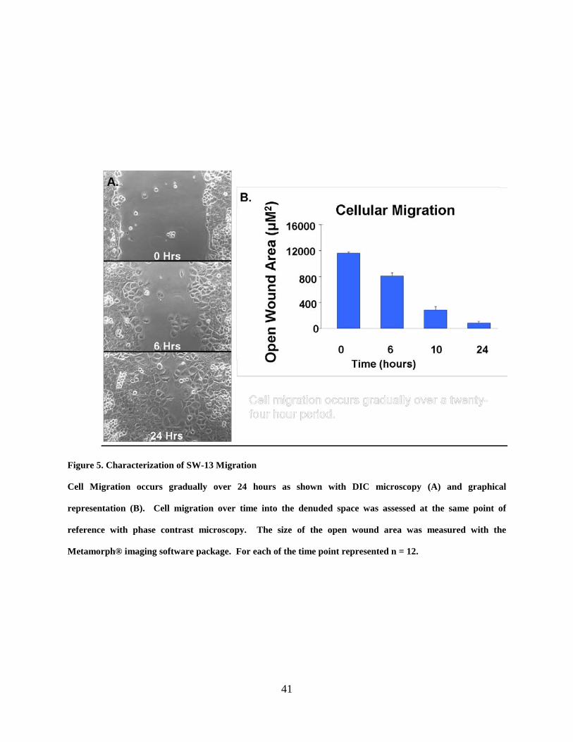
41
Figure 5. Characterization of SW-13 Migration
Cell Migration occurs gradually over 24 hours as shown with DIC microscopy (A) and graphical
representation (B). Cell migration over time into the denuded space was assessed at the same point of
reference with phase contrast microscopy. The size of the open wound area was measured with the
Metamorph® imaging software package. For each of the time point represented n = 12.

42
Figure 6. Connexin 43 gap junctions in SW-13 cells.
Immunocytochemistry and fluorescence microscopy of connexin 43 gap junction plaques and annular gap
junctions (red) in Sw-13 cells. Note the cortical actin cytoskeleton (green) is stained with phallodin outlining
and distinguishing individual cells. Nuclei (n) are stained blue.

43
Figure 7. Fluoresecence microscopy of SW-13 cells.
SW-13 cells were fixed and immunostained with anti-Cx43 antibodies at specified time points after
wounding. Note the gap junction plaques (red) are present between migrating cells that are at the
wound edge throughout migration (a-c).

44
Figure 8. The average number of gap junction plaques per cell.
The average number of plaques per cell at the wound margin and recessed areas (4-6 cell layers away from
the edge) are represented at specified time points during SW-13 cell migration. The average numbers of
plaques between cells at the margin and recess were not significantly different. For each experimental group
10 fields analyzed.

45
Figure 9. Average Plaque Size
The average gap junction plaque size between SW-13 cells that were at the wound margin and cells in the
recessed areas did not significantly differ from one another. For this set of experiments 10 fields were
analyzed per group.

46
Figure 10. Cx43-GFP expression in cells.
Cx43-GFP was expressed in SW13 cells and formed gap junction plaques and annular vesicles as seen here
with fluorescence microscopy (A) and with corresponding DIC - fluorescent overlay (B).

47
Figure 11. Gap Junction Dynamics during SW-13 Migration.
Cx43-GFP gap junction plaques were retained throughout cell migration even as the relative positions of cell
1 and cell 2 shifted with respect to one another (a-d) or stayed the same (e-h). Bar, 30µM.

48
Figure 12. Functional coupling during migration
Intercellular communication of the gap junction permeable dye Lucifer Yellow (green) occurred between
migrating SW-13 cells 5 – 40 minutes following wounding (a-c).
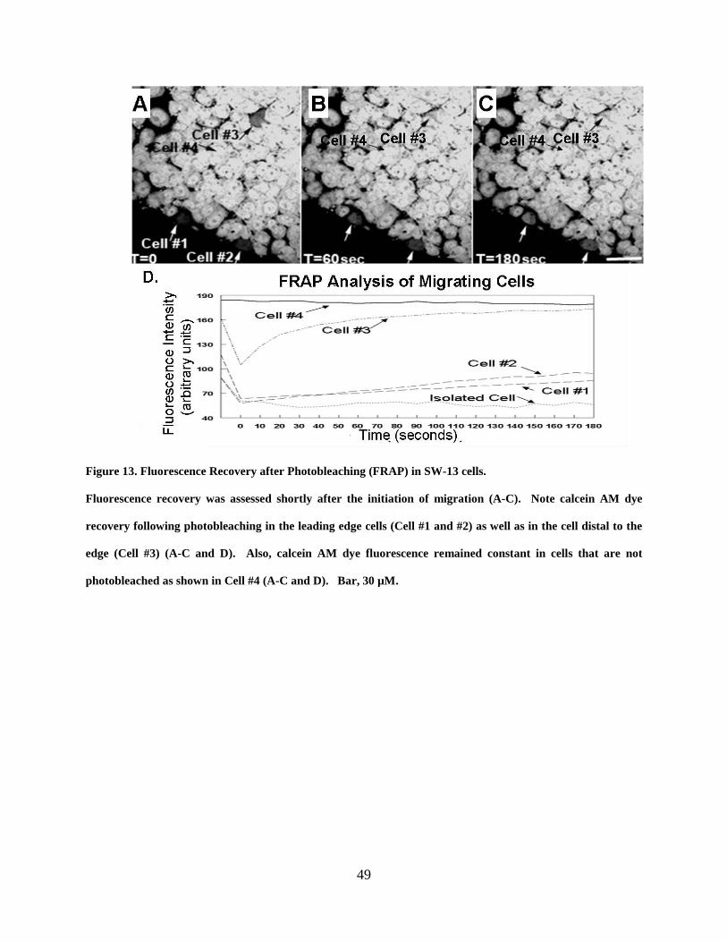
49
Figure 13. Fluorescence Recovery after Photobleaching (FRAP) in SW-13 cells.
Fluorescence recovery was assessed shortly after the initiation of migration (A-C). Note calcein AM dye
recovery following photobleaching in the leading edge cells (Cell #1 and #2) as well as in the cell distal to the
edge (Cell #3) (A-C and D). Also, calcein AM dye fluorescence remained constant in cells that are not
photobleached as shown in Cell #4 (A-C and D). Bar, 30 µM.

50
Figure 14. Gap junction communication during cell migration.
Intercellular communication of fluorescent dye was maintained between migrating cells for over six hours
following the initiation of migration, as assessed with FRAP. The fluorescence recovered by SW-13 cells at
each time point is expressed as the percentage of dye that was recovered by the photobleached cell compared
to its pre-bleach status. Fluorescence recovery was assessed in two cells per each time point.

51
3.4 DISCUSSION
Previous studies are consistent with the importance of cell-cell communication in regulating the
directionality, speed and sequence of events that are required for cell migration and it is therefore
a reasonable suggestion that the mediators of direct intercellular communication must persist as
the cells migrate (Fushiki et al., 2003; Huang et al., 1998; Kwak et al., 2001; Lo 1999; Richards
et al. 2004; Tate et al., 2006; Xu et al., 2006; Xu et al., 2001; Ashton et al., 1999; Menezes et al.
2002; Marins et al., 2009). In this study, immunocytochemistry and time-lapse imaging were
utilized to monitor the presence and position of gap junction plaques at various time points
during in vitro two-dimensional migration. Sheet migration was observed throughout the
population, and extended from cells proximal to the wound edge to cells that bordered the edge
of the tissue culture surface. Gap junction plaques were also evident between cells leading edge
as well as their contacting neighbors. In fact, the average number of gap junction plaques was
not significantly different in cells at the leading edge when compared to cells that were some
distance from the leading edge. This finding was somewhat unexpected since cells at the leading
edge had fewer neighboring cells with which to potentially form cell-cell contacts. This finding
may be indicative of an increase in gap junction formation or a decrease in gap junction turnover
in cells located at the leading edge. One possible explanation is suggested by observations of
monolayer cultures wherein the number of gap junction plaques increases as the number of
neighboring cells increases; however the gap junctions do not occur at every site of possible
contact. It is possible that the number and size of gap junctions are regulated by factors that
interact with the connexin molecule. For example, it has been demonstrated that gap junction
plaques formed with the green fluorescent protein tagged Cx43 (Cx43-GFP) which fail to bind
ZO-1, are larger than those formed from native Cx43 in which ZO-1 binding is intact (Girao and

52
Pereira, 2007). This observation has lead to the suggestion that ZO-1 plays a role in gap junction
size and turnover (Giepmans et al., 2001). Each cell may be therefore capable of autonomously
regulating gap junction plaque numbers and size and independently of neighboring cells.
Regardless of these considerations, our data are consistent with migrating cells retaining gap
junction plaques that are in position to facilitate cell migration.
The observation of retained of gap junction gap junctions in both live and fixed
migrating cells addresses the possibility that dividing rather than migrating cells were the source
of cells that filled in the denuded space. Live cell imaging of SW-13 cells revealed that the
majority of the cells extended membrane followed the movement of the cell body into the
denuded space. The retention of gap junction plaques by migrating cells, also observed with live
imaging, suggest that the plaques were not removed, which is what would have been the case if
cells had been dividing. Gap junction plaques were also seen on the surface of cells that were
fixed and processed for immunocytochemistry at specified times during migration. Furthermore,
previous characterizations of several cell models describe collective migration as a process
wherein cells of the leading edge migrate into an open area and are followed by adjacent cells
that are modulated by contact inhibition (Weijer, 2009 rev.) Thus, the contribution of dividing
cells, to the fill in the denuded space, is a plausible concern. Nevertheless, the findings presented
are evidence that the source of cells comprising the denuded space is largely from migrating
cells.
The necessity for gap junctions has been previously demonstrated in migrating
cells however these studies fail to clarify their functional status. It might be suggested, for
example, that cells simply tolerate or utilize gap junctions as structural components during
migration, while the junctions themselves do not participate in intercellular communication to

53
coordinate multicellular migration (Elias et al., 2007). We report, however, that molecules
continue to diffuse between cells during migration, as assessed with FRAP analysis. Therefore,
the transfer of regulatory molecules between cells while migrating is possible, and even at the
leading edge where the cells are the most active in altering their relative positions. It should be
noted that calcein-AM dye used in the FRAP analysis is a membrane permeable substrate that is
hydrolyzed by non-specific esterases into a non-membrane permeable fluorescent polyanionic
calcein that is retained in the cytoplasm (Czyz et al., 2000). This fluorescent form of calcein is
gap junction channel permeable, but cannot permeate through other plasma membrane channels
and is therefore widely used to study gap junction- mediated communication (Czyz et al., 2000;
Pollmann et al., 2005; Tenopoulou et al., 2007; Wade et al., 1986). In our study, all of the cells
in the population were initially labeled with the calcien and all cells including those at the
leading edge were able to recover fluorescence after photobleaching. Cells that weren’t in
contact with other cells (thus lacking the capacity for gap junction mediated communication) did
not recover fluorescence following photobleaching. The failure of such isolated cells to recover
fluorescence confirms the need for cell-cell contact as determined from our FRAP studies and
continued gap junction mediated communication between migrating cells.
The gap junction protein Cx43 has the potential to interact with numerous
cytoskeletal and signaling molecules, and the physical structure of gap junctions therefore may
serve as a platform for assembly of cytoplasmic elements for cell migration. In support of such a
possibility the cytoplasmic domains of gap junction proteins have been demonstrated to interact
with: the actin-binding protein, drebrin; microtubules; as well as the tight junction, ZO-1; PKC
and src (Butkevich et al., 2004; Giepmans et al., 2004; Giepmans, 2006; Majoul et al., 2007;
Giepmans et al., 2001; Levy and Holzbaur, 2007; Giepmans and Moolenaar, 1998; Li et al.,
54
2004; Chung et al., 2007, DeVuyst et al., 2007; Duffy et al., 2007; Giepmans et al., 2001; Lin et
al., 2006). In addition to gap junction interactions with cytoskeletal and membrane structures to
produce a platform for binding components needed for migration, gap junction hemichannels in
the membrane can be opened or closed by a number of factors, and have been shown to release
molecules such as ATP from the cell (Ramachandran et al., 2007; Retamal et al., 2007; Spray et
al., 2006; Srinivas et al., 2006; Stout et al., 2002; Zhao et al., 2005). The release molecules from
hemichannels combined with the physical structure of gap junctions and the well-known role of
gap junction mediated intercellular communication could all play a pivotal role in providing
information about the structural platforms needed by migrating cells. Our data do not rule out
the possibilities of hemichannels or gap junction physical structured as the mechanism. However
the data are consistent with gap junction channels as being capable of facilitating cell-cell
communication as cells migrate.
In summary, the results of our correlated immunocytochemical, time-lapse and
migration studies are consistent with gap junction plaques persisting between cells that migrate
as a sheet. The fact that migrating cells retain their gap junctions would suggest that the capacity
for cell-cell movement of regulatory molecules is critical in physiological processes such as
wound healing. The consequence of altering gap junctions was investigated and will be
presented in Chapter Four.

55
4.0 ALTERED CONNEXIN 43 EXPRESSION AND GAP JUNCTION
COMMUNICATION REDUCS CELL MIGRATION
4.1 ABSTRACT
To investigate if gap junction structure and intercellular communication are required for
coordinating migrating cells we utilized an in vitro, wound healing, model for initiating and
monitoring cell movement. To assess the effect of altering intercellular communication on cell
migration, we introduced, into cell cultures, mimetic peptides (Gap 26 and 27) that specifically
inhibit gap junction coupling. Functional dye coupling was confirmed with the diffusion of the
gap junction permeable dye, Lucifer yellow and the assessment of fluorescence recovery after
photobleaching (FRAP). Gap junction plaques were reduced with siRNA treatments,
specifically targeting the mRNA of the gap junction protein connexin 43 (Cx43). Cx43 protein
expression and gap junction plaque formation were confirmed with western blot and
immunofluorescence, respectively. Reduced gap junction mediated dye coupling was observed
in cells treated with mimetic peptides. In communication impaired cells migration was slowed,
thus confirming the necessity for gap junction communication in efficient cell migration.
Furthermore, cell motility was drastically hindered when Cx43 gap junction plaque structures

56
were reduced. Our results are consistent with gap junction plaques and intercellular
communication playing a pivotal role in cell migration.
4.2 INTRODUCTION
The majority of cells that comprise the tissues of our bodies are capable of migration, an event
that is thought to rely on the ability of individual cells, in a group, to communicate with one
another (Pepper and Meda, 1992; Matic et al., 1997; Nadarajah et al., 1997; Saitoh et al., 1997,
Civitelli et al., 1998, Winterhager et al., 1999; Ashton et al., 1999; Cina et al., 2007; Leaphart et
al., 2007). A number of these cell populations also often express one or more types of proteins
that can potentially form gap junction intercellular communication channels that may function in
relaying signals that help guide the cells during migration (Davis and Trinkhaus, 1981; Lo et al.,
1997; Bruzzone et al., 1996; Kumar, 1999). Developmental defects, associated with mutation of
gap junction proteins, may result from cells that failed to communicate and therefore, could not
migrate properly to target sites (Lecanda et al., 2000; Reaume et al.,1995; Law, 2002). Thus, it
is possible that perturbation of gap junction expression and/or channel function will alter cellular
migration.
Ideally, the need for gap junction mediated communication , may be straightforward, e.g.
gap junctions function solely for the purpose of communicating vital spatial and temporal cues
during migration, and therefore, mutations that inhibit connexin expression or alter protein
structure would, hypothetically, impair migration. This possibility was suggested from studies in
which the directionality and rate of cell migration were significantly reduced in cardiac neural
crest cells (CNC) that were isolated from Cx43 -/- mice compared to wild type (WT) and Cx43

57
restored (CMV43) mutants (Xu et al., 2006; Huang et al., 1998). Accordingly, gap junction
communication may facilitate cell motility and therefore altered gap junction communication
and/or expression may inhibit migration.
The implications of this specific response to gap junction communication, is
indicative of the complex role of gap junction channels play in cell physiology, particularly
migration. However, there has yet to be a systematic assessment of the effect of gap junction
communication on cell migration. In the previous chapter we demonstrated that gap junction
structure and function were retained in migrating cells. To assess the effect of altering the status
of gap junctions on migrating cells, chemical and molecular approaches were utilized to either
inhibit intercellular communication or reduce Cx43 expression. To determine whether gap
junctions and or intercellular communication are required for cell migration, Cx43 expression
was altered and the effects on cell migration were assessed.
4.3 RESULTS
4.3.1 Migration in Cells with Varied Amounts of Connexin 43 Expression
Three adrenal cortical derived cell lines were examined: human (SW-13), bovine (SBAC) and
mouse (Y-1) in order to determine the migration characteristic of different cell types that express
differing levels of connexin and to select which of the cell types to use for the various studies.
The SW13 and SBAC cell populations form numerous and large gap junction plaques and have
an abundance of cytoplasmic staining presumed to be annular gap junction vesicles. The clone
of Y-1 cells, in our possession do not express connexin proteins and therefore do not form gap

58
junction plaques. The cell populations were subject to a wound healing assay and migration was
monitored with phase contrast microscopy, over a time course (0, 2, 4, 6 and 24 hours). As
described in Chapter Three, the SW13 cells migrated in sheet-like, gradual manner, and closed
the denuded space within the twenty-four hour period (Fig. 5 A and B). There was an initial lag
of six hours observed after wounding before SBAC migration occurred into the denuded area
(Fig. 15 A and B). The Y-1 mouse cells did not migrate into the denuded space after twenty-four
hours (Fig. 16 A and B) or after forty-eight hours (not shown). Since, SW-13 has the most
abundant gap junction plaques this cell line was chosen for further studies of the effects of
decreasing or increasing gap junctions on cell migration.
4.3.2 Altered Gap Junctions and Cell Migration
To effectively decrease connexin protein expression, siRNA specifically targeted against Cx43
mRNA were introduced into SW-13 cells. Decreased Cx43 protein expression was confirmed
quantitatively with western blot (Fig. 17 B) and qualitatively with immunofluorescence (Fig. 17
A). Gap junction mediated intercellular dye coupling was assessed with Lucifer yellow dye and
fluorescence recovery after photobleaching (FRAP) analyses. Both Lucifer Yellow (Fig. 18) and
FRAP (Fig. 19) analyses were consistent with a significant decrease in dye coupling in cells with
reduced Cx43 expression. A significant decrease in the percent change in migration was
observed in cells with reduced Cx43 protein expression (Fig. 20).

59
4.3.3 Altered Gap Junction Communication and Cell Migration
In addition to the siRNA approach, to determine the effect of reduced gap junction
communication on cell migration Cx43 specific mimetic peptides (Gap 26 and 27) were used to
inhibit gap junction intercellular communication (Fig. 21). Mimetic peptides are short protein
sequences that bind to the extracellular loops of connexons, thereby inhibiting channel pore
formation. Intercellular dye communication and cellular migration were assessed in cell cultures
treated with either Gap 26 or Gap 27. Treatment with the mimetic peptides reduced gap junction
mediated communication of Lucifer yellow dye (Fig. 21 G). Migration was slowed in these
communication impaired cells compared to control cells (Fig. 22 G).
4.3.4 Increasing Gap Junctions and Cellular Migration
To address the relationship between gap junction expression and cell migration, SW13 cell
cultures were pre-treated with an analog of cAMP, dibutyryl cAMP (dbcAMP). Cyclic AMP
(cAMP) has been shown in SW-13 cells to increase the size of gap junction plaques while
reducing the number of annular structures, thereby increasing the number gap junctions on the
plasma membrane (Burghardt et al., 1995; Wang and Rose, 1995). In addition, cAMP increases
connexin synthesis and assembly (Wang and Rose, 1995). A wound healing assay was used to
assess migration of dbcAMP treated cells. Migration into the wound area was gradual for both
control and dbcAMP treated cells (Figure 23 A and B). However, the rate of migration was
reduced in cells treated with dbcAMP compared to control cells (Figure 23 B).

60
A
B

61
Figure 15. Characterization of SBAC cell migration.
An initial lag is followed by migration that is completed by 24 hours (A and B). Cell migration over time into
the denuded space was assessed at the same point of reference with phase contrast microscopy. The size of
the open wound area was measured with the Metamorph® imaging software package. For each time point
represented n = 12.

62
A
B
63
Figure 16. Characterization of Y-1 cell Migration.
Y-1 cells, which do not express connexins to form gap junctions did not migrate into the open wound area.
For each time point represented n = 10

64
Figure 17. Connexin 43 Knockdown and Gap Junction Expression.
Immunocytochemistry (A and B) and Western Blot (C) of Cx43 siRNA transfected and control transfection
(Mock) SW-13 cell cultures. Note the reduction in Cx43 protein expression in Cx43 siRNA treated cells (B,
C).

65
Figure 18. Functional coupling in connexin 43 reduced cells.
Lucifer Yellow dye transfer was measured in control and Cx43 siRNA transfected SW-13 cells. Note the
significantly reduced dye transfer in Cx43 siRNA treated cells compared to control cultures. For each group
represented 12 fields were analyzed.

66
Figure 19. Fluorescent dye recovery following photobleaching.
The percent fluorescence recovery in SW-13 cells compared to initial fluorescence values was compared. The
percent fluorescence recovery in cells with decreased Cx43 expression (43 ± 9%) was significantly reduced
compared to Mock (69 ± 12%) and non-treated (NT) (70 ± 10%) controls. For assessment of FRAP: NT: n =
15; Mock: n = 21; Cx43 siRNA n= 31.
Percent Recovery of Fluorescence Recovery after Photobleaching
0102030405060708090
NT Mock Cx43 siRNA

67
Figure 20. Comparison of percent change in cell migration.
The percent change in SW-13 cell migration was compared in control (Mock) and Cx43 siRNA transfected
cell cultures. Note the significant decrease in percent change in migration of cells transfected with Cx43
siRNA compared to the control.
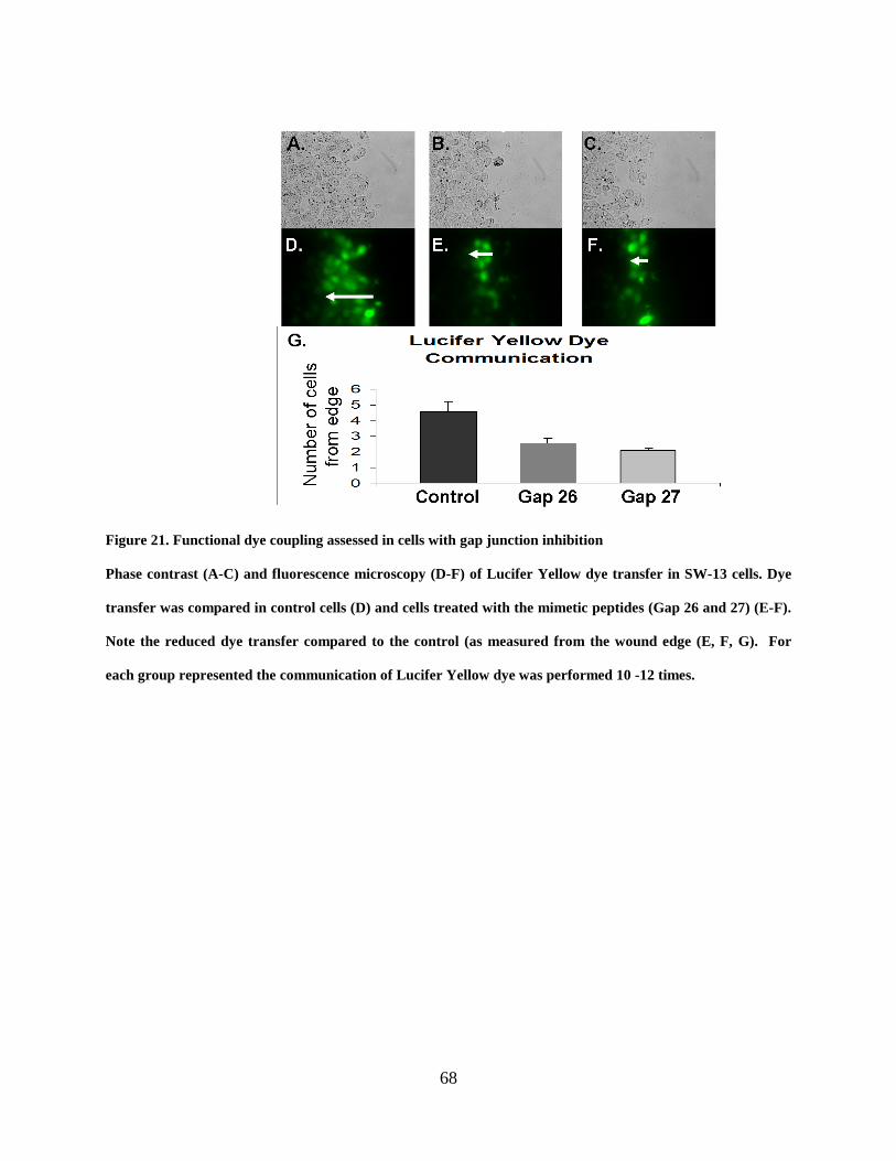
68
Figure 21. Functional dye coupling assessed in cells with gap junction inhibition
Phase contrast (A-C) and fluorescence microscopy (D-F) of Lucifer Yellow dye transfer in SW-13 cells. Dye
transfer was compared in control cells (D) and cells treated with the mimetic peptides (Gap 26 and 27) (E-F).
Note the reduced dye transfer compared to the control (as measured from the wound edge (E, F, G). For
each group represented the communication of Lucifer Yellow dye was performed 10 -12 times.

69
Figure 22. Comparison of migration in cells with reduced communication
Cellular migration was assessed in control cells (A, D) and cells treated with Cx43 specific mimetic peptides
(Gap 26 and 27) (B, E and C, F). Note migration into the denuded space was reduced with the mimetic
peptides as compared to the control. The percent change in migration in cells treated with Gap 27 was
significantly reduced compared to non-peptide treated controls. For each group represented, the assessment
of cell migration was performed 6-8 times.

70
A
B
Control DbcAMP

71
Figure 23. Migration in cells with increased gap junctions
Migration of SW-13 cells treated with DbcAMP is gradual though reduced compared to the control. For each
of the groups represented: control n = 12 and DbcAMP n = 6.

72
4.4 DISCUSSION
The goal of this study was to evaluate the role of gap junction communication channels in cell
migration by altering the gap junctions. We initially characterized migration in cells that
expressed varying levels of the gap junction protein connexin 43 (Cx43). SW13 and SBAC cells
expressed Cx43, formed gap junction plaques and also migrated gradually within twenty-four
hours. The migration of SW13 and SBAC cells which express an abundance of gap junction
proteins and are therefore capable of intercellular communication suggests that gap junctions
may have been involved in regulating cellular migration. The Y-1 cells did not express Cx43
and did not migrate. Y-1 cells are steroid producing cells that appear flat and adherent in
culture (Whitehouse et al., 2002). It was observed that an increase in the cell population was
marked by stacking or piling up of the cells on top of one another rather than spreading out and
forming a monolayer. The failure of Y-1 cells to migrate does not necessarily indicate that the
lack of motility was due to lack of gap junctions; it is known that the shape and function of the
Y-1 cytoskeletal proteins are controlled by hormones (Han and Rubin, 1996). However, Y-1 cell
responsiveness to activators of cyclic AMP –dependent kinase (PKA) signaling is associated
with changes in cell morphology from flat and attached to round and non-adherent (Whitehouse
et al., 2002). It is therefore unlikely that Y-1 cells grown in the presence of increased
concentrations of hormones and growth factors in the culture media would be morphologically
poised for migration.
The expression of Cx43 in Y-1 cells, however may influence the migration process. For
example, it has been suggested from studies in Cx43 knockout mice that polarization required for
epicardial cell migration may require interaction between connexin 43 and the cytoskeletal

73
element tubulin (Rhee et al., 2009). Furthermore, Cx43 has been demonstrated to interact with
several cytoskeletal elements that aid in producing the force at the leading edge that propels the
cell body forward during migration (Butkevich et al., 2004; Horwitz amd Webb, 2003). It was
also recently suggested by Elias and co-workers (2007) that the migration of radial glial cells
may depend on gap junction interaction with cytoskeletal components. It is thus possible that the
expression of exogenous connexins (whether or not they form functional gap junction channels)
may stabilize or reorganize the cytoskeleton thereby affect the ability of Y-1 cells to migrate.
These issues will be addressed in future experiments.
The migration of the gap junction expressing cells is, however, consistent with other in
vivo and in vitro studies demonstrating increased Cx43 protein and mRNA levels associated with
sustained cell-cell communication during wound healing (Larson and Haudenschild, 1988;
Pepper et al., 1989; Pepper et al., 1992). While the migration of SW-13 and SBAC cells
supports our hypothesis that gap junctions play an important role in cell migration, altering gap
junctions by specifically targeting connexins will ultimately indicate the importance of gap
junctions in cell migration.
In this study, we altered gap junction plaques and assessed the effects on SW-13 cell
migration by either using siRNA techniques to reduce the availability of Cx43 protein to form
gap junction channels and plaques, and also with mimetic peptides that inhibited the functional
coupling of gap junction channels. Cell migration was reduced in cells cultures treated with
mimetic compounds, and the decrease in migration was even more pronounced in cells treated
with Cx43 specific siRNA. Importantly, SW-13 migration was inhibited in cells transfected with
the Cx43 specific siRNA but it was not inhibited in cells transfected with control Cx43 (a non-
knockdown sequence) or GAPDH specific siRNA (not shown). An important caveat considered

74
in using the siRNA approach was the possibility of creating a non-specific effect that could
affect migration. To address this concern, the status of the actin cytoskeleton in control and
Cx43 siRNA knockdown cells was assessed with immunocytochemistry and fluorescence
microscopy. Preliminary analysis revealed control and knockdown with similar appearing
cytoskeletal arrangement. The reduced migration in SW13 cells with decreased connexin 43
expression is consistent with studies showing reduced migration in neural crest cells isolated
from connexin 43 null mouse embryos (Ruanvoravat and Lo, 1992; Lo et al., 1997).
Furthermore, our findings that reduced cell migration correlates with decreased gap junction
coupling suggests that intercellular communication may be necessary for efficient cell migration.
One may suggest, on the other hand, that it is possible that treatment with connexin 43 specific
mimetic compounds, while inhibiting cell-cell communication, may also inadvertently alter the
protein conformation of connexin monomers and thus the interaction of connexin with the
cytoskeleton and thereby, affect migration (Yamane et al., 2002). If so, one might suggest that
gap junction channels may also play a role as structural components that aid in cell migration. In
a recent study, connexin subunits incapable of pore function were expressed in neocortical radial
glia and were found to be sufficient for cellular migration (Elias et al., 2007). On the other hand,
reduced cellular migration of enterocytes and neural crest cells has been attributed to the
disruption of gap junction coupling with non-specific inhibitors, such as oleamide (Huang et al.,
1998; Leaphart et al., 2007; Bannerman et al., 2000). The suggestion that migration, in some
cells, may be attributed to gap junctions as mediators of adhesion rather than mediators of
communication reveals the complexities of the relationship between gap junction channels and
the cells that they inhabit. For example, gap junctions may function solely as communication
channels that relay important messages for migration or as adhesion factors or in both capacities.
75
The findings following siRNA treatment would not rule out either possibility. Nevertheless, our
findings demonstrate, for the first time, that direct targeting of connexin 43 expression with
siRNA results in decreased cell migration. In addition, the specific inhibition of gap junction
coupling with mimetic compounds Gap 26 and 27 resulted in reduced cellular migration. These
findings are consistent with the suggestion that altered gap junction channels and intercellular
communication reduce cellular migration.
Hitherto we have demonstrated that decreased gap junctions and intercellular
communication results in reduced cellular migration. Therefore, to determine the effect of
increasing gap junctions on migration cells were treated with the compound dbcAMP.
Accordingly, the migration of SW-13 cells was reduced in populations treated with the cAMP
analog dibutyryl cAMP (dbcAMP). However, one limitation in interpreting this data is the non-
specifc effect of dbcAMP on adrenal cells that has been demonstrated to decrease proliferation,
and increase steroid production (Oyoyo et al., 1997; Murray and Taylor, 1988). Furthermore,
dbcAMP treatment may have effected cell migration by altering many factors such as focal
adhesion contractile strength, inhibiting RHO kinase dependent adhesion or modifying the actin
cytoskeleton (Leader et al., 1983; Pereyra et al., 2006; Laudanna et al., 1997). Treatment with
dbcAMP has been demonstrated to sustain protein kinase A activation (PKA) and interestingly
prolonged (PKA) activity in ameboid PKA mutants resulted in an inability to alter the
cytoskeleton thereby and reduced chemotaxis (Zhang et al., 2003; Pereyra et al., 2006). Based
our studies we can conclude that, in cells treated with dbcAMP to increase gap junction plaques,
cell migration is reduced though the relationship between these finding remains to be elucidated.
Future studies designed to restore gap junctions in cells that either express few or no endogenous
connexin are needed to further discern the role of gap junctions in cell migration. Recent studies
76
suggest that gap junction are key participants in cell migration via interaction with cytoskeletal
components; thus, our understanding of the role of gap junctions in cellular migration may also
be aided by assessing the influence of sustained PKA signaling on increased gap junction
plaques and how this may correlate with cell shape and adhesion (Elias et al., 2007; as reviewed
in Olk et al., 2009).
Our findings are consistent with the hypothesis that gap junction channels and
intercellular communication are necessary for cell migration. Cell migration was effectively
reduced in cells with altered gap junction expression or intercellular communication. The
reduced migration associated with increased gap junction plaques suggests that gap junction
internalization may also be necessary for cell migration. The mechanism of gap junction
internalization will be addressed in the following Chapter Five: The Mechanism of Gap Junction
Channel Internalization.

77
5.0 THE MECHANISM OF GAP JUNCTION CHANNEL INTERNALIZATION
5.1 ABSTRACT
Gap junction-mediated intercellular communication has been implicated in the development,
differentiation and function of most cells of the body. While gap junction channel assembly has
been extensively studied, mechanisms involved in the disassembly of gap junction plaques are
not well understood. Yet, the rate of gap junction plaque removal from the cell surface is thought
to be critical to gap junction function, and alterations in removal may be related to a number of
disease processes. This study focused on gap junction protein (connexin 43) removal from
the cell membrane, specifically gap junction plaque internalization, which leads to the formation
of cytoplasmic double-membraned structures referred to as “annular” gap junctions. The process
of gap junction plaque removal is an “endoexocytic” process in which one cell internalizes
membrane (endocytosis) and the the adjoining cell contributes membrane (exocytosis). To study
the fundamental cellular mechanisms involved in gap junction plaque endoexocytosis, a
combination of immunocytochemistry, live cell imaging, quantum dot immuno-electron
microscopy and computer reconstruction techniques were used. A detailed examination of the
spatial and temporal-dependent redistribution of connexin 43-GFP, revealed that gap junction
78
plaques internalized preferentially into one of the two contacting cells. Gap junction plaque
endoexocytosis, rather than plaque component (hemichannel) dispersion within the membrane,
appeared to be the dominant method of gap junction removal. Gap junctional invaginations and
annular gap junction vesicles at the cell surface were coated with short electron-dense bristles
similar in appearance to the clathrin coated vesicles of nonjunctional membrane. To determine if
this associated clathrin-like coat was composed of moleculed involved in facilitating
endoexocytosis, clathrin and its adaptor proteins were imaged with quantum dot immuno-
electron microscopic and immunocytochemical light microscopic techniques. Clathrin was
observed to colocalize with some of the connexin 43 gap junction plaques, as well as, the annular
gap junction profiles. The clathrin adaptor protein, AP-2, which is thought to play a major role
in the formation of clathrin-coated vesicles, also colocalized with some of the annular gap
junction vesicles and gap junction plaques. An important finding was that clathrin preferentially
localized to the intracellular surface of the gap junction plaque in one of the two contacting cells.
Our findings provide evidence that clathrin, with the assistance of AP-2, may serve as the
machinery essential for gap junction plaque endoexocytosis.
5.2 INTRODUCTION
One important method of gap junction regulation involves the removal of gap junction plaques
from the cell surface. Gap junctions differ from most cell surface ion channels by exhibiting
high turnover rates (1-5 hrs. as opposed to > 20 hrs.) and it has been proposed that the removal of
gap junctions from the plasma membrane may serve to regulate intercellular communication and
thereby, physiological processes (Musil et al., 2005; Fallon and Goodenough,1981; Piehl et al.,

79
2007; mer et al., 1973). For example, it was reported that the appearance of internalized gap
junctions coincided with the spontaneous release of individual cells from a monolayer into the
media (Murray et al., 1981, Larsen et al., 1983). The mechanism of gap junction removal is
thought to involve several steps including: the endocytosis of channels and membrane
components of both cells into one of the connected cells, the formation of double membrane
annular vesicle and the movement of the annular vesicle to the interior of the receiving cell
(Murray et al., 1981). During migration, the loss of gap junctions via internalization may serve a
multitude of purposes including: freeing certain regions of the plasma membrane to participate in
the migratory process (e.g. protrusion or adhesion) or an opportunity to replace the ‘older’ gap
junctions with new ones. Spatial shifting of the plasma membrane and gap junction function and
dynamics has been characterized in live cells during migration (DeFranco et al., 2008).
However, the mechanism underlying gap junction removal from the surface of migrating cells
has yet to be determined.
The observation of cytoplasmic vesicles with distinct pentalaminar profiles in many
cell types has motivated presumptions that gap junctions at the cell surface may be targeted for
internalization via an endocytic mechanism rather than dispersal within the plasma membrane
(Larsen et al., 1979; Larsen and Tung, 1978). Early transmission electron microscopy (TEM)
studies described short electron dense bristles, thought to be clathrin, emanating from
invaginating surface gap junctions and cytoplasmic annular gap junction vesicles (Larsen et al.,
1979); suggesting the possibility that gap junctions are removed from the cell surface via a
clathrin dependent mechanism.
In a recent study, combined microscopy and immunocytochemistry approaches were
used to demonstrate subcellular association between clathrin and Cx43-GFP transfected gap
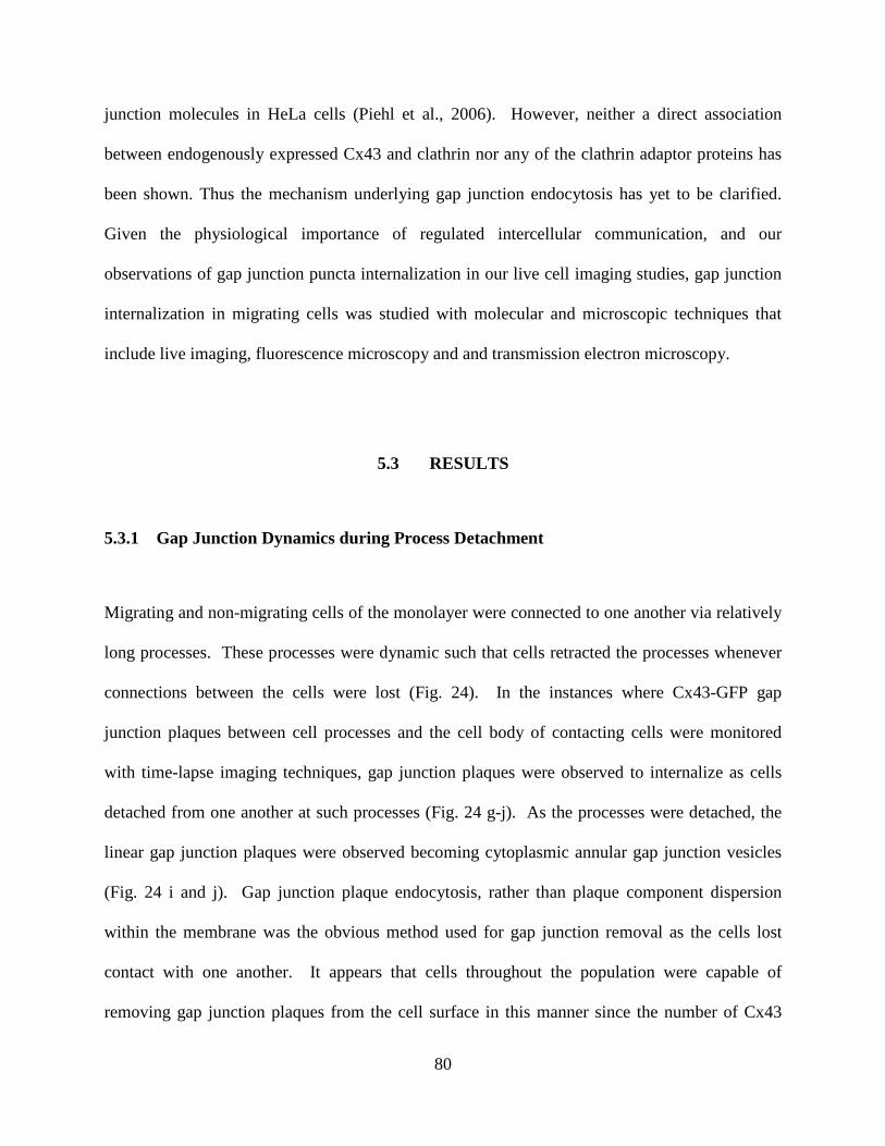
80
junction molecules in HeLa cells (Piehl et al., 2006). However, neither a direct association
between endogenously expressed Cx43 and clathrin nor any of the clathrin adaptor proteins has
been shown. Thus the mechanism underlying gap junction endocytosis has yet to be clarified.
Given the physiological importance of regulated intercellular communication, and our
observations of gap junction puncta internalization in our live cell imaging studies, gap junction
internalization in migrating cells was studied with molecular and microscopic techniques that
include live imaging, fluorescence microscopy and and transmission electron microscopy.
5.3 RESULTS
5.3.1 Gap Junction Dynamics during Process Detachment
Migrating and non-migrating cells of the monolayer were connected to one another via relatively
long processes. These processes were dynamic such that cells retracted the processes whenever
connections between the cells were lost (Fig. 24). In the instances where Cx43-GFP gap
junction plaques between cell processes and the cell body of contacting cells were monitored
with time-lapse imaging techniques, gap junction plaques were observed to internalize as cells
detached from one another at such processes (Fig. 24 g-j). As the processes were detached, the
linear gap junction plaques were observed becoming cytoplasmic annular gap junction vesicles
(Fig. 24 i and j). Gap junction plaque endocytosis, rather than plaque component dispersion
within the membrane was the obvious method used for gap junction removal as the cells lost
contact with one another. It appears that cells throughout the population were capable of
removing gap junction plaques from the cell surface in this manner since the number of Cx43

81
cytoplasmic gap junction packets, measured in cells at the leading edge and cells located at a
distance from the edge were not significantly different at 2, 4, 6 or 24 hours post wounding. For
example at 4 hours, the number of cytoplasmic packets in cells at the leading edge was (7.95 ±
0.72) versus the number of packets in relatively distant cells, which was (10.1 ± 1.1).
Furthermore, the Cx43-GFP cytoplasmic packets and plaques were similar in size to that of the
endogenous Cx43 gap junction packets and plaques. Small puncta or long linear arrays of Cx43
gap junctions were observed at the sites of cell-cell contact throughout the population.
Previous analyses of internalized annular gap junction vesicles with TEM revealed
pentalaminar profiles and in some cases faint bristle-like coats, thought to be clathrin, emanating
from discrete locations along the outer membrane of the vesicle, similar to those described in
earlier studies (Murray et al., 1981).
5.3.2 Immunocytochemistry Analysis of Gap Junction Internalization
To elucidate these internalized gap junction structures further, immunocytochemical techniques
were used. Subcellular colocalization of gap junction structures and clathrin was assessed by
probing fixed cells with specific antibodies for Cx43 and the clathrin heavy chain and then
labeling specimens with fluorescent secondary antibodies. Clathrin expression was abundant in
SW13 cells and co-localization with Cx43 molecules occurred at gap junction plaques and
annular gap junction vesicles (Fig. 25).
Since clathrin does not directly bind to its cargo, but instead uses adaptor proteins, we
investigated the association of gap junction proteins with clathrin adaptor proteins: AP2, Dab2
and Epsin. We have found that the clathrin adaptor protein 2 (AP-2) is abundant in SW13 cells
and also colocalized with gap junction plaques and annular vesicles (Fig. 26). Co-

82
immunoprecipitation analysis, is not indicative of a direct protein-protein association between
AP-2 and Cx43 (not shown). The status of clathrin adaptor proteins Disabled 2 (Dab2) and
Epsin were also assessed and found in relatively less abundance than AP-2 (Figs. 27 and 28).
Furthermore, there was no indication of any significant colocalization with Cx43 and Dab2 or
Epsin.
5.3.3 Transmission Electron Microscopy Analysis of Gap Junction Internalization
Resolution at the light microscopy level was not sufficient for determining with certainty, the
association between Cx43 and clathrin. To further characterize the relationship between clathrin
and gap junctions fixed cells labeled with quantum dots that are specific for Cx43 or clathrin and
then processed the samples for TEM analysis. Quantum dots (QD) are 10-50 nm nanocrystals
that offer the advantage being excited by laser and emitting visible light and also being visible as
spheres at the electron micropscopic level. Clathrin specific quantum dots specifically labeled
gap junctional regions of the plasma membrance and not non-junctional membrane (Fig. 29).
Interestingly, clathrin specific quantum dots labeled plaques and annular gap junctions on one
side of the membrane (Fig. 30). The clathrin label sometimes coated the entire annular vesicle or
was seen in discrete areas along the vesicular membrane (Fig. 5-7).

83
Figure 24. Gap Junction internalization occurs during cell process detachment.
Pseudo-fluorescent (left) and phase contrast (right) imaging of SW-13 cells shows Cx43-GFP plaque
invagination, annular gap junction formation and subsequent internalization into one of the two contacting
cells.
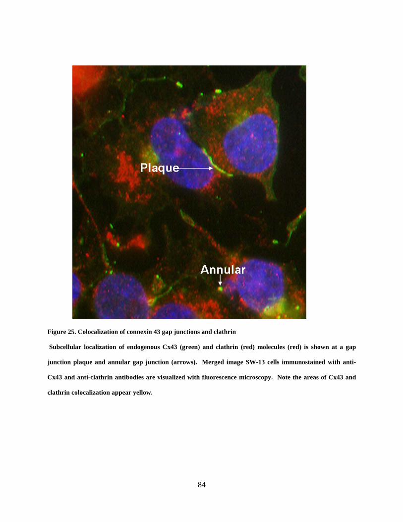
84
Figure 25. Colocalization of connexin 43 gap junctions and clathrin
Subcellular localization of endogenous Cx43 (green) and clathrin (red) molecules (red) is shown at a gap
junction plaque and annular gap junction (arrows). Merged image SW-13 cells immunostained with anti-
Cx43 and anti-clathrin antibodies are visualized with fluorescence microscopy. Note the areas of Cx43 and
clathrin colocalization appear yellow.

85
Figure 26. Subcellular localization of gap junctions and the clathrin adaptor protein AP-2
Colocalization of endogenous Cx43 (green) and the clathrin adaptor protein, AP-2 (red) is demonstrated at
gap junction plaque and cytoplasmic annular structures (shown with arrows). Merged image SW-13 cells
immunostained with anti-Cx43 and anti-AP-2 antibodies are visualized with fluorescence microscopy. Note
the areas of Cx43 and AP-2 subcellular colocalization appear yellow.

86
Figure 27. Subcellular localization of connexin 43 gap junctions and Dab2
Immunofluorescence of Cx43 (green) gap junction proteins and the adaptor protein Dab2 (red) is shown.
Merged image SW-13 cells immunostained with anti-Cx43 and anti-Dab2 antibodies are visualized with
fluorescence microscopy. Note the lack of yellow as a sign of colocalization of Cx43 with Dab2.
Annular
Plaque

87
Figure 28. Subcellular colocalization of gap junctions and Epsin
Immunofluorescence of Cx43 gap junction plaques and annulars (green) and the adaptor protein Epsin
(red) is demonstrated. Merged image SW-13 cells immunostained with anti-Cx43 and anti-Epsin antibodies
are visualized with fluorescence microscopy. Connexin 43 gap junctions and annulars are present however
there is no sign of colocalization with Epsin.
Plaque

88
A
B
Figure 29. Subcellular colocalization of gap junctions and clathrin assessed with transmission electron
microscopy
Quantum dot immunoelectron-microscopy demonstrating the accumulation of clathrin on one side of the gap
junction plaque (white arrow) (A) in SW-13 cells. Localization of Cx43 quantum dots at a gap junction
plaque (B). Note: the Cx43 quantum dots do not label non junctional membrane (B).

89
Figure 30. Quantum dot transmission immunoelectron microscopy of SW-13 cells.
The arrows designate clathrin surrounding internalized annular gap junction vesicles. Bar, 0.1 µm.

90
5.4 DISCUSSION
The maintenance of physiological processes requires electrical and metabolic coupling cells and
it is likely therefore, that the regulation of gap junction internalization would affect intercellular
communication and thereby, physiological processes (Gumpert et al., 2008). Cells internalize
gap junctions in response to numerous stimuli and gap junction internalization may serve a
multitude of purposes including: freeing certain regions of the plasma membrane to participate in
the migratory process, liberation of cells for the purpose of growth and/or mitosis and also to
replace the ‘older’ gap junctions with new ones. For example, gap junctions are often targeted
for degradation in cancerous cells such that ultimately, these cells are no longer in
communication with neighboring cells and are therefore able to grow and proliferate out of
control (Fiorini et al., 2008) Furthermore, increased gap junction internalization has also been
reported to coincide with mitosis in SW13 cells (unpublished data). Connexin 43 and 31.1 gap
junction expression were downregulated in epidermal cells during wound healing (Goliger and
Paul, 1995). We have also demonstrated that gap junctions are internalized in regions of
membrane detachment during cell migration (DeFranco et al., 2008); however the mechanism of
plaque removal is not completely understood.
We show with live cell imaging of migrating cells that neighboring cells may detach
partially from the adjacent cell and when a plaque is in the vicinity, portions (or even the entire
structure) are removed and internalized into one of the two cells. The internalized gap junction
plaque forms a double membrane or pentalaminar cytoplasmic vesicle that is referred to as an
annular gap junction. Transmission electron microscopy (TEM) revealed putative clathrin
association as a bristle-like coat at discrete segments along annular gap junction vesicles. Our
analysis of plaque and annular structures, with immunocytochemistry and fluorescence

91
microscopy, revealed that the clathrin heavy chain colocalizes with both gap junction plaques
and annular gap junction vesicles. Analyses of clathrin-specific quantum-dots/TEM are
consistent with clathrin association with one side of the gap junction plaque and with the
outermost membrane of annular gap junction vesicles. Based on findings that clathrin associates
with gap junction plaques and annular vesicles, one might suggest that clathrin is a mediator of a
unique endoexocytic mechanism that removes Cx43 gap junction plaques from the plasma
membrane (Figure 5-8).
We have shown several clathrin associated adaptor proteins, including AP2, Dab2 and
epsin are expressed in SW13 adrenal cells. The AP2 adaptor protein, in particular, colocalizes
with gap junction plaques and annular vesicles. Interestingly, we also observed in a majority of
cells that AP2 lies along one-side of the plaque, suggesting perhaps, a mechanism of gap
junction plaque invagination and removal from the plasma membrane in which AP-2 would
determine the direction of internalization. Co-immunoprecipitation analysis, however, was not
indicative of a direct protein-protein association between AP2 and Cx43. This may be a
reflection the fragile nature of the Cx43/AP2 interaction or the stringency of our methods to pull
down the complex. Overall, our analyses made with clathrin-specific quantum-dots/TEM are
consistent with clathrin association with one side of the gap junction plaque and with the
outermost membrane of annular gap junction vesicles.
Early TEM studies of annular gap junctions identified a short electron dense bristle coat
on the outermost membrane that was thought to be clathrin. Furthermore, the bristle coat was
also associated with an invaginating gap junction plaque suggesting that the mechanism of gap
junction internalization is clathrin dependent (Larsen and Tung, 1978; Larsen et al., 1979).
Recently immunocytochemistry studies with clathrin specifc antibodies were used to

92
demonstrate subcellular colocalization between clathrin and Cx43-GFP gap junction plaques in
HeLa cells and at the TEM level electron dense patches identified with annular gap junction
vesicles (Piehl et al., 2007). Furthermore, gap junction plaque internalization was inhibited with
the depletion and /or inhibition of clathrin, AP2 and Dab2 (Gumpert et al., 2008). Overall, these
studies are consistent with the internalization of gap junctions via a clathrin dependent
mechanism.
Our studies suggest that the internalization of Cx43 gap junction plaques in migrating
cells occurs via a clathrin dependent mechanism. This is first time that migration as the stimulus
for gap junction endocytosis has been linked to a specific mechanism. Importantly, the
association with gap junction plaques and annular vesicles indicates that clathrin may be an
important mediator of a unique endoexocytic mechanism that removes Cx43 gap junction
plaques from the plasma membrane.

93
Figure 31. Model of clathrin mediated gap junction plaque endoexocytosis.
Plaque invagination is stimulated and the adaptor protein complex, AP-2 is recruited to one side of the plaque
followed by clathrin (1). A clathrin coated annular pit is formed (2) that ultimately becomes an annular gap
junction vesicle (3). In the final step the annular vesicle is internalized into one of the two cells (4).

94
6.0 DISCUSSION
We demonstrate that cells that endogenously expressed the gap junction protein connexin 43
migrated as a sheet, i.e. the cells maintained connection with one another throughout migration.
Our finding that gap junction structure and function are retained in migrating cells would be
consistent with the suggestion that the communication of molecules between cells occurs during
migration. The retention of intercellular communication is indicative of a conserved mechanism
through which, cells may remain synchronized for proper migration, and it is, therefore,
reasonable to suggest that gap junction plaques would be retained during migration, which is
what we found. Furthermore, we found that when communication was decreased with connexin
43 specific inhibitory mimetic peptides, the migration was reduced. Consistent with this
finding, when gap junction protein expression was targeted and significantly decreased with
Cx43 specific siRNA, cell migration was significantly decreased. These findings are in
agreement with studies that show reduced neural crest cell migration in cells treated with non-
specific inhibitors of gap junction communication (e.g. glycyrrhetinic acid) (Bannerman et al.,
2000). Importantly, the accumulation of evidence in this body of work and by others suggests
that gap junction mediated communication is a necessary process for synchronizing cell
populations during migratory events; and expands upon the current model of cell migration.

95
The preservation of gap junction channels and intercellular communication in migrating
cells are significant findings in the field of cell migration research. Hitherto, cell migration has
been characterized as a set of repeated steps, which are set in motion by an external stimulus
(Horwitz and Webb, 2003; Jaffe and Hall, 2005). However, this characterization describes the
migration of a single cell rather than groups of cells moving together in a cell population, which,
in some cases may be more physiologically relevant. The question of how cells are able to
migrate as a group is more complicated and must depend upon the cooperation among individual
cells. Gap junction intercellular channels have been implicated as facilitators of cooperative
(sheet-like) migration in several cell types including neural crest cell populations and epithelial
cells (Xu and Lo, 2006; Richards et al., 2004; Pepper et al., 1989; Pepper et al., 1992). In fact,
the lack of gap junctions has been linked to serious developmental deficiencies, including
occulodentodigital dysplasia (OCDDD) and malformations of the heart and brain (Britz-
Cunningham., 1995; Paznekas et al., 2003 ). While these studies provide evidence that supports
the importance of gap junctions in cell migration, they do not address several important
questions regarding the structure, dynamics and functional status of gap junction channels in
migrating cells. The answers to these questions (do gap junction channels uncouple; do the
plaques disperse into plasma membrane; or do they go away and return; are the channels even
functional?) may provide evidence for recognizing the importance of gap junction channels and
communication in cell migration.
This body of work provides evidence that supports the role of gap junction channels as
mediators of cooperation between cells during migration. This study demonstrated that cells
remain coupled via gap junctions that persist, though on occasion are modified, during migration.
The inhibition of gap junction communication directly correlates with decreased cell migration

96
and thus supports the evidence that intercellular communication is also necessary for gap
junction communication. A significant decrease in cell migration was associated with the
specific targeting and down regulation of Cx43 expression. A detailed examination of the spatial
and temporal-dependent redistribution of connexin 43-GFP, revealed that gap junction plaques
internalized preferentially into one of the two contacting cells. Our subsequent analysis also
provides evidence that clathrin, with the assistance of AP-2, may serve as the machinery
essential for gap junction plaque endoexocytosis. The fact that gap junction plaques and
annulars associated with clathrin and AP-2 endocytic machinery, may suggest that the
mechanism underlying plaque removal from the plasma membrane is clathrin mediated.
Therefore, gap junction plaques are dynamic and resilient structures that are necessary for
mediating intercellular communication during migration.
It has now been well established that gap junction channels are specialized membrane
pores that facilitate the mutual and direct exchange of electrical and chemical information
between adjacent cells in tissues and organs (Loewenstein, 1966; Gilula et al., 1971; Sheridan,
1971). The information that is exchanged is in the form of small molecules and includes
secondary messengers, amino acids, nucleic acids and ions such as cyclic AMP (cAMP), calcium
and nitric oxide (Simpson et al., 1977; Lowenstein, 1978; Saez et al., 1989; Kumar and Gilula,
1996 rev.). Based on our findings, one may propose that gap junction channels are conduits of
such signals that are in turn capable of regulating gap junction communication and/or
coordinating the steps of migration.
97
6.1 FUTURE STUDIES
There are questions yet to be addressed that will expand our knowledge of gap junctions and
their role in cellular migration. Numerous potential binding partners, particularly the very same
small molecules that transit the gap junction channels, and opportunities that exist for interaction
with gap junction proteins imply that regulation of gap junctions may be targeted for therapeutic
development. Furthermore, future characterization of connexin interaction with these small
molecules may reveal unique mechanisms that are involved in regulating migration (Tribulova et
al., 2008). Such possibilities to be addressed in future studies include connexin interactions with
nitric oxide (NO), calcium and cyclic AMP. Our studies have now demonstrated that gap
junctions are retained in migrating cells and perhaps the next level of questions may ask whether
the sustained presence of gap junctions in migrating cells is regulated by intercellular
communication.
Gap junction permeable small molecules are key participants in maintaining cellular
physiology and therefore, may also affect cell migration (Anand et al., 2008; Ridley et al., 2003).
The free radical gas, nitric oxide (NO) is one the molecules capable of transiting gap junction
channels as well as affecting gap junctions. For example, NO was shown to not only increase
connexin 43 expression, but also gap junction permeability in mesangial cells (Yao et al., 2005).
It was recently demonstrated in vivo that NO increased the open status of gap junctions in
vascular tissues, however, NO also inhibited gap junction communication between enterocytes
(Anand et al., 2008, Gonczi et al., 2009). Nitric oxide signaling also plays a role in regulating
numerous physiological processes including cell migration (Jadeski et al., 2003; Kang et al.,
2004; Gupta and Vlahakis, 2009; Zhou and Zhu, 2009). For example, NO signaling is thought to
98
be involved in the regulation of α9β1 integrin dependent cell adhesion during migration (Gupta
and Vlahakis, 2009). Accordingly, NO is a small molecule capable of transiting gap junction
channels, as well as regulating the discrete steps involved in cell migration such as adhesion and
intercellular communication. The sustained presence of gap junctions in migrating cells
therefore may be regulated by the actual passage NO through gap junctions and subsequent
targeting gap junction proteins. Such possibilities will be addressed in future studies.
Calcium is another small molecule that is able to transit gap junction channels and inhibit
the channel pore at increased intracellular concentrations. Incidentally, mechanical stimulation
has been shown to trigger an increased propagation of calcium through gap junction channels
(Anand et al., 2008; Hahn et al., 1992). The process of cell migration during wound repair
depends on calcium gradients and waves (Boitano et al., 1994). Importantly, directional
movement during migration has been shown to require intracellular calcium that is well
organized in space, time and concentration (Ridley et al., 2003 rev.; Hahn et al 1992; Wei et al.,
2009). One might therefore suggest that the retention of functional gap junctions during
migration is a way to regulate calcium concentrations of cells within a population and thereby
coordinate the movement of contacting cells. While the role of calcium communication remains
to be addressed it is necessary to assess the potential role other small signaling molecules in
regulating gap junctions and cell migration.
The gap junction permeable second messenger cyclic adenosine monophosphate (cAMP),
is an important intracellular signaling mediator and yet another small molecule that has the
potential to regulate a number of target proteins including the gap junction protein, connexin
(Pierre et al., 2009). For example cAMP was shown to increase the permeability of gap
99
junctions in human granulosa cells, on the other hand; cAMP suppressed the permeability of gap
junctions in retinal cells (Furger et al., 1996; Hidaka, 2008). Increased gap junction plaque size
along with reduced internalized packets has been demonstrated in cells treated with cAMP
(Wynn et al., 2002). The migration of vascular smooth muscle cells and macrophages was
reduced when the cells were treated with cAMP (Newman et al., 2003; Block et al., 1979).
Alternatively, the directional migration of keratinocytes during wound healing was shown to
require cAMP (Pullar and Isseroff, 2005). One may propose that, depending on the cell
population and given function, small molecules are capable of regulating gap junction
permeability and expression and in this manner affecting cellular migration. Furthermore, the
communication of these molecules may be necessary not only to regulate gap junctions to remain
on the cell surface in migrating cells but also synchronized the connected cells for mass
migration.
6.2 SUMMARY
Cells do not generally act in isolation, but coordinate their activities with surrounding
cells (Sanderson et al., 1994). Gap junctions have a unique and dual role as membrane channels
and intercellular junctions, and both functions are necessary for coordinating the cells that
comprise tissues or the purpose of maintaining proper physiology. Accordingly, altered gap
junction expression, function and remodeling have been identified as underlying causes of
diseases such as cancer and cardiac arrythmias; deformities such as limb syndactyly and heart
malformations; and even deafness (Paznekas et al., 2003; Britz-Cunningham., 1995; Tribulova et
al., 2008; Leibovici et al., 2008). There are questions yet to be addressed that will expand our

100
knowledge of gap junctions and their role in cellular migration. Particularly, what signals are
being communicated between migrating cells, and are the same signals capable of altering the
very gap junction channels they transit, thereby having an effect on cell migration? As
previously discussed, there are several small molecules that are capable of transiting gap
junctions and potentially binding connexins. Opportunities for interaction with gap junction
proteins suggests that gap junction proteins can be targeted for therapeutic development and
future characterization of connexin interactions with these small molecules may reveal unique
mechanisms that are involved in regulating migration (Tribulova et al., 2008).
Cell migration is a multistep process that requires integration of cytoskeletal and
signaling components and coordination of multiple cells for forward movement. Gap junctions
are necessary mediators of intercellular communication for the purpose of synchronizing cell
populations. Gap junctions are retained and are functional throughout migration of cells that
migrate in a sheet like manner. The sustained presence of gap junctions in migrating cells may
be regulated by small molecules i.e. NO and calcium that pass through gap junctions and are
involved in cell migration. Gap junction permeable small molecules are key participants in
maintaining cellular physiology and therefore, may also affect cell migration. Accordingly, gap
junctions should be included as key players in the discussion of cell migration, though there are
still questions yet to be addressed that will expand our knowledge of gap junctions and their role
as therapeutic targets.

101
Figure 32. Gap junctions and intercellular communication are retained as cells migrate
Leading and trailing cells are coupled through gap junctions (A). The gap junction plaque and intercellular
communication of small molecules are retained as shown (B). As cells migrate, the plaque may be remodeled
via clathrin dependent endocytosis to form an annular gap junction in one of the two contacting cells.
Cell Migration and Gap Junctions
Direction of Cell Migration
Leading Cell Trailing Cell
Clathrin
Annular Gap Junction
Small Molecule ≤ 1 Kd
Gap Junction Channel

102
APPENDIX A
LIST OF ABBREVIATAIONS
CX Connexin
DbcAMP Dibutyryl cyclic Adenosine Monophosphate
FRAP Fluorescence Recovery after Photobleaching
SBAC Bovine Adrenal Cortical derived Cell Line
siRNA small interfering Ribonucleic Acid
SW13 Human Adrenal Carcinoma Cell Line
TEM Transmission Electron Microscopy
Q-Dots Quantum Dots

103
Y-1 Mouse Adrenalcortical Tumor Cell Line

104
APPENDIX B
LIST OF PUBLICATIONS AND ABSTRACTS
DeFranco BH, Nickel BM, Baty CJ, Martinez JS, Gay VL, Sandulache VC, Hackam DJ, Murray SA. “Migrating Cells Retain Gap Junction Plaque Structure and Function.” Cell Commun. Adhes. 2008 Sep; 15(3):273-88.
Nickel BM, DeFranco BH, Gay VL, Murray SA. “Clathrin and Cx43 Gap Junction Plaque Endoexocytosis.” Biochem. Biophys. Res. Commun. 2008 Oct 3; 374(4): 679-82. Epub 2008 Jul 31.
Mazie AR, Spix JK, Block ER, Achebe HB, Klarlund JK. “Epithelial cell motility is triggered by activation of the EGF receptor through phosphatidic acid signaling.” J. Cell Sci. 2006 Apr 15; 119(Pt 8): 1645-54. Epub 2006 Mar 28.
Decreased Connexin 43 and Gap Junction Communication Alter Cell Migration. B. Hewa DeFranco, Beth Nickel and Sandra A. Murray. 48th Annual American Society for Cell Biology, San Francisco, CA.
The Effects of DbcAMP on Gap Junction Dynamics in Migrating Cell Populations. B. Hewa Achebe, Beth M. Nickel and Sandra A. Murray. 45th Annual American Society for Cell Biology, San Francisco, CA.
Activation of Phospholipase D through a Positive Feed-back Mechanism Promotes Migration of Epithelial Cells. BH Achebe, AR Matela, ER Block, and JK Klarlund. 44th Annual American Society for Cell Biology, Washington, D.C.

105
BIBLIOGRAPHY
Abney, J. R., Braun, J., Owicki, J. C., 1987. Lateral interactions among membrane proteins. Implications for the organization of gap junctions. Biophys J. 52, 441-54.
Albertini, D. F., Anderson, E., 1974. The appearance and structure of intercellular connections
during the ontogeny of the rabbit ovarian follicle with particular reference to gap junctions. J Cell Biol. 63, 234-50.
Anand, R. J., Dai, S., Rippel, C., Leaphart, C., Qureshi, F., Gribar, S. C., Kohler, J. W., Li, J.,
Stolz, D. B., Sodhi, C., Hackam, D. J., 2008. Activated macrophages inhibit enterocyte gap junctions via the release of nitric oxide. Am J Physiol Gastrointest Liver Physiol. 294, G109-19.
Anand, R. J., Gribar, S. C., Li, J., Kohler, J. W., Branca, M. F., Dubowski, T., Sodhi, C. P.,
Hackam, D. J., 2007a. Hypoxia causes an increase in phagocytosis by macrophages in a HIF-1alpha-dependent manner. J Leukoc Biol. 82, 1257-65.
Anand, R. J., Hackam, D. J., 2005. The role of gap junctions in health and disease. Crit Care
Med. 33, S535-8. Anand, R. J., Kohler, J. W., Cavallo, J. A., Li, J., Dubowski, T., Hackam, D. J., 2007b. Toll-like
receptor 4 plays a role in macrophage phagocytosis during peritoneal sepsis. J Pediatr Surg. 42, 927-32; discussion 933.
Anand, R. J., Leaphart, C. L., Mollen, K. P., Hackam, D. J., 2007c. The role of the intestinal
barrier in the pathogenesis of necrotizing enterocolitis. Shock. 27, 124-33. Andries, L., Harrisson, F., Hertsens, R., Vakaet, L., 1985. Cell junctions and locomotion of the
blastoderm edge in gastrulating chick and quail embryos. J Cell Sci. 78, 191-204. Ashton, A. W., Yokota, R., John, G., Zhao, S., Suadicani, S. O., Spray, D. C., Ware, J. A., 1999.
Inhibition of endothelial cell migration, intercellular communication, and vascular tube formation by thromboxane A(2). J Biol Chem. 274, 35562-70.
Azarnia, R., Larsen, W. J., Loewenstein, W. R., 1974. The membrane junctions in
communicating and noncommunicating cells, their hybrids, and segregants. Proc Natl Acad Sci U S A. 71, 880-4.

106
Azarnia, R., Reddy, S., Kmiecik, T. E., Shalloway, D., Loewenstein, W. R., 1988. The cellular
src gene product regulates junctional cell-to-cell communication. Science. 239, 398-401. Baltscheffsky, H., Baltscheffsky, M., 1974. Electron transport phosphorylation. Annu Rev
Biochem. 43, 871-97. Bannerman, P., Nichols, W., Puhalla, S., Oliver, T., Berman, M., Pleasure, D., 2000. Early
migratory rat neural crest cells express functional gap junctions: evidence that neural crest cell survival requires gap junction function. J Neurosci Res. 61, 605-15.
Bauer, R., Lehmann, C., Fuss, B., Eckardt, F., Hoch, M., 2002. The Drosophila gap junction
channel gene innexin 2 controls foregut development in response to Wingless signalling. J Cell Sci. 115, 1859-67.
Benedetti, E. L., Emmelot, P., 1965. Electron microscopic observations on negatively stained
plasma membranes isolated from rat liver. J Cell Biol. 26, 299-305. Benedetti, E. L., Emmelot, P., 1968. Hexagonal array of subunits in tight junctions separated
from isolated rat liver plasma membranes. J Cell Biol. 38, 15-24. Beningo, K. A., Dembo, M., Wang, Y. L., 2004. Responses of fibroblasts to anchorage of dorsal
extracellular matrix receptors. Proc Natl Acad Sci U S A. 101, 18024-9. Bennett, M. V., Contreras, J. E., Bukauskas, F. F., Saez, J. C., 2003. New roles for astrocytes:
gap junction hemichannels have something to communicate. Trends Neurosci. 26, 610-7. Beyer, E. C., Paul, D. L., Goodenough, D. A., 1987. Connexin43: a protein from rat heart
homologous to a gap junction protein from liver. J Cell Biol. 105, 2621-9. Bindschadler, M., McGrath, J. L., 2007. Sheet migration by wounded monolayers as an emergent
property of single-cell dynamics. J Cell Sci. 120, 876-84. Block, L. H., Aloni, B., Biemesderfer, D., Kashgarian, M., Bitensky, M. W., 1978. Macrophage
migration inhibition factor: interactions with calcium, magnesium, and cyclic AMP. J Immunol. 121, 1416-21.
Boitano, S., Sanderson, M. J., Dirksen, E. R., 1994. A role for Ca(2+)-conducting ion channels in
mechanically-induced signal transduction of airway epithelial cells. J Cell Sci. 107 ( Pt 11), 3037-44.
Bretscher, M. S., 1996. Moving membrane up to the front of migrating cells. Cell. 85, 465-7. Britz-Cunningham, S. H., Shah, M. M., Zuppan, C. W., Fletcher, W. H., 1995. Mutations of the
Connexin43 gap-junction gene in patients with heart malformations and defects of laterality. N Engl J Med. 332, 1323-9.

107
Bryant, P. J., Fraser, S. E., 1988. Wound healing, cell communication, and DNA synthesis during imaginal disc regeneration in Drosophila. Dev Biol. 127, 197-208.
Buck, R. C., 1979. Cell migration in repair of mouse corneal epithelium. Invest Ophthalmol Vis
Sci. 18, 767-84. Bukauskas, F. F., Elfgang, C., Willecke, K., Weingart, R., 1995a. Biophysical properties of gap
junction channels formed by mouse connexin40 in induced pairs of transfected human HeLa cells. Biophys J. 68, 2289-98.
Bukauskas, F. F., Elfgang, C., Willecke, K., Weingart, R., 1995b. Heterotypic gap junction
channels (connexin26-connexin32) violate the paradigm of unitary conductance. Pflugers Arch. 429, 870-2.
Campbell, F. R., 1982. Intercellular contacts between migrating blood cells and cells of the
sinusoidal wall of bone marrow. An ultrastructural study using tannic acid. Anat Rec. 203, 365-74.
Campbell, F. R., 1983. Intercellular contacts of lymphocytes during migration across high-
endothelial venules of lymph nodes. An electron microscopic study. Anat Rec. 207, 643-52.
Clauss, S. B., Walker, D. L., Kirby, M. L., Schimel, D., Lo, C. W., 2006. Patterning of coronary
arteries in wildtype and connexin43 knockout mice. Dev Dyn. 235, 2786-94. Corsaro, C. M., Migeon, B. R., 1977a. Comparison of contact-mediated communication in
normal and transformed human cells in culture. Proc Natl Acad Sci U S A. 74, 4476-80. Corsaro, C. M., Migeon, B. R., 1977b. Contact-mediated communication of ouabain resistance in
mammalian cells in culture. Nature. 268, 737-9. Cruciani, V., Mikalsen, S. O., 2002. Connexins, gap junctional intercellular communication and
kinases. Biol Cell. 94, 433-43. D'Hondt, C., Ponsaerts, R., De Smedt, H., Bultynck, G., Himpens, B., 2009. Pannexins, distant
relatives of the connexin family with specific cellular functions? Bioessays. 31, 953-74. Daniel, R. J., Groves, R. W., 2002. Increased migration of murine keratinocytes under hypoxia is
mediated by induction of urokinase plasminogen activator. J Invest Dermatol. 119, 1304-9.
Das Sarma, J., Wang, F., Koval, M., 2002. Targeted gap junction protein constructs reveal
connexin-specific differences in oligomerization. J Biol Chem. 277, 20911-8. Davy, A., Bush, J. O., Soriano, P., 2006. Inhibition of gap junction communication at ectopic
Eph/ephrin boundaries underlies craniofrontonasal syndrome. PLoS Biol. 4, e315.

108
Defranco, B. H., Nickel, B. M., Baty, C. J., Martinez, J. S., Gay, V. L., Sandulache, V. C.,
Hackam, D. J., Murray, S. A., 2008. Migrating cells retain gap junction plaque structure and function. Cell Commun Adhes. 15, 273-88.
Dermietzel, R., Traub, O., Hwang, T. K., Beyer, E., Bennett, M. V., Spray, D. C., Willecke, K.,
1989. Differential expression of three gap junction proteins in developing and mature brain tissues. Proc Natl Acad Sci U S A. 86, 10148-52.
Diez, J. A., Ahmad, S., Evans, W. H., 1999. Assembly of heteromeric connexons in guinea-pig
liver en route to the Golgi apparatus, plasma membrane and gap junctions. Eur J Biochem. 262, 142-8.
Dipasquale, A., 1975. Locomotory activity of epithelial cells in culture. Exp Cell Res. 94, 191-
215. Dormann, D., Weijer, C. J., 2006. Imaging of cell migration. Embo J. 25, 3480-93. Driggers, P. H., Segars, J. H., 2002. Estrogen action and cytoplasmic signaling pathways. Part II:
the role of growth factors and phosphorylation in estrogen signaling. Trends Endocrinol Metab. 13, 422-7.
Elfgang, C., Eckert, R., Lichtenberg-Frate, H., Butterweck, A., Traub, O., Klein, R. A., Hulser,
D. F., Willecke, K., 1995. Specific permeability and selective formation of gap junction channels in connexin-transfected HeLa cells. J Cell Biol. 129, 805-17.
Elias, L. A., Wang, D. D., Kriegstein, A. R., 2007. Gap junction adhesion is necessary for radial
migration in the neocortex. Nature. 448, 901-7. Elias, P. M., Friend, D. S., 1976. Vitamin-A-induced mucous metaplasia. An in vitro system for
modulating tight and gap junction differentiation. J Cell Biol. 68, 173-88. Emmelot, P., Visser, A., Benedetti, E. L., 1968. Studies on plasma membranes. VII. A leucyl-
beta-naphthylamidase-containing repeating unit on the surface of isolated liver and hepatoma plasma membranes. Biochim Biophys Acta. 150, 364-75.
Evans, W. H., Boitano, S., 2001. Connexin mimetic peptides: specific inhibitors of gap-
junctional intercellular communication. Biochem Soc Trans. 29, 606-12. Falk, M. M., Buehler, L. K., Kumar, N. M., Gilula, N. B., 1997. Cell-free synthesis and assembly
of connexins into functional gap junction membrane channels. Embo J. 16, 2703-16. Falk, M. M., Gilula, N. B., 1998. Connexin membrane protein biosynthesis is influenced by
polypeptide positioning within the translocon and signal peptidase access. J Biol Chem. 273, 7856-64.

109
Falk, M. M., Kumar, N. M., Gilula, N. B., 1994. Membrane insertion of gap junction connexins: polytopic channel forming membrane proteins. J Cell Biol. 127, 343-55.
Fallon, R. F., Goodenough, D. A., 1981. Five-hour half-life of mouse liver gap-junction protein.
J Cell Biol. 90, 521-6. Farahani, R., Pina-Benabou, M. H., Kyrozis, A., Siddiq, A., Barradas, P. C., Chiu, F. C.,
Cavalcante, L. A., Lai, J. C., Stanton, P. K., Rozental, R., 2005. Alterations in metabolism and gap junction expression may determine the role of astrocytes as "good samaritans" or executioners. Glia. 50, 351-61.
Farooqui, R., Fenteany, G., 2005. Multiple rows of cells behind an epithelial wound edge extend
cryptic lamellipodia to collectively drive cell-sheet movement. J Cell Sci. 118, 51-63. Faucheux, N., Dufresne, M., Nagel, M. D., 2002. Organization of cyclic AMP-dependent
connexin 43 in Swiss 3T3 cells attached to a cellulose substratum. Biomaterials. 23, 413-21.
Fiorini, C., Gilleron, J., Carette, D., Valette, A., Tilloy, A., Chevalier, S., Segretain, D., Pointis,
G., 2008. Accelerated internalization of junctional membrane proteins (connexin 43, N-cadherin and ZO-1) within endocytic vacuoles: an early event of DDT carcinogenicity. Biochim Biophys Acta. 1778, 56-67.
Foote, C. I., Zhou, L., Zhu, X., Nicholson, B. J., 1998. The pattern of disulfide linkages in the
extracellular loop regions of connexin 32 suggests a model for the docking interface of gap junctions. J Cell Biol. 140, 1187-97.
Franco, S. J., Rodgers, M. A., Perrin, B. J., Han, J., Bennin, D. A., Critchley, D. R.,
Huttenlocher, A., 2004. Calpain-mediated proteolysis of talin regulates adhesion dynamics. Nat Cell Biol. 6, 977-83.
Friedl, P., Hegerfeldt, Y., Tusch, M., 2004. Collective cell migration in morphogenesis and
cancer. Int J Dev Biol. 48, 441-9. Friend, D. S., Gilula, N. B., 1972a. A distinctive cell contact in the rat adrenal cortex. J Cell Biol.
53, 148-63. Friend, D. S., Gilula, N. B., 1972b. Variations in tight and gap junctions in mammalian tissues. J
Cell Biol. 53, 758-76. Fujimoto, K., Nagafuchi, A., Tsukita, S., Kuraoka, A., Ohokuma, A., Shibata, Y., 1997.
Dynamics of connexins, E-cadherin and alpha-catenin on cell membranes during gap junction formation. J Cell Sci. 110 ( Pt 3), 311-22.
Furger, C., Cronier, L., Poirot, C., Pouchelet, M., 1996. Human granulosa cells in culture exhibit
functional cyclic AMP-regulated gap junctions. Mol Hum Reprod. 2, 541-8.
110
Gabbiani, G., Chaponnier, C., Huttner, I., 1978. Cytoplasmic filaments and gap junctions in epithelial cells and myofibroblasts during wound healing. J Cell Biol. 76, 561-8.
Garfield, R. E., Kannan, M. S., Daniel, E. E., 1980. Gap junction formation in myometrium:
control by estrogens, progesterone, and prostaglandins. Am J Physiol. 238, C81-9. Gipson, I. K., Kiorpes, T. C., Brennan, S. J., 1984. Epithelial sheet movement: effects of
tunicamycin on migration and glycoprotein synthesis. Dev Biol. 101, 212-20. Gomes, E. R., Jani, S., Gundersen, G. G., 2005. Nuclear movement regulated by Cdc42, MRCK,
myosin, and actin flow establishes MTOC polarization in migrating cells. Cell. 121, 451-63.
Gonczi, M., Papp, R., Kovacs, M., Seprenyi, G., Vegh, A., 2009. Modulation of gap junctions by
nitric oxide contributes to the anti-arrhythmic effect of sodium nitroprusside? Br J Pharmacol. 156, 786-93.
Goodenough, D. A., Revel, J. P., 1970. A fine structural analysis of intercellular junctions in the
mouse liver. J Cell Biol. 45, 272-90. Goodenough, D. A., Revel, J. P., 1971. The permeability of isolated and in situ mouse hepatic
gap junctions studied with enzymatic tracers. J Cell Biol. 50, 81-91. Gordillo, G. M., Sen, C. K., 2003. Revisiting the essential role of oxygen in wound healing. Am
J Surg. 186, 259-63. Gumpert, A. M., Varco, J. S., Baker, S. M., Piehl, M., Falk, M. M., 2008. Double-membrane gap
junction internalization requires the clathrin-mediated endocytic machinery. FEBS Lett. 582, 2887-92.
Gupta, S. K., Vlahakis, N. E., 2009. Integrin {alpha}9{beta}1 mediates enhanced cell migration
through nitric oxide synthase activity regulated by Src tyrosine kinase. J Cell Sci. 122, 2043-54.
Hahn, K., DeBiasio, R., Taylor, D. L., 1992. Patterns of elevated free calcium and calmodulin
activation in living cells. Nature. 359, 736-8. Hand, A. R., Gobel, S., 1972. The structural organization of the septate and gap junctions of
Hydra. J Cell Biol. 52, 397-408. Hidaka, S., 2008. Intracellular cyclic-amp suppresses the permeability of gap junctions between
retinal amacrine cells. J Integr Neurosci. 7, 29-48. Hunt, T. K., Ellison, E. C., Sen, C. K., 2004. Oxygen: at the foundation of wound healing--
introduction. World J Surg. 28, 291-3.

111
Hurtley, S. M., Helenius, A., 1989. Protein oligomerization in the endoplasmic reticulum. Annu Rev Cell Biol. 5, 277-307.
Jiang, J. X., Siller-Jackson, A. J., Burra, S., 2007. Roles of gap junctions and hemichannels in
bone cell functions and in signal transmission of mechanical stress. Front Biosci. 12, 1450-62.
Jongen, W. M., Fitzgerald, D. J., Asamoto, M., Piccoli, C., Slaga, T. J., Gros, D., Takeichi, M.,
Yamasaki, H., 1991. Regulation of connexin 43-mediated gap junctional intercellular communication by Ca2+ in mouse epidermal cells is controlled by E-cadherin. J Cell Biol. 114, 545-55.
Jordan, K., Chodock, R., Hand, A. R., Laird, D. W., 2001. The origin of annular junctions: a
mechanism of gap junction internalization. J Cell Sci. 114, 763-73. Kalimi, G. H., Lo, C. W., 1988. Communication compartments in the gastrulating mouse
embryo. J Cell Biol. 107, 241-55. Kamiguchi, H., Lemmon, V., 2000. Recycling of the cell adhesion molecule L1 in axonal growth
cones. J Neurosci. 20, 3676-86. Kamiguchi, H., Yoshihara, F., 2001. The role of endocytic l1 trafficking in polarized adhesion
and migration of nerve growth cones. J Neurosci. 21, 9194-203. Kanaseki, T., Kadota, K., 1969. The "vesicle in a basket". A morphological study of the coated
vesicle isolated from the nerve endings of the guinea pig brain, with special reference to the mechanism of membrane movements. J Cell Biol. 42, 202-20.
Kanemitsu, M. Y., Lau, A. F., 1993. Epidermal growth factor stimulates the disruption of gap
junctional communication and connexin43 phosphorylation independent of 12-0-tetradecanoylphorbol 13-acetate-sensitive protein kinase C: the possible involvement of mitogen-activated protein kinase. Mol Biol Cell. 4, 837-48.
Kanemitsu, M. Y., Loo, L. W., Simon, S., Lau, A. F., Eckhart, W., 1997. Tyrosine
phosphorylation of connexin 43 by v-Src is mediated by SH2 and SH3 domain interactions. J Biol Chem. 272, 22824-31.
Kang, Y. C., Kim, P. K., Choi, B. M., Chung, H. T., Ha, K. S., Kwon, Y. G., Kim, Y. M., 2004.
Regulation of programmed cell death in neuronal cells by nitric oxide. In Vivo. 18, 367-76.
Koizumi, M., Matsuzaki, T., Ihara, S., 2005. Expression of P-cadherin distinct from that of E-
cadherin in re-epithelialization in neonatal rat skin. Dev Growth Differ. 47, 75-85. Koval, M., Harley, J. E., Hick, E., Steinberg, T. H., 1997. Connexin46 is retained as monomers
in a trans-Golgi compartment of osteoblastic cells. J Cell Biol. 137, 847-57.

112
Krause, M., Dent, E. W., Bear, J. E., Loureiro, J. J., Gertler, F. B., 2003. Ena/VASP proteins: regulators of the actin cytoskeleton and cell migration. Annu Rev Cell Dev Biol. 19, 541-64.
Krysko, D. V., Leybaert, L., Vandenabeele, P., D'Herde, K., 2005. Gap junctions and the
propagation of cell survival and cell death signals. Apoptosis. 10, 459-69. Kumar, N. M., Friend, D. S., Gilula, N. B., 1995. Synthesis and assembly of human beta 1 gap
junctions in BHK cells by DNA transfection with the human beta 1 cDNA. J Cell Sci. 108 ( Pt 12), 3725-34.
Kumar, N. M., Gilula, N. B., 1996. The gap junction communication channel. Cell. 84, 381-8. Kurosaka, S., Kashina, A., 2008. Cell biology of embryonic migration. Birth Defects Res C
Embryo Today. 84, 102-22. Kwak, B. R., Hermans, M. M., De Jonge, H. R., Lohmann, S. M., Jongsma, H. J., Chanson, M.,
1995. Differential regulation of distinct types of gap junction channels by similar phosphorylating conditions. Mol Biol Cell. 6, 1707-19.
Laird, D. W., 2006. Life cycle of connexins in health and disease. Biochem J. 394, 527-43. Lane, N. J., Swales, L. S., 1980. Dispersal of junctional particles, not internalization, during the
in vivo disappearance of gap junctions. Cell. 19, 579-86. Larsen, W. J., 1977. Structural diversity of gap junctions. A review. Tissue Cell. 9, 373-94. Larsen, W. J., 1983. Biological implications of gap junction structure, distribution and
composition: a review. Tissue Cell. 15, 645-71. Larsen, W. J., Tung, H. N., Murray, S. A., Swenson, C. A., 1979. Evidence for the participation
of actin microfilaments and bristle coats in the internalization of gap junction membrane. J Cell Biol. 83, 576-87.
Lau, A. F., Kanemitsu, M. Y., Kurata, W. E., Danesh, S., Boynton, A. L., 1992. Epidermal
growth factor disrupts gap-junctional communication and induces phosphorylation of connexin43 on serine. Mol Biol Cell. 3, 865-74.
Laudanna, C., Campbell, J. J., Butcher, E. C., 1997. Elevation of intracellular cAMP inhibits
RhoA activation and integrin-dependent leukocyte adhesion induced by chemoattractants. J Biol Chem. 272, 24141-4.
Lauf, U., Giepmans, B. N., Lopez, P., Braconnot, S., Chen, S. C., Falk, M. M., 2002. Dynamic
trafficking and delivery of connexons to the plasma membrane and accretion to gap junctions in living cells. Proc Natl Acad Sci U S A. 99, 10446-51.

113
Leader, W. M., Stopak, D., Harris, A. K., 1983. Increased contractile strength and tightened adhesions to the substratum result from reverse transformation of CHO cells by dibutyryl cyclic adenosine monophosphate. J Cell Sci. 64, 1-11.
Leaphart, C. L., Dai, S., Gribar, S. C., Richardson, W., Ozolek, J., Shi, X. H., Bruns, J. R.,
Branca, M., Li, J., Weisz, O. A., Sodhi, C., Hackam, D. J., 2008. Interferon-gamma inhibits enterocyte migration by reversibly displacing connexin43 from lipid rafts. Am J Physiol Gastrointest Liver Physiol. 295, G559-69.
Leaphart, C. L., Qureshi, F., Cetin, S., Li, J., Dubowski, T., Baty, C., Beer-Stolz, D., Guo, F.,
Murray, S. A., Hackam, D. J., 2007. Interferon-gamma inhibits intestinal restitution by preventing gap junction communication between enterocytes. Gastroenterology. 132, 2395-411.
Leithe, E., Brech, A., Rivedal, E., 2006a. Endocytic processing of connexin43 gap junctions: a
morphological study. Biochem J. 393, 59-67. Leithe, E., Rivedal, E., 2004a. Epidermal growth factor regulates ubiquitination, internalization
and proteasome-dependent degradation of connexin43. J Cell Sci. 117, 1211-20. Leithe, E., Rivedal, E., 2004b. Ubiquitination and down-regulation of gap junction protein
connexin-43 in response to 12-O-tetradecanoylphorbol 13-acetate treatment. J Biol Chem. 279, 50089-96.
Leithe, E., Sirnes, S., Omori, Y., Rivedal, E., 2006b. Downregulation of gap junctions in cancer
cells. Crit Rev Oncog. 12, 225-56. Li, W. E., Waldo, K., Linask, K. L., Chen, T., Wessels, A., Parmacek, M. S., Kirby, M. L., Lo,
C. W., 2002. An essential role for connexin43 gap junctions in mouse coronary artery development. Development. 129, 2031-42.
Lloyd, C. W., Rees, D. A., Smith, C. G., Judge, F. J., 1976. Mechanisms of cell adhesion: early-
forming junctions between aggregating fibroblasts. J Cell Sci. 22, 671-84. Lo, C. W., Gilula, N. B., 1979. Gap junctional communication in the preimplantation mouse
embryo. Cell. 18, 399-409. Locascio, A., Nieto, M. A., 2001. Cell movements during vertebrate development: integrated
tissue behaviour versus individual cell migration. Curr Opin Genet Dev. 11, 464-9. Loewenstein, W. R., 1966. Permeability of membrane junctions. Ann N Y Acad Sci. 137, 441-
72. Loewenstein, W. R., 1979. Junctional intercellular communication and the control of growth.
Biochim Biophys Acta. 560, 1-65.

114
Loewenstein, W. R., 1987. The cell-to-cell channel of gap junctions. Cell. 48, 725-6. Loewenstein, W. R., Azarnia, R., 1988. Regulation of intercellular communication and growth
by the cellular src gene. Ann N Y Acad Sci. 551, 337-45; discussion 345-6. Madri, J. A., Pratt, B. M., Yannariello-Brown, J., 1988. Matrix-driven cell size change modulates
aortic endothelial cell proliferation and sheet migration. Am J Pathol. 132, 18-27. Marins, M., Xavier, A. L., Viana, N. B., Fortes, F. S., Froes, M. M., Menezes, J. R., 2009. Gap
junctions are involved in cell migration in the early postnatal subventricular zone. Dev Neurobiol. 69, 715-30.
Martin, F. J., Prince, A. S., 2008. TLR2 regulates gap junction intercellular communication in
airway cells. J Immunol. 180, 4986-93. Mazzoleni, G., Camplani, A., Telo, P., Pozzi, A., Tanganelli, S., Elfgang, C., Willecke, K.,
Ragnotti, G., 1996. Effect of tumor-promoting and anti-promoting chemicals on the viability and junctional coupling of human HeLa cells transfected with DNAs coding for various murine connexin proteins. Comp Biochem Physiol C Pharmacol Toxicol Endocrinol. 113, 247-56.
McNutt, N. S., Weinstein, R. S., 1970. The ultrastructure of the nexus. A correlated thin-section
and freeze-cleave study. J Cell Biol. 47, 666-88. Mehta, P. P., Bertram, J. S., Loewenstein, W. R., 1986. Growth inhibition of transformed cells
correlates with their junctional communication with normal cells. Cell. 44, 187-96. Merk, F. B., Albright, J. T., Botticelli, C. R., 1973. The fine structure of granulosa cell nexuses
in rat ovarian follicles. Anat Rec. 175, 107-25. Meyer, R. A., Laird, D. W., Revel, J. P., Johnson, R. G., 1992. Inhibition of gap junction and
adherens junction assembly by connexin and A-CAM antibodies. J Cell Biol. 119, 179-89.
Milks, L. C., Kumar, N. M., Houghten, R., Unwin, N., Gilula, N. B., 1988. Topology of the 32-
kd liver gap junction protein determined by site-directed antibody localizations. Embo J. 7, 2967-75.
Mori, R., Power, K. T., Wang, C. M., Martin, P., Becker, D. L., 2006. Acute downregulation of
connexin43 at wound sites leads to a reduced inflammatory response, enhanced keratinocyte proliferation and wound fibroblast migration. J Cell Sci. 119, 5193-203.
Moriyama, N., Ishihara, S., Hirose, M., Watanabe, S., Sato, N., Kinoshita, Y., 2001. E-cadherin
is essential for gastric epithelial restitution in vitro: a study using the normal rat gastric mucosal cell line RGM1. J Lab Clin Med. 138, 236-42.

115
Moyer, K. E., Davis, A., Saggers, G. C., Mackay, D. R., Ehrlich, H. P., 2002. Wound healing: the role of gap junctional communication in rat granulation tissue maturation. Exp Mol Pathol. 72, 10-6.
Murray, S. A., Larsen, W. J., Trout, J., Donta, S. T., 1981. Gap junction assembly and
endocytosis correlated with patterns of growth in a cultured adrenocortical tumor cell (SW-13). Cancer Res. 41, 4063-74.
Murray, S. A., Williams, S. Y., Dillard, C. Y., Narayanan, S. K., McCauley, J., 1997.
Relationship of cytoskeletal filaments to annular gap junction expression in human adrenal cortical tumor cells in culture. Exp Cell Res. 234, 398-404.
Musil, L. S., Goodenough, D. A., 1991. Biochemical analysis of connexin43 intracellular
transport, phosphorylation, and assembly into gap junctional plaques. J Cell Biol. 115, 1357-74.
Musil, L. S., Goodenough, D. A., 1993. Multisubunit assembly of an integral plasma membrane
channel protein, gap junction connexin43, occurs after exit from the ER. Cell. 74, 1065-77.
Musil, L. S., Le, A. C., VanSlyke, J. K., Roberts, L. M., 2000. Regulation of connexin
degradation as a mechanism to increase gap junction assembly and function. J Biol Chem. 275, 25207-15.
Nagy, J. I., Li, W. E., Roy, C., Doble, B. W., Gilchrist, J. S., Kardami, E., Hertzberg, E. L., 1997.
Selective monoclonal antibody recognition and cellular localization of an unphosphorylated form of connexin43. Exp Cell Res. 236, 127-36.
Naus, C. C., Hearn, S., Zhu, D., Nicholson, B. J., Shivers, R. R., 1993. Ultrastructural analysis of
gap junctions in C6 glioma cells transfected with connexin43 cDNA. Exp Cell Res. 206, 72-84.
Navolotski, A., Rumjnzev, A., Lu, H., Proft, D., Bartholmes, P., Zanker, K. S., 1997. Migration
and gap junctional intercellular communication determine the metastatic phenotype of human tumor cell lines. Cancer Lett. 118, 181-7.
Nayal, A., Webb, D. J., Horwitz, A. F., 2004. Talin: an emerging focal point of adhesion
dynamics. Curr Opin Cell Biol. 16, 94-8. Newman, W. H., Castresana, M. R., Webb, J. G., Wang, Z., 2003. Cyclic AMP inhibits
production of interleukin-6 and migration in human vascular smooth muscle cells. J Surg Res. 109, 57-61.
Nickel, B. M., DeFranco, B. H., Gay, V. L., Murray, S. A., 2008. Clathrin and Cx43 gap junction
plaque endoexocytosis. Biochem Biophys Res Commun. 374, 679-82.

116
Oh, S. Y., Grupen, C. G., Murray, A. W., 1991. Phorbol ester induces phosphorylation and down-regulation of connexin 43 in WB cells. Biochim Biophys Acta. 1094, 243-5.
Olk, S., Zoidl, G., Dermietzel, R., 2009. Connexins, cell motility, and the cytoskeleton. Cell
Motil Cytoskeleton. Oviedo-Orta, E., Errington, R. J., Evans, W. H., 2002. Gap junction intercellular communication
during lymphocyte transendothelial migration. Cell Biol Int. 26, 253-63. Oyoyo, U. A., Shah, U. S., Murray, S. A., 1997. The role of alpha1 (connexin-43) gap junction
expression in adrenal cortical cell function. Endocrinology. 138, 5385-97. Panicker, A. K., Buhusi, M., Erickson, A., Maness, P. F., 2006. Endocytosis of beta1 integrins is
an early event in migration promoted by the cell adhesion molecule L1. Exp Cell Res. 312, 299-307.
Paul, D. L., 1986. Molecular cloning of cDNA for rat liver gap junction protein. J Cell Biol. 103,
123-34. Pepper, M. S., Montesano, R., el Aoumari, A., Gros, D., Orci, L., Meda, P., 1992. Coupling and
connexin 43 expression in microvascular and large vessel endothelial cells. Am J Physiol. 262, C1246-57.
Pepper, M. S., Spray, D. C., Chanson, M., Montesano, R., Orci, L., Meda, P., 1989. Junctional
communication is induced in migrating capillary endothelial cells. J Cell Biol. 109, 3027-38.
Peracchia, C., Girsch, S. J., 1985. Permeability and gating of lens gap junction channels
incorporated into liposomes. Curr Eye Res. 4, 431-9. Pereyra, E., Ingerfeld, M., Anderson, N., Jackson, S. L., Moreno, S., 2006. Mucor rouxii
ultrastructure: cyclic AMP and actin cytoskeleton. Protoplasma. 228, 189-99. Perkins, G. A., Goodenough, D. A., Sosinsky, G. E., 1998. Formation of the gap junction
intercellular channel requires a 30 degree rotation for interdigitating two apposing connexons. J Mol Biol. 277, 171-7.
Phelan, P., 2005. Innexins: members of an evolutionarily conserved family of gap-junction
proteins. Biochim Biophys Acta. 1711, 225-45. Piehl, M., Lehmann, C., Gumpert, A., Denizot, J. P., Segretain, D., Falk, M. M., 2007.
Internalization of large double-membrane intercellular vesicles by a clathrin-dependent endocytic process. Mol Biol Cell. 18, 337-47.
Pollard, T. D., Borisy, G. G., 2003. Cellular motility driven by assembly and disassembly of
actin filaments. Cell. 112, 453-65.

117
Pollmann, M. A., Shao, Q., Laird, D. W., Sandig, M., 2005. Connexin 43 mediated gap junctional communication enhances breast tumor cell diapedesis in culture. Breast Cancer Res. 7, R522-34.
Pullar, C. E., Isseroff, R. R., 2005. Cyclic AMP mediates keratinocyte directional migration in an
electric field. J Cell Sci. 118, 2023-34. Qin, H., Shao, Q., Curtis, H., Galipeau, J., Belliveau, D. J., Wang, T., Alaoui-Jamali, M. A.,
Laird, D. W., 2002. Retroviral delivery of connexin genes to human breast tumor cells inhibits in vivo tumor growth by a mechanism that is independent of significant gap junctional intercellular communication. J Biol Chem. 277, 29132-8.
Rappoport, J. Z., Simon, S. M., 2003. Real-time analysis of clathrin-mediated endocytosis during
cell migration. J Cell Sci. 116, 847-55. Revel, J. P., Karnovsky, M. J., 1967. Hexagonal array of subunits in intercellular junctions of the
mouse heart and liver. J Cell Biol. 33, C7-C12. Richards, T. S., Dunn, C. A., Carter, W. G., Usui, M. L., Olerud, J. E., Lampe, P. D., 2004.
Protein kinase C spatially and temporally regulates gap junctional communication during human wound repair via phosphorylation of connexin43 on serine368. J Cell Biol. 167, 555-62.
Ridgway, P. F., Ziprin, P., Peck, D. H., Darzi, A. W., 2005. Hypoxia increases reepithelialization
via an alphavbeta6-dependent pathway. Wound Repair Regen. 13, 158-64. Ridley, A. J., Schwartz, M. A., Burridge, K., Firtel, R. A., Ginsberg, M. H., Borisy, G., Parsons,
J. T., Horwitz, A. R., 2003. Cell migration: integrating signals from front to back. Science. 302, 1704-9.
Risek, B., Klier, F. G., Phillips, A., Hahn, D. W., Gilula, N. B., 1995. Gap junction regulation in
the uterus and ovaries of immature rats by estrogen and progesterone. J Cell Sci. 108 ( Pt 3), 1017-32.
Risinger, M. A., Larsen, W. J., 1981. Endocytosis of cell-cell junctions and spontaneous cell
disaggregation in a cultured human ovarian adenocarcinoma. (COLO 316). Tissue Cell. 13, 413-30.
Rivedal, E., Leithe, E., 2005. Connexin43 synthesis, phosphorylation, and degradation in
regulation of transient inhibition of gap junction intercellular communication by the phorbol ester TPA in rat liver epithelial cells. Exp Cell Res. 302, 143-52.
Rivedal, E., Mollerup, S., Haugen, A., Vikhamar, G., 1996. Modulation of gap junctional
intercellular communication by EGF in human kidney epithelial cells. Carcinogenesis. 17, 2321-8.

118
Robertson, J. D., 1963. The Occurrence of a Subunit Pattern in the Unit Membranes of Club Endings in Mauthner Cell Synapses in Goldfish Brains. J Cell Biol. 19, 201-21.
Saez, J. C., Connor, J. A., Spray, D. C., Bennett, M. V., 1989. Hepatocyte gap junctions are
permeable to the second messenger, inositol 1,4,5-trisphosphate, and to calcium ions. Proc Natl Acad Sci U S A. 86, 2708-12.
Sanderson, M. J., Charles, A. C., Boitano, S., Dirksen, E. R., 1994. Mechanisms and function of
intercellular calcium signaling. Mol Cell Endocrinol. 98, 173-87. Shao, Q., Wang, H., McLachlan, E., Veitch, G. I., Laird, D. W., 2005. Down-regulation of Cx43
by retroviral delivery of small interfering RNA promotes an aggressive breast cancer cell phenotype. Cancer Res. 65, 2705-11.
Shapiro, F., 1997. Variable conformation of GAP junctions linking bone cells: a transmission
electron microscopic study of linear, stacked linear, curvilinear, oval, and annular junctions. Calcif Tissue Int. 61, 285-93.
Sheridan, J. D., 1971. Dye movement and low-resistance junctions between reaggregated
embryonic cells. Dev Biol. 26, 627-36. Sikerwar, S. S., Downing, K. H., Glaeser, R. M., 1991. Three-dimensional structure of an
invertebrate intercellular communicating junction. J Struct Biol. 106, 255-63. Simpson, I., Rose, B., Loewenstein, W. R., 1977. Size limit of molecules permeating the
junctional membrane channels. Science. 195, 294-6. Singh, D., Solan, J. L., Taffet, S. M., Javier, R., Lampe, P. D., 2005. Connexin 43 interacts with
zona occludens-1 and -2 proteins in a cell cycle stage-specific manner. J Biol Chem. 280, 30416-21.
Solan, J. L., Fry, M. D., TenBroek, E. M., Lampe, P. D., 2003. Connexin43 phosphorylation at
S368 is acute during S and G2/M and in response to protein kinase C activation. J Cell Sci. 116, 2203-11.
Solan, J. L., Lampe, P. D., 2005. Connexin phosphorylation as a regulatory event linked to gap
junction channel assembly. Biochim Biophys Acta. 1711, 154-63. Sosinsky, G., 1995. Mixing of connexins in gap junction membrane channels. Proc Natl Acad
Sci U S A. 92, 9210-4. Subak-Sharpe, H., Burk, R. R., Pitts, J. D., 2002. Metabolic co-operation between biochemically
marked mammalian cells in tissue culture. 1969. Rev Med Virol. 12, 69-78; discussion 79-80.

119
Takeda, A., Hashimoto, E., Yamamura, H., Shimazu, T., 1987. Phosphorylation of liver gap junction protein by protein kinase C. FEBS Lett. 210, 169-72.
Tandara, A. A., Mustoe, T. A., 2004. Oxygen in wound healing--more than a nutrient. World J
Surg. 28, 294-300. Thomas, T., Jordan, K., Simek, J., Shao, Q., Jedeszko, C., Walton, P., Laird, D. W., 2005.
Mechanisms of Cx43 and Cx26 transport to the plasma membrane and gap junction regeneration. J Cell Sci. 118, 4451-62.
Tonoli, H., Flachon, V., Audebet, C., Calle, A., Jarry-Guichard, T., Statuto, M., Rousset, B.,
Munari-Silem, Y., 2000. Formation of three-dimensional thyroid follicle-like structures by polarized FRT cells made communication competent by transfection and stable expression of the connexin-32 gene. Endocrinology. 141, 1403-13.
Torok, K., Stauffer, K., Evans, W. H., 1997. Connexin 32 of gap junctions contains two
cytoplasmic calmodulin-binding domains. Biochem J. 326 ( Pt 2), 479-83. Tribulova, N., Knezl, V., Okruhlicova, L., Slezak, J., 2008. Myocardial gap junctions: targets for
novel approaches in the prevention of life-threatening cardiac arrhythmias. Physiol Res. 57 Suppl 2, S1-S13.
VanSlyke, J. K., Musil, L. S., 2000. Analysis of connexin intracellular transport and assembly.
Methods. 20, 156-64. Vasilyev, A., Liu, Y., Mudumana, S., Mangos, S., Lam, P. Y., Majumdar, A., Zhao, J., Poon, K.
L., Kondrychyn, I., Korzh, V., Drummond, I. A., 2009. Collective cell migration drives morphogenesis of the kidney nephron. PLoS Biol. 7, e9.
Vaughan, R. B., Trinkaus, J. P., 1966. Movements of epithelial cell sheets in vitro. J Cell Sci. 1,
407-13. Vrionis, F. D., Wu, J. K., Qi, P., Waltzman, M., Cherington, V., Spray, D. C., 1997. The
bystander effect exerted by tumor cells expressing the herpes simplex virus thymidine kinase (HSVtk) gene is dependent on connexin expression and cell communication via gap junctions. Gene Ther. 4, 577-85.
Wang, Y., Mehta, P. P., Rose, B., 1995. Inhibition of glycosylation induces formation of open
connexin-43 cell-to-cell channels and phosphorylation and triton X-100 insolubility of connexin-43. J Biol Chem. 270, 26581-5.
Wang, Y., Rose, B., 1995. Clustering of Cx43 cell-to-cell channels into gap junction plaques:
regulation by cAMP and microfilaments. J Cell Sci. 108 ( Pt 11), 3501-8.
120
Webb, D. J., Donais, K., Whitmore, L. A., Thomas, S. M., Turner, C. E., Parsons, J. T., Horwitz, A. F., 2004. FAK-Src signalling through paxillin, ERK and MLCK regulates adhesion disassembly. Nat Cell Biol. 6, 154-61.
Wei, C., Wang, X., Chen, M., Ouyang, K., Song, L. S., Cheng, H., 2009. Calcium flickers steer
cell migration. Nature. 457, 901-5. Weijer, C. J., 2009. Collective cell migration in development. J Cell Sci. 122, 3215-23. Weir, M. P., Lo, C. W., 1984. Gap-junctional communication compartments in the Drosophila
wing imaginal disk. Dev Biol. 102, 130-46. White, F. H., Thompson, D. A., Gohari, K., 1984. Ultrastructural morphometric of gap junctions
during differentiation of stratified squamous epithelium. J Cell Sci. 69, 67-85. Wong, C. W., Christen, T., Kwak, B. R., 2004. Connexins in leukocytes: shuttling messages?
Cardiovasc Res. 62, 357-67. Wong, E. V., Kenwrick, S., Willems, P., Lemmon, V., 1995. Mutations in the cell adhesion
molecule L1 cause mental retardation. Trends Neurosci. 18, 168-72. Wright, C. S., van Steensel, M. A., Hodgins, M. B., Martin, P. E., 2009. Connexin mimetic
peptides improve cell migration rates of human epidermal keratinocytes and dermal fibroblasts in vitro. Wound Repair Regen. 17, 240-9.
Wu, J. C., Tsai, R. Y., Chung, T. H., 2003. Role of catenins in the development of gap junctions
in rat cardiomyocytes. J Cell Biochem. 88, 823-35. Wynn, J., Shah, U., Murray, S. A., 2002. Redistribution of connexin 43 by cAMP: a mechanism
for growth control in adrenal cells. Endocr Res. 28, 663-8. Xu, X., Francis, R., Wei, C. J., Linask, K. L., Lo, C. W., 2006. Connexin 43-mediated
modulation of polarized cell movement and the directional migration of cardiac neural crest cells. Development. 133, 3629-39.
Yamane, Y., Shiga, H., Asou, H., Ito, E., 2002. GAP junctional channel inhibition alters actin
organization and calcium propagation in rat cultured astrocytes. Neuroscience. 112, 593-603.
Yao, J., Hiramatsu, N., Zhu, Y., Morioka, T., Takeda, M., Oite, T., Kitamura, M., 2005. Nitric
oxide-mediated regulation of connexin43 expression and gap junctional intercellular communication in mesangial cells. J Am Soc Nephrol. 16, 58-67.
Yeager, M., Gilula, N. B., 1992. Membrane topology and quaternary structure of cardiac gap
junction ion channels. J Mol Biol. 223, 929-48.

121
Yeager, M., Harris, A. L., 2007. Gap junction channel structure in the early 21st century: facts and fantasies. Curr Opin Cell Biol. 19, 521-8.
Yeager, M., Unger, V. M., Falk, M. M., 1998. Synthesis, assembly and structure of gap junction
intercellular channels. Curr Opin Struct Biol. 8, 517-24. Zhang, H., Heid, P. J., Wessels, D., Daniels, K. J., Pham, T., Loomis, W. F., Soll, D. R., 2003.
Constitutively active protein kinase A disrupts motility and chemotaxis in Dictyostelium discoideum. Eukaryot Cell. 2, 62-75.
Zhang, J. T., Nicholson, B. J., 1989. Sequence and tissue distribution of a second protein of
hepatic gap junctions, Cx26, as deduced from its cDNA. J Cell Biol. 109, 3391-401. Zhang, W., Couldwell, W. T., Simard, M. F., Song, H., Lin, J. H., Nedergaard, M., 1999. Direct
gap junction communication between malignant glioma cells and astrocytes. Cancer Res. 59, 1994-2003.
Zhou, L., Zhu, D. Y., 2009. Neuronal nitric oxide synthase: structure, subcellular localization,
regulation, and clinical implications. Nitric Oxide. 20, 223-30.


![Cell-Cell Junctions and Epithelial Differentiation · mechanical strength to epithelial tissue as well as in cardiac muscle and meninges that are nonepithelial [13]. Gap Junctions](https://static.fdocuments.in/doc/165x107/5f84c8c5d6650a3df1488e8a/cell-cell-junctions-and-epithelial-differentiation-mechanical-strength-to-epithelial.jpg)