The Role of Activity in Synaptic Degeneration in a Protein ...
10
The Role of Activity in Synaptic Degeneration in a Protein Misfolding Disease, Prion Disease Matteo Caleo 1 * . , Laura Restani 1. , Eleonora Vannini 2 , Zuzana Siskova 3¤ , Hussain Al-Malki 3 , Ruth Morgan 3 , Vincent O’Connor 3 , V. Hugh Perry 3 1 National Research Council Neuroscience Institute, Pisa, Italy, 2 Scuola Normale Superiore, Pisa, Italy, 3 Centre for Biological Sciences, University of Southampton, Southampton, United Kingdom Abstract In chronic neurodegenerative diseases associated with aggregates of misfolded proteins (such as Alzheimer’s, Parkinson’s and prion disease), there is an early degeneration of presynaptic terminals prior to the loss of the neuronal somata. Identifying the mechanisms that govern synapse degeneration is of paramount importance, as cognitive decline is strongly correlated with loss of presynaptic terminals in these disorders. However, very little is known about the processes that link the presence of a misfolded protein to the degeneration of synapses. It has been suggested that the process follows a simple linear sequence in which terminals that become dysfunctional are targeted for death, but there is also evidence that high levels of activity can speed up degeneration. To dissect the role of activity in synapse degeneration, we infused the synaptic blocker botulinum neurotoxin A (BoNT/A) into the hippocampus of mice with prion disease and assessed synapse loss at the electron microscopy level. We found that injection of BoNT/A in naı ¨ve mice caused a significant enlargement of excitatory presynaptic terminals in the hippocampus, indicating transmission impairment. Long-lasting blockade of activity by BoNT/A caused only minimal synaptic pathology and no significant activation of microglia. In mice with prion disease infused with BoNT/A, rates of synaptic degeneration were indistinguishable from those observed in control diseased mice. We conclude that silencing synaptic activity neither prevents nor enhances the degree of synapse degeneration in prion disease. These results challenge the idea that dysfunction of synaptic terminals dictates their elimination during prion-induced neurodegeneration. Citation: Caleo M, Restani L, Vannini E, Siskova Z, Al-Malki H, et al. (2012) The Role of Activity in Synaptic Degeneration in a Protein Misfolding Disease, Prion Disease. PLoS ONE 7(7): e41182. doi:10.1371/journal.pone.0041182 Editor: Anna Dunaevsky, University of Nebraska Medical Center, United States of America Received March 29, 2012; Accepted June 18, 2012; Published July 16, 2012 Copyright: ß 2012 Caleo et al. This is an open-access article distributed under the terms of the Creative Commons Attribution License, which permits unrestricted use, distribution, and reproduction in any medium, provided the original author and source are credited. Funding: This work was supported by the Italian Ministry of Health (Progetti di Rilevante Interesse Nazionale – 2008; MC) the Tuscany region (Health Research Program 2009; MC) and the Medical Research Council, United Kingdom (G0501636). HAM was supported by the Ministry of Education, Kingdom of Saudi Arabia. The funders had no role in study design, data collection and analysis, decision to publish, or preparation of the manuscript. Competing Interests: The authors have declared that no competing interests exist. * E-mail: [email protected] ¤ Current address: Deutsches Zentrum fu ¨ r Neurodegenerative Erkrankungen e.V., University of Bonn Medical Center, Life & Brain, Bonn, Germany . These authors contributed equally to this work. Introduction Since the first suggestion that synaptic loss is associated with the degree of cognitive decline in Alzheimer’s disease (AD) [1] there has been a growing body of evidence to show that synapse loss is an early component of the neuropathology of chronic neurode- generative diseases associated with the accumulation of misfolded proteins such as AD, Parkinson’s disease, Huntington’s disease and prion disease. In some animal models that mimic aspects of the amyloid accumulation in AD there is loss of synapses but not in all models [2,3]. In humans with amyotrophic lateral sclerosis (ALS) and animal models of the disease there is loss of neuromuscular junction synapses prior to the degeneration of the neuronal cell body [4,5]. In laboratory models of prion disease there is well documented degeneration of synapses from the hippocampus prior to the degeneration of neuronal cell bodies [6,7]. Despite the obvious importance of synapse degeneration in these diseases we know remarkably little about the underlying cellular and molecular events by which a misfolded protein or peptide leads to synapse degeneration. In the ME7 model of prion disease, a tractable laboratory model of chronic neurodegeneration, synapse degeneration in the stratum radiatum of the hippocampus is characterised by the degeneration of the pre-synaptic compartment prior to the loss or degeneration of the post-synaptic dendritic spine. Biochemical analysis has shown there is a loss of synaptic vesicle proteins, cysteine string protein (CSP), VAMP-2, and synapsin, that precedes the reduction of proteins in the post-synaptic compart- ment [8]. Electron microscopy studies also reveal degeneration of the pre-synaptic compartment prior to the post-synaptic dendritic spine [6,7]. These changes are readily quantified as the degenerating presynaptic terminals appear electron dense, there is loss of vesicle integrity and the post-synaptic density (PSD) becomes progressively curved as it envelops the degenerating pre- synaptic terminal [7]. The early loss of synaptic vesicle protein CSP is of particular interest since it has been shown that mice lacking this protein are susceptible to a synaptic degeneration phenotype [9]. Further- more, it is the most active neurons with a high firing rate, GABAergic neurons, in which the synaptic loss is most apparent PLoS ONE | www.plosone.org 1 July 2012 | Volume 7 | Issue 7 | e41182
Transcript of The Role of Activity in Synaptic Degeneration in a Protein ...

The Role of Activity in Synaptic Degeneration in a ProteinMisfolding Disease, Prion DiseaseMatteo Caleo1*., Laura Restani1., Eleonora Vannini2, Zuzana Siskova3¤, Hussain Al-Malki3,
Ruth Morgan3, Vincent O’Connor3, V. Hugh Perry3
1National Research Council Neuroscience Institute, Pisa, Italy, 2 Scuola Normale Superiore, Pisa, Italy, 3Centre for Biological Sciences, University of Southampton,
Southampton, United Kingdom
Abstract
In chronic neurodegenerative diseases associated with aggregates of misfolded proteins (such as Alzheimer’s, Parkinson’sand prion disease), there is an early degeneration of presynaptic terminals prior to the loss of the neuronal somata.Identifying the mechanisms that govern synapse degeneration is of paramount importance, as cognitive decline is stronglycorrelated with loss of presynaptic terminals in these disorders. However, very little is known about the processes that linkthe presence of a misfolded protein to the degeneration of synapses. It has been suggested that the process followsa simple linear sequence in which terminals that become dysfunctional are targeted for death, but there is also evidencethat high levels of activity can speed up degeneration. To dissect the role of activity in synapse degeneration, we infusedthe synaptic blocker botulinum neurotoxin A (BoNT/A) into the hippocampus of mice with prion disease and assessedsynapse loss at the electron microscopy level. We found that injection of BoNT/A in naıve mice caused a significantenlargement of excitatory presynaptic terminals in the hippocampus, indicating transmission impairment. Long-lastingblockade of activity by BoNT/A caused only minimal synaptic pathology and no significant activation of microglia. In micewith prion disease infused with BoNT/A, rates of synaptic degeneration were indistinguishable from those observed incontrol diseased mice. We conclude that silencing synaptic activity neither prevents nor enhances the degree of synapsedegeneration in prion disease. These results challenge the idea that dysfunction of synaptic terminals dictates theirelimination during prion-induced neurodegeneration.
Citation: Caleo M, Restani L, Vannini E, Siskova Z, Al-Malki H, et al. (2012) The Role of Activity in Synaptic Degeneration in a Protein Misfolding Disease, PrionDisease. PLoS ONE 7(7): e41182. doi:10.1371/journal.pone.0041182
Editor: Anna Dunaevsky, University of Nebraska Medical Center, United States of America
Received March 29, 2012; Accepted June 18, 2012; Published July 16, 2012
Copyright: ! 2012 Caleo et al. This is an open-access article distributed under the terms of the Creative Commons Attribution License, which permitsunrestricted use, distribution, and reproduction in any medium, provided the original author and source are credited.
Funding: This work was supported by the Italian Ministry of Health (Progetti di Rilevante Interesse Nazionale – 2008; MC) the Tuscany region (Health ResearchProgram 2009; MC) and the Medical Research Council, United Kingdom (G0501636). HAM was supported by the Ministry of Education, Kingdom of Saudi Arabia.The funders had no role in study design, data collection and analysis, decision to publish, or preparation of the manuscript.
Competing Interests: The authors have declared that no competing interests exist.
* E-mail: [email protected]
¤ Current address: Deutsches Zentrum fur Neurodegenerative Erkrankungen e.V., University of Bonn Medical Center, Life & Brain, Bonn, Germany
. These authors contributed equally to this work.
Introduction
Since the first suggestion that synaptic loss is associated with thedegree of cognitive decline in Alzheimer’s disease (AD) [1] therehas been a growing body of evidence to show that synapse loss isan early component of the neuropathology of chronic neurode-generative diseases associated with the accumulation of misfoldedproteins such as AD, Parkinson’s disease, Huntington’s disease andprion disease. In some animal models that mimic aspects of theamyloid accumulation in AD there is loss of synapses but not in allmodels [2,3]. In humans with amyotrophic lateral sclerosis (ALS)and animal models of the disease there is loss of neuromuscularjunction synapses prior to the degeneration of the neuronal cellbody [4,5]. In laboratory models of prion disease there is welldocumented degeneration of synapses from the hippocampus priorto the degeneration of neuronal cell bodies [6,7]. Despite theobvious importance of synapse degeneration in these diseases weknow remarkably little about the underlying cellular andmolecular events by which a misfolded protein or peptide leadsto synapse degeneration.
In the ME7 model of prion disease, a tractable laboratory modelof chronic neurodegeneration, synapse degeneration in thestratum radiatum of the hippocampus is characterised by thedegeneration of the pre-synaptic compartment prior to the loss ordegeneration of the post-synaptic dendritic spine. Biochemicalanalysis has shown there is a loss of synaptic vesicle proteins,cysteine string protein (CSP), VAMP-2, and synapsin, thatprecedes the reduction of proteins in the post-synaptic compart-ment [8]. Electron microscopy studies also reveal degeneration ofthe pre-synaptic compartment prior to the post-synaptic dendriticspine [6,7]. These changes are readily quantified as thedegenerating presynaptic terminals appear electron dense, thereis loss of vesicle integrity and the post-synaptic density (PSD)becomes progressively curved as it envelops the degenerating pre-synaptic terminal [7].The early loss of synaptic vesicle protein CSP is of particular
interest since it has been shown that mice lacking this protein aresusceptible to a synaptic degeneration phenotype [9]. Further-more, it is the most active neurons with a high firing rate,GABAergic neurons, in which the synaptic loss is most apparent
PLoS ONE | www.plosone.org 1 July 2012 | Volume 7 | Issue 7 | e41182

[10]. There is also evidence that neuronal activity may play a rolein the formation of toxic protein aggregates as in cellular models ofspinocerebellar ataxia type 3 [11], in the release of amyloid-ß (Aß)peptides [12,13] and in Aß mediated spine loss in hippocampalslices [14]. Studies on the SOD1 mouse a model of familialamyotrophic lateral sclerosis (ALS) show that it is the fast-fatigueable neuronal population, those with the highest firing rate,that are the first to degenerate [15]. These data suggest thatneuronal activity is involved in degeneration in protein misfoldingdiseases and synapses with high rates of exo/endocytosis may beparticularly vulnerable to disease-induced death. On the otherhand, as discussed above, it is evident that disruption of synapticfunction (‘‘synaptopathies’’) represents a major early component ofneurodegenerative conditions. In this scenario, synapses withreduced or impaired activity may be selectively targeted fordegeneration and removed from the neuronal network during theearly stages of the disease. If this view were correct, pharmaco-logical impairment of synaptic transmission should speed up andaggravate synapse degeneration. Altogether, the role of activity insynapse degeneration in diseases associated with accumulation ofmisfolded protein remains undefined.In order to understand whether synaptic activity is an important
determinant of synapse degeneration we have investigatedwhether prolonged blockade of synaptic activity impacts onsynapse degeneration in prion disease. In previous studies wehave shown that the injection of botulinum toxin serotypes A andE (BoNT/A and BoNT/E) into the rodent hippocampus producesa sustained blockade of synaptic transmission via cleavage of thesynaptic protein SNAP-25 (synaptosomal-associated protein of 25kDa). While the effects of BoNT/E are relatively short-lasting(about two weeks, [16,17,18]), the action of BoNT/A is muchmore persistent and lasts for several months [17,19]. Due to itslong-lasting effects, BoNT/A is widely used in the clinical settingfor the treatment of pathologies characterized by hyper-excitabilityof peripheral nerve terminals [20,21,22,23,24]. We thus injectedBoNT/A into the hippocampus of animals at the early stages ofthe well-defined model of ME7 prion disease and studied how theblockade of synaptic activity affected the extent of synapticdegeneration.
Materials and Methods
Ethics statementAll procedures were performed according to the guidelines of
the Italian Ministry of Health for care and maintenance oflaboratory animals (law 116/92), and in strict compliance with theEuropean Communities Council Directive n. 86/609/EEC.Animal experimentation at the CNR Neuroscience Institute wasapproved by the Italian Ministry of Health (authorization # 129/20002A). Specifically, the experiments described in this studywere authorized by the Italian Ministry of Health via decree #185/2009-B, released on November 4, 2009. All surgicalprocedures were performed under deep anesthesia and all effortswere made to ameliorate suffering of animals.
Animals and surgeryC57BL/6N mice were bred and group housed in the animal
facility of the CNR Neuroscience Institute in Pisa (Italy). When themice were 11 to 12 weeks old surgery was performed as previouslydescribed [25]. Mice were anesthetized by intraperitoneal in-jection of Avertin (2,2,2-tribromoethanol solution) (20 ml/kg) andmounted in a stereotaxic frame (David Kopf Instruments,Tujunga, CA). In 8 mice, 1 ml injections of a ME7 prion agent-infected brain (ME7-animals) were made bilaterally into the dorsal
hippocampus with a 10-ml Hamilton syringe. The suspension wasslowly infused and the needle was left in place for 2 minutes beforebeing slowly withdrawn. Mice were placed in a heated recoverychamber and when fully recovered re-housed in groups andchecked daily. At 4 weeks after the initiation of disease, 4 micewere injected into the left hippocampus with BoNT/A (0.2 ml ofa 1 nM solution) as described below; 4 additional animals receivedinjection of vehicle solution (2% rat serum albumin in PBS). Micewith ME7 prion disease plus vehicle (ME7+vehicle) or withBoNT/A injection (ME7+BoNT/A animals) were killed at12 weeks after disease initiation and tissues prepared for electronmicroscopy as described below.
Botulinum neurotoxin injectionsIn C57BL/6N mice a stereotaxically guided injection of BoNT/
A (0.2 ml of a 1 nM solution) or vehicle (2% rat serum albumin inPBS) was made into the dorsal hippocampus using fine glassmicropipettes as previously described [16,17]. Animals were killedat 2, 4 and 8 weeks after BoNT/A injections and perfused forelectron microscopy as described below. Three animals per group(vehicle and BoNT/A) were taken at each time point. A furthergroup of 8 animals were injected in the hippocampus with BoNT/A (n= 4) or vehicle (n = 4) and killed at 8 weeks. Under deepanesthesia, animals were perfused through the heart with freshlyprepared 4% paraformaldehyde and the brains processed forimmunocytochemistry (see below). In all BoNT/A injectedanimals, including those previously injected with ME7, recoverywas uneventful and no overt behavioural abnormalities wereobserved.
Electron microscopyFor electron microscopy mice were perfused with fixative
containing 3.4% paraformaldehyde, 1.25% glutaraldehyde, 0.2%picric acid in 0.1 M phosphate buffer (final pH 7.2 to 7.4)immediately after a short perfusion with heparinized saline. Tissueblocks were cut on a vibratome and the area of CA1 pyramidallayer and stratum radiatum was dissected out. Some adjacentvibratome sections through the dorsal hippocampus were collectedin PBS and treated with a reducing agent (0.1 M boric acid,pH 8.5) for 10 min before being processed for flee-floatingimmunostaining for BoNT/A-truncated SNAP-25.Microdissected areas were postfixed in 1% osmium tetroxide,
dehydrated and embedded in TAAB resin as previously described[7]. Semithin (0.5 to 1 mm) sections were stained (1% v/v toluidineblue in 1% w/v borax) and used to guide further cutting of thespecimen block into ultra-thin sections (60 to 70 nm). Ultra-thinsections were picked up onto thin bar mesh copper palladium gridsand stained in Reynolds lead stain for 5 minutes. Grids wereexamined using a Hitachi H7000 transmission electron micro-scope with a MegaView III digital camera (Soft Imaging System)and subsequently processed using Adobe Photoshop software(Adobe Systems Incorporated, San Jose, CA).
ImmunocytochemistryTissue blocks from mice eight weeks after BoNT/A injection
(n= 4) and their vehicle controls (n = 4) were sectioned at 40 mmon a freezing microtome (Leica). Sections were through the dorsalhippocampus at approximately 2 mm posterior to bregma,comparable to the region studied by electron microscopy. Sectionswere stained with an antibody that specifically recognizes theBoNT/A-truncated form of SNAP-25 [17,26,27]. Briefly, sectionswere sections were blocked with 10% normal horse serum in PBScontaining 0.25% Triton X-100 and then incubated overnight atroom temperature with the anti-cleaved SNAP-25 antibody (1:500
Neural Activity and Synapse Degeneration
PLoS ONE | www.plosone.org 2 July 2012 | Volume 7 | Issue 7 | e41182
dilution; a kind gift of Dr. Rossetto, University of Padua). On thefollowing day, sections were incubated with Rhodamine Red X-conjugated secondary antibody (1:400; Jackson ImmunoResearch)for 2 h at room temperature. For Iba-1 immunostaining, sectionswere blocked in 10% BSA, 0.3% Triton X-100 in PBS for 1.5 hrat room temperature, then incubated with 1:500 anti-Iba-1 rabbitpolyclonal antibody (Wako, Japan), 1% BSA, 0.1% Triton X-100in PBS, overnight at room temperature. The signal was revealedby incubation in 1:400 anti-rabbit secondary antibody conjugatedto Rhodamine Red-X (Jackson ImmunoResearch, USA), 1%BSA, 0.1% Triton X-100 in PBS, for 2.5 hr at room temperature.Sections were mounted on glass slides using VectaShield mountingmedium (Vector Laboratories, USA). Images of cleaved SNAP-25and Iba-1 immunoreactivity in the hippocampus were acquiredwith a confocal laser scanning microscope (Leica Microsystems,Germany).
Quantitative analysisElectron microscopy images were taken from the stratum
radiatum at a magnification of x20,000 or x40,000. The imageswere sampled in a quasi-random fashion as previously described[7] but avoiding blood vessels, the major dendrites and rare cellsomas within the stratum radiatum. Measurements of the PSD andpre-synaptic terminal areas were performed on asymmetricsynapses at 2, 4 and 8 weeks after injection with BoNT/A (orvehicle) on images taken at x20,000 magnification. The area ofa PSD was measured only if synaptic vesicles were present in thesame section and typical membrane apposition between pre-synaptic and post-synaptic element was present. A total of 115–636 PSDs and pre-synaptic terminals were measured for each timepoint from three to six different sections at least ten sections apartfor both BoNT/A and vehicle injected animals. The areas of pre-synaptic terminals containing synaptic vesicles and PSDs wereincluded only if synaptic terminal profiles were clearly visible. Ina subset of the fields taken at x40,000 the size (equivalentdiameter), circularity and density of synaptic vesicles was measuredin asymmetric synapses from BoNT/A-treated and controlanimals at 4 weeks after injection. Density of vesicles wasmeasured in 30 synaptic terminals per group from 3 vehicle-and 3 BoNT/A-injected mice. Circularity and vesicle diameterwere determined from 1962 and 927 synaptic vesicles fromBoNT/A- and vehicle-treated terminals. Measurement of PSDareas, presynaptic terminal areas, vesicle area and circularity wereperformed using ImageJ software (U.S. National Institutes ofHealth, http://rsb.info.nih.gov/ij/download.html). The identitiesof images were coded and only revealed to the observer after thedata analysis was complete.To examine synaptic degeneration, images from prion animals
were taken from the stratum radiatum at a magnification ofx20,000. The analysis was conducted in ME7-only animals (10,12, 16 and 18 weeks after injection; n= 3 per time point) and ME7mice treated with vehicle (ME7+vehicle; n = 4) or BoNT/A(ME7+BoNT/A; n= 4). For each image (52–136 images percondition) we determined the number of degenerating and healthysynapses. We scored as degenerating terminals those boutons witha typical dark appearance and a curved PSD [7]. Results wereexpressed either as the percentage of degenerating/healthysynapses in each condition or as the mean number of degenerat-ing/healthy synapses per image.The density of Iba-1-positive cells was quantified by means of
the Stereo Investigator software (MicroBrightField, USA), usingthree-dimensional counting boxes (100 mm6100 mm630 mm)positioned in the stratum radiatum of the CA1 region. For each
animal, at least five sections were examined with the operatorblind to experimental treatment.
StatisticsStatistical analysis was performed with SigmaPlot (version 11).
Differences between two groups were assessed with Student’s t-testfor data normally distributed, and with Mann-Whitney rank sumtest for data non-normally distributed. Differences between fourgroups were evaluated with one way analysis of variance(ANOVA) followed by Holm-Sidak test or Dunn’s test. Normalityof distributions was assessed with a Kolmogorov-Smirnov test.
Results
Effect of BoNT/A on synaptic morphology in naıve miceThe long-term effects of BoNT/A on synapses of the CNS
following direct injection into the brain parenchyma have not beenpreviously studied, although there have been reports on the impactof BoNT/A and tetanus neurotoxin on neurons of the peripheralnervous system [28,29]. We addressed this issue by injecting adultmice into the left hippocampus with BoNT/A (1 nM solution,0.2 ml; n = 13 animals) or vehicle solution (0.2 ml of 2% rat serumalbumin in PBS; n= 13). Animals were killed 2, 4 and 8 weeksafter injection and the CA1 stratum radiatum of the treated sidewas processed for electron microscopy analysis.The most striking feature of the BoNT/A-treated tissue was the
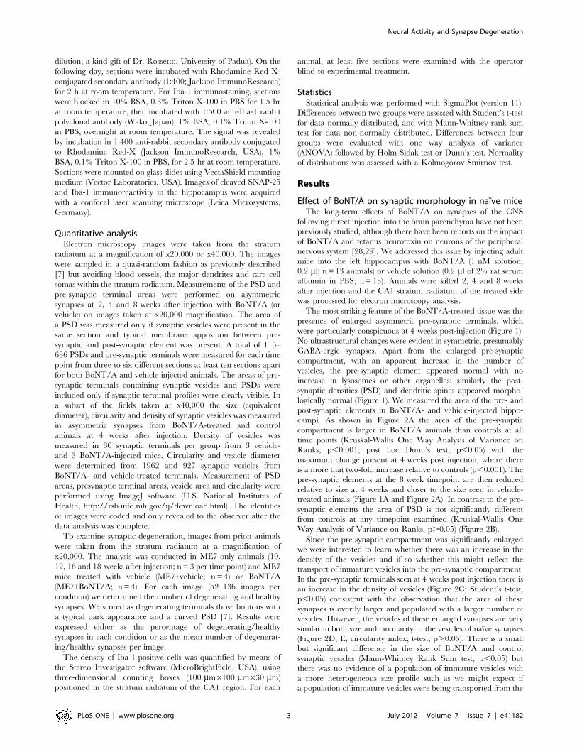
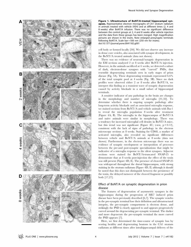
presence of enlarged asymmetric pre-synaptic terminals, whichwere particularly conspicuous at 4 weeks post-injection (Figure 1).No ultrastructural changes were evident in symmetric, presumablyGABA-ergic synapses. Apart from the enlarged pre-synapticcompartment, with an apparent increase in the number ofvesicles, the pre-synaptic element appeared normal with noincrease in lysosomes or other organelles: similarly the post-synaptic densities (PSD) and dendritic spines appeared morpho-logically normal (Figure 1). We measured the area of the pre- andpost-synaptic elements in BoNT/A- and vehicle-injected hippo-campi. As shown in Figure 2A the area of the pre-synapticcompartment is larger in BoNT/A animals than controls at alltime points (Kruskal-Wallis One Way Analysis of Variance onRanks, p,0.001; post hoc Dunn’s test, p,0.05) with themaximum change present at 4 weeks post injection, where thereis a more that two-fold increase relative to controls (p,0.001). Thepre-synaptic elements at the 8 week timepoint are then reducedrelative to size at 4 weeks and closer to the size seen in vehicle-treated animals (Figure 1A and Figure 2A). In contrast to the pre-synaptic elements the area of PSD is not significantly differentfrom controls at any timepoint examined (Kruskal-Wallis OneWay Analysis of Variance on Ranks, p.0.05) (Figure 2B).Since the pre-synaptic compartment was significantly enlarged
we were interested to learn whether there was an increase in thedensity of the vesicles and if so whether this might reflect thetransport of immature vesicles into the pre-synaptic compartment.In the pre-synaptic terminals seen at 4 weeks post injection there isan increase in the density of vesicles (Figure 2C; Student’s t-test,p,0.05) consistent with the observation that the area of thesesynapses is overtly larger and populated with a larger number ofvesicles. However, the vesicles of these enlarged synapses are verysimilar in both size and circularity to the vesicles of naıve synapses(Figure 2D, E; circularity index, t-test, p.0.05). There is a smallbut significant difference in the size of BoNT/A and controlsynaptic vesicles (Mann-Whitney Rank Sum test, p,0.05) butthere was no evidence of a population of immature vesicles witha more heterogeneous size profile such as we might expect ifa population of immature vesicles were being transported from the
Neural Activity and Synapse Degeneration
PLoS ONE | www.plosone.org 3 July 2012 | Volume 7 | Issue 7 | e41182
cell body or formed locally [30]. We did not observe any increasein dense core vesicles, also associated with synapse development, inthe BoNT/A treated animals (data not shown).There was no evidence of neuronal/synaptic degeneration in
the EM sections analyzed 2 or 8 weeks after BoNT/A injection.However, in the animals sacrificed at 4 weeks, we detected a subsetof dark, electron-dense synapses with ‘‘curved’’ PSDs, thatresemble degenerating terminals seen in early stages of priondisease (Fig. 3A). These degenerating terminals represented 6.6%of the total synaptic pool at 4 weeks (Fig. 3B). Since no darkprofiles were observed either 2 or 8 weeks after BoNT/A, weinterpret this finding as a transient wave of synaptic degenerationcaused by activity blockade in a small subset of hippocampalterminals.A sensitive indicator of any pathology in the brain are changes
in the morphology and number of microglia [31,32]. Todetermine whether there is ongoing synaptic pathology afterlong-term activity blockade and an associated microglia response,we stained sections from BoNT/A and vehicle animals with Iba-1to reveal the microglia population 8 weeks after treatment(Figure 4A, B). The microglia in the hippocampus of BoNT/Aand naıve animals were similar in morphology. There wasa tendency for increased microglial cell density in BoNT/A mice,but this trend was not significant (Figure 4C; t-test, p = 0.12),consistent with the lack of signs of pathology in the electronmicroscopy sections at 8 weeks. Staining for CD68, a marker ofactivated microglia, also revealed no significant differencesbetween vehicle and BoNT/A animals at 8 weeks (data notshown). Furthermore, in the electron microscopy there was noevidence of synaptic envelopment or interposition of processesbetween the pre-and post-synaptic specializations that might beindicative of a microglia response to the silent synapses. Adjacentsections were stained for BoNT/A-truncated SNAP-25 todemonstrate that at 8 weeks post-injection the effect of the toxinwas still present (Figure 4D, E). The presence of cleaved SNAP-25was widespread throughout the dorsal hippocampus, with strongstaining in the stratum radiatum (Figure 4D, E) although it shouldbe noted that this does not distinguish between the persistence ofthe toxin, the delayed turnover of the cleaved fragment or possiblyboth [17,27].
Effect of BoNT/A on synaptic degeneration in priondiseaseThe features of degeneration of asymmetric synapses in the
hippocampus during the progression of ME7 induced priondisease have been previously described [6,7]. The synaptic vesiclesin the pre-synaptic terminal lose their definition and ultrastructuralintegrity, the pre-synaptic compartment is electron dense, andstrikingly the PSD is closely apposed to and appears progressivelycurved around the degenerating pre-synaptic terminal. The darkerand more degenerate the pre-synaptic terminal the more curvedthe PSD appears [7].Here, we first determined the time-course of synaptic loss by
scoring healthy and degenerating boutons in the CA1 stratumradiatum at different times after intrahippocampal delivery of the
Figure 1. Ultrastructure of BoNT/A-treated hippocampal syn-apses. Representative electron micrographs of CA1 stratum radiatumin animals treated with vehicle (VEH) and at different times (2, 4 and8 weeks) after BoNT/A infusion. There was no significant differencebetween the control groups at 2, 4 and 8 weeks after vehicle injectionand the data from these groups has been merged. High magnificationpictures are shown in the insets. Note enlarged presynaptic terminalsfollowing BoNT/A. Scale bar = 500 nm (250 nm for insets).doi:10.1371/journal.pone.0041182.g001
Neural Activity and Synapse Degeneration
PLoS ONE | www.plosone.org 4 July 2012 | Volume 7 | Issue 7 | e41182
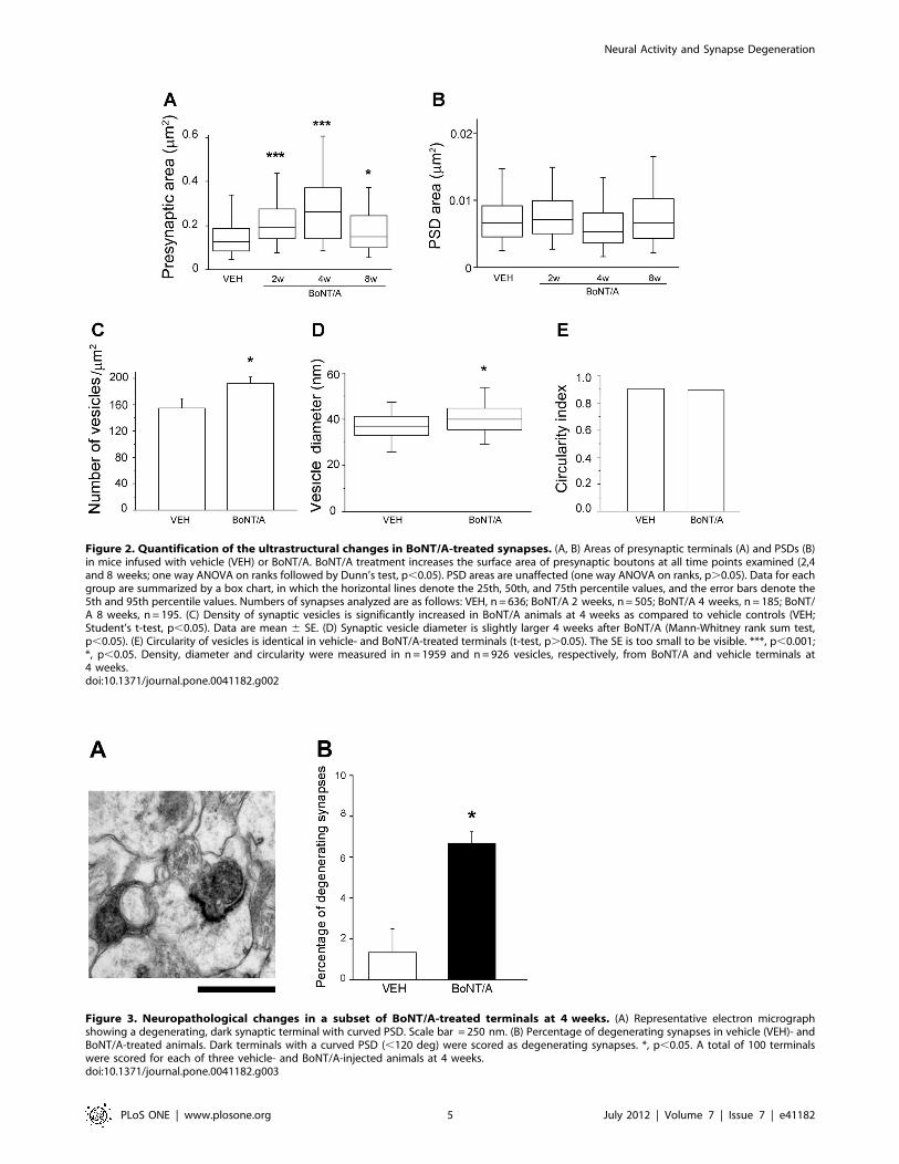
Figure 2. Quantification of the ultrastructural changes in BoNT/A-treated synapses. (A, B) Areas of presynaptic terminals (A) and PSDs (B)in mice infused with vehicle (VEH) or BoNT/A. BoNT/A treatment increases the surface area of presynaptic boutons at all time points examined (2,4and 8 weeks; one way ANOVA on ranks followed by Dunn’s test, p,0.05). PSD areas are unaffected (one way ANOVA on ranks, p.0.05). Data for eachgroup are summarized by a box chart, in which the horizontal lines denote the 25th, 50th, and 75th percentile values, and the error bars denote the5th and 95th percentile values. Numbers of synapses analyzed are as follows: VEH, n = 636; BoNT/A 2 weeks, n = 505; BoNT/A 4 weeks, n = 185; BoNT/A 8 weeks, n = 195. (C) Density of synaptic vesicles is significantly increased in BoNT/A animals at 4 weeks as compared to vehicle controls (VEH;Student’s t-test, p,0.05). Data are mean 6 SE. (D) Synaptic vesicle diameter is slightly larger 4 weeks after BoNT/A (Mann-Whitney rank sum test,p,0.05). (E) Circularity of vesicles is identical in vehicle- and BoNT/A-treated terminals (t-test, p.0.05). The SE is too small to be visible. ***, p,0.001;*, p,0.05. Density, diameter and circularity were measured in n = 1959 and n=926 vesicles, respectively, from BoNT/A and vehicle terminals at4 weeks.doi:10.1371/journal.pone.0041182.g002
Figure 3. Neuropathological changes in a subset of BoNT/A-treated terminals at 4 weeks. (A) Representative electron micrographshowing a degenerating, dark synaptic terminal with curved PSD. Scale bar = 250 nm. (B) Percentage of degenerating synapses in vehicle (VEH)- andBoNT/A-treated animals. Dark terminals with a curved PSD (,120 deg) were scored as degenerating synapses. *, p,0.05. A total of 100 terminalswere scored for each of three vehicle- and BoNT/A-injected animals at 4 weeks.doi:10.1371/journal.pone.0041182.g003
Neural Activity and Synapse Degeneration
PLoS ONE | www.plosone.org 5 July 2012 | Volume 7 | Issue 7 | e41182
ME7 prion agent. Sections were taken from ME7 mice ofa previous study [7]. We found that on average about 30% of theboutons were degenerating at 10 weeks. As the disease progresses,the ratio of degenerating synapses increased to reach a maximum16–18 weeks after ME7 (Fig. 5A). In parallel, the proportion ofhealthy terminals decreased steadily from 10 to 18 weeks (Fig. 5A).On the basis of these findings, we selected the 12 weeks time-pointfor the analysis of the effects of synaptic blockade on synapsedegeneration. Indeed, this time-point corresponds to an in-termediate phase of the degeneration process that allows us tomeasure BoNT/A-mediated decreases or increases in the degreeof synaptic loss.We have previously shown that immediately after the injection
of ME7-infected or normal brain homogenate into the mousehippocampus there is an acute inflammatory response to thedelivery of a high concentration of protein but this resolves by4 weeks [33]. We thus injected ME7-animals with BoNT/A (n= 4)or vehicle (n = 4) into the dorsal hippocampus at 4 weeks after theinitiation of the disease. All animals were killed 12 weeks after
ME7. Since the BoNT/A serotype has very long lasting effects[17,27] we reasoned that silencing synaptic activity for manyweeks prior to the onset of significant synaptic degeneration ratherthan acutely would provide the most likely scenario for an impacton synapse survival.We found that at 12 weeks after the initiation of disease the
ratio of degenerating to intact synapses was the same (Student’s t-test, p = 0.67) in both the ME7 animals from Siskova et al. (2009)and the ME7+vehicle animals used in this study (Fig. 5A, B). InME7+BoNT/A animals it was apparent that there were asym-metric synapses undergoing degeneration with the same features asthose seen in ME7+vehicle animals, with loss of vesicle integrity,electron dense pre-synaptic compartment and curvature of thePSD around the degenerating pre-synaptic component (Fig. 6).The post-synaptic dendritic spine showed no signs of degeneration(Fig. 6). Quantification of the ratio of degenerating synapses tototal synapses revealed no significant differences between ME7+-vehicle animals (33.765.4%) and ME7+BoNT/A mice(33.8611.5%; Student’s t-test, p = 0.97; Fig. 5B). We also analyzed
Figure 4. Lack of significant microglial activation 8 weeks after BoNT/A. (A, B) Representative immunostaining for the microglial marker Iba-1 in the CA1 stratum radiatum of mice treated with vehicle (A, VEH) and BoNT/A (B). Scale bar = 100 mm. (C) Cell countings reveal no significantdifferences in microglial density between vehicle and BoNT/A-infused animals (t-test, p.0.05). (D, E) Immunostaining for cleaved SNAP-25 (red)demonstrates strong labeling in the CA1 stratum radiatum of BoNT/A-treated animals (E). No labeling is evident in vehicle mice (D). Green, Yoyo-1nuclear counterstaining. Pyr, stratum pyramidale; s.r., stratum radiatum. Scale bar = 100 mm.doi:10.1371/journal.pone.0041182.g004
Neural Activity and Synapse Degeneration
PLoS ONE | www.plosone.org 6 July 2012 | Volume 7 | Issue 7 | e41182
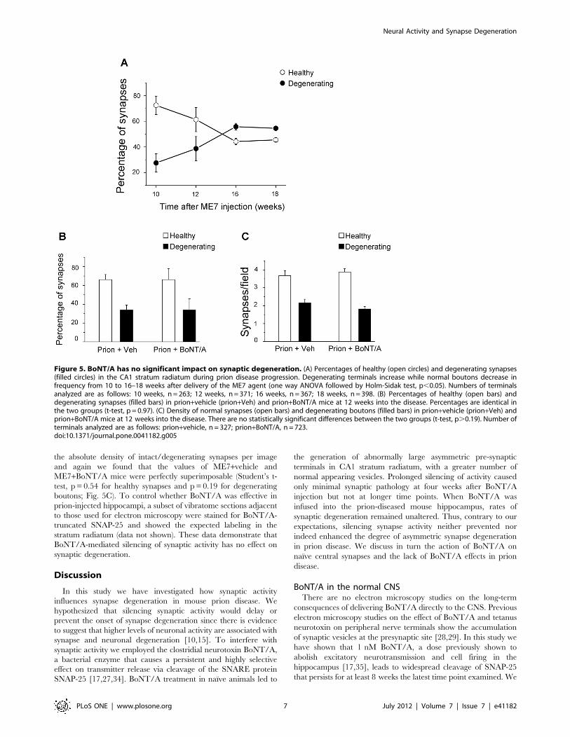
the absolute density of intact/degenerating synapses per imageand again we found that the values of ME7+vehicle andME7+BoNT/A mice were perfectly superimposable (Student’s t-test, p = 0.54 for healthy synapses and p=0.19 for degeneratingboutons; Fig. 5C). To control whether BoNT/A was effective inprion-injected hippocampi, a subset of vibratome sections adjacentto those used for electron microscopy were stained for BoNT/A-truncated SNAP-25 and showed the expected labeling in thestratum radiatum (data not shown). These data demonstrate thatBoNT/A-mediated silencing of synaptic activity has no effect onsynaptic degeneration.
Discussion
In this study we have investigated how synaptic activityinfluences synapse degeneration in mouse prion disease. Wehypothesized that silencing synaptic activity would delay orprevent the onset of synapse degeneration since there is evidenceto suggest that higher levels of neuronal activity are associated withsynapse and neuronal degeneration [10,15]. To interfere withsynaptic activity we employed the clostridial neurotoxin BoNT/A,a bacterial enzyme that causes a persistent and highly selectiveeffect on transmitter release via cleavage of the SNARE proteinSNAP-25 [17,27,34]. BoNT/A treatment in naıve animals led to
the generation of abnormally large asymmetric pre-synapticterminals in CA1 stratum radiatum, with a greater number ofnormal appearing vesicles. Prolonged silencing of activity causedonly minimal synaptic pathology at four weeks after BoNT/Ainjection but not at longer time points. When BoNT/A wasinfused into the prion-diseased mouse hippocampus, rates ofsynaptic degeneration remained unaltered. Thus, contrary to ourexpectations, silencing synapse activity neither prevented norindeed enhanced the degree of asymmetric synapse degenerationin prion disease. We discuss in turn the action of BoNT/A onnaıve central synapses and the lack of BoNT/A effects in priondisease.
BoNT/A in the normal CNSThere are no electron microscopy studies on the long-term
consequences of delivering BoNT/A directly to the CNS. Previouselectron microscopy studies on the effect of BoNT/A and tetanusneurotoxin on peripheral nerve terminals show the accumulationof synaptic vesicles at the presynaptic site [28,29]. In this study wehave shown that 1 nM BoNT/A, a dose previously shown toabolish excitatory neurotransmission and cell firing in thehippocampus [17,35], leads to widespread cleavage of SNAP-25that persists for at least 8 weeks the latest time point examined. We
Figure 5. BoNT/A has no significant impact on synaptic degeneration. (A) Percentages of healthy (open circles) and degenerating synapses(filled circles) in the CA1 stratum radiatum during prion disease progression. Degenerating terminals increase while normal boutons decrease infrequency from 10 to 16–18 weeks after delivery of the ME7 agent (one way ANOVA followed by Holm-Sidak test, p,0.05). Numbers of terminalsanalyzed are as follows: 10 weeks, n = 263; 12 weeks, n = 371; 16 weeks, n = 367; 18 weeks, n = 398. (B) Percentages of healthy (open bars) anddegenerating synapses (filled bars) in prion+vehicle (prion+Veh) and prion+BoNT/A mice at 12 weeks into the disease. Percentages are identical inthe two groups (t-test, p = 0.97). (C) Density of normal synapses (open bars) and degenerating boutons (filled bars) in prion+vehicle (prion+Veh) andprion+BoNT/A mice at 12 weeks into the disease. There are no statistically significant differences between the two groups (t-test, p.0.19). Number ofterminals analyzed are as follows: prion+vehicle, n = 327; prion+BoNT/A, n = 723.doi:10.1371/journal.pone.0041182.g005
Neural Activity and Synapse Degeneration
PLoS ONE | www.plosone.org 7 July 2012 | Volume 7 | Issue 7 | e41182

detected ultrastructural changes in asymmetric terminals in thestratum radiatum, the vast majority of which arise from axons ofCA3 pyramidal neurons. Following BoNT/A treatment, CA1terminals were not only enlarged but had a larger complement ofsynaptic vesicles. These synaptic vesicles had a normal shape and
size, and are suggestive of a continuing supply of mature vesiclesarriving at synapses and unable to fuse with the plasma membraneto release their neurotransmitter. There was no evidence ofimmature pleiomorphic vesicles [30] at the pre-synaptic terminal.Presynaptic terminal areas were maximally increased 4 weeks afterBoNT/A, with a significant relaxation at 8 weeks. The return ofthe presynaptic boutons towards their normal size at 8 weeks mayreflect an adaptation of the neuron to the lack of activity, as therewas clear evidence of persistence of high levels of cleaved SNAP-25 in the hippocampus at 8 weeks (present results and [17]). Ofinterest is that while the pre-synaptic terminals were enlarged, thePSDs did not show a compensatory change in the absence ofneurotransmitter release. It is not the case that PSDs can notenlarge since this is seen during progression of prion disease andinterpreted as a reactive response to the loss of synapses in thestratum radiatum [36].The increase in the density of synaptic vesicles in BoNT/A-
treated terminals is consistent with previous ultrastructural workon peripheral neurons incubated with clostridial toxins [28,29,34].Recently, Wree and collaborators reported axonal swellings andvaricosities following injection of BoNT/A into the rat striatum.These swellings might correspond to enlarged synaptic terminalsas they contained the biosynthetic enzymes for acetylcholine anddopamine [37].In addition to enlargement of terminals, BoNT/A caused the
appearance of a small percentage of dark, degenerating synapses4 weeks after injection. Since these dark profiles were not detectedat other times (2 and 8 weeks), we interpret this finding asa transient wave of degeneration triggered by the activity blockadein a subset of sensitive terminals. It is interesting to note that darksynapses have been seen in association with hypoglossal neuronsfollowing injection of BoNT/A into the tongue [38]. Recentevidence showing that BoNT/A may be transferred transneur-onally [17,27] suggests that the toxin may access the presynapticneuron and cause degeneration in some synapses innervating thehypoglossal motor neurons.In the long-term (8 weeks), BoNT/A caused no synaptic
degeneration and, consistently, there was the absence of a signif-icant microglia response as shown by normal microglial celldensities. It is interesting that the microglia do not respond to thewidespread silencing of synaptic activity in the CNS since recentstudies have suggested that microglia monitor synaptic health andactivity [39,40]. However, we cannot exclude that more subtlechanges in microglia morphology and/or function (not detectablewith Iba-1 labeling) occur as a result of synaptic silencing.
Activity and synaptic degenerationThe degeneration of asymmetric terminals is a conspicuous
feature of the early pathology in the ME7 model of prion diseaseand precedes any detectable degeneration of cell bodies of CA3 orCA1 pyramidal neurons [7,8]. The precise mechanisms leading tosynapse degeneration are poorly understood but as discussedelsewhere [8] do not appear to be simply related to theaccumulation of protease resistant prion protein (PrPSc). There isgrowing body or evidence to show in prion disease that theaccumulation of PrPSc is neither necessary or sufficient to lead toneuronal degeneration [41,42], but by analogy with studies inAlzheimer’s disease soluble oligomeric species of misfolded proteinmay be critical [43,44]. How the misfolded protein targets the pre-synaptic terminal leading to degeneration is unclear but earlychanges in the synaptic vesicle proteins [8] suggest that uptake ofthe misfolded peptide during the exocytosis-endocytosis cycle maybe involved. In keeping with this idea, elevated neural activity hasbeen shown to enhance synapse vulnerability in a mouse model of
Figure 6. Ultrastructural hallmarks of prion disease are notimpacted by BoNT/A treatment. Electron micrographs of CA1stratum radiatum of the hippocampus illustrating control, vehicle-exposed synapses (A, VEH) and neuropathological changes inprion+vehicle (prion + VEH; B) and prion+BoNT/A animals (C) at12 weeks following delivery of the ME7 agent. High magnificationpictures are shown in the insets. Note degenerating boutons withcurved PSDs in both prion+vehicle and prion+BoNT/A samples. Scalebar = 500 nm (250 nm for insets).doi:10.1371/journal.pone.0041182.g006
Neural Activity and Synapse Degeneration
PLoS ONE | www.plosone.org 8 July 2012 | Volume 7 | Issue 7 | e41182
synaptic degeneration [10]. In Alzheimer’s patients, brain areasthat develop the most Aß plaques also have the highest restingactivity [45]. We thus hypothesized that blockade of synapticactivity may arrest or delay synapse loss during chronicneurodegeneration.To test this idea, we employed the ME7 mouse model of prion
disease that has been previously characterized in detail [46]. Theadvantage of this model is that the neuropathology can be spatiallyand temporally controlled by intracerebral delivery of the prionagent. Injection of the ME7 agent into the hippocampus triggersa transient inflammatory response that resolves by 4 weeks [33],and synapse loss with associated behavioural impairments isdetected in the stratum radiatum by 12 weeks [47]. Synapticdegeneration increases between 10 and 12 weeks after ME7injection and reaches a plateau at 16–18 weeks (see Figure 5). Wethus chose to infuse BoNT/A in the prion-injected hippocampusat 4 weeks, many weeks prior to first significant loss of synapses butafter the first inflammatory phase, and assessed numbers ofhealthy/degenerating synapses in the CA1 stratum radiatum at12 weeks into the disease. Thus, synaptic exocytosis was blockedearly in disease evolution and the evidence indicates, from stainingfor cleaved SNAP-25, that even eight weeks after injection thetoxin is still active and blocking vesicle-mediated release ofneurotransmitter.We found no evidence that BoNT/A protected synapses in the
stratum radiatum from neurodegeneration, indeed the ratio ofdegenerating to intact synapses was the same in BoNT/A- andvehicle-treated animals (Fig. 5B). Absolute numbers of normal/dark terminals were also perfectly comparable between groups(Fig. 5C), ruling out an acceleration/slowing down of thedegeneration process over the weeks preceding the analysis. Amodel in which the misfolded protein requires neural activity tokill presynaptic terminals is clearly not tenable. In the case of Aßoligomers, it has been hypothesized that their direct binding toplasma membranes of neurons could perturb the fine structure ofthe phospholipid bilayer [44], thus resulting in signaling changesand toxic effects proceeding via pathways largely independent of
electrical activity. The lack of impact of BoNT/A on synapticdegeneration also suggests that toxic species of normal prionprotein (PrPc) or PrPSc must initiate neurodegeneration viaa receptor interaction that is not activity dependent. Similarly,our data provide little support for a sequential model, in which thedisease-associated neurotoxic agent (i.e. the prion agent) firstcauses synaptic dysfunction and then progresses through todegeneration of the same terminals.PrPSc may access the presynaptic element by pathways that are
not dependent on vesicle exo/endocytosis. It has been shown invitro that PrPc is rapidly internalized from the cell surface and re-cycled back again in association with LRP1 [48] and theconversion of PrPc to potentially toxic PrPSc may take place atthe plasma membrane before being internalized by clathrinindependent endocytosis [49]. If PrPc also mediates the impair-ment of synaptic plasticity of Aß oligomers [50,51], a similarmechanism of synaptic loss may operate in AD, although there islittle evidence of degenerating synapses in experimental studies onthe action of Aß peptides. It is of interest that the major effects ofAß studied to date interfere with synaptic efficacy at the post-synaptic site and inhibit long-term potentiation or enhance long-term depression [52]. Our data suggest that toxicity leading todegeneration involves different processes or perhaps differentpeptide from those interfering with transmission. The identifica-tion of these different species and the mechanisms of synapticdegeneration remains a significant challenge in protein misfoldingdiseases.
Acknowledgments
We thank Anton Page and Jan Collier for their excellent assistance withelectron microscopy.
Author Contributions
Conceived and designed the experiments: MC VHP. Performed theexperiments: MC LR ZS HAM VHP. Analyzed the data: LR EV ZS RM.Wrote the paper: MC VOC VHP.
References
1. Terry RD, Masliah E, Salmon DP, Butters N, DeTeresa R, et al. (1991) Physicalbasis of cognitive alterations in Alzheimer’s disease: synapse loss is the majorcorrelate of cognitive impairment. Ann Neurol 30: 572–580.
2. Dickey CA, Loring JF, Montgomery J, Gordon MN, Eastman PS, et al. (2003)Selectively reduced expression of synaptic plasticity-related genes in amyloidprecursor protein + presenilin-1 transgenic mice. J Neurosci 23: 5219–5226.
3. Rutten BP, Van der Kolk NM, Schafer S, van Zandvoort MA, Bayer TA, et al.(2005) Age-related loss of synaptophysin immunoreactive presynaptic boutonswithin the hippocampus of APP751SL, PS1M146L, and APP751SL/PS1M146L transgenic mice. Am J Pathol 167: 161–173.
4. Fischer LR, Culver DG, Tennant P, Davis AA, Wang M, et al. (2004)Amyotrophic lateral sclerosis is a distal axonopathy: evidence in mice and man.Exp Neurol 185: 232–240.
5. Frey D, Schneider C, Xu L, Borg J, Spooren W, et al. (2000) Early and selectiveloss of neuromuscular synapse subtypes with low sprouting competence inmotoneuron diseases. J Neurosci 20: 2534–2542.
6. Jeffrey M, Halliday WG, Bell J, Johnston AR, MacLeod NK, et al. (2000)Synapse loss associated with abnormal PrP precedes neuronal degeneration inthe scrapie-infected murine hippocampus. Neuropathol Appl Neurobiol 26: 41–54.
7. Siskova Z, Page A, O’Connor V, Perry VH (2009) Degenerating synapticboutons in prion disease: microglia activation without synaptic stripping.Am J Pathol 175: 1610–1621.
8. Gray BC, Siskova Z, Perry VH, O’Connor V (2009) Selective presynapticdegeneration in the synaptopathy associated with ME7-induced hippocampalpathology. Neurobiol Dis 35: 63–74.
9. Fernandez-Chacon R, Wolfel M, Nishimune H, Tabares L, Schmitz F, et al.(2004) The synaptic vesicle protein CSP alpha prevents presynaptic de-generation. Neuron 42: 237–251.
10. Garcia-Junco-Clemente P, Cantero G, Gomez-Sanchez L, Linares-Clemente P,Martinez-Lopez JA, et al. (2010) Cysteine string protein-alpha prevents activity-dependent degeneration in GABAergic synapses. J Neurosci 30: 7377–7391.
11. Koch P, Breuer P, Peitz M, Jungverdorben J, Kesavan J, et al. (2011) Excitation-induced ataxin-3 aggregation in neurons from patients with Machado-Josephdisease. Nature 480: 543–546.
12. Cirrito JR, Kang JE, Lee J, Stewart FR, Verges DK, et al. (2008) Endocytosis isrequired for synaptic activity-dependent release of amyloid-beta in vivo. Neuron58: 42–51.
13. Cirrito JR, Yamada KA, Finn MB, Sloviter RS, Bales KR, et al. (2005) Synapticactivity regulates interstitial fluid amyloid-beta levels in vivo. Neuron 48: 913–922.
14. Wei W, Nguyen LN, Kessels HW, Hagiwara H, Sisodia S, et al. (2010) Amyloidbeta from axons and dendrites reduces local spine number and plasticity. NatNeurosci 13: 190–196.
15. Pun S, Santos AF, Saxena S, Xu L, Caroni P (2006) Selective vulnerability andpruning of phasic motoneuron axons in motoneuron disease alleviated byCNTF. Nat Neurosci 9: 408–419.
16. Antonucci F, Di Garbo A, Novelli E, Manno I, Sartucci F, et al. (2008)Botulinum neurotoxin E (BoNT/E) reduces CA1 neuron loss and granule celldispersion, with no effects on chronic seizures, in a mouse model of temporallobe epilepsy. Exp Neurol 210: 388–401.
17. Antonucci F, Rossi C, Gianfranceschi L, Rossetto O, Caleo M (2008) Long-distance retrograde effects of botulinum neurotoxin A. J Neurosci 28: 3689–3696.
18. Caleo M, Restani L, Gianfranceschi L, Costantin L, Rossi C, et al. (2007)Transient synaptic silencing of developing striate cortex has persistent effects onvisual function and plasticity. J Neurosci 27: 4530–4540.
19. Bozzi Y, Costantin L, Antonucci F, Caleo M (2006) Action of botulinumneurotoxins in the central nervous system: antiepileptic effects. Neurotox Res 9:197–203.
20. Davletov B, Bajohrs M, Binz T (2005) Beyond BOTOX: advantages andlimitations of individual botulinum neurotoxins. Trends Neurosci 28: 446–452.
21. Johnson EA (1999) Clostridial toxins as therapeutic agents: benefits of nature’smost toxic proteins. Annu Rev Microbiol 53: 551–575.
Neural Activity and Synapse Degeneration
PLoS ONE | www.plosone.org 9 July 2012 | Volume 7 | Issue 7 | e41182
22. Montecucco C, Molgo J (2005) Botulinal neurotoxins: revival of an old killer.Curr Opin Pharmacol 5: 274–279.
23. Simpson DM, Blitzer A, Brashear A, Comella C, Dubinsky R, et al. (2008)Assessment: Botulinum neurotoxin for the treatment of movement disorders (anevidence-based review): report of the Therapeutics and Technology AssessmentSubcommittee of the American Academy of Neurology. Neurology 70: 1699–1706.
24. Simpson DM, Gracies JM, Graham HK, Miyasaki JM, Naumann M, et al.(2008) Assessment: Botulinum neurotoxin for the treatment of spasticity (anevidence-based review): report of the Therapeutics and Technology AssessmentSubcommittee of the American Academy of Neurology. Neurology 70: 1691–1698.
25. Cunningham C, Wilcockson DC, Campion S, Lunnon K, Perry VH (2005)Central and systemic endotoxin challenges exacerbate the local inflammatoryresponse and increase neuronal death during chronic neurodegeneration.J Neurosci 25: 9275–9284.
26. Matak I, Bach-Rojecky L, Filipovic B, Lackovic Z (2011) Behavioral andimmunohistochemical evidence for central antinociceptive activity of botulinumtoxin A. Neuroscience 186: 201–207.
27. Restani L, Antonucci F, Gianfranceschi L, Rossi C, Rossetto O, et al. (2011)Evidence for anterograde transport and transcytosis of botulinum neurotoxin A(BoNT/A). J Neurosci 31: 15650–15659.
28. Hunt JM, Bommert K, Charlton MP, Kistner A, Habermann E, et al. (1994) Apost-docking role for synaptobrevin in synaptic vesicle fusion. Neuron 12: 1269–1279.
29. Kao I, Drachman DB, Price DL (1976) Botulinum toxin: mechanism ofpresynaptic blockade. Science 193: 1256–1258.
30. Ahmari SE, Buchanan J, Smith SJ (2000) Assembly of presynaptic active zonesfrom cytoplasmic transport packets. Nat Neurosci 3: 445–451.
31. Kreutzberg GW (1996) Microglia: a sensor for pathological events in the CNS.Trends Neurosci 19: 312–318.
32. Ransohoff RM, Perry VH (2009) Microglial physiology: unique stimuli,specialized responses. Annu Rev Immunol 27: 119–145.
33. Betmouni S, Perry VH (1999) The acute inflammatory response in CNSfollowing injection of prion brain homogenate or normal brain homogenate.Neuropathol Appl Neurobiol 25: 20–28.
34. Schiavo G, Matteoli M, Montecucco C (2000) Neurotoxins affectingneuroexocytosis. Physiol Rev 80: 717–766.
35. Capogna M, McKinney RA, O’Connor V, Gahwiler BH, Thompson SM (1997)Ca2+ or Sr2+ partially rescues synaptic transmission in hippocampal culturestreated with botulinum toxin A and C, but not tetanus toxin. J Neurosci 17:7190–7202.
36. Siskova Z, Sanyal NK, Orban A, O’Connor V, Perry VH (2010) Reactivehypertrophy of synaptic varicosities within the hippocampus of prion-infectedmice. Biochem Soc Trans 38: 471–475.
37. Wree A, Mix E, Hawlitschka A, Antipova V, Witt M, et al. (2011) Intrastriatalbotulinum toxin abolishes pathologic rotational behaviour and induces axonalvaricosities in the 6-OHDA rat model of Parkinson’s disease. Neurobiol Dis 41:291–298.
38. Sumner BE (1977) Ultrastructural responses of the hypoglossal nucleus to thepresence in the tongue of botulinum toxin, a quantitative study. Exp Brain Res30: 313–321.
39. Graeber MB (2010) Changing face of microglia. Science 330: 783–788.40. Tremblay ME, Lowery RL, Majewska AK (2010) Microglial interactions with
synapses are modulated by visual experience. PLoS Biol 8: e1000527.41. Barron RM, Campbell SL, King D, Bellon A, Chapman KE, et al. (2007) High
titers of transmissible spongiform encephalopathy infectivity associated withextremely low levels of PrPSc in vivo. J Biol Chem 282: 35878–35886.
42. Lasmezas CI, Deslys JP, Robain O, Jaegly A, Beringue V, et al. (1997)Transmission of the BSE agent to mice in the absence of detectable abnormalprion protein. Science 275: 402–405.
43. Haass C, Selkoe DJ (2007) Soluble protein oligomers in neurodegeneration:lessons from the Alzheimer’s amyloid beta-peptide. Nat Rev Mol Cell Biol 8:101–112.
44. Selkoe DJ (2011) Resolving controversies on the path to Alzheimer’stherapeutics. Nat Med 17: 1060–1065.
45. Buckner RL, Snyder AZ, Shannon BJ, LaRossa G, Sachs R, et al. (2005)Molecular, structural, and functional characterization of Alzheimer’s disease:evidence for a relationship between default activity, amyloid, and memory.J Neurosci 25: 7709–7717.
46. Perry VH (2010) Contribution of systemic inflammation to chronic neurode-generation. Acta Neuropathol 120: 277–286.
47. Cunningham C, Deacon R, Wells H, Boche D, Waters S, et al. (2003) Synapticchanges characterize early behavioural signs in the ME7 model of murine priondisease. Eur J Neurosci 17: 2147–2155.
48. Parkyn CJ, Vermeulen EG, Mootoosamy RC, Sunyach C, Jacobsen C, et al.(2008) LRP1 controls biosynthetic and endocytic trafficking of neuronal prionprotein. J Cell Sci 121: 773–783.
49. Goold R, Rabbanian S, Sutton L, Andre R, Arora P, et al. (2011) Rapid cell-surface prion protein conversion revealed using a novel cell system. NatCommun 2: 281.
50. Gimbel DA, Nygaard HB, Coffey EE, Gunther EC, Lauren J, et al. (2010)Memory impairment in transgenic Alzheimer mice requires cellular prionprotein. J Neurosci 30: 6367–6374.
51. Lauren J, Gimbel DA, Nygaard HB, Gilbert JW, Strittmatter SM (2009) Cellularprion protein mediates impairment of synaptic plasticity by amyloid-betaoligomers. Nature 457: 1128–1132.
52. Ondrejcak T, Klyubin I, Hu NW, Barry AE, Cullen WK, et al. (2010)Alzheimer’s disease amyloid beta-protein and synaptic function. Neuromole-cular Med 12: 13–26.
Neural Activity and Synapse Degeneration
PLoS ONE | www.plosone.org 10 July 2012 | Volume 7 | Issue 7 | e41182


![ReviewHeat shock protein 90 in neurodegenerative diseases€¦ · induced motor-neuron degeneration as well as progres-sive motor-neuron degeneration models [18]. In various cellular](https://static.fdocuments.in/doc/165x107/60c102d180a92345cc5a648c/reviewheat-shock-protein-90-in-neurodegenerative-diseases-induced-motor-neuron-degeneration.jpg)