THE PRACTICAL GUIDE TO CDG FAMILIES - Gua metab³lica
96
1 THE PRACTICAL GUIDE TO CDG FAMILIES: A project done in collaboration between families, researchers and Health Care experts
Transcript of THE PRACTICAL GUIDE TO CDG FAMILIES - Gua metab³lica

1
THE PRACTICAL GUIDE
TO CDG FAMILIES:
A project done in collaboration
between families, researchers
and Health Care experts

2
"For you, wonderful human beings, owners of a smile that make us believe
in dreams, that fill us with hope and give us the" driving force "needed to fight ..."
For you, the person with whom I have silent conversations, that put sense
in my life and for whom I feel a misunderstood love”.
AD-T
Vanessa Ferreira

3
Index
Prologue………………………………………………………………………………………………………5
Acknowledgements……………………………………………………………………………………...7
Chapter 1. The Molecular and Cellular CDG point of view………………………..11
Chapter 2. The CDG genetic landscape……………………………………………………..24
Chapter 3. PMM-CDG (CDG-Ia): Clinical manifestations and their
management
Introduction………………………………………………………………………………………………..33
3.1. Neurologic Clinical Issues………………………………………………………………….37
3.2. Gastrointestinal………………………………………………………………………………..41
3.3. Hepathology……………………………………………………………………………………...45
3.4. Kidney……………………………………………………………………………………………...46
3.5. Coagulopathy…………………………………………………………………………………….47
3.6. Endocrinology…………………………………………………………………………………..48
3.7. Ophthalmic manifestations……………………………………………………………….50
3.8. Orthopedics, The musculoskeletal system in CDG…………………………….57
3.9. Cardiology……………………………………………………………………..………………….60
3.10. Hematology……………………………………………………………………………………….62
3.11. Immunology……………………………………………………………………………………...64
3.12. General concerns………………………………………………………………………………65
Chapter 4. CDG and the Hospital urgency…………………………………………………….68
Chapter 5. Diagnostic tools…………………………………………………………………………70
Chapter 6. Treatment and therapies……………………………………………………………73
4
Chapter 7. CDG management of development…………………………………………….75
Chapter 8. The importance of research and communication and
dissemination in Rare Diseases………………………………………………………………….84
Glossary………………………………………………………………………………………………………90
The empowerment of CDG patient’s voice…………………………………………………..93

5
Prologue
When a person faces, for the first time, the words “Congenital Disorders of
Glycosylation”, “CDG”, and “Rare Genetic Disease”, many concerns are raised. To
have a rare genetic disease means having a diagnosis that is not well recognized or
understood by the medical and scientific communities as there are so many
different disorders. In fact, it is estimated that there are between 6,000-8,000
different rare diseases. Obtaining the correct diagnosis very often means many
years of waiting! Beyond the diagnosis, treatment for symptoms which can
improve the quality and even the life expectancy of an affected individual, can take
years.
It is important to bear in mind that usually these diseases are chameleon-
like. Thus, they are characterized by a broad range of symptoms that vary not only
from disease to disease but also from patient to patient suffering from the same
disease. Relatively common symptoms can hide underlying rare diseases, leading
to misdiagnosis.
The Portuguese Association for CDG and other Rare Metabolic Diseases
(Associação Portuguesa CDG e outras Doenças Metabólica sRaras (APCDG-DMR),
together with the Spanish Association for the same disorder aims to educate
families and the public about Rare Metabolic Diseases, mainly focused on
Congenital Disorders of Glycosylation. In addition, we will advocate for the rights
of this group, and aim to improve the quality of life of individuals with CDG and
other rare metabolic diseases. Being part of the Association represents an
opportunity to unite our voices in one direction: to help families with the same
problems and to contribute to increasing the medical and scientific knowledge
about these disorders.
It is my honor and privilege to present to you this project that I have wanted
to establish for a longtime, meanwhile I started to write my thesis! Everything
started with an e-mail directed to Professors Jaak Jaeken and Gert Matthijs, Dr. Paz
Briones, Dr. Rafael Artuch, Dr. Célia Pérez-Cerdá and Dr. Belén Pérez Dueñas sent
the 26th of July 2010 (12:46 am!). Their response was positive and extremely

6
supportive. Furthermore, other collaborators demonstrated their willingness to
participate in this project.
We aim to share many perspectives on Congenital Disorders of
Glycosylation with the CDG community and the broader society.
Think Metabolic, Think CDG!
Vanessa Ferreira, PhD
(Portuguese Association for CDG and other Rare Metabolic Diseases)

7
Acknowledgements
This "Practical Guide for CDG Families” is the direct result of many people
who have participated in meetings, the exchange of e-mails, the elaboration of
texts, multiple corrections and translations, and provided comments and
proofreading for final approval!
To all of you, I want to express my deepest and sincere THANKS!
I want to especially express my sincere gratitude to Dr. Donna Krasnewich,
who offered invaluable comments, enthusiastic revision and input to this
important resource targeted to CDG families!
I sincerely thank Dr. Maria Antonia Vilaseca, Dr. Belén Pérez Dueñas,
Professor Jaak Jaeken, Andrea Berarducci and Merell Liddle, for their support,
patience and continued interest during the preparation of this work.
I would like to extend my gratitude to CDG families, because many of these
questions were compiled by the Spanish and Portuguese parents through an
informal survey, and many others are based on my personal curiosities.
Finally, I want to thank all people who believe in the voice of individuals
with CDG!
To all of you, THANK YOU!

8
© Original Idea, Project Coordinator, Design and Layout:
Vanessa Ferreira (Associação Portuguesa CDG e outras Doenças Metabólicas
Raras).
Translation
Revision of overall translation and content: Donna Krasnewich M.D., Ph.D.
(Program Director, NIGMS, NIH, USA).
Translation: Belén Pérez-Dueñas, Melisa Stitzman, Merell Liddle, Andrea
Berarducci and Vanessa Ferreira.
List of collaborators who participated as volunteers in the preparation of this
Guide
(listed in the order of appearance in the chapters of the Guide:
Maria Antonia Vilaseca Ph.D. (Guia Metabólica, Hospital Sant Joan de Déu,
Barcelona, España).
Vanessa Ferreira, Ph.D. (Associação Portuguesa CDG e outras Doenças Metabólicas
Raras, Portugal) and Liliana’s sister.
Célia Pérez-Cerdá, Ph.D. (Centro de Diagnóstico de Enfermedades Moleculares,
Centro de Biología Molecular, CIBERER, Universidad Autónoma de Madrid, Spain).
Belén Pérez González ,Ph.D. (Centro de Diagnóstico de Enfermedades Moleculares,
Centro de Biología Molecular, CIBERER, Universidad Autónoma de Madrid, Spain).
Jaak Jaeken, M.D., Ph.D. (Center for Metabolic Disease, Katholieke Universiteit
Leuven, Belgium).
Belén Pérez Dueñas, M.D., Ph.D. (Neurology Department Hospital Sant Joan de
Déu, Barcelona, España).
Ruth García Romero, M.D. (Gastroenterology, Hepatology and Child nutrition
Section, Metabolic Diseases Unity, Hospital Sant Joan de Déu, Barcelona).

9
Mercedes Serrano, M.D., Ph.D. (Neurology Department Hospital Sant Joan de Déu,
Barcelona, Spain).
Daisy Rymen (CDG Ph.D. student), Center for Human Genetics, Gert Matthijs
Laboratory, Leuven, Bélgica).
Luis Terricabras Carol, M.D., Ph.D. (Orthopedic Unity, Hospital Sant Joan de Déu,
Barcelona)
Mario Sanz Cuesta, M.D. (Pediatrician Unity, Parc Sanitari Sant Joan de Déu, Sant
Boi de Llobregat).
Donna Krasnewich M.D., Ph.D. (Program Director at the National Institute of
General Medical Sciences, USA)
Mercedes Pineda Marfà, M.D., Ph.D.( Pediatrician Unity ,Hospital Sant Joan de Déu,
Barcelona, España).
Paz Briones Godino, Ph.D. (IBC. Secció d’Errors Congènits del Metabolisme,
Hospital Clínic, CSIC, España).
Rafael Artuch Iriberri, M.D., Ph.D. (Unidad de Bioquímica Clínica, Hospital Sant Joan
de Déu, Barcelona).
Merell Liddle, Australian CDG patient representative and Morgan’s mother.
Andrea Berarducci, USA CDG patient representative and Bianca’s mother.
Beatriz Sanz, Child Occupational Therapist, Professor at the Rey Juan Carlos
University. Alcorcón. Madrid. Spain)
Paula Davila Martinez (Physiotherapist specialized in the Bobath Concept (babies,
child and adult). C.P.E.E. Princesa Sofia (Madrid)
María Luisa Pendas Sánchez, Psychologist specialized in Early Attention and
Psychomototricity, Therapeutic Pedagogical Teacher.
Mafalda Araújo, Ph.D. (Basic researcher at Instituto de Biologia Molecular e Celular,
Porto, Portugal.IBMC, Porto, Portugal).
10
Sebastián Sánchez, (PhD in Information Science. Bachelor in Communication and
History. Research Group on Disability and Communication (GIDyC). Professor at
the University of Valencia).

11
This guide focuses on PMM2-CDG (CDG-Ia).
Chapter 1. The Molecular and Cellular CDG point of view
Authors and translators: Maria Antonia Vilaseca Ph.D. (Guia Metabólica, Hospital Sant Joan de Déu, Barcelona, España) and Vanessa Ferreira, Ph.D. (Associação Portuguesa CDG e outras Doenças Metabólicas Raras)
Illustrations: Vanessa Ferreira, Ph.D. (Associação Portuguesa CDG e outras Doenças Metabólicas Raras)
What is the DNA?
DNA is a complex molecule that contains the instructions needed to synthesize
proteins. Proteins are components of cells and organs, and have a huge variety of
different functions. Therefore, the proper functioning of a cell and an organ
depends on the sequence of DNA. DNA is in the nucleus of a cell (named genomic
DNA) and is like a book, the information is contained in genes, which are like
words in the book. Information in DNA is inherited from our parents and
determines who we are and how our body functions.
12
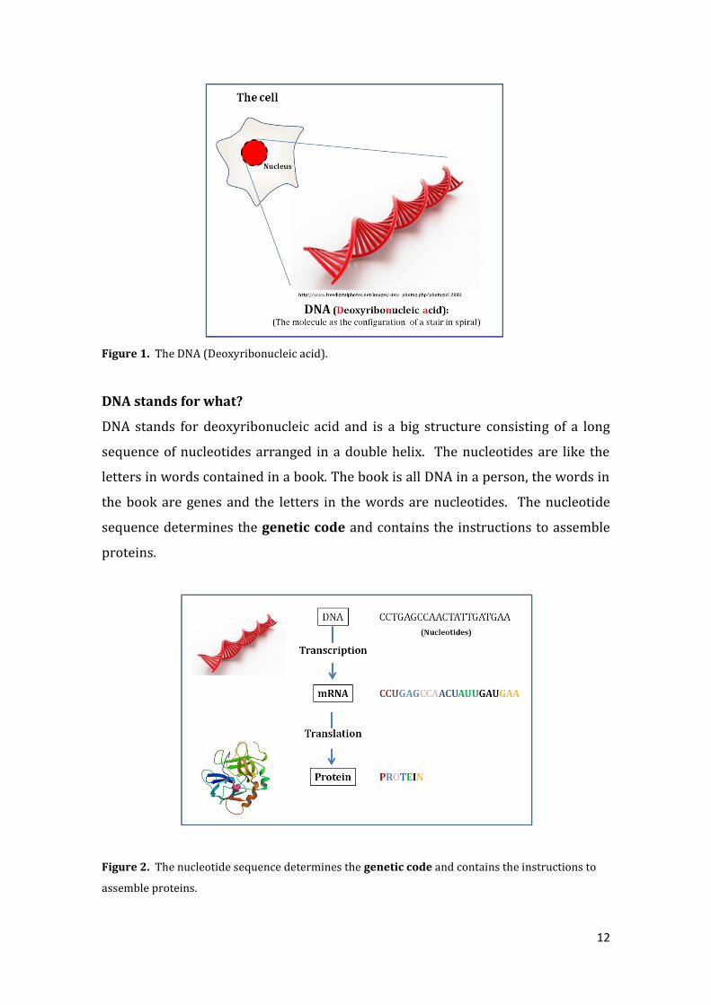
Figure 1. The DNA (Deoxyribonucleic acid).
DNA stands for what?
DNA stands for deoxyribonucleic acid and is a big structure consisting of a long
sequence of nucleotides arranged in a double helix. The nucleotides are like the
letters in words contained in a book. The book is all DNA in a person, the words in
the book are genes and the letters in the words are nucleotides. The nucleotide
sequence determines the genetic code and contains the instructions to assemble
proteins.
Figure 2. The nucleotide sequence determines the genetic code and contains the instructions to
assemble proteins.

13
What is a gene?
The minimal DNA sequence that is capable of encoding a protein (cellular function)
is called a gene. It is comparable to the words made with the letters (nucleotides)
and located in the book (genomic DNA). Humans have about 20,000 genes coded
in their entire DNA sequence.
Figure 3. The gene is the minimal DNA sequence that is capable of encoding a protein.
Locus and Allele:
Locus is the unique chromosomal location defining the position of an individual
gene within the entire DNA sequence, in other words, where the word (gene) is
located in the book (genomic DNA). An allele of a gene is the version of the
sequence at that locus. There may be a few different sequences that code for a
protein that function normally, these would all be considered normal alleles. They
are not exactly the same sequence but the protein they code functions normally.
There may be changes in sequence that would code for a non-functional allele.
These sequence changes are sometimes called mutations.
14
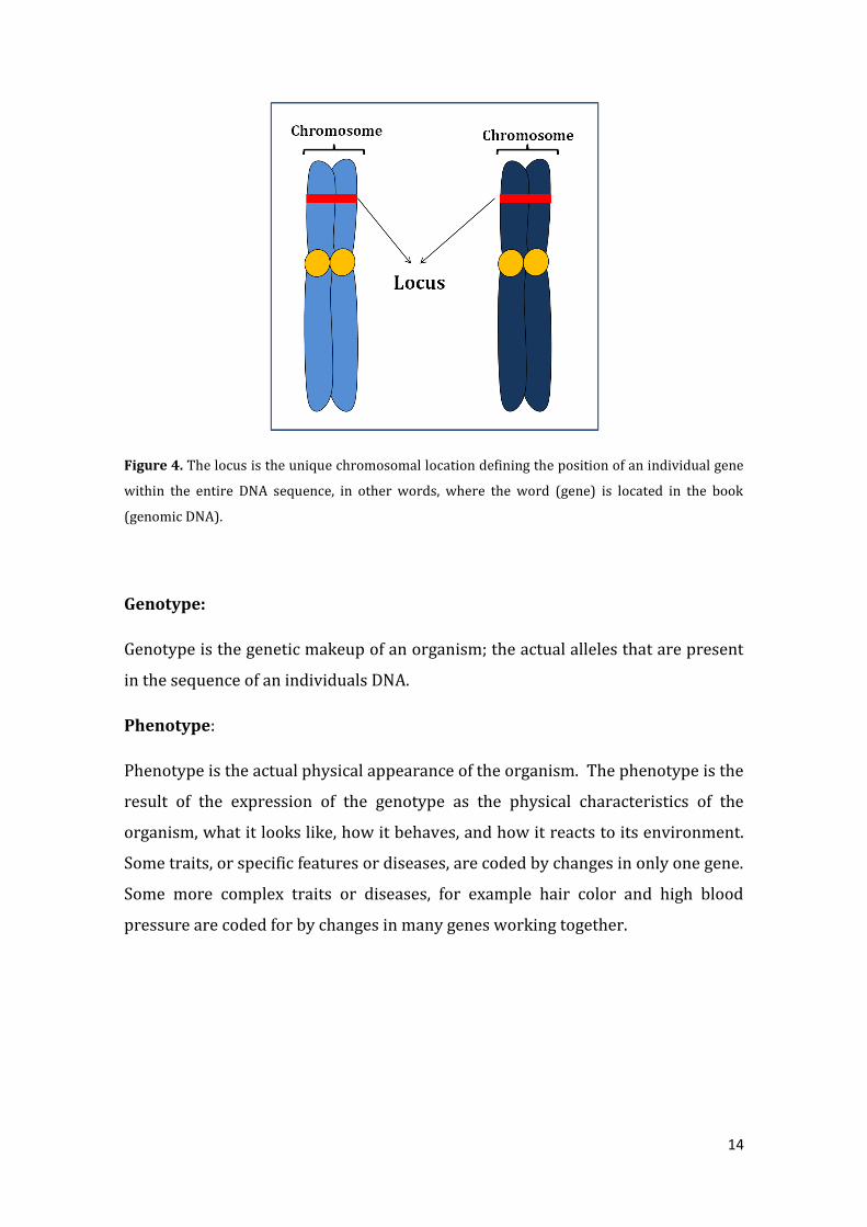
Figure 4. The locus is the unique chromosomal location defining the position of an individual gene
within the entire DNA sequence, in other words, where the word (gene) is located in the book
(genomic DNA).
Genotype:
Genotype is the genetic makeup of an organism; the actual alleles that are present
in the sequence of an individuals DNA.
Phenotype:
Phenotype is the actual physical appearance of the organism. The phenotype is the
result of the expression of the genotype as the physical characteristics of the
organism, what it looks like, how it behaves, and how it reacts to its environment.
Some traits, or specific features or diseases, are coded by changes in only one gene.
Some more complex traits or diseases, for example hair color and high blood
pressure are coded for by changes in many genes working together.

15
Figure 5. The genotype and phenotype.
How is the DNA organized?
DNA is organized on chromosomes, the organized packages of DNA or genetic information in the nucleus of the cell. There are 23 pairs of chromosomes (46 chromosomes in total), 23 of which come from the father's sperm and 23 from the mother's egg. These chromosomes contain the many genes that will largely determine the characteristics of an individual. There are 2 of the 46 chromosomes that define the sex of an individual, XX for female, XY for male, these are called the sex chromosomes. The other 44 chromosomes are called autosomes.
Figure 6. The chromosomes contain the many genes that will largely determine the characteristics of an individual.

16
What is a mutation?
It is a stable change in a DNA sequence or gene, which will cause an alteration in the synthesis of an encoded protein, thus it will change the function of that coded protein.
Figure 7. A mutation is a stable change in a DNA sequence or gene.
What is a protein?
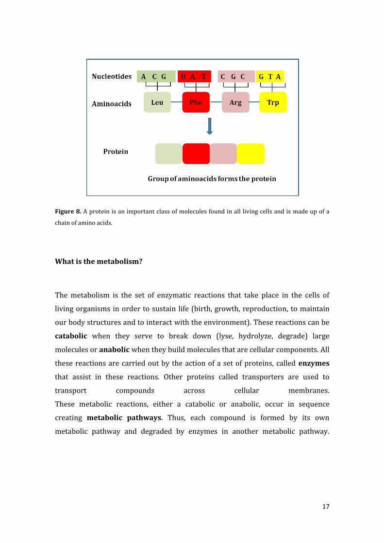
A protein is an important class of molecules found in all living cells and is made up
of a chain of amino acids. The protein’s amino acid sequence corresponds to the
DNA sequence of the gene that encodes that protein. The proteins can have
different functions in the cells of our body: it can be an enzyme, a transporter, a
hormone, a structural protein or a membrane protein.
When there is a mutation or change in the sequence of a gene that encodes a
protein there are several possible outcomes. , One possibility is that the protein
would not be synthesized or that the protein shape would be changed which would
change or abolish the function of the protein.
17
Figure 8. A protein is an important class of molecules found in all living cells and is made up of a
chain of amino acids.
What is the metabolism?
The metabolism is the set of enzymatic reactions that take place in the cells of
living organisms in order to sustain life (birth, growth, reproduction, to maintain
our body structures and to interact with the environment). These reactions can be
catabolic when they serve to break down (lyse, hydrolyze, degrade) large
molecules or anabolic when they build molecules that are cellular components. All
these reactions are carried out by the action of a set of proteins, called enzymes
that assist in these reactions. Other proteins called transporters are used to
transport compounds across cellular membranes.
These metabolic reactions, either a catabolic or anabolic, occur in sequence
creating metabolic pathways. Thus, each compound is formed by its own
metabolic pathway and degraded by enzymes in another metabolic pathway.

18
Figure 9. The metabolism is the set of enzymatic reactions that take place in the cells of living
organisms in order to sustain life.
What are Inborn Errors of Metabolism (IEM)?
The IEM are a large group of human diseases found in low frequency in the
population. They are therefore considered to be RARE and because often little is
known about them are called, ORPHAN DISEASES. They are caused by inherited
changes in the DNA (mutations) that code for “modified” proteins, which do not
function correctly. These poorly functioning proteins cause the malfunction of
cells and ultimately organs.
What happens when there is an Inborn Error of Metabolism (IEM) in an
individual?
When there is an inborn error in metabolism the catabolic or anabolic pathways of
the cell do not function well. Sometimes compounds that are typically broken
down in a catabolic pathway actually accumulate. If compounds are accumulated in
an IEM they can be toxic in the short or long term. On the other hand, in an
anabolic pathway with an inborn error of metabolism the products of the
pathways are not synthesized correctly. This means that when that product is
needed to build a cell or perform a function, it will not be available. In addition, the

19
metabolic pathways may also be altered when the IEM leads to a non-functional
protein that is a transporter of a compound across the membrane, leading to build
up of a compound in the cell. Each of these are a different IEM and each causes
different set of symptoms in an individual.
Figure 10. The Inborn Errors of Metabolism (IEM).
What is inheritance?
An inherited trait is one that is genetically determined. It is the transmission of the
information that we carry in the genes or DNA that we pass on to our offspring.
Each of us carries two copies of each gene, also called alleles, one that carries our
father’s genetic information and the other that carries our mother’s genetic
information. Together these two gene copies will produce two alleles, one from the
mother and one from the father. If both alleles are identical it is said that the
individual is homozygous for this allele and if they are different, he/she is
heterozygous for this allele.

20
Are there different kinds of inheritance?
Of our 23 pairs of human chromosomes, 22 pairs are autosomes and one pair are
called sex chromosomes, XX for a female, XY for a male. Inheritance can be
autosomal, if the gene that codes for the trait is located in the autosomal
chromosomes (non sex chromosomes). If the gene that codes for a trait is
located on the X-chromosome we know that it will be inherited by X-linked
inheritance- Inheritance can also be maternal or mitochondrial, when the gene is
located in the DNA of the mitochondria, named mitochondrial DNA. This is a
very special and rare type of inheritance pattern.
Depending on the expression of the trait in an individual and their family, a trait
can be dominant, or recessive. In case of a dominantly inherited traits or
diseases, the trait or disease can be present when only one parent carries a
mutation in the causative gene. In a recessive trait or disease, both copies of the
allele in the affected individual must carry a mutation or pathogenic change. In a
recessively inherited trait or disease, one changed gene copy is from the mother,
who is a carrier, and one changed gene copy is from the father who is also a carrier.
What is a cell?
A cell is the smallest unit that composes organisms. There are two kind of cells: the
prokaryotic (such as bacterial cells), and the eukaryotic cells (such as human cells).
Which are the components of the cell?
o Cell (plasma) membrane: a wall that surrounds the cell and is in charge of
controlling entry of compounds into and out of the cell.
o Cytoplasm: substance within the cell containing the different cell
components and organelles.
o Endoplasmic reticulum: network of tubules within cells that are the place
where the synthesis of complex molecules and other biochemical reactions
occur.

21
o Golgi apparatus: Organelle where proteins are synthesized and processed.
The golgi are also tubes and are connected to the endoplasmic reticulum.
o Lysosome: It is the digestion organelle of the cell.
o Microtubule: small tubes that support and shape the cell.
o Mitochondria: Organelle responsible for the energy synthesis often called
the “cellular powerhouse”.
o Nuclear membrane: Membrane inside the cell that surrounds the nucleus.
o Nucleus: Organelle containing the genetic material (DNA).
o Chromosomes: Organized package of DNA found in the nucleus of the cell.
Humans have 23 pairs of chromosomes
o Ribosomes: A cellular particle made of RNA and protein that is the site of
protein synthesis in the cell.
Figure 11. The components of the cell.
22
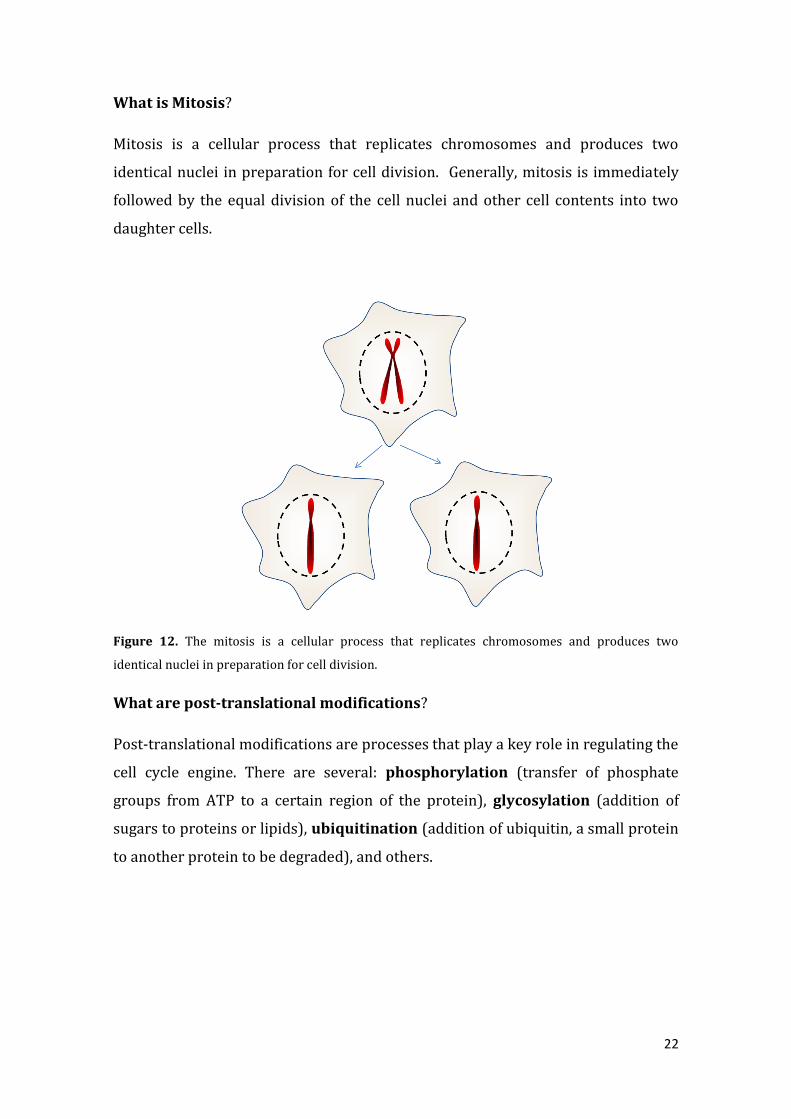
What is Mitosis?
Mitosis is a cellular process that replicates chromosomes and produces two
identical nuclei in preparation for cell division. Generally, mitosis is immediately
followed by the equal division of the cell nuclei and other cell contents into two
daughter cells.
Figure 12. The mitosis is a cellular process that replicates chromosomes and produces two
identical nuclei in preparation for cell division.
What are post-translational modifications?
Post-translational modifications are processes that play a key role in regulating the
cell cycle engine. There are several: phosphorylation (transfer of phosphate
groups from ATP to a certain region of the protein), glycosylation (addition of
sugars to proteins or lipids), ubiquitination (addition of ubiquitin, a small protein
to another protein to be degraded), and others.

23
Figure 13. The post-translational modifications.

24
Chapter 2. The CDG genetic landscape
Authors: Célia Pérez Cerdá and Belén Pérez González (Centro de Diagnóstico de
Enfermedades Moleculares, Centro de Biología Molecular, CIBERER, Universidad
Autónoma de Madrid, Spain)
Translator and Reviewer: Vanessa Ferreira, Ph.D. (Associação Portuguesa CDG e outras Doenças Metabólicas Raras) and Maria Antonia Vilaseca, Ph.D. (Guia Metabólica, Hospital Sant Joan de Déu, Barcelona, España)
What genes are changed in each type of the group of disorders called the
Congenital Disorders of Glycosylation (CDG)?
Although the number of patients with CDGs is not high, because it is a group
of rare diseases, there are still more than 50 different genetic defects or CDG types
have been reported, most of them affecting protein glycosylation (Jaeken and
Matthijs, 2009). Seventeen are involved in the pathway of N-glycosylation of
proteins (Haeuptle and Hermetists, 2009), eleven in the biosynthesis of different
types of O-glycans. Nineteen combined diseases are caused by defects of the N-
and O-glycosylation or other pathways or proteins involved in glycosylation,
including defects in the COG complex which includes many different subunits
(Zeevaert et al2008). These nineteen also include defects in the synthesis of
dolichol-phosphate (Denecke et al, 2009), the defect in the ATP6V0A2-CDG, which

25
has the phenotype of cutis laxa type II. Another type is called Barsy syndrome
(Morava et al2009). There are three genetic defects described in the synthesis of
glycosphingolipids. In the website http://www.ncbi.nlm.nih.gov/omim
information is available about each of these defects. To access this information you
can use the OMIM number that corresponds to each CDG see the table at the end of
the Practical Guide. There is also information about CDG on the website
www.genetests.org.
The first clinical description was done by Professor Dr. Jaak Jaeken in 1980
(Jaeken etal, 1980), and the first enzymatic and genetic description was done in the
early 1990s. (Van Schaftingen et al, 1995)
The most common type of CDG is PMM2-CDG (CDG-Ia), caused by mutations
in the gene PMM2 encoding for the phosphomannomutase enzyme responsible for
the conversion of mannose-6-P into mannose-1-P. The deficiency of the
phosphomannomutase enzyme leads to a decrease in the production of GDP-
mannose and Dol-P-mannose, both mannose donors in the biosynthesis of the
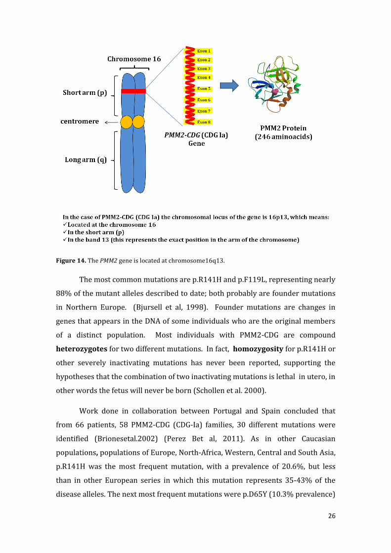
sugar chain or oligosaccharide that binds to proteins. The PMM2 gene is located on
chromosome16q13. At present approximately 800 PMM2-CDG (CDG-Ia) patients
have been identified around the world. 112 different mutations have been
described in the PMM2 gene causing CDG-Ia (https://portal.biobase-
international.com/hgmd/pro/genesearch.php) (Haeuptle and Hennet, 2009).
26
Figure 14. The PMM2 gene is located at chromosome16q13.
The most common mutations are p.R141H and p.F119L, representing nearly
88% of the mutant alleles described to date; both probably are founder mutations
in Northern Europe. (Bjursell et al, 1998). Founder mutations are changes in
genes that appears in the DNA of some individuals who are the original members
of a distinct population. Most individuals with PMM2-CDG are compound
heterozygotes for two different mutations. In fact, homozygosity for p.R141H or
other severely inactivating mutations has never been reported, supporting the
hypotheses that the combination of two inactivating mutations is lethal in utero, in
other words the fetus will never be born (Schollen et al. 2000).
Work done in collaboration between Portugal and Spain concluded that
from 66 patients, 58 PMM2-CDG (CDG-Ia) families, 30 different mutations were
identified (Brionesetal.2002) (Perez Bet al, 2011). As in other Caucasian
populations, populations of Europe, North-Africa, Western, Central and South Asia,
p.R141H was the most frequent mutation, with a prevalence of 20.6%, but less
than in other European series in which this mutation represents 35-43% of the
disease alleles. The next most frequent mutations were p.D65Y (10.3% prevalence)
27
and p.T237M ( 7.6% prevalence), while p.F119L and p.E139K are the most
frequent changes in Scandinavian and French populations respectively, these
mutations were not found in these patients. Thirteen out of these 30 identified
mutations have only been reported among Iberian PMM2-CDG patients. The most
common genotype was [p.R141H]/[p.T237M].
What are the advantages of knowing the genes involved in CDG?
PMM2-CDG (CDG-Ia) is the most frequent type of CDG, but there are at least
50 different diseases or types due to a defect in protein and/or lipid glycosylation.
In a very small number of types there is a specific treatment, so knowing the type
will not change clinical management. Knowing which type or gene is involved in a
specific affected individual in a family leads to more informative genetic
counseling. For example, the specific gene and mutation must be identified before
prenatal diagnosis can be offered. Some individuals are known to have CDG, only
because they have abnormal transferrin glyco-analysis, yet the genetic defect
cannot be found. These individuals carry the diagnosis of CDG-X, until a genetic
basis can be found for their CDG. The scientific community; including medical
doctors, biochemists, biologists, geneticists, are joining efforts to characterize
these unknown genetic defects by studying the DNA of patients through the
massive sequencing technique of the whole genome or through the regions that
code the exons called exome sequencing.
What is the role of a genetic counselor?
A genetic counselor is an important member of the medical team caring for
patients and families at risk for or affected by genetic disorders such as CDG. The
counselor provides advice about the risks of disease recurrence, the possibilities of
prenatal diagnosis, as well different techniques for assisted reproduction. Genetic
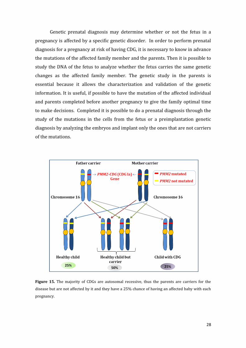
counselors inform families that the majority of CDGs are autosomal recessive. This
means that the parents are carriers for the disease but are not affected by it and
that they have a 25% chance of having an affected baby with each pregnancy.
28
Genetic prenatal diagnosis may determine whether or not the fetus in a
pregnancy is affected by a specific genetic disorder. In order to perform prenatal
diagnosis for a pregnancy at risk of having CDG, it is necessary to know in advance
the mutations of the affected family member and the parents. Then it is possible to
study the DNA of the fetus to analyze whether the fetus carries the same genetic
changes as the affected family member. The genetic study in the parents is
essential because it allows the characterization and validation of the genetic
information. It is useful, if possible to have the mutation of the affected individual
and parents completed before another pregnancy to give the family optimal time
to make decisions. Completed it is possible to do a prenatal diagnosis through the
study of the mutations in the cells from the fetus or a preimplantation genetic
diagnosis by analyzing the embryos and implant only the ones that are not carriers
of the mutations.
Figure 15. The majority of CDGs are autosomal recessive, thus the parents are carriers for the
disease but are not affected by it and they have a 25% chance of having an affected baby with each
pregnancy.

29
What is the best medical care for an individual with CDG and their family?
When introduced to a patient suspected to have CDG the following should
be considered:
An interview with the parents including the clinical history of the patient
and a family history;
A physical examination: weight, height, body mass index and vital signs
Complementary laboratory evaluation including tests to assess specific
organ systems, such as the kidney or liver as well as special genetic tests
that will determine whether the individual has CDG and what type of CDG is
present. The diagnosis and management plan is based on the clinical history
information obtained, on the physical examination, and also on the results
of different laboratory tests and other additional tests, for example, imaging
studies. Information should be given by the medical team, if known, about
the natural evolution of the disease, prognosis and treatment if available.
The information obtained from the patient will hopefully benefit to the
medical community. In the long-term as they seek to better understand the
natural history of the disease.
How technology will help to find new genes in CDG?
CDG has been classified into 2 different groups: CDG type I and CDG type II
depending on the glycosylation analysis profile of the sialotransferrin. Yet,
knowing whether an individual has type I or type II does not identify the genetic
defect. In fact, knowing the exact gene affected, if it has not already been
identified, is a very complex task, because it is thought that there are more than
300 genes involved and so far only 20% are known to be involved in identified
CDG types. Using genotype or SNP arrays and whole genome or whole exome
sequencing may give us more information. Optimally, the biochemistry of each of
these newly identified genes should also be worked out to help us to understand
the cell biology and perhaps find new therapeutic options (Kuhlenbaumer et al
2011). This work can be very expensive to undertake yet, genetic sequencing is

30
becoming more efficient and less expensive. With the cooperation of physicians,
patients and scientists there is a bright future ahead in research on CDG.
Do we know whether CDG is more common in Europe compared with other
continents?
The CDGs are a rapidly growing genetic disease family with a relatively
recent characterization. During the 1990s the identification of numerous genetic
defects that affect glycosylation started to be identified and the diagnosis of a
growing group of patients in which the primary cause was unknown, increased. In
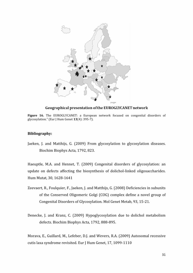
Europe, thanks to the efforts and devotion of Professor Jaak Jaeken and Professor
Gert Matthjis, two important European projects were established: EUROGLYCAN
and EUROGLYCANET, funded by the European Commission from 2000 to 2009.
Both boosted CDG early diagnosis by offering the diagnostic tools for screening as
well as for expert analysis and raised awareness. By the end of 2009, the network
involved 29 participating clinical and basic research centers from 17 countries and
established referral laboratories in the European centers. At the same time,
EUROGLYCAN and EUROGLYCANET integrated research activities in the field, and
worked towards the development of therapies for CDG and related disorders. The
ultimate goal of the project was to be able to precisely diagnose all cases of CDG, to
get a complete inventory of the enzymatic defects that cause protein glycosylation
defects and to extend the therapeutic tools available to treat CDG.
In the USA there are also research and clinical groups involved in the early
diagnosis and management of individuals with CDG, they collaborate with the
European groups. Thus, this network contributed to the early diagnosis of many
patients, and it might be the main reason, on why it seems that there are more CDG
patients in Europe. In fact, the PMM2-CDG is a pan-ethnic disease, with patients
having a European, North and South America origin, Asiatic, and most other
continents.
31
Figure 16. The EUROGLYCANET: a European network focused on congenital disorders of glycosylation." (Eur J Hum Genet 13(4): 395-7).
Bibliography:
Jaeken, J. and Matthijs, G. (2009) From glycosylation to glycosylation diseases.
Biochim Biophys Acta, 1792, 823.
Haeuptle, M.A. and Hennet, T. (2009) Congenital disorders of glycosylation: an
update on defects affecting the biosynthesis of dolichol-linked oligosaccharides.
Hum Mutat, 30, 1628-1641
Zeevaert, R., Foulquier, F., Jaeken, J. and Matthijs, G. (2008) Deficiencies in subunits
of the Conserved Oligomeric Golgi (COG) complex define a novel group of
Congenital Disorders of Glycosylation. Mol Genet Metab, 93, 15-21.
Denecke, J. and Kranz, C. (2009) Hypoglycosylation due to dolichol metabolism
defects. Biochim Biophys Acta, 1792, 888-895.
Morava, E., Guillard, M., Lefeber, D.J. and Wevers, R.A. (2009) Autosomal recessive
cutis laxa syndrome revisited. Eur J Hum Genet, 17, 1099-1110
32
Bjursell C, Wahlstrom J, Berg K, Stibler H, Kristiansson B, Matthijs G, Martinsson T.
1998. Detailed mapping of the phosphomannomutase 2 (PMM2) gene and
mutation detection enable improved analysis for Scandinavian CDG type I families.
Eur J Hum Genet 6(6):603-11
Schollen E, Kjaergaard S, Legius E, Schwartz M, Matthijs G. 2000. Lack of Hardy-
Weinberg equilibrium for the most prevalent PMM2 mutation in CDG-Ia
(congenital disorders of glycosylation type Ia). Eur J Hum Genet 8(5):367-71
Briones P, Vilaseca MA, Schollen E, Ferrer I, Maties M, Busquets C, Artuch R, Gort L,
Marco M, van Schaftingen E and others. 2002. Biochemical and molecular studies
in 26 Spanish patients with congenital disorder of glycosylation type Ia. J Inherit
Metab Dis 25(8):635-46
B. Pérez, P. Briones, D. Quelhas, R. Artuch, A.I. Vega, E. Quintana, L. Gort, M.J. Ecay,
G. Matthijs,M. Ugarte, and C. Pérez-Cerdá. 2011. The Molecular Landscape of
PhosphomannoseMutase Deficiency in Iberian Peninsula: Identification of 15
Population-Specific Mutations. J Inherit Metab Dis On Line DOI:
10.1007/8904_2011_26
Quelhas D, Quental R, Vilarinho L, Amorim A, Azevedo L. 2007. Congenital disorder
of glycosylation type Ia: searching for the origin of common mutations in PMM2.
Ann Hum Genet 71 (Pt 3): 348-53
Jaeken, J et al. Familial psychomotor retardation with markedly fluctuating serum
proteins, FSH and GH levels, partial TBG-deficiency, increased serum
arylsulphatase A and increased CSF protein: a new syndrome? Pediatric research
14:179 (1980)
Jaeken J et al. Carbohydrate-deficient glycoprotein syndrome type II: a deficiency
in Golgi localised N-acetyl-glucosaminyltransferase II. Arch Dis Child71:123-127
(1994)
Van Schaftingen E and Jaeken J.Phosphomannomutase deficiency is the cause of
carbohydrate-deficient glycoprotein syndrome type I. FEBS letter 377:318-320
(1995)

33
Chapter 3. PMM-CDG (CDG-Ia): Clinical manifestations and their
management
Introduction
Author and Illustrations: Vanessa Ferreira, Ph.D. (Associação Portuguesa CDG e outras Doenças Metabólicas Raras)
Translator Reviewer: Maria Antonia Vilaseca, Ph.D. (Guia Metabólica, Hospital Sant Joan de Déu, Barcelona, España)
Carbohydrates, Saccharides or Sugars
The chemical formula of any sugar is Cm (H2O)n. Carbohydrates are the main
source of energy. Over 56% of the energy, the body receives from carbohydrates,
the rest - at the expense of protein and fat.
Glycosylation
Carbohydrate (also known as Saccharides or Sugars) molecules get attached to
proteins, lipids or other organic molecules at specific cellular environment – this
process is termed glycosylation. Hundreds of enzymatic steps are involved in the
glycosylation pathway. In nature, a glycosylated protein has multiple complex
carbohydrates attach to the protein structure.

34
Figure 17. The sugars have structural (glucose) or energetic (sucrose) functions.
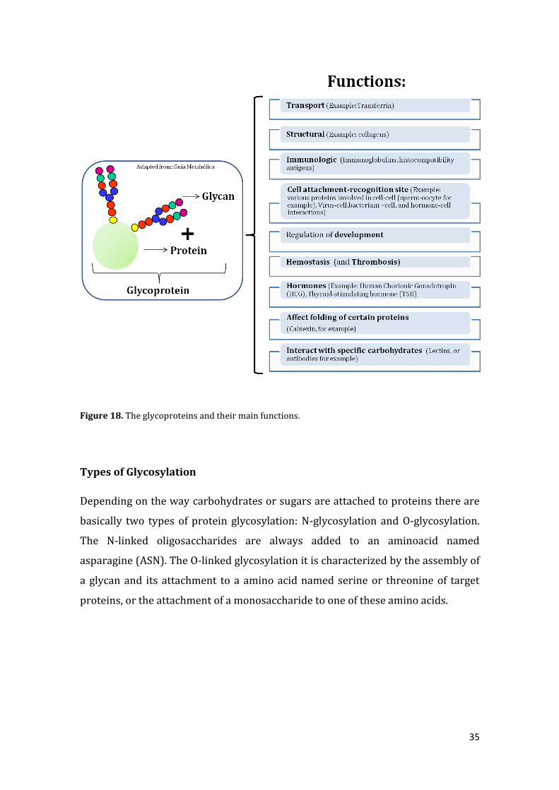
Glycosylation: why is it important?
The correct transfer of glycans, chain of sugars, to proteins or lipids is essential for
their biological function and the sugar chains act as biologic signals for cell-cell
communication, intracellular signalling, protein folding or targeting of proteins.
Given the overall importance of glycosylation, it is not surprising, that a disruption
of the glycosylation machinery can lead to multisystemic and serious diseases.
35
Figure 18. The glycoproteins and their main functions.
Types of Glycosylation
Depending on the way carbohydrates or sugars are attached to proteins there are
basically two types of protein glycosylation: N-glycosylation and O-glycosylation.
The N-linked oligosaccharides are always added to an aminoacid named
asparagine (ASN). The O-linked glycosylation it is characterized by the assembly of
a glycan and its attachment to a amino acid named serine or threonine of target
proteins, or the attachment of a monosaccharide to one of these amino acids.

36
Figure 19. There are two types of protein glycosylation: N-glycosylation and O-glycosylation.
How many cases of CDG are known worldwide?
Author: Jaak Jaeken, M.D., Ph.D. (Center for Metabolic Disease, Katholieke
Universiteit Leuven, Belgium)
We have no exact figures but the published cases figure around 600 for the N-
glycosylation disorders. There are many more patients reported with a known O-
glycosylation disorder (multiple exostoses, Walker-Warburg syndrome, muscle-
eye brain disease) but again it is very difficult to obtain exact figures. All together
for typed and untyped CDG I guess that an incidence of 1 in 5000 births may be a
minimum estimate.
37
3.1. Neurologic Clinical Issues
Author: Jaak Jaeken, M.D., Ph.D. (Center for Metabolic Disease, Katholieke
Universiteit Leuven, Belgium).
Ataxia
Ataxia is a consequence of brain disease, particularly of the cerebellum,
causing a lack of coordination of limb movements and of regulation of
body posture. There is no specific medication to treat ataxia.
Mechanical aids such as walkers or adapted utensils may be helpful.
Since there is no amelioration of the brain disease in CDG, no
significant improvement of the ataxia is to be expected.
Seizures
What is the difference between seizures and epilepsy?
Seizures (or convulsions) are uncontrollable (involuntary) movements
that are part of many forms of epilepsy. Indeed, some forms of epilepsy
do not show seizures but only loss of consciousness.
Does ketogenic diet help to treat CDG patients that have epilepsy?
A ketogenic diet is a difficult diet that might be tried in a few very rare
CDG patients with epilepsy that cannot be controlled by medication.
This has to be decided by your physician. However, as a rule this does

38
not apply to the classical PMM2-CDG (CDG-Ia). In these patients,
epilepsy is controllable by medication.
Stroke-like episodes:
What is a stroke-like episode?
A stroke-like episode is an acute event that very much resembles a
stroke. A stroke is a sudden loss of consciousness due to an acute
vascular disturbance caused by the rupture of an artery in the brain
or its obstruction by a blood clot (embolism or thrombosis); we
think that in CDG these episodes are due to a transient local
thickening of blood. It can present in several ways: drowsiness,
dullness, subcoma, coma, loss of vision, paralysis on one side, which
is called hemiparesis or hemiplegia) or the paralysis may be on both
sides.
What is the difference between a stroke-like episode and a
seizure?
A stroke is a vascular problem; a stroke-like episode has the clinical
appearance of a vascular event. A seizure is the expression of an
abnormal electrical activity in the brain but it can very much
resemble a stroke. Other words for “seizures” are “epilepsy” and
“convulsions”.
Who is likely to have a stroke-like episode and when are they
likely to occur?
Initially we thought that teens were the most likely to have stroke –
like episodes, however, they have been reported in young children as
well. These episodes are most likely to occur on occasion of an
infection (viral or bacterial); so these episodes are often
accompanied by fever.
In the CDG field all the patients with PMM2-CDG (CDG-Ia) have an
increased risk for thrombosis because their blood platelets have an

39
increased tendency to stick together and to stick to the wall of blood
vessels; for the other CDG patients this risk for stroke-like episodes
is probably also increased but this has not yet formally been proven
also because we know only a small number of these patients.
Are there ever any long lasting effects?
These episodes can last for hours, days or sometimes even longer.
However, the positive thing is that, as a rule, they are transient and
the affected individual recovers to their previous functional level.
What action should parents take during one of these episodes?
If a child has a stroke-like episode the parents should ask a
physician to examine your child as soon as possible in order to make
the diagnosis and to take appropriate measures including fever
control, hydration and assessment for infection if necessary.
Is there a suggested therapy to prevent or help these episodes?
Medical treatment and prevention are possible but it is up to the
treating physician to decide about the treatment.
Author: Belén Pérez Dueñas, M.D., Ph.D. (Neurology Department Hospital Sant
Joan de Déu, Barcelona, España).
Peripheral neuropathy
What is peripheral neuropathy?
Peripheral neuropathy refers to a disorder of peripheral nerves. As a
consequence patients manifest a reduction in muscle strength and gait
difficulties. In the PMM2-CDG (CDG-Ia), the peripheral neuropathy may
appear in the first years of life, and tends to stabilize in late childhood and
adulthood.
40
How is it detected?
There is a decrease or disappearance the osteotendinous reflexes, reduced
muscle strength and muscle atrophy. Neurophysiological studies including
electromyography and neurography confirms the diagnosis.
Cerebellar atrophy or hypoplasia
What is cerebellar atrophy or hypoplasia?
A frequent clinical manifestation found in CDG patients, typically those with
PMM2-CDG, is a defect in the formation of the cerebellum, termed hypoplasia.
The cerebellum is small and poorly differentiated. Additionally,
histopathological and serial neuroimaging studies in some patients detect
progressive volume loss due to neuronal loss and reactive gliosis during the life
of the affected individual. This suggests that hypoplasia and/or cerebellar
atrophy cause cerebellar ataxia in CDG.
Why do CDG patients when they are tired, shake so much?
The majority of individuals with CDG suffer cerebellar ataxia as a consequence
of the cerebellar disturbances. One clinical manifestation is a delay in motor
development, hypotonia and dysequilibrium. Children achieve head control, sit
unassisted and walk very late when compared with normally developing
children. Another manifestation of cerebellar ataxia is intentional tremor. In
general, tremor increases during intensive exercise due to muscle fatigue.
Why CDG children have difficulties concentrating when they are doing
an activity, like studying or swimming?
Individuals with CDG may have mild to severe cognitive dysfunction depending
on their type and other factors. Cognitive dysfunction is frequently associated
with poor attention span and poor scholastic performance. Cognitive
difficulties together with physical impairment may also prevent them from
playing sports as well as their peers.

41
3.2. Gastrointestinal
Author: Ruth Garcia (Gastroenterology, Hepatology and Child nutrition Section,
Metabolic Diseases Unity, Hospital Sant Joan de Déu, Barcelona)
Translator: Belén Pérez Dueñas, M.D., Ph.D. (Neurology Department Hospital Sant
Joan de Déu, Barcelona, España).
Failure to thrive
My son does not eat, only drinks the feeding bottle and with difficulty.
He does not have much appetite and vomits frequently. What should I
do?
CDG is often associated with failure to thrive in infancy and early
childhood. It is characterized by a low weight and height percentiles for age.
If untreated, failure to thrive may be associated with malnutrition. In CDG,
the causes are not well understood and seem to be multifactorial.
Treatment consists of hypercaloric foods that in small quantities,

42
concentrate large amounts of nutrients and calories.
If this is not enough, enteral formulas will be used to supplement the diet.
In some children a nasogastric tube or gastrostomy will improve nutrition
and decrease the stress of feeding in a family. Elemental formulas may be
useful during the first years of life if there is malnutrition and/or significant
gastro-esophageal reflux.
Gastro-esophogeal reflux
What is gastro-esophogeal reflux?
Gastro-esophageal reflux (GER) is an important factor related with failure
to thrive. It is the involuntary return of gastric contents into the mouth,
usually just following eating. It occurs in 18% of healthy breast feeding
children but in children with neurological symptoms and hypotonia it is
more common. When this phenomenon is increased in frequency and
intensity it may exceed the defensive capacity of the mucous of the
esophagus and produce a disease by RGE (ERGE) can have many
presentations from subtle to very obvious. Typical symptoms include:
regurgitation, vomiting, colic, irritability, crying, failure to thrive, thoracic
pain and even blood while vomiting. Other atypical symptoms include
chronic respiratory processes, rumination, and dental disorders.
When children have GER, vomiting exposes the esophagus to the stomach
acid causing mucosal irritation (esophagitis). This esophagitis is
manifested as pain and irritability mostly associated with the meals, and
therefore infants reject feeding. Treatment consists of postural measures;
avoid laying down after eating and elevating the head of the bed. Avoid
irritating foods and consider thickening the texture of foods as well as
hypoallergenic or elemental formulas for infants. Medical treatment
includes antacids, acid suppressants and prokinetics and, if these treatment
are not successful, surgical intervention (Nissen) may be necessary.
A Nissen fundoplication surgery may be performed in children that have

43
symptoms after the medical management is not successful after a discussion
with the family and treating physicians. The Nissen fundoplication is a
widely used surgical procedure.
What is a gastrostomy and in what situations is it used?
Gastrostomy means the insertion of a tube into the stomach through the
abdominal wall. It is a treatment option to ensure feeding in patients unable
to feed by mouth (due to dysphagia) or in patients with inadequate oral
feeding. It is a safe method considered in situations of prolonged artificial
nutrition by nasogastric tube (NGT), because it prevents the most common
complications that typically occur including obstruction and frequent
replacement. It is also easy to use by caregivers.
Another cause of food refusal and possible consideration of a gastrostomy
may be dysphagia (difficulty swallowing). This is common in children with
neurological disorders and hypotonia. Symptoms include coughing during
meals, recurrent respiratory difficulties and failure to thrive. Treatment
options are thickening and/or enteral feeding (nasogastric tube or
gastrostomy).
Protein-losing enteropathy
What is protein-losing enteropathy?
Protein-losing enteropathy (PLE) is characterized by an excessive loss of
proteins through the stool and a decrease of the proteins levels in plasma. It
occurs in MPI-CDG (CDG-Ib) and a few other rarer types of CDG.
The most relevant sign of PLE is edema due to hypoproteinemia (decrease
blood protein levels). Sometimes, patients also present with diarrhea and
abdominal pain. Measurement of alpha-1-antitrypsin in the stools of the
patient is important for the diagnosis of PLE. In CDG patients, protein-
losing enteropathy is thought to be due to mucosal dysfunction caused by a
disorder in the cellular synthesis.

44
The treatment of PLE is supportive care, fluids and sometimes
protein/albumin infusions. In patients with PMI-CDG (CDG-Ib) this the
clinical problems improve after the administration of mannose. Affected
children gain weight, have stable glucose levels and decreased protein
losing enteropathy. The long term clinical story of individuals with PMI-
CDG (CDG-Ib) treated with mannose has yet to be determined.
45
3.3. Hepathology
Author: Ruth Garcia (Gastroenterology, Hepatology and Child nutrition Section,
Metabolic Diseases Unity, Hospital Sant Joan de Déu, Barcelona)
Translator: Belén Pérez Dueñas, M.D., Ph.D. (Neurology Department Hospital Sant
Joan de Déu, Barcelona, España).
Why are transaminases high? How do they normalize? How
transaminases are detected? Currently, there is a treatment?
In CDG there is a multisystem involvement affecting various organs and
structures, and one of them is the liver. The measurement of transaminases
in the blood reflects the breakdown of cells from the liver. Fortunately, the
liver has lots of reserve so slightly high transaminase levels need to be
watched and assessed but are typically well tolerated. In most cases of CDG
the transaminases may go rise and fall. As the children grow older they
remain normal but may sometimes elevate when the children become ill.
There are, however, some affected individuals that have problems with
elevated transaminases for longer times. In some types of CDG affected
individuals may present with an enlarged liver, fibrosis and problems with
the biliary pathway. Currently there is not effective treatment for the liver
involvement in CDG, except for MPI-CDG (CDG-Ib) patients where oral
mannose treatment is currently considered standard of care.
Is CDG considered in the group of inherited metabolic diseases that
associate liver involvement?
Liver involvement is not a constant finding in all CDG patients, but in most
of the CDG types increased transaminases is a frequent finding.
46
3.4. Kidney
Author: Mercedes Serrano, M.D., Ph.D. (Neurology Department Hospital Sant Joan
de Déu, Barcelona, Spain).
What kidney symptoms are seen in individuals with CDG?
In infants and children, bilateral hyperechogenic kidney signal (increased
signal in the kidneys seen by echography) may be seen in a variety of medical
diseases including some metabolic disorders. Renal anomalies(abnormal renal
anatomy) has been rarely described in children with PMM2-CDG (CDG-Ia) and
has never been reported in adults. The most frequent finding in individuals
affected with PMM2-CDG is increased signal in the echographic images of the
kidney that correspond to cysts and microcysts (very small cysts), with grossly
preserved renal function. On the other hand, there are rare anecdotal reports of
patients with PMM2-CDG (CDG-Ia) with nephrotic syndrome, a problem where
proteins are abnormally lost in urine and may damage the glomerulus
(important functional elements of the kidney) requiring specific treatment.
Nephrotic syndrome may appear as edema (swelling of the hands and feet).

47
3.5. Coagulopathy
Author: Jaak Jaeken, M.D., Ph.D. (Center for Metabolic Disease, Katholieke
Universiteit Leuven, Belgium).
Are Low platelets related to CDG?
As a rule low platelets are not a feature of CDG.
Is it dangerous to give aspirin to a CDG patient? Or an anti-
inflammatory?
In general, it is not more dangerous to give aspirin or an anti-inflammatory
to CDG patients than to unaffected people. Low dose aspirin is even helpful
as preventive therapy in patients with frequent stroke-like episodes. When
to start it and in which dose has to be decided by your physician.
What should be checked if a CDG patient has to undergo surgery?
Platelet number, platelet function and coagulation/anticoagulation factors
including the coagulation cascade, Protein C, Protein S and Anti-thrombin
III should be measured.
What are the signs of deep venous thrombosis (DVT)?
DVT is the formation of a blood clot in a deep vein, usually in a leg. It can
cause pain and swelling. The blood clot can also be detached from the vessel
wall and travel to other organs causing damage, for example to the lungs.
How are bleeding disorders manifested and how should they be
managed?
The clinical signs of a bleeding disorder depends of course on the place of
the bleeding. For example, when the bleeding occurs in the brain, it can
cause limb paralysis (on the opposite side), seizures, and or loss of
consciousness. Some individual with bleeding disorders have excessive
bleeding in surgery, even dental surgery, so the physician or dentist should
be aware of this possibility so that it can be managed if it occurs.
Which coagulation factors should be measured and how often?
The coagulation factors that are most decreased in PMM2-CDG (CDG-Ia) are
factor XI, antithrombin III, protein C and protein S. It is not necessary to
measure all of these factors regularly because they will not change much in
childhood. In adults they tend to increase and even normalize.

48
3.6. Endocrinology
Author: Jaak Jaeken, M.D., Ph.D. (Center for Metabolic Disease, Katholieke
Universiteit Leuven, Belgium).
Thyroid function
The thyroid gland secretes thyroid hormones that are very important
for many functions particularly growth and psychomotor
development.
Why are thyroid function test very often abnormal in children with
PMM2-CDG (CDG-Ia)?
Because the transporter for thyroid hormones (called TBG: thyroxin
binding globulin) is glycosylated.
How is thyroid function tested?
By measuring thyroid hormones in the blood.
Is there a recommended medication if there is a problem with the
thyroid?
As a rule, these patients should not be treated since the ‘active’ hormone
usually is normal. The hormone that is transported by TBG is not active.
Assessment by an endocrinologist will help to determine whether
treatment is necessary.
Growth hormone
Should growth hormone be administrated and when?
In general, growth hormone should not be given since there is no deficiency
of growth hormone, with rare exceptions.
Is it possible to give growth hormones when there are growth
problems? What happens, if the CDG patient has growth problems but
the plasma levels of those hormones are not deficient?
The patient may have short stature.
Is it possible to get the approval from the Ethical Committee taking
into account that the first condition to give the authorization, are low
plasma levels of these growth hormones?

49
The EC will not give approval when growth hormone tests are normal.
Taking into account that many hormones are glycosylated or are
transported by glycosylated proteins, how is puberty and its hormonal
changes affected in patients with CDG?
As a consequence of this, there is in some individuals with CDG, such as
those with PMM2-CDG (CDG-Ia), who have no or very limited pubertal
development.
During puberty, will there be changes in attitude and mood and will
they be pronounced?
In my experience, the changes in mood and attitude are generally not
different from those in pubertal patients who have no CDG.
Blood sugar levels and hypoglycemia
Do CDG patients usually have low blood sugars levels?
No, except in MPI-CDG (CDG-Ib)
What are the symptoms of low blood sugar?
This can manifest as drowsiness or even loss of consciousness. Other
possible symptoms are jitteriness or seizures.
How is this treated?
First, it should be shown that blood glucose is indeed low (using a properly
functioning glucometer). Treatment consists of administering a glucose-
containing drink, by mouth (if the child is able to drink) or by giving glucose
intravenously which must be performed in the hospital).
Can hyperinsulinism can affect my child? How it is detected?
In MPI-CDG (CDG-Ib) hyperinsulinism can cause low blood sugar levels
(see above.). It is detected by finding a blood insulin level that is too high in
relation to the blood sugar level. .

50
3.7. Ophthalmic manifestations of congenital disorders of glycosylation
Author: Daisy Rymen, M.D., PhD student concerning CDG (Center of Human
Genetics – Prof Gert Matthijs laboratory, Catholic university of Leuven, Belgium).
Introduction
Ophthalmological abnormalities are frequently encountered in patients with
congenital disorders of glycosylation. The most reported findings are shown in
table 1.
In order to understand these ophthalmologic abnormalities, it is important to
have a basic knowledge of how vision operates. What happens when we look at
an object? First we move our eyes to fixate the object. Doing this our extra-
ocular muscles have to work together in an extremely coordinated way. They
can achieve this difficult task under the command of the cerebellum (Figure 20).
Figure 20. First we move our eyes to fixate the object. Doing this our extra-ocular muscles have
to work together in an extremely coordinated way. They can achieve this difficult task under the
command of the cerebellum.
Table 1: Ocular findings in CDG
Strabismus
Retinitis pigmentosa
Nystagmus
Refractive errors
51
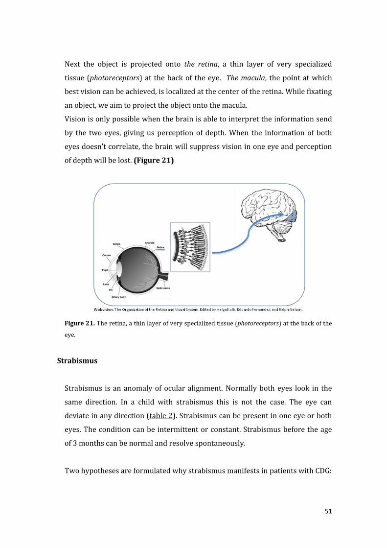
Next the object is projected onto the retina, a thin layer of very specialized
tissue (photoreceptors) at the back of the eye. The macula, the point at which
best vision can be achieved, is localized at the center of the retina. While fixating
an object, we aim to project the object onto the macula.
Vision is only possible when the brain is able to interpret the information send
by the two eyes, giving us perception of depth. When the information of both
eyes doesn’t correlate, the brain will suppress vision in one eye and perception
of depth will be lost. (Figure 21)
Figure 21. The retina, a thin layer of very specialized tissue (photoreceptors) at the back of the
eye.
Strabismus
Strabismus is an anomaly of ocular alignment. Normally both eyes look in the
same direction. In a child with strabismus this is not the case. The eye can
deviate in any direction (table 2). Strabismus can be present in one eye or both
eyes. The condition can be intermittent or constant. Strabismus before the age
of 3 months can be normal and resolve spontaneously.
Two hypotheses are formulated why strabismus manifests in patients with CDG:

52
1. Being part of the general muscle weakness, i.e. the muscle is too weak to get
into the right position.
2. As a consequence of cerebellar hypoplasia, i.e. the muscle doesn’t receive the
right commands.
Symptoms
When strabismus is intermittent, the child can experience diplopia (double
vision).
When strabismus is constant and no treatment is started, the child will lose the
ability of depth perception. Because the brain suppresses the images send by
the diverging eye, this eye will become functionally blind (amblyopia).
Treatment
Strabismus can be corrected when treatment is attempted at young age: glasses,
eye patches, certain drugs, and, when this fails, surgery (Figure 22). When
there is already loss of function (loss of depth perception or amblyopia), surgery
will not restore these functions. In that case, surgery has only cosmetic reasons.
Figure 22. The strabismus is an anomaly of ocular alignment.

53
Retinitis Pigmentosa
Retinitis Pigmentosa entails the progressive degeneration of the retina with
subsequent loss of vision. More specifically it defines a loss of photoreceptors
with pigment deposits in the retina. One can assume that Retinitis Pigmentosa
in individuals with CDG is caused by a glycosylation defect within parts of the
retina.
The degeneration starts at the periphery of the retina and moves towards the
center, i.e. the macula (Figure 23). Initially only peripheral vision is
compromised, leading to tunnel vision. This means that the patient only can see
things that happen right in front of him, and not the things that happen aside.
One can compare this by looking through a narrow tube (Figure 23). When
disease progresses also central vision can become compromised, leading to
diminished vision or eventually total blindness. Currently the known adults
with PMM2-CDG (CDG-Ia) have compromised night vision but their daytime
vision is functional.
Symptoms
The first clinical symptom of Retinitis Pigmentosa is night blindness, related to
the destruction of photoreceptors. Next a progressive loss of peripheral vision
will take place, leading to tunnel vision (Figure 23). Initially patients and their
caregivers are not aware of this problem, because there is no functional impact.
It is important to remember that night blindness and loss of peripheral vision
can lead to anxiety, because the patient isn’t aware of what is happening around
him. Eventually the disease can lead to diminished vision or blindness, though
not all patients evolve to this stage.

54
Figure 23. Retinitis Pigmentosa entails the progressive degeneration of the retina with
subsequent loss of vision.
Photophobia (i.e. light bothers the eyes) and cataracts (i.e. clouding of the lens in
the eye) may develop. Because cataracts can lead to diminished central vision, it
should be corrected by surgery.
Treatment
No proven treatment exists.
Nystagmus
Nystagmus is a rhythmic, involuntary oscillation of one or both eyes. It can
occur continuously or intermittently. It also can be evoked by some maneuvers
(certain head position, gaze in certain direction,…). One can assume that in
patients with CDG, nystagmus is caused by an erroneous input from the
cerebellum.

55
Symptoms
As the patient isn’t able to fixate an object, he can experience blurred vision. A
patient can assume a strange head position to minimize nystagmus.
Treatment
Surgery can change positions of the extra-ocular muscles, so that a gaze position
is achieved in which nystagmus is diminished or absent. Although nystagmus
will still occur with lateral gaze.
Refractive errors
A refractive error is present when an object is not sharply projected onto the
retina. Patients with myopia are not able to see things sharply that are far away.
With this condition the eye is too long. The focusing point lies in front of the
retina, instead of onto it (Figure 24).
In CDG, myopia is the most common refractive error. One can assume that
because of reduced glycosylation the wall of the eye is less firm, leading to
elongation of the eye. Although this hypothesis has not been proven.
Treatment
Glasses.

56
Figure 24. A refractive error is present when an object is not sharply projected onto the retina.
Conclusion
The high prevalence of ophthalmologic abnormalities in CDG emphasizes the
importance of a thorough ophthalmologic screening. A routine annual follow-up
is recommended.

57
3.8. Orthopedics, The musculoskeletal system in CDG
Author: Luis Terricabras Carol, M.D., Ph.D. (Orthopedic Unity, Hospital Sant Joan
de Déu, Barcelona)
Translation: Melisa Stitzman (Orthopedic Unity, Hospital Sant Joan de Déu,
Barcelona)
The child is not a small adult, and we do not present the same morphology
at the birth as we eventually develop as an adult. In fact, our bodies are constantly
being modified, this is especially true for the skeletal and muscular system.
The newborn has a proportion of limbs and head different from adults. In
addition, the shape and consistency of the bones are also different. Together, it is
interesting that even our walk is different, and we do not develop an “adult” walk
until we are about three years old.
So, everything that interferes with a normal locomotor development will be
the cause for a difference in the growth of the bones, joints and even the muscle,
tendons and fascia.
For example, if the patient doesn’t walk and the gluteus muscles are
insufficient it will cause a coxavalga, which is a underdevelopment of the
acetabulum favoring the sub-dislocation and dislocation of the hip. A lack of
standing or walking, may cause osteopenia, or light bones, which may be a
precursor for osteoporosis. Osteopenia is a decrease in the bone mineral density
that is regulated by multiple endocrinologic, renal, nutritional and mechanical
factors. For example, if astronauts in the space did not do specific exercises, they
can develop osteopenia due to lack of gravity.
Or, if a bone doesn’t have a certain load, when it is immobilized, it can develop
osteopenia.
Scoliosis is a deviation of the spine but with rotation of the vertebral bodies,
therefore it is a structured deformity that in some cases is progressive.

58
A scoliotic shape of the spine may occur when there is an asymmetry
between both lower extremities (for example if there is one leg shorter than the
other) or when we are seated there is a pelvic obliquity (the two buttocks are not
completely symmetrical and the pelvis rises more in one side than in the other
side).
If the paravertebral muscle does not contract symmetrically it can cause a
scoliotic shape of the spine. To correct the scoliosis it may be useful to use raised
insole, molded seats or wheelchairs with good lateral supports.
The raised insole and molded seats are less useful and are used only to
compensate in some cases for the off center of gravity. In some processes corsets
may be useful and in the case of severe curvature surgery is indicated This type of
surgery of the spine varies but it is intended to prevent progressive deformity).
In a lateral view of the spine there is a curvature called kyphosis, if this
curvature is too great it may provoke important respiratory problems or skeletal
muscle pains in this area and even eating disorders.
An inward curvature of a portion of the lumbar and cervical vertebral
column is known as lordosis. Hyperlordosisis, an exaggerated curve in the lower
spine, may cause significant pain and difficulty in proper positioning of the pelvis
when standing.
The development of bones and joints is not always typical in individuals
with CDG and the mechanisms underlying these issues are not well known.
The dysostosis multiplex, is a group of generalized skeletal abnormalities.
Some of the milder features of dysostosis multiplex may be seen in individuals
with CDG. The radiographic findings of dysostosis multiplex include: skull
abnormalities, vertebral abnormalities and changes in the ribs and long bones.
59
It is important to monitor individuals with CDG to assure that they maintain
their joint range of motion, especially of the knees, hips and elbows. Decreased of
range of motion may be due to: skeletal changes, contractures and fibrosis of the
tendons, ligaments and soft tissue. Also, if the foot is not balanced it may lead to
problems with the knees and hips. Therefore, it is important that orthotics are
used to center the foot if needed to minimize the risk of problems to other joints.

60
3.9. Cardiology
Author: Mario Sanz Cuesta, M.D. (Pediatrician Unity, Parc Sanitari Sant Joan de
Déu, Sant Boi de Llobregat).
Translator Reviewer: Belén Pérez Dueñas, M.D., Ph.D. (Neurology Department
Hospital Sant Joan de Déu, Barcelona, España).
Introduction
The heart, and specifically the cardiac muscle may be affected in a few patients
with CDG. More often individuals with CDG, especially PMM2-CDG (CDG-Ia) have
pericardial effusions which are not life threatening. However, because it is a vital
organ, its dysfunction affects a patient’s quality of life.
Pericardial effusion in CDG
Pericardial effusion, diagnosed by an echocardiogram, is not uncommon in
individuals with some types of CDG. Typically the children do not have symptoms
from these effusions and they are diagnosed and followed and eventually
disappear without problems. Sometimes these pericardial effusions caused
pressure on the cardiac muscle if they become large enough. This is a very rare
occurrence in CDG but if it does happen it requires hospitalization and
management by a cardiologist.

61
Cardiomyopathy
Cardiomyopathy is seen in rare cases of CDG. There are two basic forms of
cardiomyopathy, dilated and hypertrophic myocardiopathy. Each of them affects
predominantly one of the two moments of the cardiac cycle, the contraction and
relaxation. In the hypertrophic form the heart muscle increases its thickness
above the normal, resulting in a vigorous contraction and also an inability to relax.
In the dilated myocardiopathy there is a dilation of the heart, which progressively
becomes weaken and impairs their ability to contract.
Dilated and hypertrophic myocardiopathy are not specific for a type of
glycosylation disorder and have been reported but only in rare cases. In these
cases there was progressive deterioration of the cardiac function resulting in
manifestations such as poor weight gain, fatigue with feeding, paleness, muscle
weakness and hypotonia , difficulty to breath, or irritability. There is no curative
treatment for myocardiopathy. Various types of medication are used to alleviate
symptoms, but they do not slow the progression of the disease. In the case of some
types of arrhythmia a pacemaker may be indicated. It is a small device implanted
in the chest under the skin of the patient.

62
3.10. Hematology
Author: Donna Krasnewich M.D., Ph.D. (Program Director at the National Institute
of General Medical Sciences, USA)
Which parameters should be checked by a hematologist? With which
frequency should be done?
In CDG the children and adults may have differences in the level of blood
proteins, or factors, that help the blood clot and help the blood break down those
clots.
Coagulation factors are named with numbers and their job is to clotg the
blood. In CDG these coagulation factors are low and may make it difficult for the
blood to clot. Typically this is not a problem in daily living but should the child or
adult have surgery or a trauma knowing about these low coagulation factors is
important for the medical team. Therefore, it is important that hematologists
have a baseline level of at least one of the coagulation factors. Factor IX levels are
typically available in most labs and they are expected to be low in people with
CDG. Once these baseline levels of Factor IX are documented, nothing needs to be
done except if an affected person is going to surgery. Then the surgeon should be
reminded of these low levels and they can be checked in the pre-surgical workup
and treatment with fresh frozen plasma can be used if bleeding occurs. These
levels could be checked every 2 years but are not expected to change with age.

63
There are three blood factors, Protein C, Protein S and Antithrombin-III,
involved in breaking down clots in the blood if they occur. In people with CDG
these three proteins may also be low which means that the blood may clot at the
wrong times and in the wrong places, blocking blood flow. While this is not a
frequent occurrence, caregivers of people with CDG should know the signs of a clot
or deep venous thrombosis (DVT) which is considered a medical emergency. Signs
of a DVT include swelling, especially of the leg(s), pain or tenderness and warmth
to the touch. It is important that a hematologist measures blood levels of Protein
C, Protein S and Antithrombin-III at the time of the diagnosis of CDG. They will not
change much over time and could be checked every 2 years.
While most individuals with CDG do not have problems with the level of red
blood cells (carry oxygen), white blood cells (fight infection) or platelets( help with
clotting) in their blood they should be monitored the same as an unaffected
person, about yearly or more often if there is concern.

64
3.11. Immunology
Author: Donna Krasnewich M.D., Ph.D. (Program Director at the National Institute
of General Medical Sciences, USA)
Why do patients with PMM2-CDG (CDG-Ia) have susceptibility to infections?
What should be done?
Children with CDG-Ia typically do not get infections more than other
children however, some seem to get sick more often. In either case, once a child
with PMM2-CDG (CDG-Ia) becomes ill it is good for them to see their doctor a bit
faster than a child without CDG. A good assessment by a physician and treatment
if necessary will minimize the risk of serious medical issues. This is especially
important if the child has an illness that causes fever or dehydration, like diarrhea
or vomiting. Affected children do best when they are well hydrated and without
fever.
What about children with other types of CDG?
There are some types of CDG where the affected individuals have more
problems with infections and response to vaccinations. The clinical assessment of
children with any type of CDG should be followed by an immunologist if they have
frequent recurrent infections or there is concern that they are not responding to
their immunizations which can be tested with a blood test.
65
3.12. General concerns
Author: Jaak Jaeken, MD, Ph.D. (Center for Metabolic Disease, Katholieke
Universiteit Leuven, Belgium) and Belén Pérez Dueñas, MD, Ph.D. (Departamento
de Neurología del Hospital Sant Joan de Déu, Barcelona, España)
Part I. Jaak Jaeken, MD, Ph.D. (Center for Metabolic Disease, Katholieke
Universiteit Leuven, Belgium)
Should I avoid high exposure to sun?
In general, they are not more sensitive to sun than normal people.
What should a parent do if their child has diarrhea?
The family and medical team should give basic care to control the diarrhea,
fever, and monitor sugar levels.
How should a parent react when this happens, and how should it be
communicated to medical doctors that this can be critical to CDG
patients?
Your physician (medical doctor) has to decide whether the situation is
serious and needs hospitalization. This applies not only to CDG but also to
other metabolic diseases. Therefore you should try to find a physician who
has experience with metabolic diseases.
66
Why are fever and diarrhea so dangerous for CDG patients?
They can be dangerous in patients who are already in a bad condition and
who have poor reserves of water, muscle mass and/or fat.
Why do patients with CDG sweat so much?
In my experience, excessive sweating is not a regular feature of (known)
CDG.
Part II. Belén Pérez Dueñas, MD, Ph.D. (Departamento de Neurología del Hospital
Sant Joan de Déu, Barcelona, España)
When is it appropriate to rule out symptoms or conditions that our
child might experience?
In general, it is known that the majority of the symptoms appear in the early
stages f development, thus, in the first year of life. In the case of elevated
transaminases from the liver, the enzymes from this organ are elevated in
the first year, and these clinical manifestations tend to decrease or
disappear over the next few years. The cerebellar ataxia and low tone is
also evident in an early stage, so we expect that once a child gains a skill,
such as rolling or sitting, that they will maintain that skill and gain more
skills over time. There is no expectation of degeneration in the
development of affected children. However, some problems might appear
later in life, such as retinopathy and neuropathy. The only way to detect
their appearance is make sure that the child or adult is seen regularly by
their physician.
What information is there related to the lifespan of a patient with
CDG?
The most severe forms of CDG effect babies very early, sometimes even
before birth. In these infants there is a high risk of early death, which has
been reported to be 25% of newborns according to the largest series that
have been published. On the other hand, patients with a late-onset forms of
67
CDG or, children with exclusively neurological manifestations have better
quality of life and less risk of lethal complications. Of concern, there are
severe medical complications that can shorter the life of patients and may
be manifested at an indeterminate age, such as cardiac manifestations,
severe infections or thromboembolisms.
Why is CDG a multisystem disorder?
Most types of CDG have many organ systems involved, this is called
multisystemic involvement. The reason for this diversity of symptoms is
because the process of glycosylation is essential for the functioning of many
proteins in the body, including enzymes, clotting factors, structural proteins
and transport proteins.

68
Chapter 4. CDG and the Hospital urgency.
Author: Donna Krasnewich M.D., Ph.D. (Program Director at the National Institute
of General Medical Sciences, USA)
Which medical exams should be done as soon as possible in case of a
hospital emergency?
If a child with CDG appears ill they should be managed according to
standard pediatric care. When an infant with CDG is hospitalized, special
attention should be paid to good hydration and assessment for infection. Liver
function tests (which may be high as a natural course of their CDG), glucose
(which may be low) and albumin (which may be low) should be drawn. Signs
of edema (swelling) may indicate fluid leaking from the blood vessels and low
albumin.
Children with CDG are also at risk for seizures and stroke like episodes.
These may be brought on by fever or infection so keeping fevers down with
medication may decrease the risk of seizures. Stroke like episodes are common
in children and teens with CDG and what is seen is that the child cannot move
an arm or a leg, is very unhappy and tired. Typically these stroke-like episodes
begin to get better in a few days to a week with gradual complete recovery over
time. If they occur they should be treated with fluids IV until the child can drink
to avoid dehydration.
Children and adults with CDG may also have problems with deep venous
thrombosis (DVT), which presents as swelling, redness and pain in a leg or
69
possibly an arm. If there is a concern about a DVT then coagulation factors and
Protein C, Protein S, and Antithrombin-III (which may be low) should be drawn
and an ultrasound and appropriate anti-coagulation medication should be
given if a clot is found.
Pericardial effusion (fluid around the heart) may occur but typically does
not cause clinical problems.
Other medical issues with CDG including failure to thrive and
developmental issues are longer term issues and most often can be monitored
in an outpatient setting.
Which medicines should be avoided?
Medicines that are broken down by the liver should be used only when
necessary. For example, acetaminophen, should not be used if an alternative is
available and helpful.
Which medical exams should be done once we get the diagnosis? What which
ones should be done annually?
Liver function tests, albumin, coagulation cascade levels and Protein C,
Protein S and Antithrombin III levels in the blood should be initially evaluated and
checked as warranted by their levels. A baseline cardiac echo, renal echo should
be done on diagnosis. Most children with CDG have a strong immune system,
however, some have more trouble fighting off infections. While baseline testing is
not really necessary, should a child have many infectious illnesses, they should be
seen by a doctor that specializes in immunology to have a more specialized
assessment. Children with CDG should receive their vaccinations unless they meet
criteria set by the pediatric specialists for not being vaccinated. Except for a blood
count no other immunologic parameters need to be done yearly, unless the child
appears to be sick more than other children.

70
Chapter 5. Diagnostic tools.
Authors: Paz Briones Godino, Ph.D. (IBC. Secció d’Errors Congènits del
Metabolisme, Hospital Clínic, CSIC, España) and Rafael Artuch Iriberri, M.D., Ph.D.
(Unidad de Bioquímica Clínica, Hospital Sant Joan de Déu, Barcelona).
Screening techniques:
Figure 25. The Isoelectrofocusing of serum transferrin Tf (Tf-IEF) is the most widely used method
to screen for N-glycosylation defects associated with sialic acid deficiency.
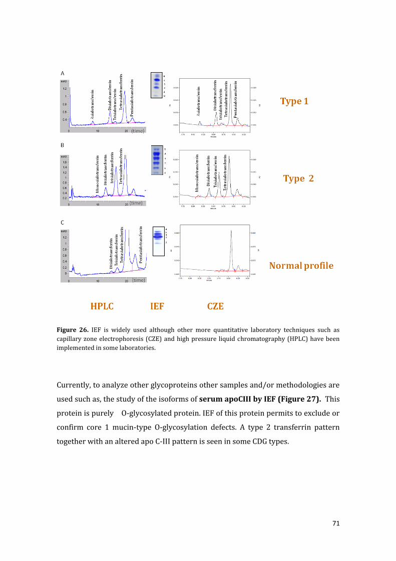
71
Figure 26. IEF is widely used although other more quantitative laboratory techniques such as
capillary zone electrophoresis (CZE) and high pressure liquid chromatography (HPLC) have been
implemented in some laboratories.
Currently, to analyze other glycoproteins other samples and/or methodologies are
used such as, the study of the isoforms of serum apoCIII by IEF (Figure 27). This
protein is purely O-glycosylated protein. IEF of this protein permits to exclude or
confirm core 1 mucin-type O-glycosylation defects. A type 2 transferrin pattern
together with an altered apo C-III pattern is seen in some CDG types.

72
Figure 27. IEF of serum apoCIII permits to exclude or confirm core 1 mucin-type O-glycosylation
defects.
Figure 28. Algorithm of the diagnostic pathway commonly used from clinical suspicion to reach a
definitive diagnosis.
73
Chapter 6. Treatment and therapies
Authors: Célia Pérez Cerdá and Belén Pérez González (Centro de Diagnóstico de
Enfermedades Moleculares, Centro de Biología Molecular, CIBERER, Universidad
Autónoma de Madrid, Spain).
Translator and Reviewer: Vanessa Ferreira, Ph.D. (Associação Portuguesa CDG e outras Doenças Metabólicas Raras) and Maria Antonia Vilaseca, Ph.D. (Guia Metabólica, Hospital Sant Joan de Déu, Barcelona, Spain).
Will a treatment for PMM2-CDG (CDG-Ia) be available soon?
There are several groups studying different possibilities. Of course, these
are recent studies, which mean that all efforts should be combined in order to
optimize their chance of success.
In our group, we are investigating the possibility of treating these patients with
mutation-specific therapies. For its application it is essential to know the
responsible gene, the mutations causing mutations the disease and the effect of
these changes in the activity and function of the corresponding proteins. The
study of the effect of the mutations identified and the specific involvement in
cell and tissue functions produced by these mutations may facilitate the
development of personalized therapeutic options for each patient.
For CDG and other inherited metabolic diseases we have developed an
antisense therapy that can be applied to a mutation type that affects the
processing of the mRNA. We have identified several of these mutations and
successfully applied the antisense therapy in cells from patients with organic
74
acidurias and congenital defects of glycosylation (Rincon et al. 2007, Vega et al.
2009; Perez et al. 2009). We think that 5-10% of the disease-causing mutations
that remain to be identified in the genes responsible for inherited metabolic
diseases could be intronic mutants that cause the exonization of sequences.
Currently, the main obstacle to the clinical application of these antisense
oligonucleotides lies in the efficacy and safety of the release of these molecules.
Consequently, the current research focuses on the use of nanoparticles or viral
vectors that allow a proper and efficient release (Perez et al, 2010).
Another type of emerging therapeutic approach we are developing is a
therapy with pharmacological chaperones used to improve the folding of
mutated mis-folded proteins (Bernier et al. 2004). The pharmacological
chaperones have been successfully tested in the so-called conformational
diseases such as phenylketonuria, Gaucher disease, cystic fibrosis, and others,
where it has been possible to achieve the recovery of the “in vitro” functional
activity of several mutants identified (Pey et al. 2008). Recently, we identified
several folding mutants in the protein phosphomannomutase (gene-PMM2),
which could be candidates to be recovered through the use of these drugs. It is
noteworthy that all patients with a deficiency in PMM2 carry at least one of
their alleles with a mutation that retains residual activity, which in most cases
corresponds to a folding mutant (Vega et al, 2011). At present our challenge is
to find a compound in a library of 10000 drugs that can be used specifically to
treat this type of mutations identified in the PMM2 protein.
Bibliography:
• Perez B, Rincon A, Jorge-Finnigan A, Richard E, Merinero B, Ugarte M, Desviat LR (2009). Hum Mutat 30: 1676-82.
•Pérez B, Rodriguez-Pascua L, Vilageliu L, Grinberg D, Ugarte M, LR. Desviat 2010. J Inher Metab Dis (in press).
• Pey AL et al. 2008. J Clin Invest 118(8):2858-67.
• Rincon A, Aguado C, Desviat LR, Sanchez-Alcudia R, Ugarte M, Perez B. 2007. Am J Hum Genet 81(6).
• Vega AI, Perez-Cerda C, Desviat LR, Matthijs G, Ugarte M, Perez B. 2009. Hum Mutat. 30, 795-803.
Vega AI, Pérez-Cerdá C, Abia D, Gámez A, Briones P, Artuch R, Desviat LR, Ugarte M, Pérez B. J Inherit Metab Dis. 2011 Aug;34(4):929-39. Epub 2011 May 4.

75
Chapter 7. CDG management of development
7.1. PMM2-CDG (CDG-Ia) Some Thoughts on Speech and Communication
Author: Merell Liddle, Australian CDG patient representative and mother.
Our daughter Morgan is 18-years-old now so we have been on the journey
to assist her develop better communication for a long time. As a young child she
was active and alert but had no speech. At that time she did not have her
diagnoses of CDG1A but we were aware that she had an underdeveloped
cerebellum and she had a diagnoses of dysarthria (a motor speech disorder) and
dyspraxia of speech (difficulty planning, initiating and executing speech sounds
and non-speech movements) in addition to severe ataxia. Research was starting
to suggest that the cerebellum is also involved in much more than motor planning
and coordination and that it is involved in a variety of linguistic and higher order
thinking functions. So from very young we were aware that communication would
be problematic for her and also that literacy could also be a challenge for a child
with speech difficulties. While working on her physical skills was very important,
we believed that working on her communication skills was probably even more
important for a happy and interesting life.
We all want our children to develop natural speech, and that is something
we have always worked towards, but we also wanted to give Morgan the tools to
communicate independently without speech. From very young we started using
Augmentative and Alternative Communication (AAC) strategies and Morgan has
76
grown up using both natural speech and AAC. I do recommend you read this article
on independent communication.
A Communication Independence Model for People with Severe Communication
Disabilities
http://www.speechpathology.com/articles/article_detail.asp?article_id=215
It took us a while to really understand the importance of independent
communication in a young child because very often you think that you can ‘work
out’ what your child wants and that AAC is not necessary and it is a lot of effort.
Also I know I used to feel secretly pleased to show what a good mother I was by
being able to understand my child! This article shows how individuals with
complex communication needs are often dependent upon significant others to
send or interpret their communication messages and it demonstrates how
individuals with severe disabilities can participate more fully and independently
once their partners alter their communication strategies and expectations.
We started to work on Morgan’s communication very early but at that time
AAC services were very limited and we implemented and supported use of her AAC
systems ourselves. These days AAC systems are much more widely available and
many more therapists are trained in their use.
Early Intervention for Young Children with Complex Communication Needs
http://aackids.psu.edu/index.php/page/show/id/1
AAC provides a way to move young children beyond simply labelling (eg
dog, spoon) and making demands (eg cookie or drink) to providing modelling
and opportunities for commenting, questioning, making observations and
statements and even singing. We wanted Morgan to move beyond being a
responder to being an initiator of conversations. It is very hard work
integrating AAC strategies into everyday life but once Morgan started using the
system and could say many more things, it became fun. We could see her
personality emerging and her frustration levels reduced as she was able to
communicate more effectively.
Literacy Instruction for Children with Disabilities
77
http://aacliteracy.psu.edu/
Because we knew that Morgan would always have difficulty with her walking
and speaking we thought it would be very important if she could read. So, from
a very early age we started using text with symbols on her AAC device to
demonstrate to her that pictures also had words and that they could be
combined to make sentences. We also did a lot traditional phonological
awareness activities, for example, rhyming words. Morgan learnt to read at age
six with her typically developing peer in a mainstream classroom. Until age
eight, she used a system with symbols and words but then moved to using a
text-to-speech device.
Moving Into Adulthood
Morgan started life relying very much on her AAC system to communicate but
during her early and middle years of school she did not need to rely on her
system so much because her teacher and the other children could generally
understand her speech and the communication demands were not so complex.
During her senior high-school years and now moving to post-secondary
education and more independent living she will again need to rely more on
using her AAC device. Morgan uses a Lightwriter SL40, a text-to-speech device
with word prediction. She recently presented a paper on communication and
dance at the International Society for Augmentative and Alternative
Communication Conference in Barcelona.
http://www.isaac-online.org/en/home.shtml
The ISAAC website will help you find a local contact in your country for more
information on AAC.
Other links:
http://www.complexchild.com/developmental.html
78
7.2. Occupational therapy helps CDG children to become independent
Author: Beatriz Sanz, Child Occupational Therapist, Professor at the Rey Juan
Carlos University. Alcorcón. Madrid. Spain)
Translation: Vanessa Ferreira, Ph.D. (Associação Portuguesa CDG e outras Doenças Metabólicas Raras)
Translation reviewer: Merell Liddle, Australian CDG patient representative and mother.
Occupational Therapy is "the social health discipline that evaluates the
ability of one person to perform activities in daily life and participate when that
capacity is at risk or damaged by any physical, cognitive, emotional or social
cause".
An Occupational Therapist for children is professionally trained and
specializes in facilitating and enabling children to participate in the activities of
childhood: play, leisure, self-care activities (feeding, dressing and hygiene) and
school activities.These activities are essential for emotional well-being of any child.
What can an occupational therapist for a child with CDG do?
1) Use games and activities of daily living to improve the manipulative skills
enabling the child to dress by himself.
2) Help the children who avoid certain textures and flavors to try new
foods, and consequently it will improve their tolerance for different foods.
3) Carry out activities that help to increase the child’s strength in the hands
and advise on the needed adaptations for the child to hold a pen or a spoon.
4) Design a chair and a table suitable for the child that does not have head
and trunk control, and can maintain the child’s position in such a way that
the child can perform work more effectively.
5) An occupational therapist will show parents of young children how to
play with them in order to facilitate the coordinated use of his eyes and
hands or head control.
6) Identify appropriate toys for children with limited vision or manipulative
difficulties to play with.
7) Recommend to parents how to handle a child with abnormal tone and

79
posture, to how to clothe, feed and play.
8) Advise parents about the necessary adaptations in the home (removal of
architectural barriers, installation of grab bars, etc.) to promote the
independence of children in this environment.
9) Facilitate the coordinated movement of the eyes when there is a squint
or any other disorder of ocular motor coordination that interferes with the
child's functional vision.
10) Collaborate with other professionals involved in child care in order to
develop joint strategies to improve their participation in all activities of
daily living.
Different adaptations are designed by an Occupational Therapist to give the
child with poor sensory-motor skills the necessary skills to eat, paint and play, for
example, by using a thicker grip on handles.
Occupational therapists work with children, parents and teachers to
overcome the barriers limiting their participation and consequently their
development of self-esteem.
Some helpful links to look for information:
o APTOCAM. Asociación Profesional de Terapeutas Ocupacionales de la
Comunidad Autónoma de Madrid. C/ Serrano 79. Bajo
Derecha.28006.Tf. 91 4115297.
www.aptocam.org
o APETO. Asociación Profesional Española de Terapia Ocupacional. C/
Modesto Lafuente 63, 3ºC. 28003 Madrid. Tf. 91 5535608.
www.apeto.com
o AEIS. Asociación Española de Integración Sensorial. Apartado de
Correos 38032. Madrid 28031. Tf. 689499568.
www.integracionsensorial.es
o AETB. Asociación Española de Terapeutas formados en el concepto
Bobath. C/ Mirador de la Reina 113-115. 28035 Madrid. Teléfono
647988249.
www.asociacionbobath.es

80
7.3. Physiotherapy in CDG children
Author: Paula Davila Martinez (Physiotherapist specialized in the Bobath Concept
(babies, child and adult). C.P.E.E. Princesa Sofia (Madrid)
Translation: Vanessa Ferreira, Ph.D. (Associação Portuguesa CDG e outras Doenças Metabólicas Raras)
Translation reviewer: Merell Liddle, Australian CDG patient representative and mother.
Physiotherapy plays an important role in the intervention with children
affected by CDG, especially by helping the child to modulate his postural tone
(hypotonia), to improve coordination and equilibrium in different functional
patterns of movement and to prevent sensory-motor developmental delay. The
main goal is to help the child learn how to organize posture and movement in
order to perform functional activities that are meaningful to their development.
However, physiotherapy involves many more areas than sensory-motor
problems. The main aim of the physiotherapist is to improve the autonomy, the
functional abilities of the child and developmental stages of life, helping him to be
as independent as possible to maximize participation in his environment (school,
play with friends, family and life.
To reach this goal, the work of the physiotherapist is not limited to physical
therapy sessions, but in addition includes:
Guidelines of physical therapy management programs
We teach the parents and other family members how to improve posture and
movement, for example: how to walk, how to sit down correctly, how to get up,
how to climb the stairs, how to use a tricycle, how to stand, etc. always looking for

81
functional situations. Proper management throughout the day is very important to
encourage the child to learn appropriate posture and movement patterns. We must
not forget that to learn it is necessary that the child practices and repeats in
different contexts.
- Program of activities at home. We develop and teach specific activities, which
are easy to perform and which extend the benefits achieved during the physical
therapy sessions.
- Recommendations about recreational programs. The physiotherapist advises
what type of leisure activity will benefit the child and will help to improve
functional abilities. For example, adaptive sports, swimming, etc.
- Choice and recommendation of assistive technology. Assistive technology is
any kind of item, piece of equipment or product system, whether acquired
commercially manufactured, modified or personalized, that is used to increase or
improve functional capacities of individuals with disabilities. For example, mobility
systems (different types of wheelchairs, walkers, adapted tricycles, etc.),
positioning systems (standing, classroom chairs, etc.), dynamic orthotic of ankle-
foot, orthoses elasticized, and others.
In everything, it is important to take into account the needs of the individual
child (type of problem presented, age, degree of impairment, cognitive level, etc.)
and also the needs of the family, with regard to the task or activity that will be
undertaken and the environment in which is performed.
And of course, to include all these aspects we need to work in full
collaboration with other professionals involved in the care of the child
(occupational therapist, speech therapist, orthopedic technician, teachers, doctors,
etc). The Physical therapy must be included as of part an interdisciplinary team in
which all its members work to achieve common goals that will benefit the child
and the family. Therefore, understanding the role of the child, parents and other

82
family members is essential to the team.
Our attention is directed to solving the problems that the child presents,
anticipating future problems and seeking to attain maximum participation with
movement quality at every stage of life.
Websites of interest:
http://www.iser.com/physical-therapy.html
http://specialchildren.about.com/od/therapies/Therapies_Learn_More_About_Ho
w_They_Help_Your_Child.htm

83
7.4. The importance of Early Attention or Intervention
Author: María Luisa Pendas Sánchez, Psychologist specialized in Early Attention
and Psychomototricity, Therapeutic Pedagogical Teacher.
Translation: Vanessa Ferreira, Ph.D. (Associação Portuguesa CDG e outras
Doenças Metabólicas Raras)
Translation reviewer: Merell Liddle, Australian CDG patient representative and mother.
WHAT IS EARLY INTERVENTION?
“Early attention or intervention”is the support services directed to children
under age 6 that have some type of developmental disorder, to their families and
their close environment. These actions must take into account the totality of the
child through the development of different aspects: motor, emotional and cognitive
optimizing the child’s integration into the family, school and society.
Who is involved in Early Intervention?
The real target of early intervention is the children and their parents. The activity
of the professional is aimed at delivering direct services to children and their
families through advice and guidance related to various bio-psycho-social aspects
from an interdisciplinary approach.
Where is Early Intervention done?
Early intervention can be done at home, school and in the child’s community and in
coordination with specialized teams responsible for the assessment, diagnosis and
intervention programs.
What are the specific objectives of Early Intervention in CDG?
- Reducing effects of CDG in the overall development of the child
- To promote optimal development of the child
-Avoid or reduce the occurrence of side effects due to the disorder
- Attend to and meet the needs of the family and child's school environment

84
- Supporting active participation of children in the community
Links:
http://www.kidsource.com/kidsource/content/early.intervention.html
http://www.kidsource.com/NICHCY/infantpub.html
http://www.earlyinterventionsupport.com/
7.5. How swimming pool activities can help my child?
Source: Article from Guia Metabólica
Inborn errors of metabolism and sports
Mercedes Serrano a, M. AntòniaVilasecab and Jaume Campistol,*
aServei de Neurologia, Unitat de MalaltiesMetabòliques, Hospital Sant Joan de
Déu, Universitat de Barcelona,
Barcelona, España
aServei de Bioquímica, Unitat de MalaltiesMetabòliques, Hospital Sant Joan de
Déu, Universitat de Barcelona,
Barcelona, España
7.6. Pet-therapy
http://www.ctac.cat/portal/
85
Chapter 8. The importance of research and communication and
dissemination in Rare Diseases
8.1. Basic Research in rare diseases
Author: Mafalda Araújo, Basic Reseacher at the IBMC, Porto, Portugal.
Basic Research in rare diseases
Rare diseases represent a challenging area of research. Affecting few
individuals, diagnosis is very difficult and symptoms are often hard to define with
many of them being common to many different diseases.
Estimates project that rare diseases affect nearly 250 million people all over
the world (Shire, biopharmaceutical company) and that 8% of people will become
afflicted with a rare disease at some point in their lives(Time magazine, Aug 2010).
The development of treatments for these diseases faces two big obstacles: the lack
of understanding of the underlying pathophysiological mechanism of the
conditions and the lack of interest of pharmaceutical companies.
Basic research is sometimes neglected and not very well understood. Some
people believe their tax money should not be spent by scientists that simply want
to know how things work but aren’t really working to find the cure for a
devastating disease. However, our understanding of genetics and heredity is
fundamentally due to the studies of Gregor Mendel (for review see Dunn 2003),
who studied pea plants in the 1860’s, and the experiments of Thomas Morgan with
the fruit flies in the beginning of the 20th century (for review Miko 2008).
According to The European Organization for Rare Diseases (EURORDIS) there are
between 5,000 and 7,000 distinct rare diseases and about 80% are genetic

86
diseases. Therefore these are just two very good examples of the value of basic
research.
Another example of the contribution of basic research to disease treatment
is the transformation of cancer treatment thanks to breakthroughs in biochemistry
and genetics. The simple discovery of molecular markers (compounds that appear
in abnormal levels or at unusual places in the organism) has contributed very
much to the diagnosis and the establishment of prognosis estimates. For rare
diseases these discoveries allow the design of new and more accurate diagnostic
tests that do not require the expertise of a geneticist and can be interpreted by any
physician.
On the other hand there is a large gap between basic scientific research
advances and applied product development, which is reflected in the lack of real
products arriving to patients. The European Rare Disease Therapeutic Initiative
(ERDITI) is a collaborative effort between academic research institutions and
pharmaceutical companies to promote therapeutic research on rare diseases.
In fact, recently big multi-national pharmaceuticals are recognizing the
long-neglected potential of the rare disease market and becoming interested in
either buying smaller companies with expertise in rare medical conditions drugs or
invest themselves by setting up a dedicated rare disease Research and
Development Unit (Shaffer, 2010).
Therefore it is crucial that public and private entities give financial support
for basic research.
Fortunately several rare disease dedicated research institutions are being
created to develop new methods for understanding, diagnosing and treating these
neglected, often called orphan, diseases. The collaboration between researchers,
physicians and patients are needed to achieve these goals.
87
Research Fellowships:
http://www.childrenshospital.org/cfapps/research/data_admin/Site2673/mainpa
geS2673P16.html
http://www.rarediseases.org/research/requests
http://www.action.org.uk/our_research/research_training_fellowship_holders
http://rarediseasefoundation.org/RDFGrants.html
http://www.tfrd.org.tw/english/laws/cont.php?kind_id=78&top1=What%20we%
20do&top2=Patient%20Activity&top3=Scholarship
http://www.marionegri.it/mn/en/sezioni/formazione/borseDiStudio/borsaCurie.
html
http://www.findaphd.com/custadverts/4year/bham/minisite09/trans-3.asp
References:
1.Is It Time We Paid More Attention to Rare Diseases?
By Frances PerraudinSaturday, Aug. 21, 2010, TIME magazine
1. Arch Dis Child Fetal Neonatal Ed. 2003 Nov;88(6):F537-9.
Gregor Mendel, OSA (1822-1884), founder of scientific genetics.Dunn
PM.Department of Child Health, University of Bristol, Southmead,
2. Miko, I. (2008) Thomas Hunt Morgan and sex linkage. Nature
Education 1(1)
3. EURORDIS
4. ERDITI, The European Rare Disease Therapeutic Initiative
5. Shaffer C., Pfizer explores rare disease path. Nature Biotechnology 28, 881-
882- 2010
88
Useful links:
(USA) Children’s r.a.r.e. disease network: http://www.crdnetwork.org/
http://www.euroglycanet.org/
http://www.eurordis.org/content/survey-patient-groups-research
http://www.orpha.net/consor/cgi-bin/index.php
RareSpace-This community is for anyone interested in learning more about rare
diseases, rare disorders and related topics.
http://www.medpedia.com/communities/274-RareSpace
The Global Genes Project (www.globalgenesproject.org), a worldwide rare disease
awareness initiative.
89
8.2. The importance of communication in Rare Diseases
Author: Sebastián Sanchéz, PhD in Information Science. Bachelor in
Communication and History. Research Group on Disability and Communication
(GIDyC). Professor at the University of Valencia.
Translation: Vanessa Ferreira, Ph.D. (Associação Portuguesa CDG e outras
Doenças Metabólicas Raras)
Translation reviewer: Merell Liddle, Australian CDG patient representative and
mother.
In the area of public health, diseases which occur in low-prevalence, known
as "Rare Diseases", have changed our understanding of universal health care. To
provide necessary care for more than 35 million people affected by nearly 8,000
different diseases in the European Union, it is necessary to develop a new
relationship between the patients and the health administrators involved in this
sensitive area. The fact that over 80% of these diseases have a genetic origin plus
the high degree of severity and difficulty of early diagnosis requires a new
multidisciplinary approach. The situation makes patients highly vulnerable in a
complex system.
As recommended by the EU Council (2009 / C 151/02), "the principles and
the essential values of universality, access to a good quality health care, equity and
solidarity, adopted in the conclusions of the Council about the values and common
principles of EU health systems, from June 2, 2006, are of high importance for
patients with rare diseases. " Therefore, it is necessary to create new synergies
between all public health agencies to improve quality of life for these patients and
provide their families with the proper tools to ensure best practice in health care.
Because many rare diseases have a genetic origin and due to the
exponential advances in basic research in other areas of scientific knowledge, after
the lengthy
process of diagnosis, those affected by these diseases must consider genetic and
clinical research as part of their future needs. It is not just the search for a rapid

90
and accurate diagnosis, it is also the expectation of a good outcome as a result
ofscientific developments and transfer of research knowledge.
All these variables place the patient at the centre of a complex social space
in which the language used sometimes lacks respect and dignity and is confusing
due to limited quality information. Therefore, patients and practitioners who
produce information for discussion in the mass media must provide the
information in an appropriate way,
The treatment that the media give to rare diseases and those who are living
with them, must be scrutinized and monitored continuously. The aim of
promoting social inclusion of people with these type of disabilities is to help
gainacceptance of disability as a normal part of life, leaving behind images of
weakness and misery.
In short, avoid stigmatization, stereotyping, superficiality, and the use of language that focuses only on the clinical picture. Instead, enhance the understanding that the "rarity" of these conditions does not come from strange symptoms, but from their low prevalence in the population. If the media is one of the main sources of the construction of reality in developed societies, rare diseases such as congenital disorders of glycosylation (CDG) need a consistent and enhanced media presence. But, there is still a long way to go. ----------------------------------------------- Useful Links on Rare Diseases - Http://www.orpha.net/consor/cgi-bin/index.php?lng=ES (portal for rare diseases and orphan drugs) - Http://www.ciberer.es/ (Biomedical Research Centre Network for Rare Diseases) - Http://ec.europa.eu/health/index_en.htm (European Commission on public health) - Http://89.97.167.162/Home.aspx (European Project on Development Plans for Rare Diseases) - Http://www.enfermedades-raras.org/ (Spanish Federation for Rare Diseases)
91
9. Glossary:
Authors: Maria Antonia Vilaseca Ph.D. (Guia Metabólica, Hospital Sant Joan de
Déu, Barcelona, España) and Belén Pérez Dueñas, MD, Ph.D. (Departamento de
Neurología del Hospital Sant Joan de Déu, Barcelona, España).
Cerebellar Hypoplasia: disorder characterized by the incomplete or
underdevelopment of the cerbellum. It may be genetic or occur sporadically.
Microcephaly: Reduced growth or detention of head’s growth that give rises to a
smaller head compared to its corresponding size for the age
Seizures: Appearance of a sudden abnormal electric activity in the brain.
Stroke-like episodes: Acute episodes that affect the blood circulation.
Ataxia: Specially, reduction of the ability to coordinate voluntary muscular
movements.
Atrophy: Is the reduction of the size of a certain organ due to the lost of its mass.
Hypotonia: Reduction of muscular tone
Protein-losing enteropathy: Disease of the digestive track due the loss of
proteins through the stool.
Deshydration: Excessive loss of body liquids.
Bleeding disorders: The tendancy of an individual with CDG to bleed more than
expected often because the clotting factor levels are low.
Reflux gastroesophagitis: As a consequence, of loss of the muscular tone of the
inferior esophagus sphincter, gastric contents ascends into the esophagus. In the
children this can have several consequences, like discomfort after the meals,
esophagitis, cough, difficulty gaining weight, colic and respiratory problems.
Cardiomyopathy: disease of the cardiac muscle.
Osteopenia: Reduction of the bone mass below the normal limits.
92
Fat pads and Lipodystrophy: Is a symptom of the cellular subcutaneous tissue.
Consequently, its volume is reduced and at the same time is accumulated in an
abnormal way in certain localizations, like the gluteus and the arm of the children
affected by CDG.
Nystagmus: Involuntary and uncontrolled movement of the eyes. It can be
horizontal, vertical, rotatory or a combination of them.
Retinitis Pigmentosa: It is a progressive disease of the retina due to the loss of
the rods and cones, the main cells of the retina. The main symptoms are the slow
but progressive reduction of the visual acuity that in the first stages affects
predominantly, the night vision and the peripheral field of vision, with the central
vision remaining.
Ocular Motor Apraxia: Inability to maintain looking at the fast follow-up of a
certain object. The child compensates this by doing fast movements of their head
that excess the objective of the look, with the posterior correction until they
achieve a media position of their head.
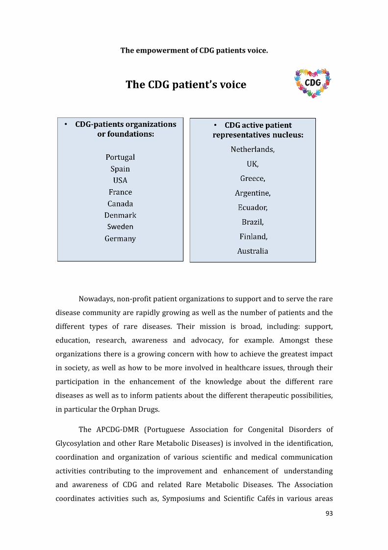
93
The empowerment of CDG patients voice.
Nowadays, non-profit patient organizations to support and to serve the rare
disease community are rapidly growing as well as the number of patients and the
different types of rare diseases. Their mission is broad, including: support,
education, research, awareness and advocacy, for example. Amongst these
organizations there is a growing concern with how to achieve the greatest impact
in society, as well as how to be more involved in healthcare issues, through their
participation in the enhancement of the knowledge about the different rare
diseases as well as to inform patients about the different therapeutic possibilities,
in particular the Orphan Drugs.
The APCDG-DMR (Portuguese Association for Congenital Disorders of
Glycosylation and other Rare Metabolic Diseases) is involved in the identification,
coordination and organization of various scientific and medical communication
activities contributing to the improvement and enhancement of understanding
and awareness of CDG and related Rare Metabolic Diseases. The Association
coordinates activities such as, Symposiums and Scientific Cafés in various areas

94
such as the nanotechnology, epidemiology, orphan drugs, etc. The aim of these
activities is to establish a relationship between scientists and society, to contribute
to scientific debate and to increase media coverage of different aspects of rare
diseases. Interestingly, several initiatives of CDG awareness and dissemination
have been established in partnership between CDG organizations, researchers and
clinicians such as the: (1) Practical guide targeted to CDG families; (2) Fairy-tale:
"Glycoland and the coloured antennas" (3) CDG awareness and dissemination tool
(4) CDG Online community (5) International CDG logo (6) CDG community store,
(7) Kit to disseminate Rare Metabolic Diseases, and others.
Nowadays empowerment is a reality for patients with rare diseases at all
levels, from research to regulatory aspects of the orphan-drug decision, including
production of awareness and educational information, and the design of study
projects involving doctors, researchers, health professionals, therapists, teachers
and society in general, in order to contribute to better healthcare, treatment and
outcomes. Thus, patients managed to engage all stakeholders in one direction: To
provide high quality up to date information about best practice in treatment and
care for their diseases.
Being part of the Association represents an opportunity to unite our voices in
one direction: to help the other families with the same problem and to provide high
quality up to date information about best practice in treatment and care for their
diseases.
Thinking of a better future for Liliana, Martim, Bruna, Elena, Aina, Ivan,
Morgan, and many other children and adults affected by a Rare Disease, give us the
reason to believe in our project and give us the strength to face the difficulties we
have at the moment in supporting and defending the rights of them at all levels of
government.

95
Part of the text is extracted from: World Orphan Drug Congress article
written by Vanessa Ferreira, President for the Portuguese Association for
Congenital Disorders of Glycosylation and other Rare Metabolic Diseases
(Associação Portuguesa CDG e outras Doenças Metabólicas Raras (APCDG-DMR).
In the following links, there are many resources for families affected by Rare
Metabolic Diseases and their Healthcare Community:
http://sindromecdg.orgfree.com/
http://www.guiametabolica.org/
http://www.rareconnect.org/en/community/cdg

96
"Menina"
Eu sei no que acredito
Quem me dera saber menina quem és tu
E o que me queres dizer
Sei que
tudo vem dentro de ti
Acredito que queres vencer
Mas na vida a tua força vai ganhar
A tua coragem vai mostrar menina que és feliz
A tua valentia
é do tamanho do mundo
talvez seja aqui que vais mostrar
O quanto a tua vida nos diz.
(Poem dedicated to Liliana Ferreira).
By Rosália Félix