THE POSTCRANIAL ELEMENTS OF PARADOLICHOPITHECUS ...
16
THE POSTCRANIAL ELEMENTS OF PARADOLICHOPITHECUS ARVERNENSIS (PRIMATES, CERCOPITHECIDAE, PAPIONINI) FROM LESVOS, GREECE* by A.A.E. VAN DER GEER** & P.Y. SONDAAR*** I. INTRODUCTION Recently, a rich Late Pliocene locality with land vertebrates was discovered along a dust road in an olive orchard near the village Vatera on Lesvos Island, Greece. The faunal assemblage of this site is characterized by, among others, a diversity in bovids, a giraffid, deer, Equus stenonis, Nyctereutes megamastoides and Gazella borbonica. In the same Vatera Formation Anancus arvernensis and Mammuthus meridionalis were found. The fauna is indicative for an open savanna mainland fauna of Middle Villafranchian age (MN 17). Besides the classical elements of the Middle Villafranchian, St. Vallier Faunal Unit, fossil remains were found of a giant tortoise and Paradolichopithecus arvernensis (DEPÉRET, 1929). This monkey is known from a few Late Pliocene localities, but only fossil material from Valea Grâunceanului (Romania) gives some information on postcranial morphology of this large extinct Old World monkey. Besides a number of postcranial elements (see Table 1), two mandibles are present among the material so far excavated at the Vatera F site. The larger mandible (PO 114) contains I 1-2 , DC, DP 3-4 , M 1-2 , the smaller one (PO 170) I 1-2 , C, P 3-4 , M 1- 3 . Until now, Paradolichopithecus was considered to have a way of life similar to that of the recent baboons and geladas with a terrestrial, quadrupedal locomotion adapted to an increased speed in the savanna. In size, Paradolichopithecus was a little larger than the largest extant baboon, Papio (C.) hamadryas, and comparable to the mandrill, Papio (P.) sphinx. The general architecture resembles that of the baboons, which observation is in agreement with SZALAY & DELSON (1979) who state that the Paradolichopithecus resembles macaques cranially and baboons postcranially. Within the Papionini, however, Paradolichopithecus can clearly be distinguished. In the present study, the material is compared with extant baboon and mandrill, and with the ankle bones of Australopithecus. * Tα ➵ετακρανιακά σκελετικά στοιχεία του Paradolichopithecus arvenensis από τη Λέσβο. ** Instituut Kern, Nonnensteeg 1-3, Universiteit Leiden, PO Box 9515, 2300 RA Leiden, The Netherlands. *** Natuurmuseum Rotterdam, PO Box 23452, 3001 KL Rotterdam, The Netherlands.
Transcript of THE POSTCRANIAL ELEMENTS OF PARADOLICHOPITHECUS ...
THE POSTCRANIAL ELEMENTS OFPARADOLICHOPITHECUS ARVERNENSIS ( PRIMATES,
CERCOPITHEC IDAE, PAPION INI) FROM LESVOS, GREECE*
by
A.A. E. VAN DER G EER** & P. Y. SO NDAAR***
I . INT RO DU CT ION
Recently, a rich Late Pliocene locality with land vertebrates was discovered along adust road in an olive orchard near the village Vatera on Lesvos Island, Greece. Thefaunal assemblage of this site is characterized by, among others, a diversity in bovids,a giraffid, deer, Equus stenonis, Nyctereutes megamastoides and Gazella borbonica. Inthe same Vatera Formation Anancus arvernensis and Mammuthus meridionalis werefound. The fauna is indicative for an open savanna mainland fauna of MiddleVillafranchian age (MN 17). Besides the classical elements of the MiddleVillafranchian, St. Vallier Faunal Unit, fossil remains were found of a giant tortoise andParadolichopithecus arvernensis (DEPÉRET, 1929). This monkey is known from a fewLate Pliocene localities, but only fossil material from Valea Grâunceanului (Romania)gives some information on postcranial morphology of this large extinct Old Worldmonkey.
Besides a number of postcranial elements (see Table 1), two mandibles arepresent among the material so far excavated at the Vatera F site. The larger mandible(PO 114) contains I1-2, DC, DP3-4, M1-2, the smaller one (PO 170) I1-2, C, P3-4, M1-3.
Until now, Paradolichopithecus was considered to have a way of life similar to thatof the recent baboons and geladas with a terrestrial, quadrupedal locomotion adaptedto an increased speed in the savanna. In size, Paradolichopithecus was a little largerthan the largest extant baboon, Papio (C.) hamadryas, and comparable to themandrill, Papio (P.) sphinx. The general architecture resembles that of the baboons,which observation is in agreement with SZALAY & DELSON (1979) who state that theParadolichopithecus resembles macaques cranially and baboons postcranially. Withinthe Papionini, however, Paradolichopithecus can clearly be distinguished. In thepresent study, the material is compared with extant baboon and mandrill, and withthe ankle bones of Australopithecus.
* Tα "ετακρανιακά σκελετικά στοιχεία του Paradolichopithecus arvenensis από τη Λέσβο.** Instituut Kern, Nonnensteeg 1-3, Universiteit Leiden, PO Box 9515, 2300 RA Leiden, The
Netherlands.*** Natuurmuseum Rotterdam, PO Box 23452, 3001 KL Rotterdam, The Netherlands.
I I . MATERIAL
The postcranial material (Table 1) from Vatera that can be attributed toParadolichopithecus arvernensis (DEPÉRET, 1929) consists of a slightly damaged butcomplete right humerus (PO 225), a left humerus without proximal part (PO 200), aright ulna (PO 229) and a right radius (PO 431), both missing the distal part, aseparate left olecranon (PO 059), an upper third right radius missing the proximalarticulation (PO 501), two upper shaft fragments of two left radii (PO 498 and PO502), an upper sixth part of a left radius (PO 630), a distal right tibia (PO 228), and aright talus (PO 157). The right humerus (PO 225), ulna (PO 229) and radius (PO 431)belong to the same individual. Though the distal tibia (PO 228) and talus (PO 157)are of one individual, they cannot be assigned with certainty to the former individual.All specimens are comparable in size, and more or less also in shape, to the extantmandrill, Papio (P.) sphinx (LINNAEUS, 1758).
Comparison material consists of five complete skeletons of the large baboon Papio(C.) hamadryas, thirteen of the mandrill Papio (P.) sphinx, three of the geladaTheropithecus gelada, casts of the Romanian postcranials (right distal humerus, rightalmost complete ulna, and right radius without articulation areas, all with number VGr350 and belonging to one individual) and casts of the distal tibia and talus of theHadar hominoid Australopithecus afarensis A.L. 288-1 («Lucy»). The materials arestored at Naturalis, National Natural History Museum, Leiden, The Netherlands, and atthe American Museum of Natural History, New York. The Paradolichopithecusspecimens are stored at the Vrissa Natural History Collection, Lesvos. For generalmeasurements of the Paradolichopithecus arvernensis postcranial material, see Table2; for a photograph of the Romanian material (humerus, radius, ulna), see SZALAY &DELSON (1979).
– 72 –
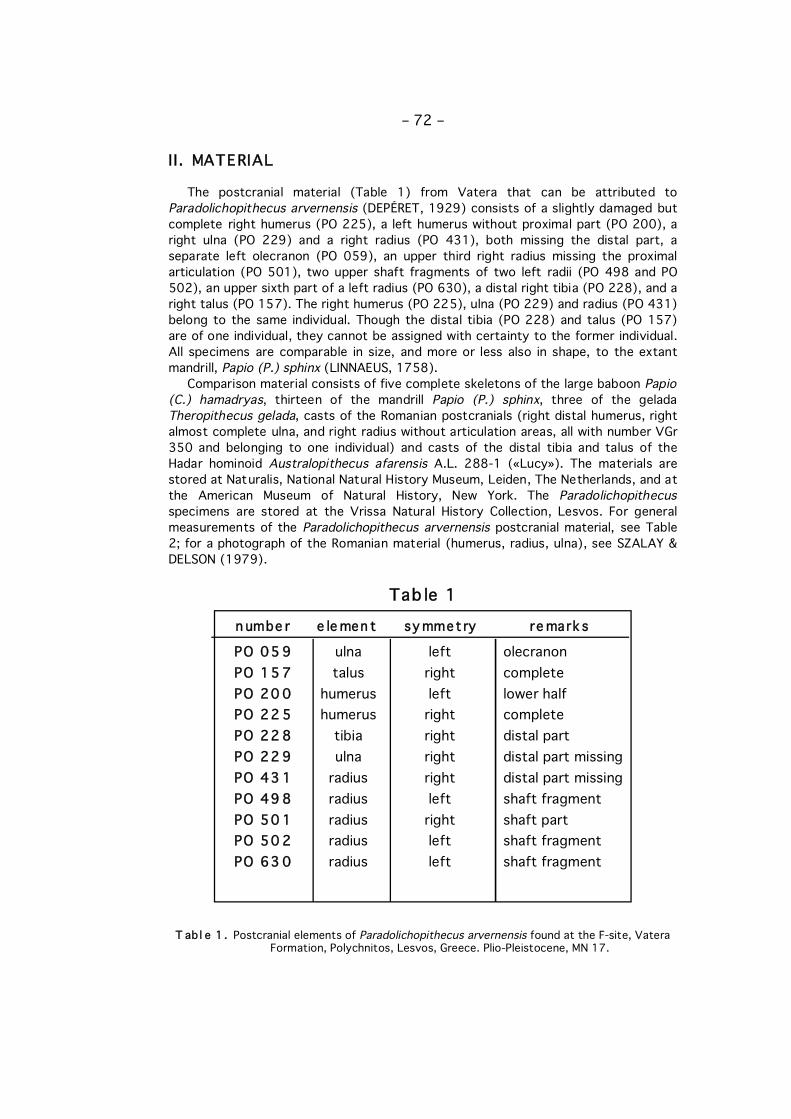
PO 05 9 ulna left olecranonPO 15 7 talus right completePO 20 0 humerus left lower halfPO 22 5 humerus right completePO 22 8 tibia right distal partPO 22 9 ulna right distal part missingPO 43 1 radius right distal part missingPO 49 8 radius left shaft fragmentPO 50 1 radius right shaft partPO 50 2 radius left shaft fragmentPO 63 0 radius left shaft fragment
n umbe r e le men t sy mmet ry re mark s
T ab l e 1 . Postcranial elements of Paradolichopithecus arvernensis found at the F-site, VateraFormation, Polychnitos, Lesvos, Greece. Plio-Pleistocene, MN 17.
Tab le 1
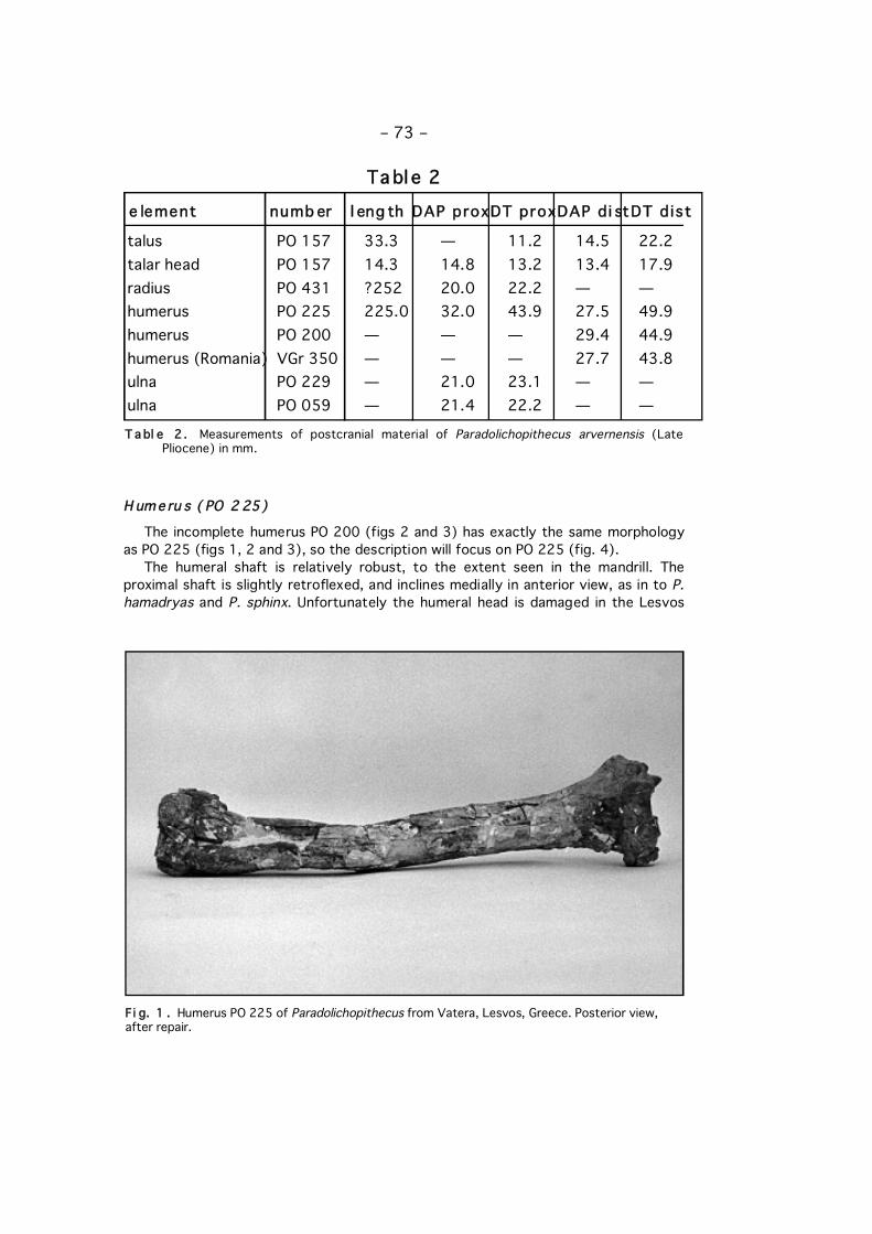
H ume ru s ( PO 2 25)
The incomplete humerus PO 200 (figs 2 and 3) has exactly the same morphologyas PO 225 (figs 1, 2 and 3), so the description will focus on PO 225 (fig. 4).
The humeral shaft is relatively robust, to the extent seen in the mandrill. Theproximal shaft is slightly retroflexed, and inclines medially in anterior view, as in to P.hamadryas and P. sphinx. Unfortunately the humeral head is damaged in the Lesvos
– 73 –
talus PO 157 33.3 — 11.2 14.5 22.2talar head PO 157 14.3 14.8 13.2 13.4 17.9radius PO 431 ?252 20.0 22.2 — —humerus PO 225 225.0 32.0 43.9 27.5 49.9humerus PO 200 — — — 29.4 44.9humerus (Romania) VGr 350 — — — 27.7 43.8ulna PO 229 — 21.0 23.1 — —ulna PO 059 — 21.4 22.2 — —
e lement numb er l eng th DAP proxDT proxDAP d i stDT d ist
Tabl e 2
T a bl e 2 . Measurements of postcranial material of Paradolichopithecus arvernensis (LatePliocene) in mm.
Fi g. 1 . Humerus PO 225 of Paradolichopithecus from Vatera, Lesvos, Greece. Posterior view,after repair.
– 74 –
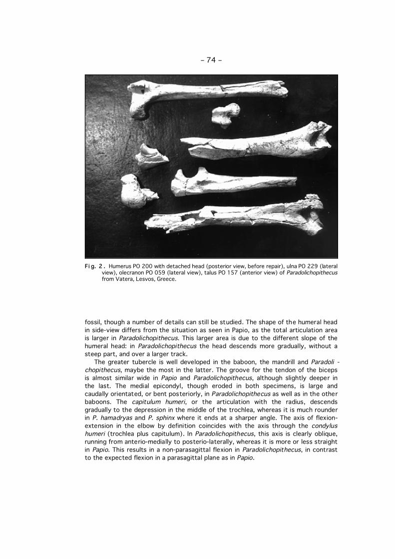
Fi g. 2 . Humerus PO 200 with detached head (posterior view, before repair), ulna PO 229 (lateralview), olecranon PO 059 (lateral view), talus PO 157 (anterior view) of Paradolichopithecusfrom Vatera, Lesvos, Greece.
fossil, though a number of details can still be studied. The shape of the humeral headin side-view differs from the situation as seen in Papio, as the total articulation areais larger in Paradolichopithecus. This larger area is due to the different slope of thehumeral head: in Paradolichopithecus the head descends more gradually, without asteep part, and over a larger track.
The greater tubercle is well developed in the baboon, the mandrill and Paradoli -chopithecus, maybe the most in the latter. The groove for the tendon of the bicepsis almost similar wide in Papio and Paradolichopithecus, although slightly deeper inthe last. The medial epicondyl, though eroded in both specimens, is large andcaudally orientated, or bent posteriorly, in Paradolichopithecus as well as in the otherbaboons. The capitulum humeri, or the articulation with the radius, descendsgradually to the depression in the middle of the trochlea, whereas it is much rounderin P. hamadryas and P. sphinx where it ends at a sharper angle. The axis of flexion-extension in the elbow by definition coincides with the axis through the condylushumeri (trochlea plus capitulum). In Paradolichopithecus, this axis is clearly oblique,running from anterio-medially to posterio-laterally, whereas it is more or less straightin Papio. This results in a non-parasagittal flexion in Paradolichopithecus, in contrastto the expected flexion in a parasagittal plane as in Papio.
– 75 –
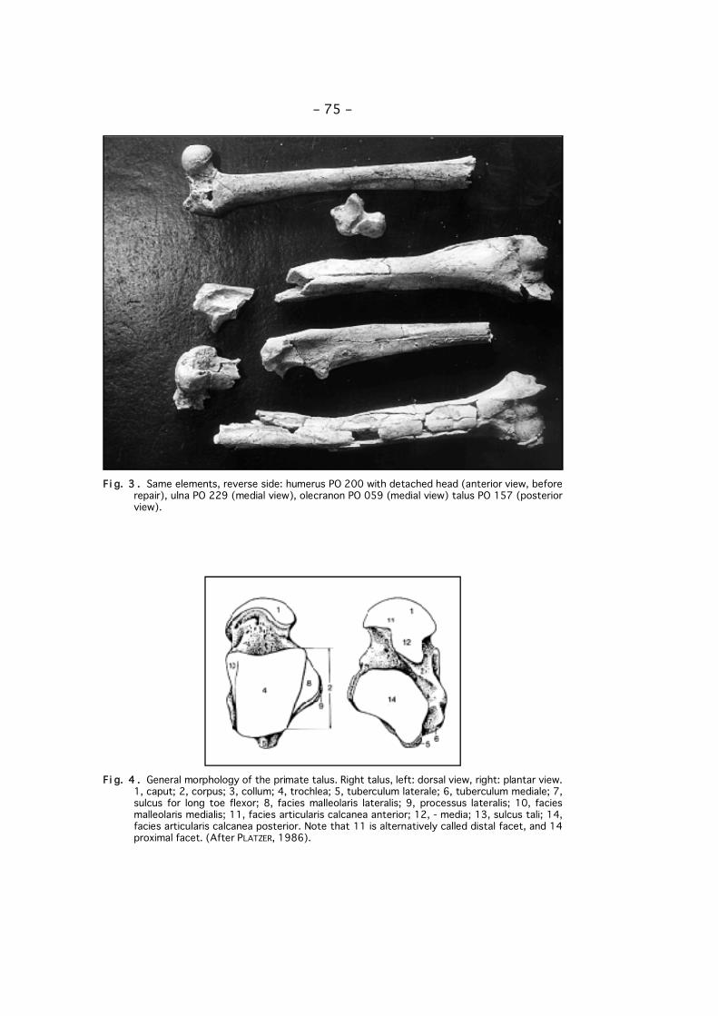
Fi g. 3 . Same elements, reverse side: humerus PO 200 with detached head (anterior view, beforerepair), ulna PO 229 (medial view), olecranon PO 059 (medial view) talus PO 157 (posteriorview).
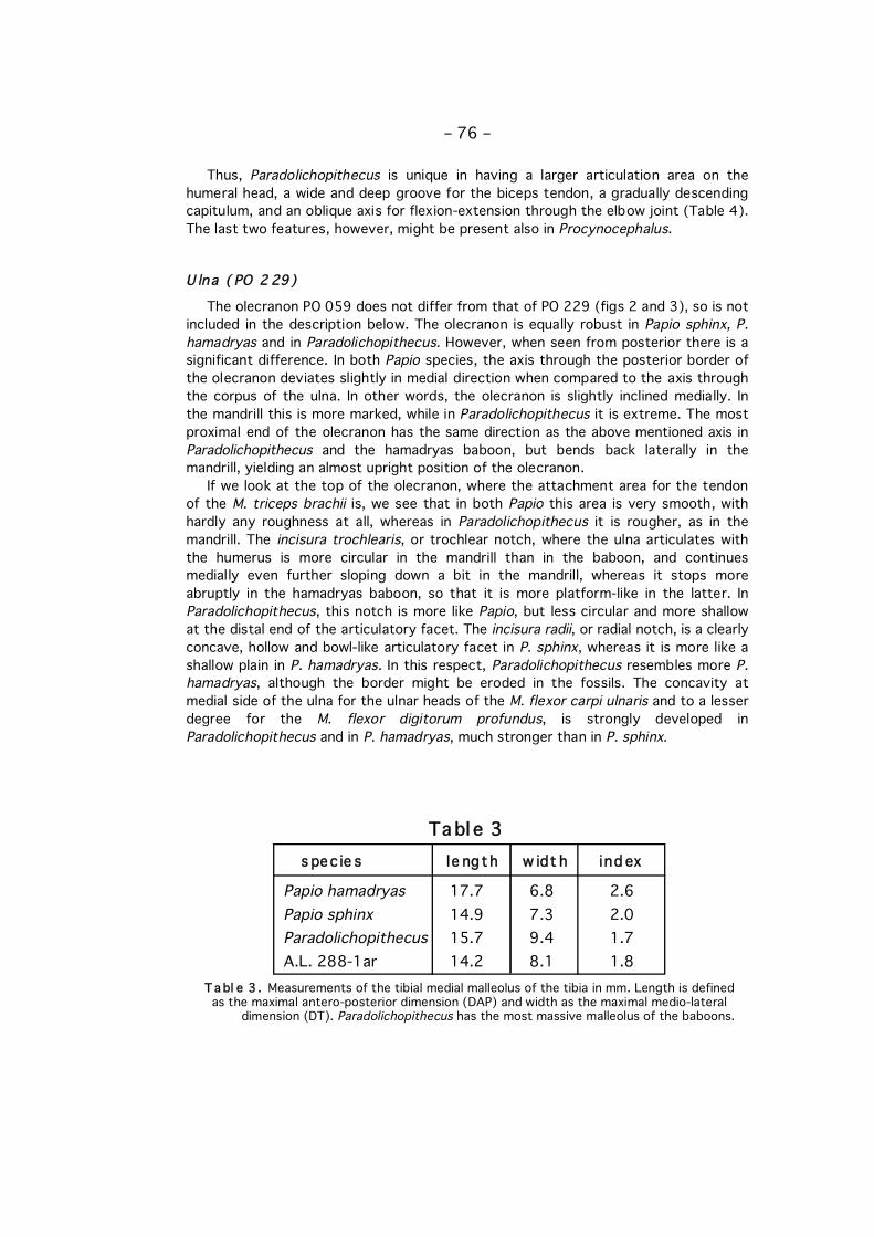
Fi g. 4 . General morphology of the primate talus. Right talus, left: dorsal view, right: plantar view.1, caput; 2, corpus; 3, collum; 4, trochlea; 5, tuberculum laterale; 6, tuberculum mediale; 7,sulcus for long toe flexor; 8, facies malleolaris lateralis; 9, processus lateralis; 10, faciesmalleolaris medialis; 11, facies articularis calcanea anterior; 12, - media; 13, sulcus tali; 14,facies articularis calcanea posterior. Note that 11 is alternatively called distal facet, and 14proximal facet. (After PLATZER, 1986).
Thus, Paradolichopithecus is unique in having a larger articulation area on thehumeral head, a wide and deep groove for the biceps tendon, a gradually descendingcapitulum, and an oblique axis for flexion-extension through the elbow joint (Table 4).The last two features, however, might be present also in Procynocephalus.
U lna ( PO 2 29)
The olecranon PO 059 does not differ from that of PO 229 (figs 2 and 3), so is notincluded in the description below. The olecranon is equally robust in Papio sphinx, P.hamadryas and in Paradolichopithecus. However, when seen from posterior there is asignificant difference. In both Papio species, the axis through the posterior border ofthe olecranon deviates slightly in medial direction when compared to the axis throughthe corpus of the ulna. In other words, the olecranon is slightly inclined medially. Inthe mandrill this is more marked, while in Paradolichopithecus it is extreme. The mostproximal end of the olecranon has the same direction as the above mentioned axis inParadolichopithecus and the hamadryas baboon, but bends back laterally in themandrill, yielding an almost upright position of the olecranon.
If we look at the top of the olecranon, where the attachment area for the tendonof the M. triceps brachii is, we see that in both Papio this area is very smooth, withhardly any roughness at all, whereas in Paradolichopithecus it is rougher, as in themandrill. The incisura trochlearis, or trochlear notch, where the ulna articulates withthe humerus is more circular in the mandrill than in the baboon, and continuesmedially even further sloping down a bit in the mandrill, whereas it stops moreabruptly in the hamadryas baboon, so that it is more platform-like in the latter. InParadolichopithecus, this notch is more like Papio, but less circular and more shallowat the distal end of the articulatory facet. The incisura radii, or radial notch, is a clearlyconcave, hollow and bowl-like articulatory facet in P. sphinx, whereas it is more like ashallow plain in P. hamadryas. In this respect, Paradolichopithecus resembles more P.hamadryas, although the border might be eroded in the fossils. The concavity atmedial side of the ulna for the ulnar heads of the M. flexor carpi ulnaris and to a lesserdegree for the M. flexor digitorum profundus, is strongly developed inParadolichopithecus and in P. hamadryas, much stronger than in P. sphinx.
– 76 –
Tabl e 3
Papio hamadryas 17.7 6.8 2.6Papio sphinx 14.9 7.3 2.0Paradolichopithecus 15.7 9.4 1.7A.L. 288-1ar 14.2 8.1 1.8
s pec ie s le ngth w idt h ind ex
T a bl e 3 . Measurements of the tibial medial malleolus of the tibia in mm. Length is defined as the maximal antero-posterior dimension (DAP) and width as the maximal medio-lateral
dimension (DT). Paradolichopithecus has the most massive malleolus of the baboons.
– 77 –
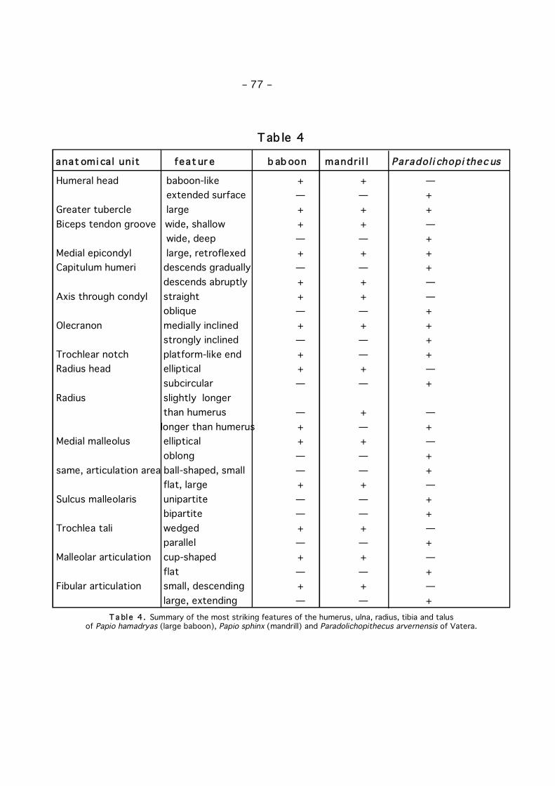
Humeral head baboon-like + + —extended surface — — +
Greater tubercle large + + +Biceps tendon groove wide, shallow + + —
wide, deep — — +Medial epicondyl large, retroflexed + + +Capitulum humeri descends gradually — — +
descends abruptly + + —Axis through condyl straight + + —
oblique — — +Olecranon medially inclined + + +
strongly inclined — — + Trochlear notch platform-like end + — +Radius head elliptical + + —
subcircular — — +Radius slightly longer
than humerus — + —longer than humerus + — +
Medial malleolus elliptical + + —oblong — — +
same, articulation area ball-shaped, small — — +flat, large + + —
Sulcus malleolaris unipartite — — +bipartite — — +
Trochlea tali wedged + + —parallel — — +
Malleolar articulation cup-shaped + + —flat — — +
Fibular articulation small, descending + + —large, extending — — +
anat omi ca l uni t feat ur e b ab oon mandr i l l Paradol i chopi thec us
T ab le 4
T a bl e 4 . Summary of the most striking features of the humerus, ulna, radius, tibia and talus of Papio hamadryas (large baboon), Papio sphinx (mandrill) and Paradolichopithecus arvernensis of Vatera.
To sum up, Paradolichopithecus differs from the baboons in having a highly inclinedolecranon, and a more shallow distal end of the trochlear notch (Table 4).
R ad iu s ( PO 4 31)
The radius is very similar to that of the mandrill, but broader and more round intransversal section. Its proximal articulation is almost circular, whereas this is moreelliptical in the mandrill and the hamadryas baboon.
The radius is much longer than the humerus in P. sphinx, and in P. hamadryas onlyslightly longer. Although the distal part is missing, we have the impression that thetotal length of the Paradolichopithecus radius PO 431 is about 25.2 cm. The righthumerus PO 225, belonging to the same individual, measures about 22.5 cm. Thismeans that the Paradolichopithecus radius of the Vatera Formation is a bit longerthan the humerus, similar to Papio sphinx. The radius fragments PO 498, PO 501, PO502 and PO 630 do not differ from the corresponding parts of PO 431.
The only, yet obvious and unique, difference between Paradolichopithecus and theother papionins is seen in the subcircular shape of the proximal articulation area (Table4).
T ib i a (P O 228)
The tibia of Paradolichopithecus is represented by a distal, right tibia. In distalview, the architecture of the articular surface is very similar to that seen in themandrill, both in shape as in size. A striking difference, however, is found in the medialmalleolus, which sets Paradolichopithecus clearly apart. The articular surface of themalleolus is clearly bowl-shaped and large in the mandrill, whereas it is almost flat andmuch smaller in Paradolichopithecus. The non-articulatory area of the malleolus isvery robust in Paradolichopithecus, oblong and tapering dorsally at the articulatoryarea bearing part. In both P. hamadryas and P. sphinx the non-articulatory area iselliptical, thus tapering at both ends. The articulatory area bearing part is larger andmore massive than the plantar part, in contrast to Paradolichopithecus where bothparts are massive and the plantar part even more so. On the whole, theParadolichopithecus malleolus is more massive (Table 3), and almost oblong.
In medial view, the malleolus also differs between Paradolichopithecus on one sideand Papio at the other side. In the latter, the slope of the distal border makes anangle of about 45 degrees in P. sphinx, about 35 degrees in P. hamadryas, but 25degrees in Paradolichopithecus. As a result, the dorsal part extends further distally,yielding a quite pointed apex, in the extant baboons than in Paradolichopithecus,where the malleolus is more equally massive with a blunt distal end. InAustralopithecus the malleolus is oblong in both distal and medial view, which makesit even more massive and more equal in all directions than in Paradolichopithecus.
Another difference with the extant baboons is the division of the sulcus malleolarison the plantar side of the malleolus in two separate parts. This bipartite sulcusoccupies the complete medial half of the plantar side. In the extant baboons, theunipartite sulcus occupies just one-sixth of the plantar side. Furthermore, thedirection of the sulcus is clearly distal in Paradolichopithecus, but medio-distal inPapio, with an angle of about 10-15 degrees.
In short, the uniqueness of the tibia of Paradolichopithecus is found in the
– 78 –
massiveness and flatness of the medial malleolus, and the bipartite groove (Table 4).
T al us (PO 157)
The talus PO 157 (figs 2 and 3) is well-preserved and fits perfectly to the distaltibia PO 228.
The talus, or astragalus, is characterized by a number of articular facets, due to itsintermediate position (fig. 4). The head carries the facet for the navicular bone, theplantar side of the body the three facets for the calcaneum and the dorsal side of thebody forms the trochlea for the tibia. On the medial and the lateral side are themalleolar facets for the malleolus fork formed by the tibia and the fibula. The maindifferences between the cercopithecoid talus and the hominoid talus are found in thepronounced lateral ridge of the trochlea in cercopithecoids, whereas the ridges are(almost) equal in the hominoids, and in the pronounced groove for the tendon of thelarge toe flexor in hominoids, whereas this groove is hardly visible in thecercopithecoids. The talus of Paradolichopithecus follows therefore clearly thecercopithecoid pattern, contrary to the clearly hominoid pattern of Australopithecus.
The talar trochlea of Paradolichopithecus is not typically cercopithecoid in wed-gedness, as it is more or less parallel, whereas it diverges from proximal to distal inthe other baboons (see also SONDAAR & VAN DER GEER, this volume). The proximalwidth of the trochlea is in Paradolichopithecus 15.8, the distal width is 17.2, resultingin an index of 1.1. In P. hamadryas these measurements are 12.1, 18.4, index = 1.5,and in P. sphinx 14.2, 19.5, index = 1.4. In a way Paradolichopithecus is comparablewith Australopithecus: 16.4, 19.2, index = 1.2.
In P. hamadryas, seen from lateral, two distinct phases are seen in the curvature ofthe proximal face. The proximal half is round, but the distal half is more gradually de-scending. The transition point is situated exactly halfway. In the mandrill and Parado -lichopithecus, however, there is only one phase that is gradually curved.
At the lateral side a large suspensory flap for articulation with the fibula is presentin Paradolichopithecus, whereas it is only small in both Papio species. Seen fromproximal, it extends 1.3 mm in P. hamadryas, 2.3 mm in P. sphinx and 3.7 mm inParadolichopithecus, who is similar to Australopithecus (3.8 mm) in this respect. Theshape of the suspensory facet for the fibular malleolus is more or less round in P.sphinx and Australopithecus, but pyramidal in P. hamadryas and Paradolichopithecus.The apex is tapering in Papio, and blunt in Paradolichopithecus. In the papionins thefacet is symmetrical, in contrast to Australopithecus where the distal part has alarger surface than the proximal part.
The curve of the articular head does not differ much between the species studied.In P. sphinx, Paradolichopithecus and Australopithecus it is elliptical. The mandrill ischaracterized by a distinct fossa at the plantar side of the talar head. P. hamadryasshows a slightly more round curvature than both mandrill and Paradolichopithecus.
The medial suspensory facet is (almost) flat in the studied tali. In both Austra -lopithecus and Paradolichopithecus, and to a lesser degree in the hamadryas baboon,the medial facet ends with a clear ridge or border where it touches the caput tali,whereas it merges gradually in the mandrill.
Another striking feature is the degree of hollowness of the posterior facet. Thisfacet is parabolic in shape in Australopithecus and Paradolichopithecus, but verycircular or round in Papio. This facet also extends further in proximo-distal direction in
– 79 –
Paradolichopithecus and Australopithecus (1.5 cm against total plantar length of 3.5cm), than in P. hamadryas (1 cm against 3 cm) or P. sphinx (1 cm against 3.5 cm).
The groove for the tendon of the large toe flexor (flexor hallucis longus) is as arule but poorly developed in the cercopithecoids, in contrast to the hominoids. Papioand Paradolichopithecus are no exception in this case, though the groove is slightlydeeper in P. hamadryas and Paradolichopithecus than in P. sphinx.
In short, the talus of Paradolichopithecus differs to a great extent from thebaboons, though it is still cercopithecoid in architecture. Unique features are theparallel trochlea, the highly extending suspensory flap for the fibula, the absence of acup-shaped depression for the tibial malleolus (Table 4), a more shallow and largerposterior suspensory facet for the calcaneum, and a clear separation between themedial suspensory facet and the talar head. These features are shared byAustralopithecus, and probably also by Procynocephalus as inferred from thedrawings (TEILHARD DE CHARDIN, 1938).
III. D IS CUSS ION
F or e -l i mbThe slight retroflexion and medial inclinement of the humerus shaft is a consistent
feature of extant cercopithecines (PILBEAM et al., 1990) which can also occur inAsian apes (BEGUN, 1992). Its functional significance is not clear, and it might be acharacteristic of quadrupedality (ROSE, 1994), although it also might be merely theancestral condition.
The more gradually curved, enlarged articulation area of the humeral head makesthe degree of arm movement in Paradolichopithecus more than in Papio. Thisindicates a freer use of the arms.
In the baboons the greater tubercle has a clear lever function. Two muscles inserton its upper part: the M. supraspinatus on the upper facet, and the M. infraspinatus onthe middle facet. Action of the first muscle is stabilization of humerus in the joint,tensor of the joint capsule, and abduction of the forelimb. Action of the second muscleis strengthening of the shoulder capsule, but its main function is exorotation of theforelimb. Both muscles add to the stability of the joint, and move the limb outward andaway from the body. It is easy to imagine that the strong infra- and supraspinatemuscles in Papio are very suitable for quick turns during swift quadrupedal runs, as itdemands considerable muscle action to give the whole body mass another direction,especially at high speed when the impulse is high. In Paradolichopithecus anotherexplanation might be valid. Here the enlarged articulation area on the head of thehumerus needs additional stability, which is solved by the joint stabilizing action ofboth muscles.
A deeper groove for the biceps tendon may, too, reflect an increased need forstability, which is the case in a loaded situation or in a situation with increasedmobility. The action of the biceps is quite complicated due to its two heads and thefact that it acts on two joints. Its main action is anteversion of the shoulder (inhumans: lifting the arm in the sagittal plain) and flexor and powerful supinator of theelbow. Paradolichopithecus seems to have used its biceps in approximately the sameorder as the extant baboons, but with more force or more freedom of movement. Atemptative explanation for the strong biceps might be its increased use for carryingthings, which is very suitable in open plains with its scattered food sources. For
– 80 –
parallels with hominids, see amongst others HEWES (1961), KORTLANDT (1967),LEAKEY & LEWIN (1979). In our opinion, however, even if Paradolichopithecus hadarms that were suitable for carrying things, it is only an additional advantage, and nota reason on itself. Locomotion is primarily an adaptation to environment in order tobe more effective in a certain habitat, as DE VOS (this volume) and DE VOS et al.(1998) demonstrate for Hipparion and Australopithecus.
The form of the capitulum informs us about the elbow joint. The articulation of thehumerus with the radius is less stable in Paradolichopithecus than in the baboons, butat the same time more mobile, which makes this joint less suitable for a loaded,quadrupedal walk. A better explanation is free movement of the lower arm. This is notonly confirmed by the more prominent supination and pronation factor indicated bythe subcircular section through the proximal radius, but also by the flexion in a non-parasagittal plane of the lower arm. In other cercopithecoids the ulna runs parallelwith the humerus during maximal flexion, whereas the ulna deviates medially inParadolichopithecus.
The lack of a prominent olecranon process excludes the lever function for the M.triceps brachii, the elbow extensor. This lever can be missed only without impact ifthe posture of the ulnar joint is the extended one, or if there is no balance neededagainst a flexing load (PREUSCHOFT, 1974). PREUSCHOFT observed that the modernpongids extend their elbows in quadrupedal positions, so that no flexing load ispresent. This makes knuckle-walking as well as fist-walking possible, as now full bodyweight can be transferred through the forelimbs. In the monkeys this is not possiblefor two reasons. Firstly, the maximal extension is less than in apes, due to thearchitecture of the olecranon and the trochlear notch in particular. Secondly, thearticular surface is too small to sustain a large force without overstressing thecartilage, although KIMURA et al. (1979) showed that primates as a rule carry themajority of their body weight on their hindlimbs. However, due to the limitedextension, the forelimbs of none of the baboons are apted for knuckle or fist-walking.This implicates that a higher degree of terrestriality cannot be expressed in thistypically ape form of quadrupedal locomotion.
The form of the trochlear notch might be explained with a greater mobility in themandrill, a lesser mobility in the baboon, and an even lesser mobility in Paradolichopi -thecus.
The difference in radial notch between the baboon and Paradolichopithecus on theone side, and the mandrill on the other side is best explained by a more restrictedulnar-radial mobility in the first, yielding a restricted but more powerful supination.
The aspect of the humeral medial epicondyl shows that Paradolichopithecus, andPapio to a lesser degree, have a strong flexion ability with abduction. On thisepicondyl, the humeral heads of two flexors arise: the M. flexor carpi ulnaris (flexionof the hand with abduction) and the M. flexor digitorum profundus (flexion of thehand and the digits). The strong development in the baboons fits perfectly well withtheir terrestrial behavior in which the flexors are used during the acceleration phase.Paradolichopithecus and Papio have the strongest development, which may be a logicconsequence for their inward directed hands. Of these two, Paradolichopithecus is thestrongest, because the medial epicondyl of the humerus is more pronounced than inPapio.
The brachial index of Paradolichopithecus and Papio is somewhere between that ofa power-orientated movement and that of a speed-orientated adaptation. A highbrachial index reflects a longer radius in relation to the humerus, and this is
– 81 –

associated with rapidity of movement at the elbow joint whereas a short radius isassociated with a power-orientated movement (ANDREWS & AIELLO, 1984). Thisindicates that Paradolichopithecus and P. hamadryas are stronger than P. sphinx inthis respect, and less fast in their arm movements.
H in d- l im bThe absence of the cup-shaped depression for the tibial malleolus on the medial
side of the talus provides a kind of locking mechanism to restrict all movements otherthan strictly dorsal and plantar flexion. A similar construction is seen inAustralopithecus. Papio has a clear depression, to enable rotation of the tibialmalleolus, and forms, together with the suspensory flap for the fibula at the lateraltalar, the axis of flexion-extension through the ankle. During flexion-extension, thetibial malleolus remains in the cup-shaped talar depression in Papio, whereas it slightlydeparts from it in Paradolichopithecus and Australopithecus. The shape of thecontact area on the medial malleolus of the tibia corresponds perfectly well with theshape of the depression on the medial side of the talus. This articulation determinesthe direction of movement in the tibio-talar joint. In the baboons, there is a degree ofinversion during maximal dorsiflexion, in contrast to Paradolichopithecus andAustralopithecus where the movement remains strictly in the sagittal plane. Thepermanent contact of tibial malleolus with the talar depression yields a very stableconstruction, with one rotation axis only. The slight departure in Paradolichopithecusand Australopithecus results in a compound movement with a rotational and atranslational component.
The massive aspect of the tibial malleolus in Paradolichopithecus, as in Australopi -thecus, indicates an ability to transfer a greater force. Such an increased force mayhave been due to a higher body mass or to frequent jumping. A higher body massneed not necessarily be absolute, but may also be relative, which occurs when alarger part of the total body mass is carried on the hindlimbs, or the percentage ofbipedal behavior increases as is suggested for Australopithecus.
The asymmetrical talar trochlea, due to a higher lateral ridge, yields a more stabletibio-talar joint in the cercopithecines, whereas it fails to do so in Australopithecus,who might be thus considered quite unstable in this respect. The advantage,however, of the quite flat trochlea is a more even distribution of body weight over theupper ankle joint. In the baboons the body weight is transferred for the greater partthrough the lateral side of the talus, the clearest in Paradolichopithecus and Papio.This means that Paradolichopithecus walked mainly on the lateral side of the foot,with the toes curved inwards.
The one-staged, gradual curvature of the lateral trochlear ridge implicates a moreevenly body weight distribution in all postures, and does not indicate a preference fora certain posture in the mandrill and Paradolichopithecus. The unique two-stagedcurvature in P. hamadryas indicates a very stable rest position.
The three articulation facets for the calcaneum are essential in transferring bodyweight from the tibia to the calcaneum, and subsequently to the ground. Thefunctional implication of the aspects of the contact area of the medial facet with thetalar head might be that in the mandrill the calcaneum and navicular bone have alarger mobility range, as the facets unite. A possible spectrum might thereforeconsist of eversion, inversion, and flexion at the lower ankle joint, typical for arborealspecies. In the others, the facets are separated, thus confining movement of thenavicular to eversion and inversion. In general, the very flat medial facet in
– 82 –
Paradolichopithecus is very similar to that of Australopithecus. Such a flat facet isvery suitable in transferring body weight evenly over the lower ankle joint. Differencesbetween the species taken into account are, however, only minimal, thus quiteprobably they all have a more or less equal calcano-talar joint.
The transfer of body weight on the heel in Paradolichopithecus andAustralopithecus is, as regards the shape of the posterior facet, very comparable, andboth are definitely plantigrade walkers. Both have a larger posterior articulatory area,that transfers the body weight not only more evenly, but is also apted to transfer alarger body weight, absolutely or relatively (see above).
Finally, Paradolichopithecus had a toe-flexion comparable to the baboon and man-drill, which is typically cercopithecine. This is one of the major differences with thehominoids, who have an abductable large toe. Australopithecus for example was ableto use the large toe in grasping things. Development of this flexor is more related topostural behavioral aspects, like holding food particles and picking up things, than topositional aspects, or locomotion.
R e la ti on t o P r oc ynocep ha lu sThe postcranial elements of Paradolichopithecus from Vatera might shed some
light on the still unclear taxonomic position of Procynocephalus wimani SCHLOSSER,1924 (Late Pliocene, China and India). This large-sized cercopithecine is consideredmacaque-like with adaptations to cursorial habits in the postcranial, and a dentitionadapted similar to that of living savanna baboons. As already suggested by JOLLY(1967) and SIMONS (1970), the genus might be synonymous withParadolichopithecus as they share those features which are used to distinguishProcynocephalus. However, till now the relationship between the two genera isunsolved, mainly due to the fact that, despite the large number of references, thelatter genus is known from very few specimens, of which the best were neverpublished and are now lost. From the depictions (TEILHARD DE CHARDIN, 1938),however, it is very clear that the unique features of the talus and distal humerus thatdistinguish Paradolichopithecus from the baboons are also shared by Procynoce -phalus. Drawings of this type are usually not conclusive, but the discriminatingfeatures are so obvious and unequivocal that a misinterpretation can be excluded.The lateral suspensory facet for the fibular malleolus in proximal view projects only inParadolichopithecus and Procynocephalus in such a remarkable degree. The paralleltrochlea tali is also a feature that cannot be drawn easily in a wrong way. Thegradually descending capitulum of the distal humerus is again a feature that cannotbe mistaken.
CONCLUS ION
Taking all elements into account, the picture emerges of a highly terrestrialmonkey. This is not surprising as many fossil cercopithecines are found in opencountry habitats and show terrestrial adaptations, such as Dinopithecus (LatePliocene, Africa), Procynocephalus (Late Pliocene, China and India),Paradolichopithecus (Pliocene, Spain and Asia), Theropithecus (Middle Pleistocene –Holocene, Africa) and, among the colobines, Paracolobus (Pliocene, East Africa) andDolichopithecus (Pliocene, Europa) (SZALAY & DELSON, 1979). In addition, the larger
– 83 –
species tend to be terrestrial (FLEAGLE, 1998), possibly as a response to predatorpressure (KAY & COVERT, 1984). This, too, makes a terrestrial adaptation of ourlarge Paradolichopithecus very probable.
Body weight was carried more posterior, as the architecture of the olecranon andthe trochlear notch are less apted for sustaining heavy load than is the case in theextant baboons. The morphology of the arm indicates an increased mobility in theelbow joint, with a departure from the sagittal plane during flexion.Paradolichopithecus could very well have used his strong arms for carrying food whilewalking or standing. Another option is the use of the arms in fights and defense.
The massive medial malleolus of the tibia also shows that a larger (part of the)body weight was carried on the hindlimbs. The suspensory facet for the fibularmalleolus indicates an increased importance of the lateral malleolus in transferringbody weight, and an increased fixation of the talus in the malleolar fork, formed byboth the malleoli together.
As to the ankle joint, a remarkable parallel is seen with Australopithecus. Uniquefeatures that distinguish Paradolichopithecus, and probably also Procynocephalus,from the other papionins are seen also in Australopithecus, though the overallarchitecture of the Paradolichopithecus talus is typically cercopithecoid (pronouncedlateral trochlear ridge, hardly developed groove for large toe flexor), whereas it istypically hominoid for Australopithecus (symmetrical trochlea, pronounced large toeflexor).
The terrestrial traits in the postcranial elements show that this large monkey wasclearly adapted to the habitat: an open savanna/bushland environment with seasonalavailability of food, and large distances between the food sources.
SUMM ARY
The faunal record of the Vatera site (Lesvos, Greece; Late Pliocene, MN 17)contains elements that are typical for a savanna environment. Savannas are openhabitats, in which a higher degree of terrestriality is advantageous in seasonalforaging. Such a higher degree is also indicated by the morphology of the postcranialmaterial attributed to the monkey Paradolichopithecus arvernensis (DEPÉRET, 1929).Though this material has a size and general architecture similar to the mandrill (Papio(P.) sphinx) and the baboon (Papio (C.) hamadryas), it shows some unique featuresthat clearly distinguish Paradolichopithecus from the other cercopithecoids. Asregards the talus and tibia, the only taxa that appear to share these unique featuresare the papionin Procynocephalus wimani SCHLOSSER, 1924 (Asia; Late Pliocene) andthe hominoid Australopithecus afarensis (Africa; Plio-Pleistocene), indicating a kind ofparallel evolution between cercopithecoids and hominoids in this respect. Thedescribed post-cranial material comprises the humerus, ulna, radius, tibia and talus.
ΠEPIΛHΨH
H πανίδα της θέσεως Bατερά (Λέσβος, Aνώτερο Πλειόκαινο, MN 17) περιέχει τυπικάστοιχεία περιβάλλοντος σαβάννας. Oι σαβάννες είναι ανοιχτά ενδιαιτή"ατα, στα οποία η
– 84 –

αυξη"ένη ικανότητα κίνησης επί του εδάφους προσφέρει ση"αντικό πλεονέκτη"α για έναείδος. Tέτοια ικανότητα δηλώνει η "ορφολογία του "ετακρανιακού σκελετικού υλικού πουαποδίδεται στον Paradolichopithecus arvernensis ( DE P É R E T, 1929). Aν και το υλικό αυτόο"οιάζει ως προς το "έγεθος και τη γενική "ορφολογία του "ε τα αρτίγονα είδη Papio sphinxκαι Papio hamadryas, ε"φανίζει ορισ"ένα ιδιαίτερα χαρακτηριστικά, τα οποία διαχωρίζουνσαφώς τον Paradolichopithecus από τους άλλους κερκοπιθήκους. Όσον αφορά τοναστράγαλο και την κνή"η, τα "όνα είδη που ε"φανίζουν ό"οιους χαρακτήρες είναι ταProcynocephalus wimani SCHLOSSER, 1924 (Aσία, Aνώτερο Πλειόκαινο) και Australopithecusafarensis (Aφρική, Πλειο-Πλειστόκαινο), δείχνοντας παράλληλη εξέλιξη σε αυτόν τον το"έα"εταξύ των κερκοπιθήκων και των ανθρωποειδών. Tο "ετακρανιακό υλικό που περιγράφεταιστην παρούσα "ελέτη περιλα"βάνει τα οστά βραχιόνιο, ωλένη , κερκίδα, κνή"η καιαστράγαλο.
ACKNOWLEDGMENTS
First of all, thanks are due to the University of Athens, especially to Prof. Dr M.D.DERMITZAKIS, Dr H. DRINIA, and G. LYRAS, the people of Lesvos, especially Mr K.TAXIDIS. Together they organized the Workshop and the excavations. Secondly, DrsE. DELSON (AMNH, New York), F. SPOOR (London), E. SARMIENTO (AMNH, New York)contributed to the present paper with remarks about the material and criticismsconcerning theoretical framework. Dr E. DELSON provided us with casts of theRomanian material. Finally, Dr J. REUMER (Natuurmuseum Rotterdam) and Dr VANBREE (Zoölogisch Museum, Amsterdam) are thanked for the loan of the recentprimate material.
REFERENCES
ANDREWS, P. & L. AIELLO (1984). An evolutionary model for feeding and positionalbehaviour. In CHIVERS, D.J., WOOD, B.A. & A. BILSBOROUGH (eds.): Food acquisi-tion and processing in primates. New York: Plenum Press, pp. 429-466.
BEGUN, D.R. (1992). Phyletic diversity and locomotion in primitive European hominids.Am. J. phys. Anthrop. 87: 311-340.
DE VOS, J. (this volume). Locomotion, brainsize and adaptive radiation of Hominids.DE VOS, J., SONDAAR, P.Y. & J.W.F. REUMER (1998). The evolution of hominid
bipedalism. Anthropologie 36 (1-2): 5-16.FLEAGLE, J.G. (1998). Primate adaptation and evolution. New York: Academic Press,
second edition.HEWES, G.W. (1961). Food transport and the origin of hominid bipedalism. American
Anthropologist 63: 687-710.JOLLY, C.J. (1967). The evolution of the baboons. In: VAGTBORG, H. (Ed.). The
baboon in medical research, vol. 2, pp. 23-50. University of Texas, Austin.KAY, R.F. & H.H. COVERT (1984). Anatomy and behaviour of extinct primates. In:
CHIVERS, D.J., WOOD, B.A. & A. BILSBOROUGH (Eds). Food acquisition andprocessing in Primates. New York: Plenum Press, pp. 467-508
KIMURA, T., OKADA, M. & H. ISHIDA (1979). Kinesiological characteristics of primate
– 85 –
walking: its significance in human walking. In: MORBECK, M.E., PREUSCHOFT, H. &N. GOMBERG (Eds). Dynamic Interactions in Primates: Environment, Behaviour andMorphology, Gustav Fischer, New York, pp. 297-312.
KORTLANDT, A. (1967). Experimentation with chimpanzees in the wild. In: D.STARCK, SCHNEIDER, R. & H.J. KUHN (Eds). Neue Ergebnisse der Primatologie,Stuttgart, pp. 208-224.
LEAKEY, R. & R. LEWIN (1979). People of the lake. Man – his origins, nature andfuture. London.
PILBEAM, D.R., ROSE, M.D., BARRY, J.C. & S.M. IBRAHIM SHAH (1990). NewSivapithecus humeri from the Chinji Formation of Pakistan and the relationship ofSivapithecus and Pongo. Nature, 348: 237-239.
PLATZER, W. (1986). Bewegingsapparaat. In: KAHLE, W., LEONHARDT, H. & W.PLATZER (eds). Sesam Atlas van de Anatomie. Baarn: Bosch & Keuning.
PREUSCHOFT, H. (1974). Body posture and mode of locomotion in fossil primates -method and example: Aegyptopithecus zeuxis. In:KONDO, S., KAWAI, M., EHARA,A. & S. KAWAMURA (eds). Proceedings from the symposia of the fifth congressof the International Primatological Society. Nagoya, Japan, August 1974. Tokyo:Japan Science Press. pp. 345-359.
ROSE, M.D. (1994). Quadrupedalism in some Miocene catarrhines. Journal of HumanEvolution, 26: 387-411.
SIMONS, E.L. (1970). The deployment and history of Old World monkeys(Cercopithecidae, Primates). In: NAPIER, J.R. & P.H. NAPIER (eds). Old WorldMonkeys. New York: Academic Press. pp. 97-137.
SONDAAR, P.Y., & A.A.E. VAN DER GEER (this volume). Arboreal and terrestrial traitsas revealed by the primate ankle joint.
SZALAY, F.S. & E. DELSON (1979). Evolutionary history of the primates. New York:Academic Press.
TEILHARD DE CHARDIN, P. (1938). The fossils from locality 12 of Choukoutien.Palœontologia Sinica, n.s., 114: 2-46.
– 86 –