The p53 Tumor Suppressor - Columbia University course-p53-2019.pdfDiscovery of p53 ☞In 1979, it...
77
The p53 Tumor Suppressor Wei Gu, Ph.D. Columbia University Medical Center [email protected] Herbert Irving Comprehensive Cancer Center Institute for Cancer Genetics
Transcript of The p53 Tumor Suppressor - Columbia University course-p53-2019.pdfDiscovery of p53 ☞In 1979, it...
The p53 Tumor Suppressor
Wei Gu, Ph.D.
Columbia University Medical Center
Herbert Irving Comprehensive Cancer Center
Institute for Cancer Genetics


The most popular genes
in the human genome.
Nature. 2017 Nov 23;551
(7681):427-431.
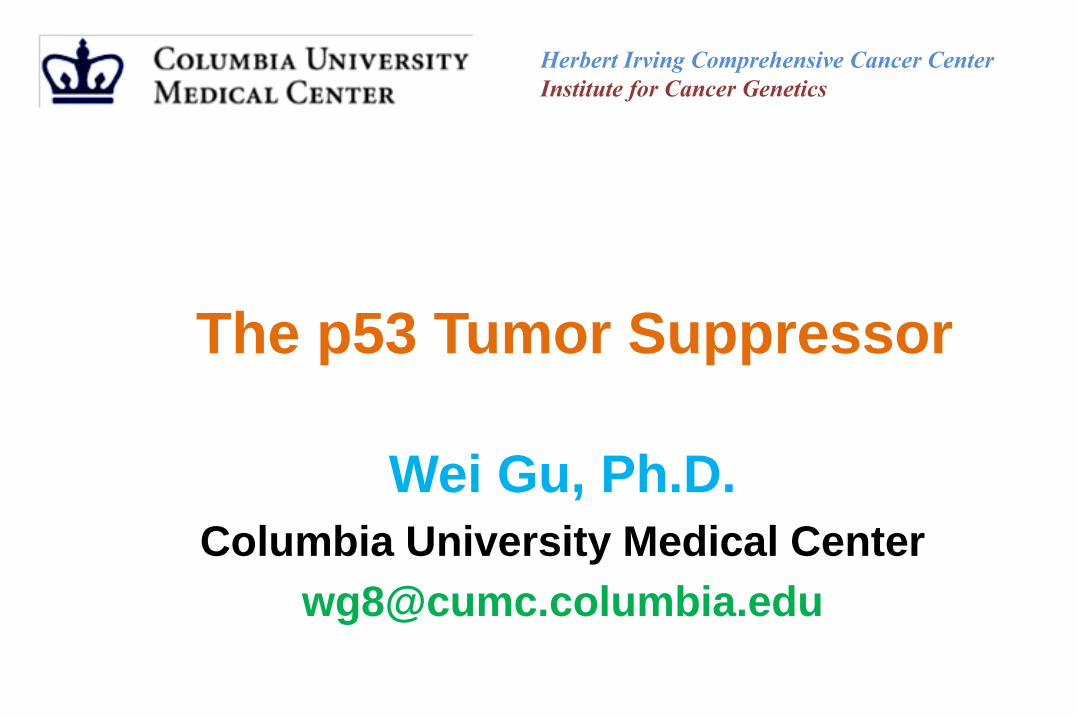
NLS I NLS II NLS III
3931 I II III IV V
Tetramerization
domain
Transactivation
domainDNA-binding domain Regulatory domain
• Discovered in 1979 (A. Levine, D. Lane)
(1988, B. Vogelstein)
• bona fide Tumor suppressor
• Transcriptional activator and repressor
• Mutated on over 50% of human tumors
• Cancer therapeutic target (traditional drugs
side effects)
p53
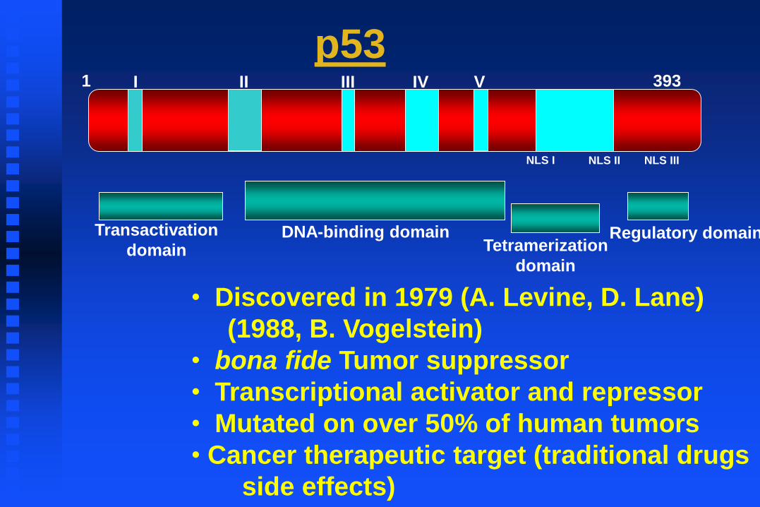
Discovery of p53☞ Studies of SV40-transformed cells show that a (50~55-kDa) protein is coprecipitated
with the large-T antigen.
☞ Linzer and Levine found that the 54-kDa protein was overexpressed in a wide variety of SV40 transformed cells, but also in uninfected embryonic carcinoma cells.

Discovery of p53☞ In 1979, it was found that animals bearing several types of tumors elicited an immune response
specific for p53.
☞ Crawford et al. first described antibodies against human p53 protein in 9% of breast cancer patient sera. Caron de Fromentel et al. later found that such antibodies were present in sera of children with a wide variety of cancers. The average frequency was 12%, but the figure was 20 % in Burkitt Lymphoma.
Discovery of p53☞ Early work on p53 suggested that it may be implicated in the promotion of cell proliferation. When
the cell was induced to grow by serum stimulation, the level of p53 mRNA and the rate of p53 protein synthesis increased markedly, reaching a peak near the G1/S boundary just prior to initiation of DNA replication (Reich and Levine 1984).
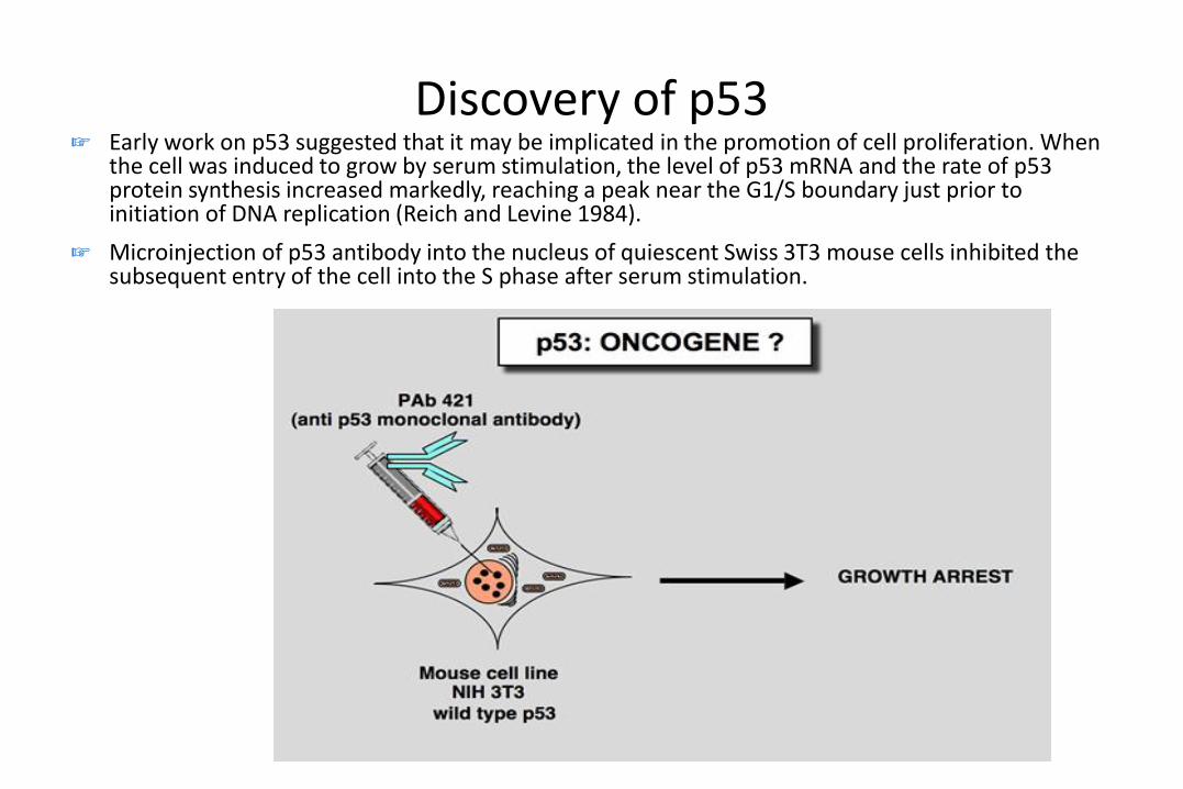
☞ Microinjection of p53 antibody into the nucleus of quiescent Swiss 3T3 mouse cells inhibited the subsequent entry of the cell into the S phase after serum stimulation.

Discovery of p53☞ Two groups reported that cotransfection of p53 with an activated c-Ha-ras oncogene could
transform REF cells in a manner similar to that observed with proto-oncogenes such as myc or E1A (Eliyahu et al. 1984; Parada et al. 1984).
☞ p53 could imortalize normal rat cells leading to cells sensitive to ras transformation (Jenkins et al. 1985; Jenkins et al. 1984).
☞ These observations resulted in the classification of p53 as a nuclear oncogene
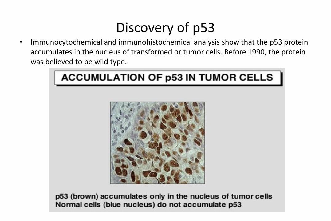
Discovery of p53• Immunocytochemical and immunohistochemical analysis show that the p53 protein
accumulates in the nucleus of transformed or tumor cells. Before 1990, the protein was believed to be wild type.

p53 is a tumor suppressor• p53 in Friend murine erythroleukemia
• In these tumors induced by the Friend virus, the p53 gene found in the tumor cells is truncated or mutated. In this tumor model, functional inactivation of the p53 gene seems to confer a selective growth advantage to the cells during the development of Friend leukemia in vivo.
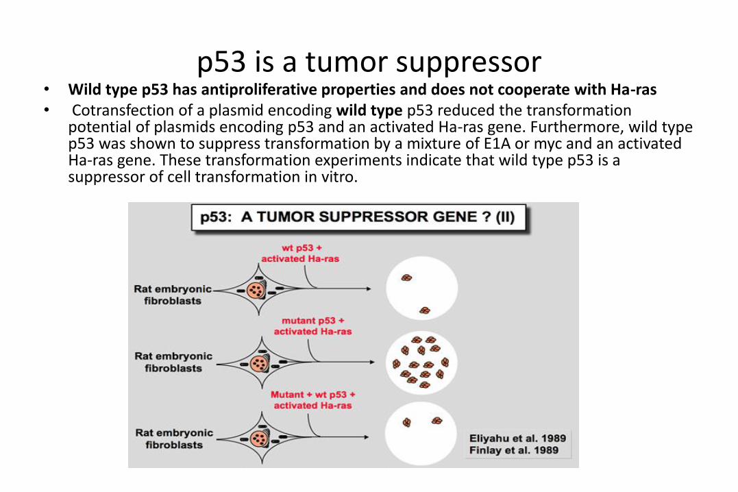
p53 is a tumor suppressor• Wild type p53 has antiproliferative properties and does not cooperate with Ha-ras• Cotransfection of a plasmid encoding wild type p53 reduced the transformation
potential of plasmids encoding p53 and an activated Ha-ras gene. Furthermore, wild type p53 was shown to suppress transformation by a mixture of E1A or myc and an activated Ha-ras gene. These transformation experiments indicate that wild type p53 is a suppressor of cell transformation in vitro.
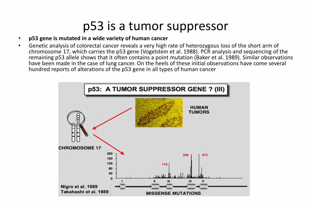
p53 is a tumor suppressor• p53 gene is mutated in a wide variety of human cancer• Genetic analysis of colorectal cancer reveals a very high rate of heterozygous loss of the short arm of
chromosome 17, which carries the p53 gene (Vogelstein et al. 1988). PCR analysis and sequencing of the remaining p53 allele shows that it often contains a point mutation (Baker et al. 1989). Similar observations have been made in the case of lung cancer. On the heels of these initial observations have come several hundred reports of alterations of the p53 gene in all types of human cancer

p53 mutations in human tumors

p53 is a tumor suppressor• Germline mutation of the p53 gene are found in Li-Fraumeni patients• This syndrome presents as a familial association of a broad spectrum of cancers including
osteosarcomas, breast cancer, soft tissue sarcoma and leukemia, appearing at a very early age. Statistical analysis predicts that 50 % of these individuals will have a tumor before the age of 30, and 90 % before the age of 70. Germ-line mutations in the p53 gene have been found in several families with this syndrome.
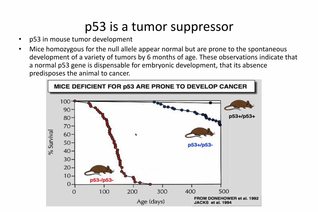
p53 is a tumor suppressor• p53 in mouse tumor development
• Mice homozygous for the null allele appear normal but are prone to the spontaneous development of a variety of tumors by 6 months of age. These observations indicate that a normal p53 gene is dispensable for embryonic development, that its absence predisposes the animal to cancer.
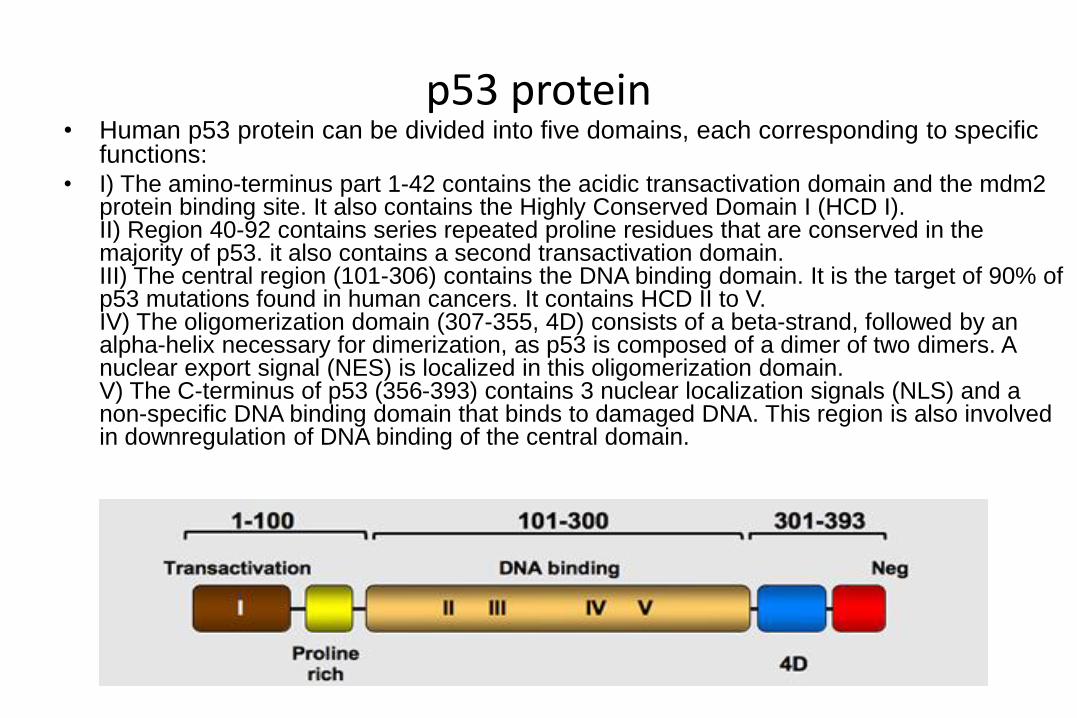
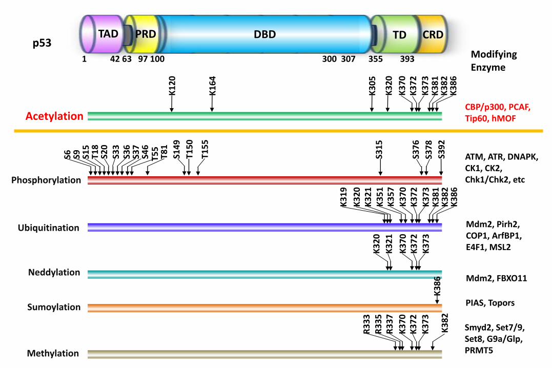
p53 protein• Human p53 protein can be divided into five domains, each corresponding to specific
functions:• I) The amino-terminus part 1-42 contains the acidic transactivation domain and the mdm2
protein binding site. It also contains the Highly Conserved Domain I (HCD I). II) Region 40-92 contains series repeated proline residues that are conserved in the majority of p53. it also contains a second transactivation domain. III) The central region (101-306) contains the DNA binding domain. It is the target of 90% of p53 mutations found in human cancers. It contains HCD II to V. IV) The oligomerization domain (307-355, 4D) consists of a beta-strand, followed by an alpha-helix necessary for dimerization, as p53 is composed of a dimer of two dimers. A nuclear export signal (NES) is localized in this oligomerization domain. V) The C-terminus of p53 (356-393) contains 3 nuclear localization signals (NLS) and a non-specific DNA binding domain that binds to damaged DNA. This region is also involved in downregulation of DNA binding of the central domain.
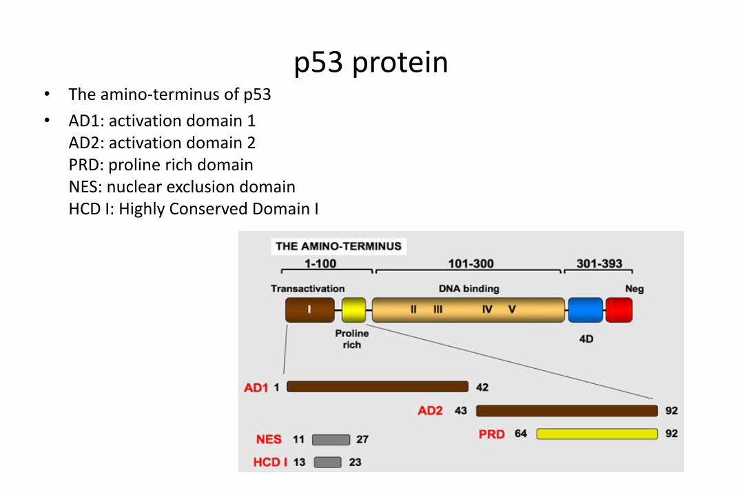
p53 protein• The amino-terminus of p53
• AD1: activation domain 1AD2: activation domain 2PRD: proline rich domainNES: nuclear exclusion domainHCD I: Highly Conserved Domain I

p53 protein• The carboxy-terminus of p53
• Tetra (4D): oligomerization domainNEG: negative regulation domainNES: nuclear exclusion domainNLS: nuclear localization domain
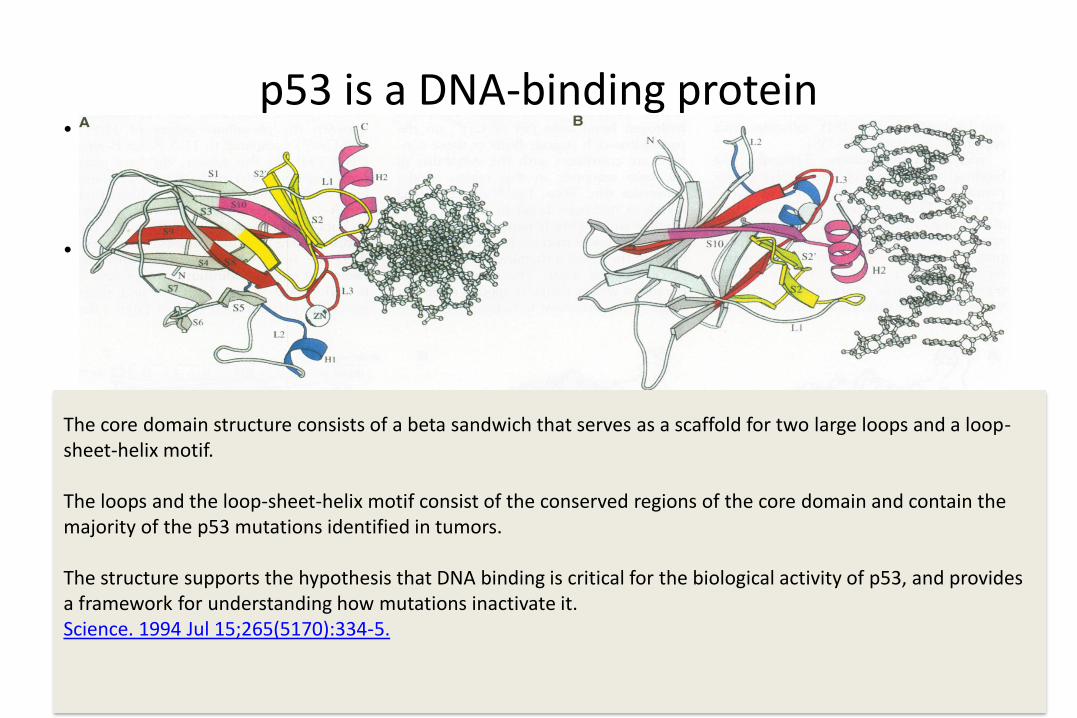
p53 is a DNA-binding protein• The core domain structure
consists of a beta sandwich that serves as a scaffold for two large loops and a loop-sheet-helix motif.
• The two loops, which are held together in part by a tetrahedrally coordinated zinc atom, and the loop-sheet-helix motif form the DNA binding surface of p53. Residues from the loop-sheet-helix motif interact in the major groove of the DNA, while an arginine from one of the two large loops interacts in the minor groove. The loops and the loop-sheet-helix motif consist of the
• Cho Y, Gorina S, Jeffrey PD, Pavletich NP. Science. 1994 Jul 15;265(5170):346-55.
The core domain structure consists of a beta sandwich that serves as a scaffold for two large loops and a loop-sheet-helix motif.
The loops and the loop-sheet-helix motif consist of the conserved regions of the core domain and contain the majority of the p53 mutations identified in tumors.
The structure supports the hypothesis that DNA binding is critical for the biological activity of p53, and provides a framework for understanding how mutations inactivate it.Science. 1994 Jul 15;265(5170):334-5.

p53 is a tumor suppressor
p53 structure
p53 tumor suppressor binds to DNA using all four of its arms.
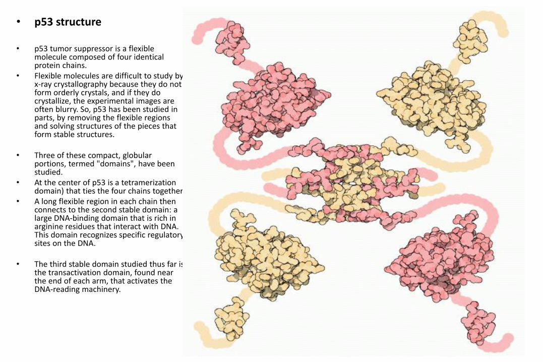
• p53 structure
• p53 tumor suppressor is a flexible molecule composed of four identical protein chains.
• Flexible molecules are difficult to study by x-ray crystallography because they do not form orderly crystals, and if they do crystallize, the experimental images are often blurry. So, p53 has been studied in parts, by removing the flexible regions and solving structures of the pieces that form stable structures.
• Three of these compact, globular portions, termed "domains", have been studied.
• At the center of p53 is a tetramerization domain) that ties the four chains together.
• A long flexible region in each chain then connects to the second stable domain: a large DNA-binding domain that is rich in arginine residues that interact with DNA. This domain recognizes specific regulatory sites on the DNA.
• The third stable domain studied thus far is the transactivation domain, found near the end of each arm, that activates the DNA-reading machinery.

• P53/DNA structure
• p53 tumor suppressor binds to DNA using all four of its arms.
• The typical binding site for the whole molecule is composed of three parts: a specific binding site for two p53 domains, a variable stretch of 0 to 13 base pairs, and a second specific binding site for the other two p53 domains. .
• The tetramerization domain is behind the helix, tying all four chains together, and the four transactivation domains extend along the DNA helix, ready to activate neighboring proteins involved in reading the DNA.
• The flexible chains that connect all four arms together allow p53 to bind to many different variants of this binding site, allowing it to regulate transcription at many places in the genome.

p53 Family Proteins☞ In 1997 and 1998, p63 and p73 have been cloned as two p53 related genes.
☞ p63 and p73 have “p53-like” function in vitro but are not frequently mutated in human tumors.
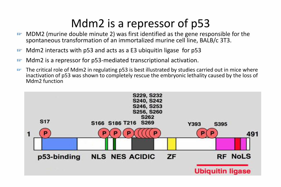
Mdm2 is a repressor of p53☞ MDM2 (murine double minute 2) was first identified as the gene responsible for the
spontaneous transformation of an immortalized murine cell line, BALB/c 3T3.
☞ Mdm2 interacts with p53 and acts as a E3 ubiquitin ligase for p53
☞ Mdm2 is a repressor for p53-mediated transcriptional activation.
☞ The critical role of Mdm2 in regulating p53 is best illustrated by studies carried out in mice where inactivation of p53 was shown to completely rescue the embryonic lethality caused by the loss of Mdm2 function
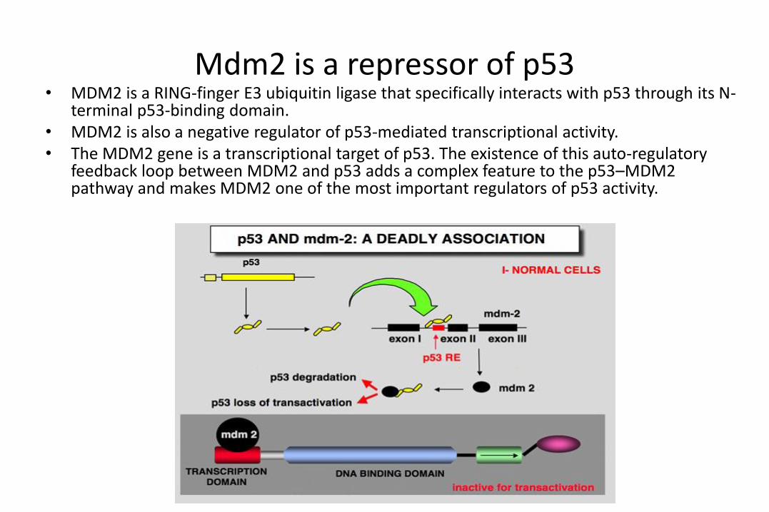
Mdm2 is a repressor of p53• MDM2 is a RING-finger E3 ubiquitin ligase that specifically interacts with p53 through its N-
terminal p53-binding domain. • MDM2 is also a negative regulator of p53-mediated transcriptional activity. • The MDM2 gene is a transcriptional target of p53. The existence of this auto-regulatory
feedback loop between MDM2 and p53 adds a complex feature to the p53–MDM2 pathway and makes MDM2 one of the most important regulators of p53 activity.

Mdm2/Mdmx (Mdm4)• MDM4 is a RING-finger protein that specifically interacts with p53 through its N-terminal p53-binding
domain. ☞ Despite its high sequence homology with Mdm2 and the presence of a RING domain, Mdmx does not
have intrinsic E3-ligase activity for p53 but was instead shown to inhibit p53-induced transcription via their interactions.
☞ Mdmx knockout mice die even in the presence of Mdm2 and this lethality is also rescued by inactivation of p53
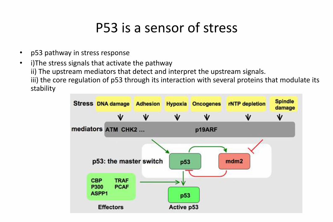
P53 is a sensor of stress
• p53 pathway in stress response
• i)The stress signals that activate the pathwayii) The upstream mediators that detect and interpret the upstream signals.iii) the core regulation of p53 through its interaction with several proteins that modulate its stability
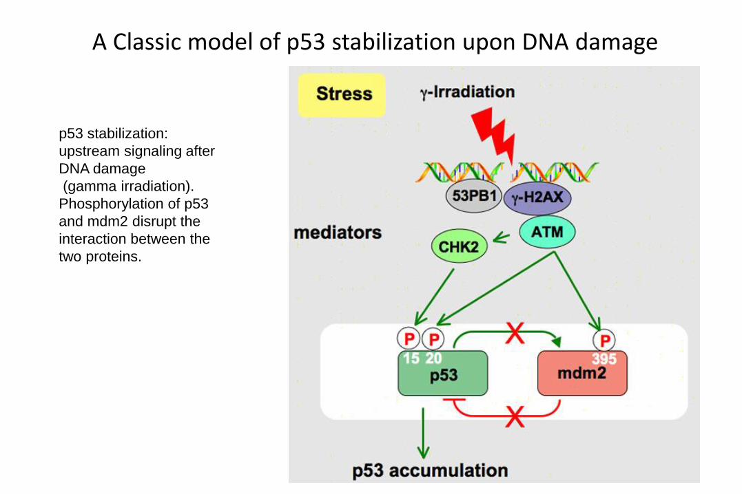
A Classic model of p53 stabilization upon DNA damage
p53 stabilization:
upstream signaling after
DNA damage
(gamma irradiation).
Phosphorylation of p53
and mdm2 disrupt the
interaction between the
two proteins.

A model of p53 activation by ARF
p53 activation by ARF
ARF (known as p14 in
humans and p19 in mice)
was originally identified
as an alternative
transcript of the
Ink4a/ARF tumor
suppressor locus, a gene
that encodes the
Ink4a/p16 inhibitor of
cyclin-dependent
kinases. ARF
suppresses aberrant cell
growth in response to
oncogenic stress mainly
by activating the p53
pathway.
MULE = ARF-BP1
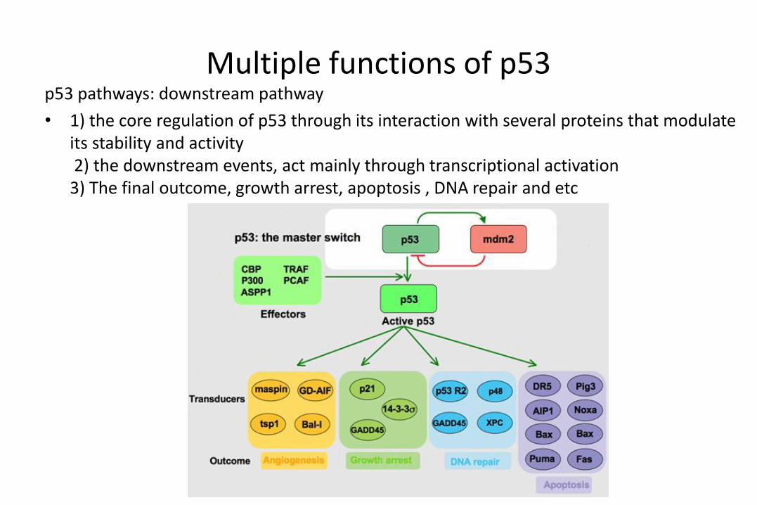
Multiple functions of p53 p53 pathways: downstream pathway
• 1) the core regulation of p53 through its interaction with several proteins that modulate its stability and activity2) the downstream events, act mainly through transcriptional activation 3) The final outcome, growth arrest, apoptosis , DNA repair and etc

A model of p53-mediated cell cycle arrest.
P53-mediated growth
arrest:
downstream signaling, G1
arrest via p21 transcription.
The CDKI p21 will prevent
Rb phosphorylation via
inhibiation of the CDK4 and
CDK2 kinases.
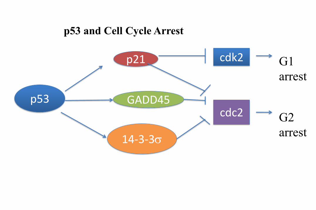
p53
p21
GADD45
14-3-3s
cdk2
cdc2
G1
arrest
G2
arrest
p53 and Cell Cycle Arrest
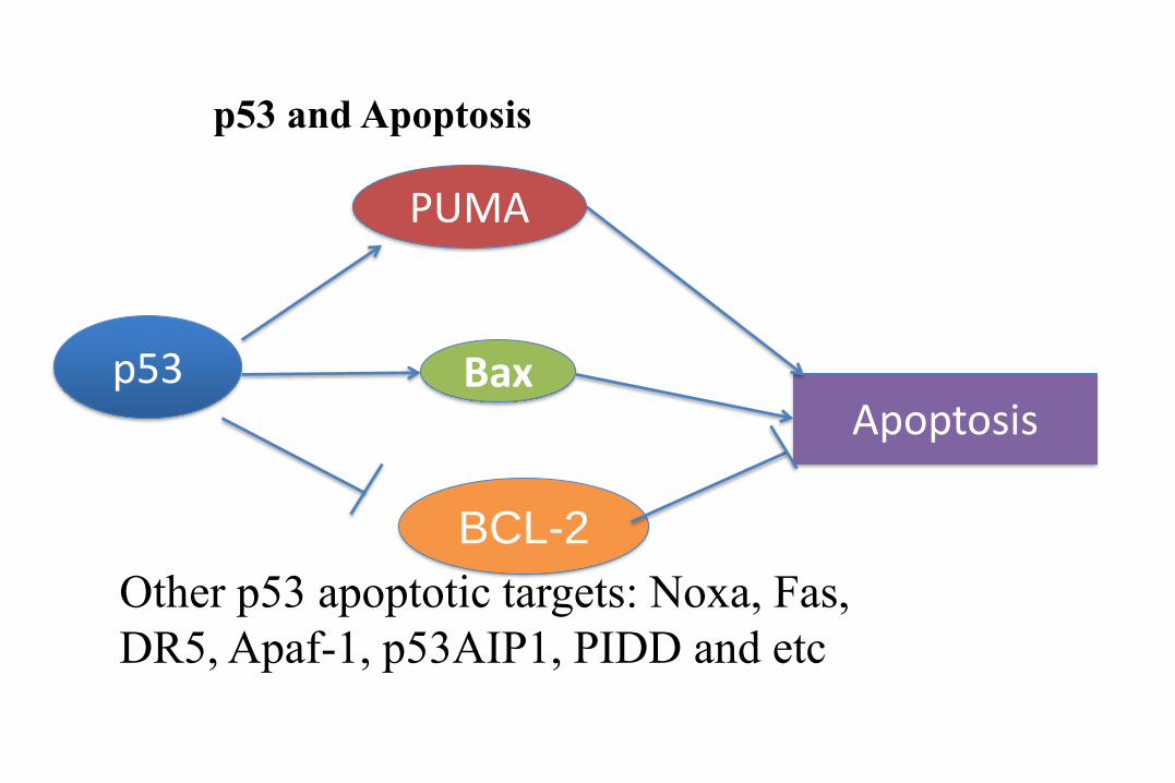
p53
PUMA
Bax
BCL-2
Apoptosis
p53 and Apoptosis
Other p53 apoptotic targets: Noxa, Fas,
DR5, Apaf-1, p53AIP1, PIDD and etc

Transcription-dependent and –independent p53 apoptotic pathways. Nuclear p53 induces expression of PUMA and Bax. In the mitochondria, p53 induces Bax and Bak oligomerization, antagonizes the Bcl-2 and Bcl-XL antiapoptotic effect, which result in marked disruption of mitochondrial membranes and subsequent apoptosis
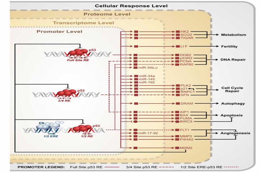
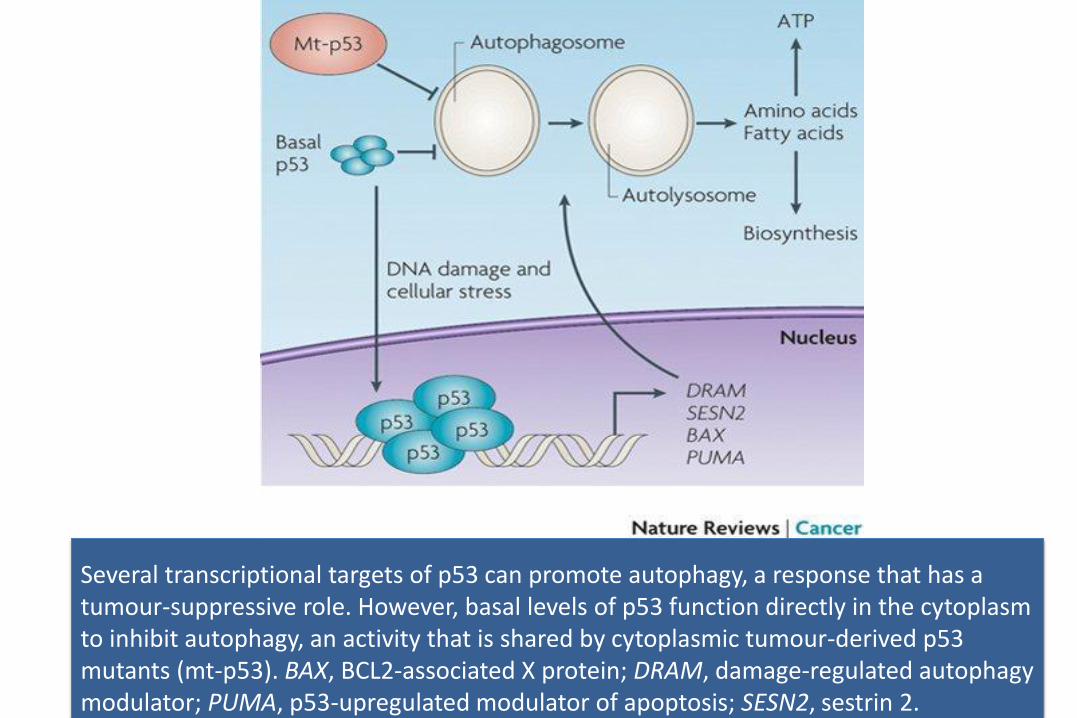
Several transcriptional targets of p53 can promote autophagy, a response that has a tumour-suppressive role. However, basal levels of p53 function directly in the cytoplasm to inhibit autophagy, an activity that is shared by cytoplasmic tumour-derived p53 mutants (mt-p53). BAX, BCL2-associated X protein; DRAM, damage-regulated autophagy modulator; PUMA, p53-upregulated modulator of apoptosis; SESN2, sestrin 2.
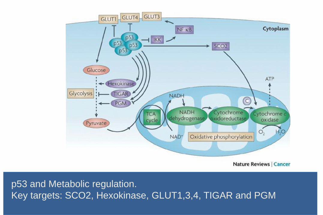
p53 and Metabolic regulation.
Key targets: SCO2, Hexokinase, GLUT1,3,4, TIGAR and PGM

p53
Tumorigenesis
Oncogenic
stress
Metabolic
Stress
DSBs
(γ-IR)
Chemical
exposure
Telomere
shortening
Senescence Metabolism
Or others ?
Transient cell
cycle arrestApoptosisDNA repair
autophagy
Mdm2
Mdmx
Sirt1HDAC1/2
HAUSPp300
Tip60hMOF
PCAF
CBP
K3
21
1 42 63 97 100 300 307 355 393
TAD PRD DBD TD CRD
S46
S6 S14
9
T15
0
T15
5
Phosphorylation
S9 S15
T18
S20
S33
S36
S37
S31
5
S37
6
S37
8
S39
2
T55
T81
Ubiquitination
K3
19
K3
70
K3
72
K3
20
K3
51
K3
57
K3
73
K3
81
K3
82
K3
86
Neddylation
K3
21
K3
20
Sumoylation
Methylation
K3
86
K3
82
R3
37
R3
35
R3
33
K3
70
K3
72
K3
73
K3
70
K3
72
K3
73
Modifying Enzyme
Mdm2, Pirh2, COP1, ArfBP1, E4F1, MSL2
ATM, ATR, DNAPK, CK1, CK2, Chk1/Chk2, etc
Mdm2, FBXO11
PIAS, Topors
Smyd2, Set7/9, Set8, G9a/Glp, PRMT5
Acetylation
K3
20
K1
20
K1
64
K3
05
K3
70
K3
72
K3
73
K3
81
K3
82
K3
86
CBP/p300, PCAF, Tip60, hMOF
p53
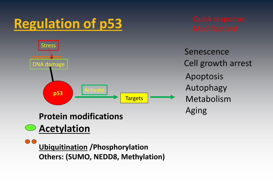
Regulation of p53
Stress
DNA damage
Protein modifications
Acetylation
Ubiquitination /Phosphorylation Others: (SUMO, NEDD8, Methylation)
p53Activate
Targets
Cell growth arrest
ApoptosisAutophagyMetabolismAging
Senescence
Quick response:Modification!

The p53-ubiquitination pathway
p53 ubiquitination:
1. Mdm2 Mono vs. Poly
(Ub—Nuclear export)
2. Deubiquitination: HAUSP
3. Mdm2-independent ubiquitination
ARF-BP1, COP1, PIRH2 and etc.
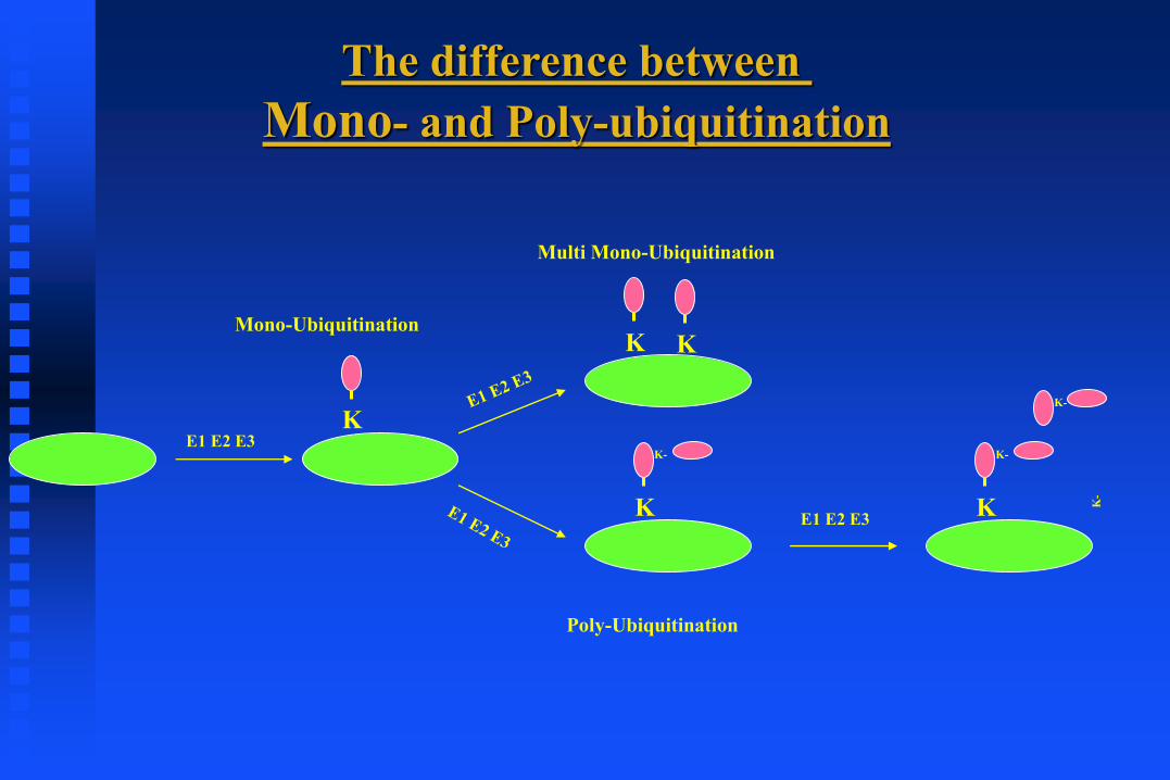
The difference between
Mono- and Poly-ubiquitination
K
Mono-Ubiquitination
E1 E2 E3
K
Multi Mono-Ubiquitination
K
Poly-Ubiquitination
K-
K K
K-
K-
E1 E2 E3

Mdm2 can induce both mono- and polyubiquitination of p53
Mdm2 (55 fmol)
His-Ubwt
_
_+ ++ +
1 2 3 4 5 6
0.2X 0.9X 1.8X 3.6X 5.4X 54X 54X
+++
7 8
p53 (poly-Ub)
p53 (mono-Ub)
p5350
250
100
p53 (55 fmol) 1X1X 1X 1X 1X 1X 1X 1X
MW (Kd)
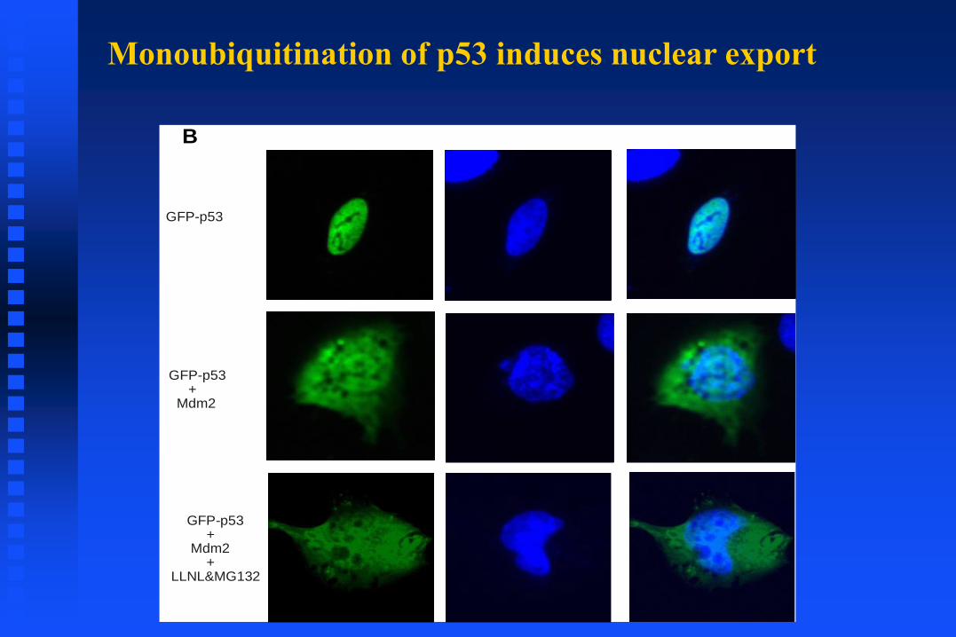
Monoubiquitination of p53 induces nuclear export
GFP-p53
GFP-p53+
Mdm2
GFP-p53+
Mdm2+
LLNL&MG132
B
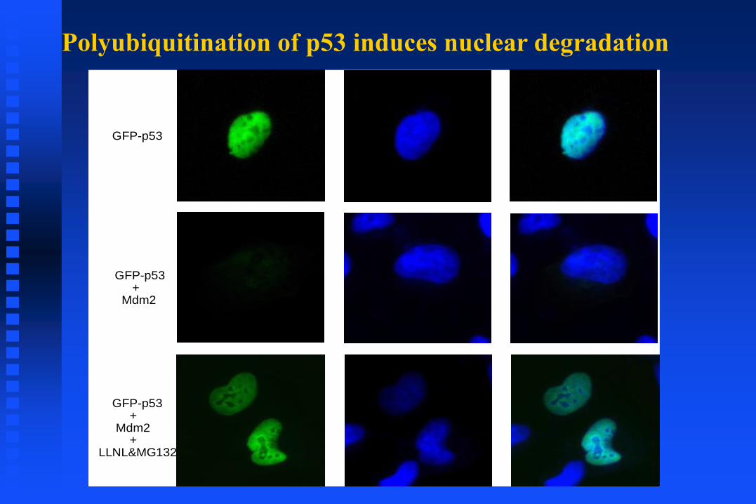
Polyubiquitination of p53 induces nuclear degradation
GFP-p53
GFP-p53+
Mdm2
GFP-p53+
Mdm2+
LLNL&MG132

p53Ub
PARC/
Cul-7
p53p53 Ub
UbUb
Ub
p53Ub
Mdm2
26S
Mdm2 dependent
??
Cytoplasm
Nucleus
p53 p53
p53Ub
p53
p53Ub
p53

P53 is degraded by both Mdm2-depedend and –independent ubiquitination
☞ Mdm2 is the major E3 ligase
☞ Other E3 ligases such as ARF-BP1/MULE, Pirh2, COP1 are also involved in
p53 degradation
(ARF-BP1)
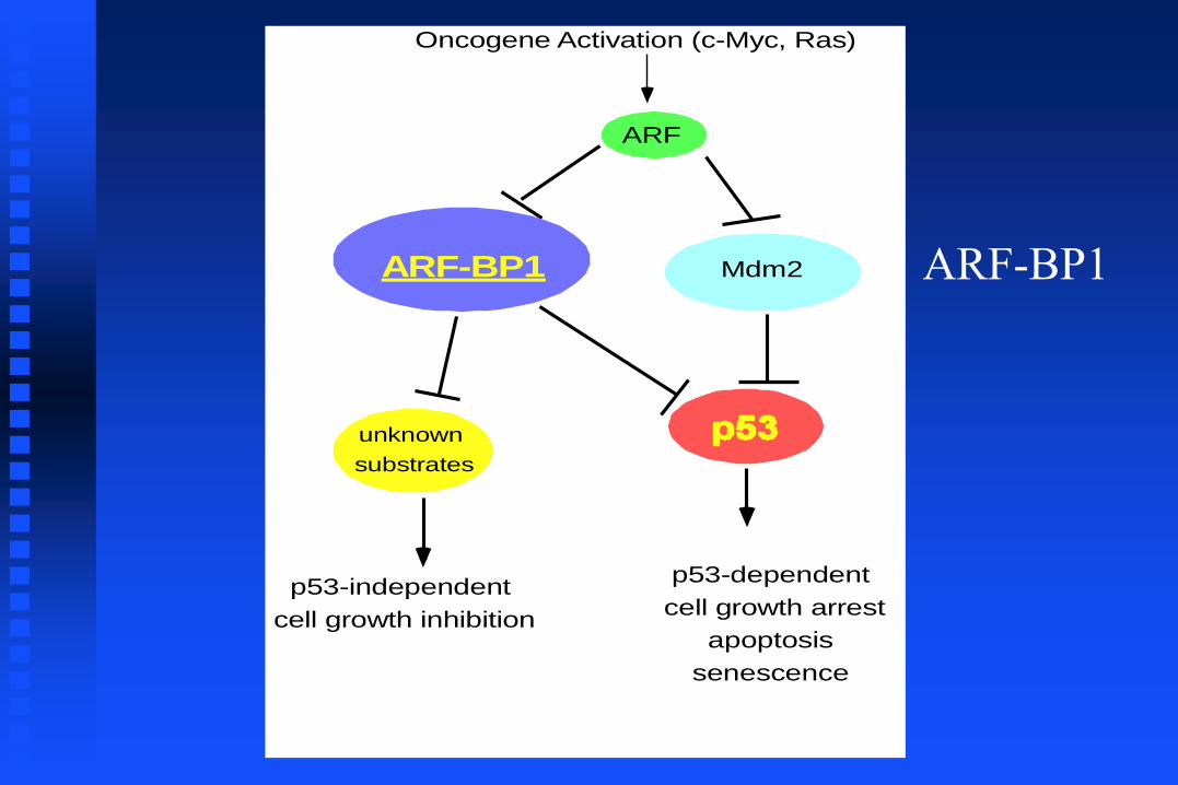
ARF
Oncogene Activation (c-Myc, Ras)
Mdm2
unknown
substrates
p53-independent
cell growth inhibition
p53-dependent
cell growth arrest
apoptosis
senescence
ARF-BP1 ARF-BP1
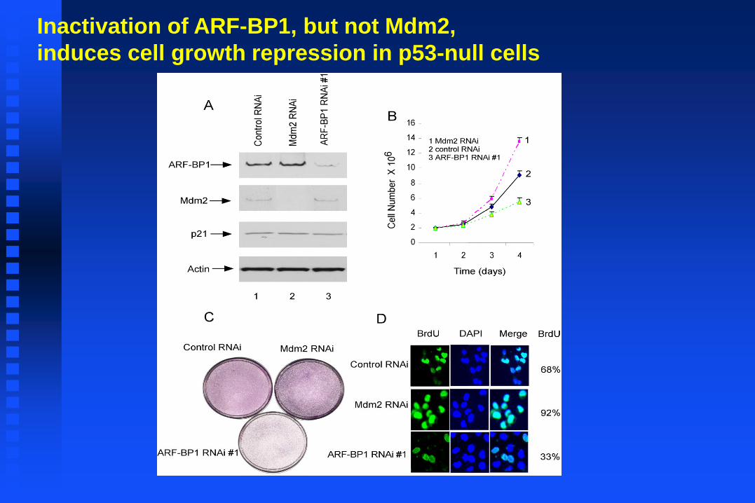
Inactivation of ARF-BP1, but not Mdm2,
induces cell growth repression in p53-null cells
P53 Caspase 3
WT
KO
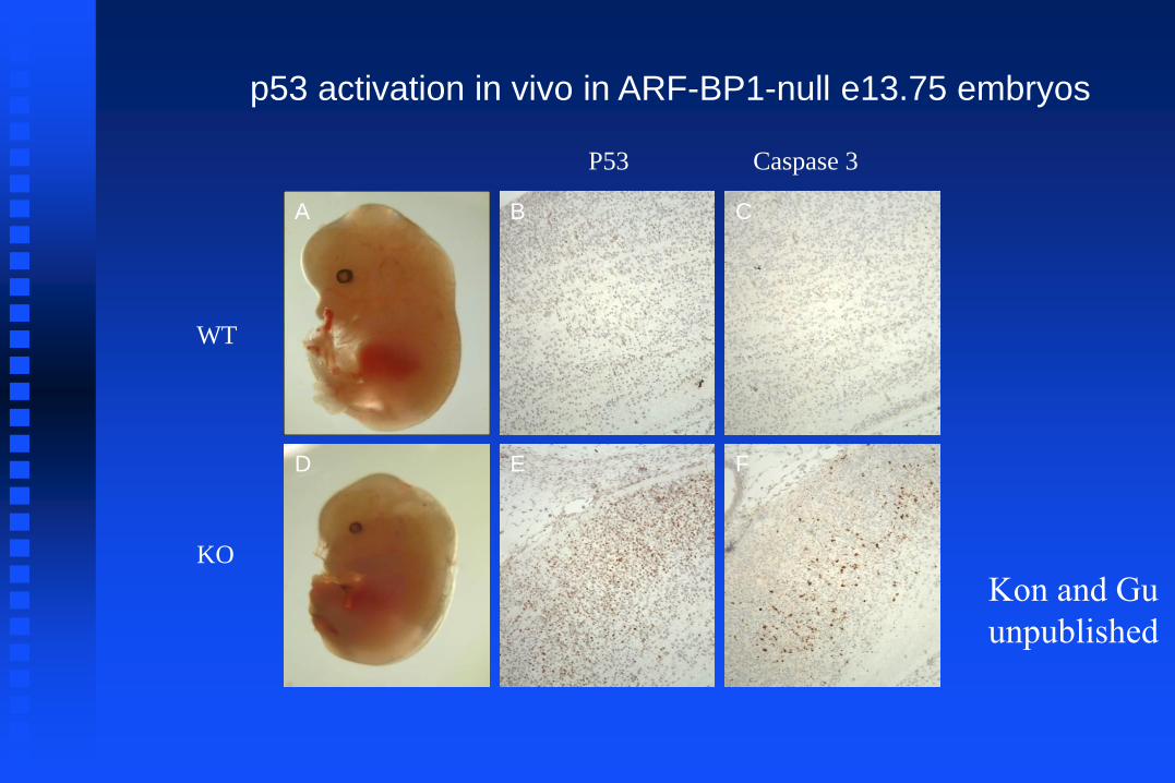
p53 activation in vivo in ARF-BP1-null e13.75 embryos
A B C
D E F
Kon and Gu
unpublished
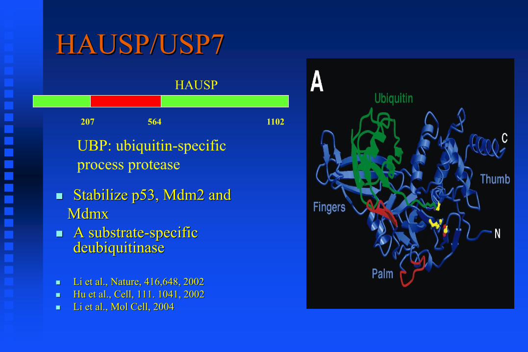
HAUSP/USP7
Stabilize p53, Mdm2 and
Mdmx
A substrate-specific deubiquitinase
Li et al., Nature, 416,648, 2002
Hu et al., Cell, 111. 1041, 2002
Li et al., Mol Cell, 2004
HAUSP
UBP: ubiquitin-specific
process protease
207 564 1102
(Shi, Y. PLoS 2006)
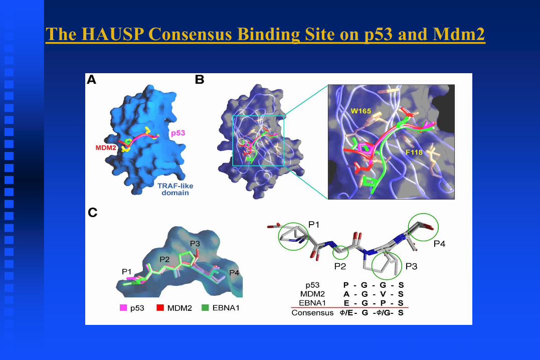
The HAUSP Consensus Binding Site on p53 and Mdm2
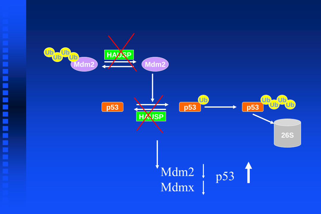
p53 p53Ub
HAUSP
Mdm2
HAUSP
Mdm2Ub
UbUb
Ub
p53 UbUb
UbUb
26S
Mdm2
Mdmx p53
HAUSP
MDM2
MDMX
p53
Actin
0 24 48 72 96+Dox
0 .5 1 2 4 8 0 .5 1 2 4 8
HAUSP
MDM2
MDMX
p53
Actin
HCT116 HAUSP wt HCT116 HAUSP -/-
CHX HRS.
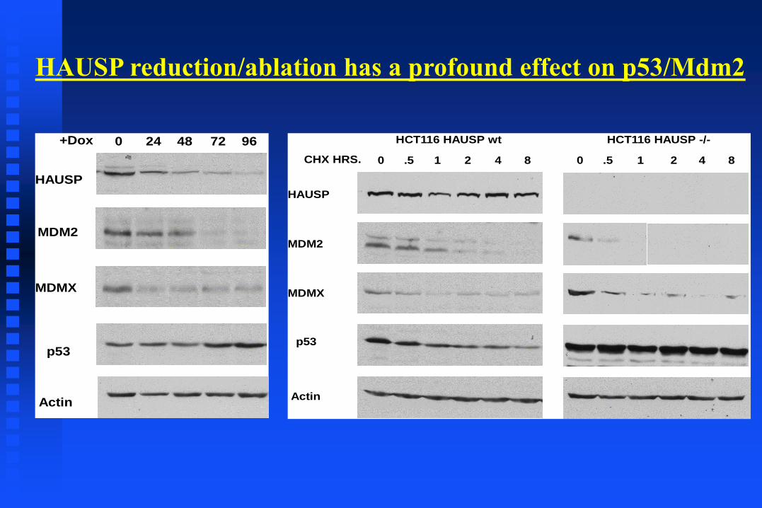
HAUSP reduction/ablation has a profound effect on p53/Mdm2

H&E Hausp p53
Hausp+/cl
Embryo
Hauspko/cl, ERT2
Embryo
Analysis of e11.5 embryos from Hausp conditional knockout
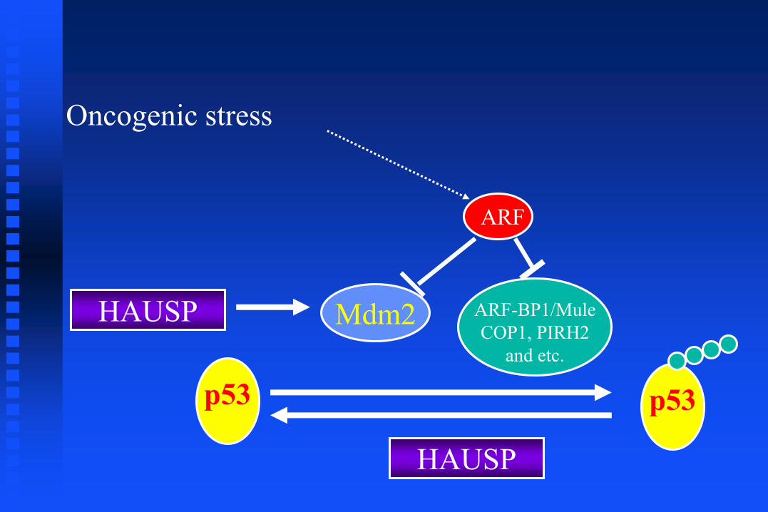
Oncogenic stress
ARF
p53
Mdm2 ARF-BP1/Mule
COP1, PIRH2
and etc.
p53
HAUSP
HAUSP

Regulation of p53 by acetylation/Deacetylation
(general mechanism for non-histone proteins)~1400 substrates
Deacetylation
Cell growth repression
Apoptosis
Senescence
p53 p53
Inactive Active
HDAC1Sirt1
A
CBP/p300 (acetylase)
Tip60/MOF
Gu et al., Cell 1997; Nature 1997.Luo et al., Nature, 2000; Brooks and Gu, 2003Luo et al., Cell, 2001; Tang et al., 2006
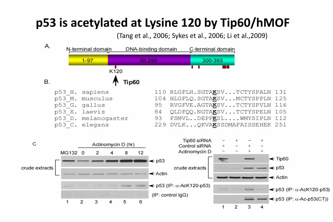
p53 is acetylated at Lysine 120 by Tip60/hMOF(Tang et al., 2006; Sykes et al., 2006; Li et al.,2009)

puma
caspase 3
actin
p53
IR: 12.5 Gy
p-p53
Wt K117R Null
- + - + - +
p21
Thymus
Fold
ch
ange
in m
RN
A l
evel
Fold
ch
ange
in m
RN
A l
evel
Fold
ch
ange
in m
RN
A l
evel
Fold
ch
ange
in m
RN
A l
evel
p21 pumamdm2 noxa
DNA damage-mediated p53 activation in the K117R mutant mouse
0
0.5
1
1.5
2
2.5
3
3.5
no treat 5Gy IR
WT K117R
0
2
4
6
8
10
12
no treat 5Gy IR
WT K117R
0
2
4
6
8
10
12
14
no treat 5Gy IR
WT K117R
02468
10121416
no treat 5Gy IR
WT K117R
Killer/Dr5
012345678
no treat 5Gy IR
WT K117R
Fold
ch
ange
in m
RN
A l
evel
No treat 5Gy IR
WT
K1
17
Rp53 (thymus)
No treat 12.5Gy IR
WT
K117R
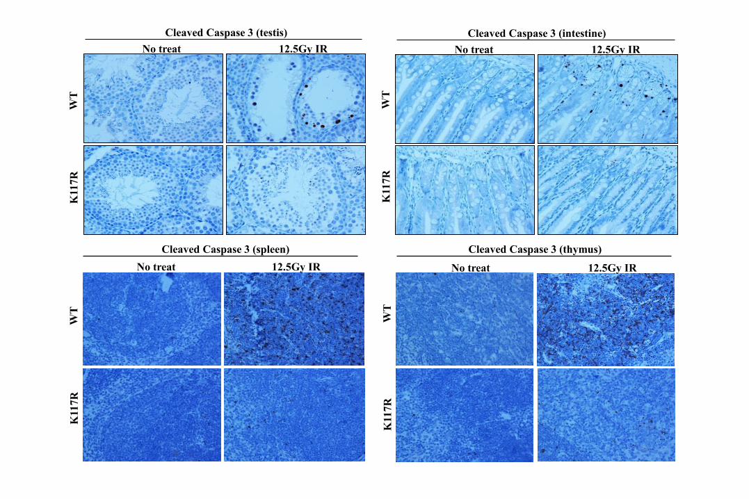
Cleaved Caspase 3 (testis)
No treat 12.5Gy IR
WT
K117R
Cleaved Caspase 3 (intestine)
No treat 12.5Gy IR
WT
K117R
Cleaved Caspase 3 (spleen)
No treat 12.5Gy IR
WT
K117R
Cleaved Caspase 3 (thymus)

Is p53-mediated apoptosis dispensable
for tumor suppression?
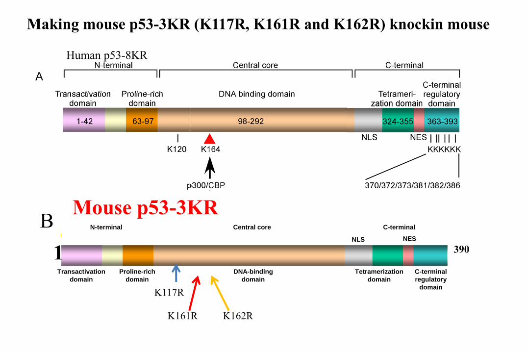
Transactivation
domain
Proline-rich
domain
DNA-binding
domain
Tetramerization
domain
C-terminal
regulatory
domain
NLS NES
N-terminal Central core C-terminal
Mouse p53-3KR
Human p53-8KR
B
Making mouse p53-3KR (K117R, K161R and K162R) knockin mouse
3901
K117R
K161R K162R
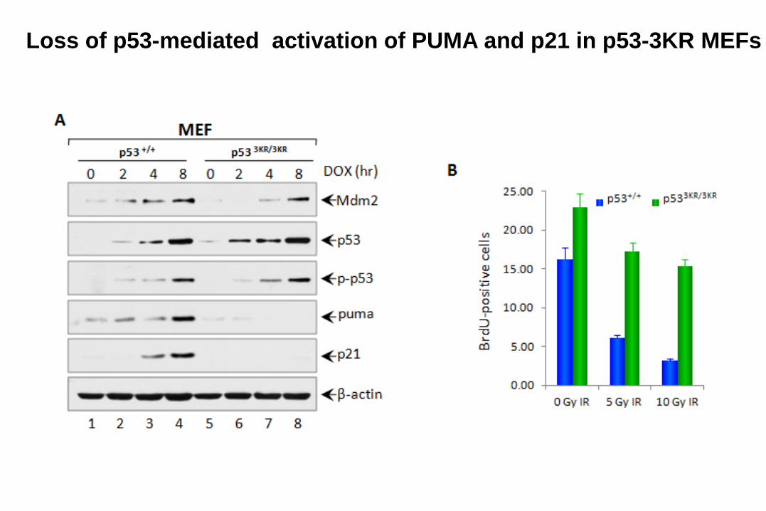
Loss of p53-mediated activation of PUMA and p21 in p53-3KR MEFs
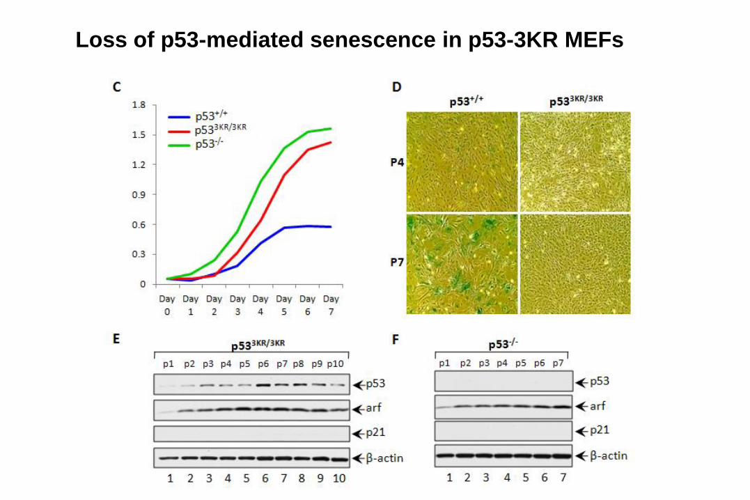
Loss of p53-mediated senescence in p53-3KR MEFs

p533KR/3KR
WT
Are p53-mediated apoptosis and senescence
absolutely required for tumor suppression?
If cell-cycle arrest, apoptosis and
senescence are not absolutely required,
what else could be important for tumor
suppression?
Big Question
Feb, 17, 2011
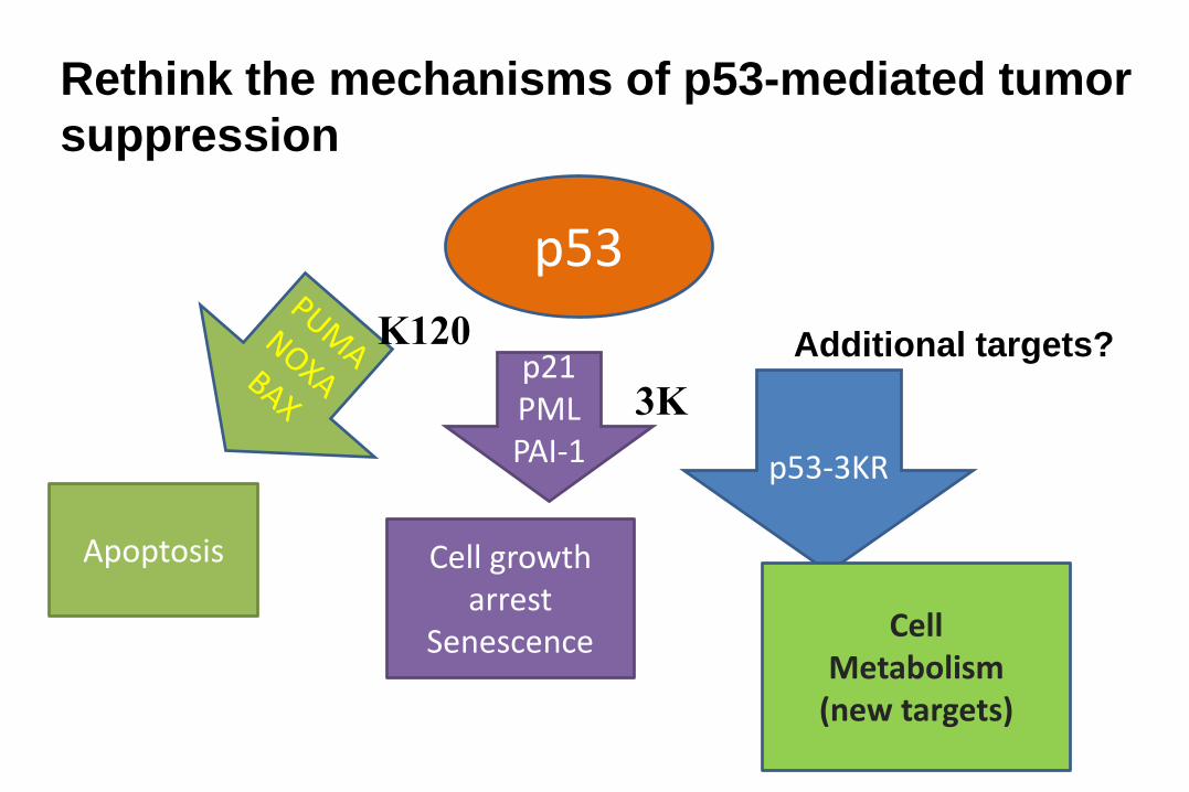
p53
p21PMLPAI-1 p53-3KR
Apoptosis Cell growth arrest
Senescence Cell Metabolism
(new targets)
Rethink the mechanisms of p53-mediated tumor
suppression
K120
3K
Additional targets?
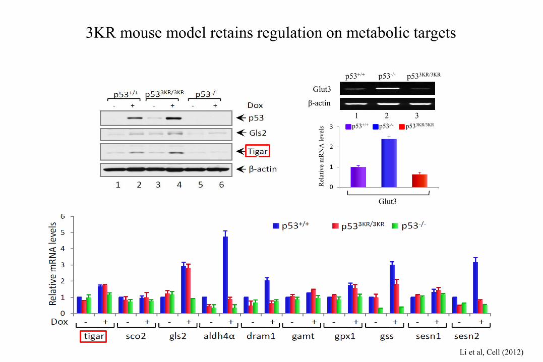
3KR mouse model retains regulation on metabolic targets
Rel
ativ
e m
RN
A l
evel
s p53+/+ p53-/- p533KR/3KR
0
1
2
3
Glut3
p53+/+ p53-/- p533KR/3KR
Glut3
β-actin
1 2 3
Li et al, Cell (2012)
.
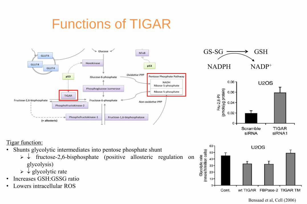
Functions of TIGAR
Tigar function:
• Shunts glycolytic intermediates into pentose phosphate shunt
fructose-2,6-bisphosphate (positive allosteric regulation on
glycolysis)
glycolytic rate
• Increases GSH:GSSG ratio
• Lowers intracellular ROS
GS-SG GSH
NADPH NADP+
Bensaad et al, Cell (2006)

Total # of
tumors
Total tumor
burden
Average
tumor size
Cheung et al., Dev Cell (2013)
TIGAR promotes tumor survival in mouse model
ROS scavenger (glutathione)
Nucleoside synthesis (allow for cell proliferation)
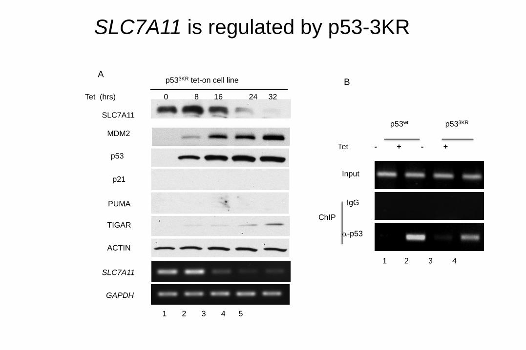
SLC7A11 is regulated by p53-3KR
B
Tet - + - +
p53wt p533KR
1 2 3 4
-p53
Input
IgG
ChIP
A
1 2 3 4 5
MDM2
SLC7A11
p53
ACTIN
Tet (hrs) 0 8 16 24 32
SLC7A11
GAPDH
TIGAR
PUMA
p21
p533KR tet-on cell line
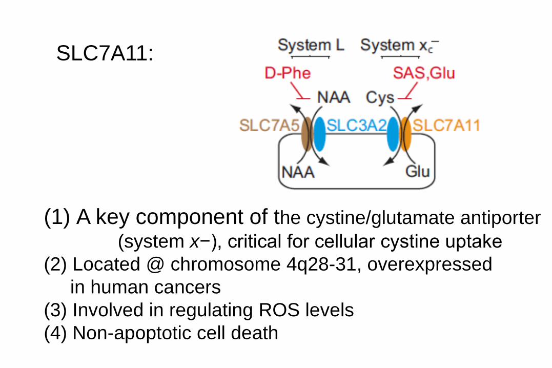
SLC7A11:
(1) A key component of the cystine/glutamate antiporter
(system x−), critical for cellular cystine uptake
(2) Located @ chromosome 4q28-31, overexpressed
in human cancers
(3) Involved in regulating ROS levels
(4) Non-apoptotic cell death
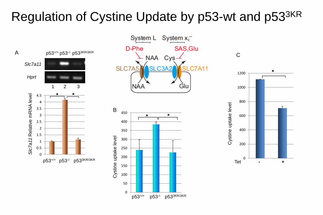
Regulation of Cystine Update by p53-wt and p533KR
A
B
0
0.5
1
1.5
2
2.5
3
3.5
4
4.5
Slc
7a
11 R
ela
tive
mR
NA
leve
l
p53+/+ p53-/- p533KR/3KR
Slc7a11
Hprt
p53+/+ p53-/- p533KR/3KRC
1 2 3
* *
0
200
400
600
800
1000
1200
Cystin
e u
pta
ke
leve
l
Tet - +
*
0
50
100
150
200
250
300
350
400
450
p53+/+ p53-/- p533KR/3KR
Cystin
eu
pta
ke
leve
l * *
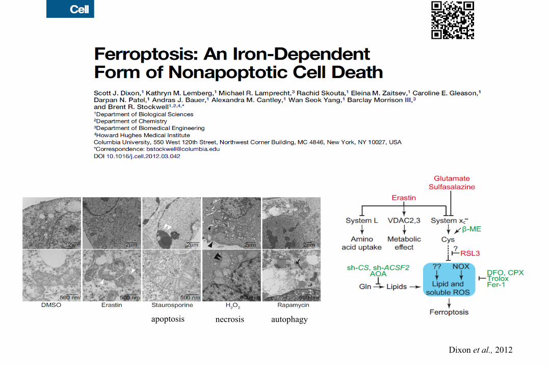
apoptosis necrosis autophagy
Dixon et al., 2012

Regulation of Ferroptosis by p53-wt and p533KR
0
20
40
60
80
100
120
140
160
180
Empty vector
SLC7A11
Tu
mo
r w
eig
ht
(mg)
p<0.05
p<0.05
k l
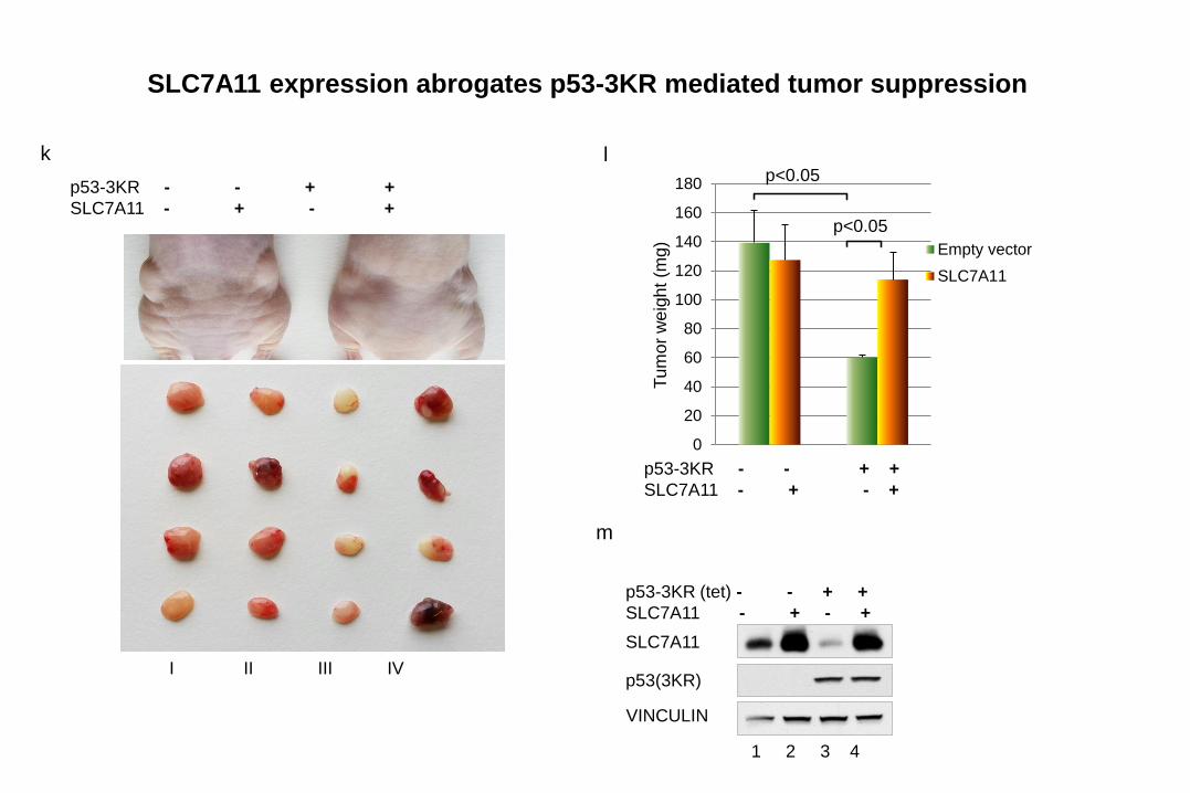
p53-3KR - - + +
SLC7A11 - + - +
p53-3KR - - + +
SLC7A11 - + - +
I II III IV
1 2 3 4
SLC7A11
p53(3KR)
VINCULIN
m
p53-3KR (tet) - - + +
SLC7A11 - + - +
SLC7A11 expression abrogates p53-3KR mediated tumor suppression

Feb, 17, 2011
p53
p21PMLPAI-1
SLC7A11and
OthersMetabolic
targetsApoptosis Cell growth
arrestSenescence Metabolism
ferroptosis
p53-mediated tumor suppression
K120
K164
K163