The Neurological Complexities and Prognosis of COVID-19
12
COVID-19 The Neurological Complexities and Prognosis of COVID-19 Inderbir Padda 1 & Nimrat Khehra 2 & Urooj Jaferi 3 & Mayur S. Parmar 4 Accepted: 17 September 2020 # Springer Nature Switzerland AG 2020 Abstract Several neurological manifestations and complications linked to SARS-CoV-2 have been reported along with well-known respiratory pathology. The global active transmission of SARS-CoV-2 and its unexplained characteristics has led to a pandemic. Since its rapid emergence from Wuhan, China, in December 2019, several studies have reported the impacts of COVID-19 on the CNS and PNS and its implications. This comprehensive review article comprises case reports, case series, metaanalysis, cohort studies, retrospective studies, and narrative reviews focusing on COVID-19-associated CNS and PNS complexities. The authors searched for over 200 articles and used 52 publications related to the neurological complexities of COVID-19 affecting the CNS and PNS as part of the literature review process. The predominant CNS symptoms noted in COVID-19 patients were headaches and dizziness, and the most common PNS symptoms were alterations in smell and taste. Case reports on headache/dizziness, intracerebral hemorrhage, acute hemorrhagic necrotizing encephalopathy, meningitis/encephalitis, encephalopathy, cerebrovas- cular events, chemosensory dysfunction, Guillain–Barre syndrome, and acute transverse myelitis/acute necrotizing myelitis in PCR-confirmed SARS-CoV-2 subjects are also reported. New-onset neurological symptoms were also observed in children with PCR-confirmed SARS-CoV-2 that developed pediatric multisystem inflammatory syndrome (PIMS). This comprehensive re- view article will assist the clinicians and researchers to gain information about the neurological manifestations and complications associated with COVID-19 and develop planning to treat these symptoms in concerned patients of all ages. However, it is unclear whether SARS-CoV2-associated neurological effects are due to primary infections or secondary response to the possible mech- anisms discussed in this review. Keywords COVID-19 . SARS-CoV-2 . Coronavirus . Mechanisms . Pathophysiology . Neurologic manifestations . Neurologic complications . CNS symptoms . PNS symptoms . Cerebrovascular disease . Encephalopathy . Viral encephalitis . Meningitis . Neurological signs and symptoms . Systematic literature review . Literature review Introduction COVID-19 is a rapidly emerging RNA virus since its first reported case in Wuhan, China, in December 2019 [1, 2]. On March 11, 2020, the World Health Organization (WHO) declared COVID-19 a global pandemic affecting over 200 countries [1, 2]. Being a novel virus at the time of its initial exposure to the public, very little was known about the mech- anisms and pathophysiology of the disease and what it ensued; the outbreak of COVID-19 quickly spread worldwide leading to a pandemic by March 2020. As of August 23, 2020, there have been over 23,057,288 confirmed cases and 800,906 deaths to date worldwide [3]. Although pulmonary and car- diovascular complications are the mainstay of SARS-CoV-2 studies, it is vital to note increasing extrapulmonary cases and the neurological complexities presenting in COVID-19 pa- tients. This virus severely affects immunocompromised pa- tients with preexisting health conditions, contributing to fur- ther challenges in management and an increased risk of mor- tality [4]. Figure 1 shows the data obtained from a systematic review and metaanalysis study [5] of clinical manifestations and evidence of neurological involvement of nearly 4700 This article is part of the Topical Collection on Covid-19 * Inderbir Padda [email protected] * Mayur S. Parmar [email protected] 1 University of Washington, Seattle, WA, USA 2 Caribbean Medical University School of Medicine, Willemstad, Curaçao 3 All Saints University School of Medicine, Roseau, Dominica 4 Dr. Kiran C. Patel College of Osteopathic Medicine, Nova Southeastern University, Tampa Bay Campus, Clearwater, FL, USA SN Comprehensive Clinical Medicine https://doi.org/10.1007/s42399-020-00527-2
Transcript of The Neurological Complexities and Prognosis of COVID-19

COVID-19
The Neurological Complexities and Prognosis of COVID-19
Inderbir Padda1 & Nimrat Khehra2 & Urooj Jaferi3 & Mayur S. Parmar4
Accepted: 17 September 2020# Springer Nature Switzerland AG 2020
AbstractSeveral neurological manifestations and complications linked to SARS-CoV-2 have been reported along with well-knownrespiratory pathology. The global active transmission of SARS-CoV-2 and its unexplained characteristics has led to a pandemic.Since its rapid emergence fromWuhan, China, in December 2019, several studies have reported the impacts of COVID-19 on theCNS and PNS and its implications. This comprehensive review article comprises case reports, case series, metaanalysis, cohortstudies, retrospective studies, and narrative reviews focusing on COVID-19-associated CNS and PNS complexities. The authorssearched for over 200 articles and used 52 publications related to the neurological complexities of COVID-19 affecting the CNSand PNS as part of the literature review process. The predominant CNS symptoms noted in COVID-19 patients were headachesand dizziness, and the most common PNS symptoms were alterations in smell and taste. Case reports on headache/dizziness,intracerebral hemorrhage, acute hemorrhagic necrotizing encephalopathy, meningitis/encephalitis, encephalopathy, cerebrovas-cular events, chemosensory dysfunction, Guillain–Barre syndrome, and acute transverse myelitis/acute necrotizing myelitis inPCR-confirmed SARS-CoV-2 subjects are also reported. New-onset neurological symptoms were also observed in children withPCR-confirmed SARS-CoV-2 that developed pediatric multisystem inflammatory syndrome (PIMS). This comprehensive re-view article will assist the clinicians and researchers to gain information about the neurological manifestations and complicationsassociated with COVID-19 and develop planning to treat these symptoms in concerned patients of all ages. However, it is unclearwhether SARS-CoV2-associated neurological effects are due to primary infections or secondary response to the possible mech-anisms discussed in this review.
Keywords COVID-19 . SARS-CoV-2 . Coronavirus .Mechanisms . Pathophysiology . Neurologicmanifestations . Neurologiccomplications . CNS symptoms . PNS symptoms . Cerebrovascular disease . Encephalopathy . Viral encephalitis . Meningitis .
Neurological signs and symptoms . Systematic literature review . Literature review
Introduction
COVID-19 is a rapidly emerging RNA virus since its firstreported case in Wuhan, China, in December 2019 [1, 2].
On March 11, 2020, the World Health Organization (WHO)declared COVID-19 a global pandemic affecting over 200countries [1, 2]. Being a novel virus at the time of its initialexposure to the public, very little was known about the mech-anisms and pathophysiology of the disease and what it ensued;the outbreak of COVID-19 quickly spread worldwide leadingto a pandemic by March 2020. As of August 23, 2020, therehave been over 23,057,288 confirmed cases and 800,906deaths to date worldwide [3]. Although pulmonary and car-diovascular complications are the mainstay of SARS-CoV-2studies, it is vital to note increasing extrapulmonary cases andthe neurological complexities presenting in COVID-19 pa-tients. This virus severely affects immunocompromised pa-tients with preexisting health conditions, contributing to fur-ther challenges in management and an increased risk of mor-tality [4]. Figure 1 shows the data obtained from a systematicreview and metaanalysis study [5] of clinical manifestationsand evidence of neurological involvement of nearly 4700
This article is part of the Topical Collection on Covid-19
* Inderbir [email protected]
* Mayur S. [email protected]
1 University of Washington, Seattle, WA, USA2 Caribbean Medical University School of Medicine,
Willemstad, Curaçao3 All Saints University School of Medicine, Roseau, Dominica4 Dr. Kiran C. Patel College of Osteopathic Medicine, Nova
Southeastern University, Tampa Bay Campus, Clearwater, FL, USA
SN Comprehensive Clinical Medicinehttps://doi.org/10.1007/s42399-020-00527-2
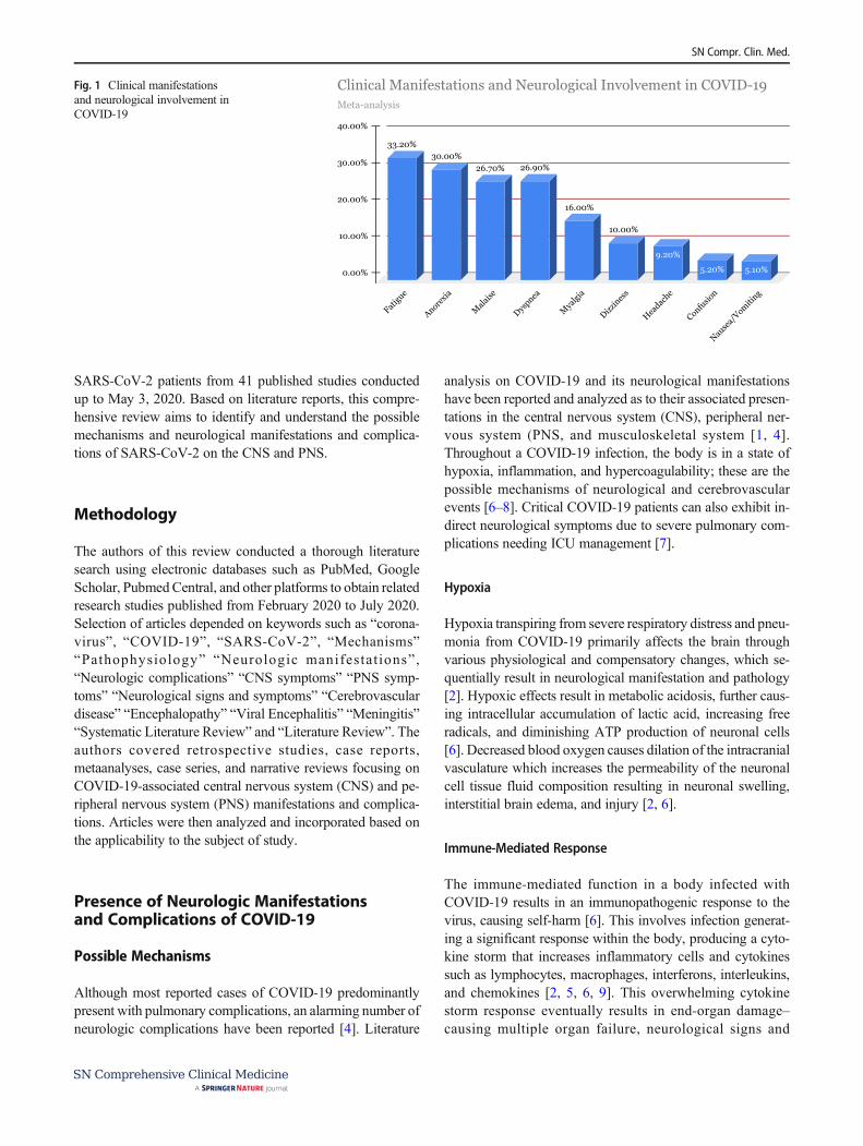
SARS-CoV-2 patients from 41 published studies conductedup to May 3, 2020. Based on literature reports, this compre-hensive review aims to identify and understand the possiblemechanisms and neurological manifestations and complica-tions of SARS-CoV-2 on the CNS and PNS.
Methodology
The authors of this review conducted a thorough literaturesearch using electronic databases such as PubMed, GoogleScholar, PubmedCentral, and other platforms to obtain relatedresearch studies published from February 2020 to July 2020.Selection of articles depended on keywords such as “corona-virus”, “COVID-19”, “SARS-CoV-2”, “Mechanisms”“Pathophysiology” “Neurologic manifestat ions” ,“Neurologic complications” “CNS symptoms” “PNS symp-toms” “Neurological signs and symptoms” “Cerebrovasculardisease” “Encephalopathy” “Viral Encephalitis” “Meningitis”“Systematic Literature Review” and “Literature Review”. Theauthors covered retrospective studies, case reports,metaanalyses, case series, and narrative reviews focusing onCOVID-19-associated central nervous system (CNS) and pe-ripheral nervous system (PNS) manifestations and complica-tions. Articles were then analyzed and incorporated based onthe applicability to the subject of study.
Presence of Neurologic Manifestationsand Complications of COVID-19
Possible Mechanisms
Although most reported cases of COVID-19 predominantlypresent with pulmonary complications, an alarming number ofneurologic complications have been reported [4]. Literature
analysis on COVID-19 and its neurological manifestationshave been reported and analyzed as to their associated presen-tations in the central nervous system (CNS), peripheral ner-vous system (PNS, and musculoskeletal system [1, 4].Throughout a COVID-19 infection, the body is in a state ofhypoxia, inflammation, and hypercoagulability; these are thepossible mechanisms of neurological and cerebrovascularevents [6–8]. Critical COVID-19 patients can also exhibit in-direct neurological symptoms due to severe pulmonary com-plications needing ICU management [7].
Hypoxia
Hypoxia transpiring from severe respiratory distress and pneu-monia from COVID-19 primarily affects the brain throughvarious physiological and compensatory changes, which se-quentially result in neurological manifestation and pathology[2]. Hypoxic effects result in metabolic acidosis, further caus-ing intracellular accumulation of lactic acid, increasing freeradicals, and diminishing ATP production of neuronal cells[6]. Decreased blood oxygen causes dilation of the intracranialvasculature which increases the permeability of the neuronalcell tissue fluid composition resulting in neuronal swelling,interstitial brain edema, and injury [2, 6].
Immune-Mediated Response
The immune-mediated function in a body infected withCOVID-19 results in an immunopathogenic response to thevirus, causing self-harm [6]. This involves infection generat-ing a significant response within the body, producing a cyto-kine storm that increases inflammatory cells and cytokinessuch as lymphocytes, macrophages, interferons, interleukins,and chemokines [2, 5, 6, 9]. This overwhelming cytokinestorm response eventually results in end-organ damage–causing multiple organ failure, neurological signs and
33.20%30.00%
26.70% 26.90%
16.00%
10.00%
9.20%
5.20% 5.10%0.00%
10.00%
20.00%
30.00%
40.00%
Fatigu
e
Anorexia
Mala
ise
Dyspnea
Mya
lgia
Dizzines
s
Headac
he
Confusio
n
Nause
a/Vomiti
ng
Meta-analysis
Clinical Manifestations and Neurological Involvement in COVID-19 Fig. 1 Clinical manifestationsand neurological involvement inCOVID-19
SN Compr. Clin. Med.

symptoms, and mortality [2, 6]. COVID-19 patients oftenhave increased WBC’s, neutrophils, and CRP observed inlaboratory reports, which poses a significant risk for cerebro-vascular events for patients with underlying comorbiditiessuch as hypertension and diabetes [6].
Hypercoagulability
The hematologic profiles of COVID-19 patients attested ahypercoagulable state with elevated D-dimer, fibrinogen,prolonged PT, and decreased antithrombin levels [10–12]. Acase series study conducted byWang et al., [11] reported threecases of COVID-19 patients summarizing prothromboticcomplexities, demonstrating transient symptomatic clinicalimprovements when managed with a tissue plasminogen acti-vator (tPA). In addition, respiratory failure associated withCOVID-19 patients displayed microthrombi on autopsy re-ports affirming a prothrombotic occlusive etiology rather thantypical findings of acute respiratory distress syndrome(ARDS) [11]. This case series also reported that 71.4% ofsubjects concluding in mortality met the InternationalSociety on Thrombosis and Haemostasis’ (ISTH) criteria fordisseminated intravascular coagulation (DIC) [11]. Theprothrombotic complexities of COVID-19 may contribute tothe occlusive cerebrovascular events in patients who may de-velop DIC [11, 12]. Anticoagulant therapy has shown to de-crease the risk of venous thromboembolism and better prog-nosis in COVID-19-related coagulopathy [13, 14].
Angiotensin-Converting Enzyme II
The SARS-CoV-2 virus enters cells via the angiotensin-converting enzyme (ACE) II receptor, which is found primar-ily within the pulmonary alveolar cells; this receptor is alsofound extrapulmonary in the vascular endothelial cells of theGI tract, heart, and brain, functioning as a vasoconstrictorregulating blood flow [15, 16]. The virus may enter the brainvia ACE II receptor and cause disruption to the blood flow andregulation in the vasculature, causing rupture of the arteries[15, 17]. ACE II has endothelial and vasoprotective effectsregulating angiotensin II (Ang II), a potent vasoconstrictor0,exerting vasodilatory effects [6, 18]. COVID-19 infectionmay decrease the ACE II receptor expression, diminishingtheir vasoprotective action [6, 18]. Subjects with COVID-19demonstrated raised levels of Ang II, which were positivelycorrelated with the severity of lung injury and viral load. AngII further stimulates a proinflammatory immune response con-tributing to atherosclerosis [6, 15].
Transsynaptic Transfer
Progressing data on the CNS invasion of SARS-CoV2 hasbeen documented for similar CoVs, suggesting a transsynaptic
transfer [16, 19]; invasion of the PNS further transferring tothe CNS by synapses [16]. Preliminary trials conducted onmice with SARS-CoV34 orMERS-CoV13 exhibited possibleentrance of the virus through the olfactory nerves via the crib-riform plate and ethmoid bone and spread to the CNS localiz-ing to areas such as the brainstem and thalamus [16, 17]. Theextrapulmonary CNS infection was also noted to be a signif-icant constituent for mortality recorded in mice [16].
Central Nervous System–Related Symptoms
The central nervous system (CNS)–related occurrences inCOVID-19 patients include headaches, dizziness, seizures,decreased awareness, ataxia, cerebrovascular accident (ische-mic or hemorrhagic), delirium, acute necrotizing encephalop-athy (ANE), acute encephalitis, and/or meningitis [1, 8, 20].Among these signs and symptoms, headaches and dizzinesswere the most predominant CNS symptoms [1].
Headaches and Dizziness
Ametaanalysis conducted on clinical, laboratory, and imagingfeatures of COVID-19 from dates January 1, 2020, toFebruary 23, 2020, reported headache as the predominantCNS symptom with a mean prevalence of 8% [21].
A retrospective, observational case series conducted fromJanuary 16, 2020, to February 18, 2020, in Wuhan, China,reported that 36.4% of patients presented neurological symp-toms, of which 24.8% constituted of CNS and 8.9% PNSmanifestations, and were more prevalent in patients who hada severe respiratory illness [22]. Among patients in the groupof CNS manifestations, 16.8% reported dizziness, and 13.1%reported headaches [22]. The study also concluded that sub-jects who developed neurological manifestations were olderpatients with a mean age of 52.7 that had underlying comorbidconditions, primarily hypertension [22]. Figure 2 illustratesdata obtained from this retrospective, observational case seriesfrom 214 patients inWuhan, China, from January 16, 2020, toFebruary 19, 2020 [22].
Another case series study of 1099 COVID-19 confirmedsubjects, 13.6% reported headache, and 14.9% reported my-algia [7]. Headache is considered to be secondary to hypoxia,causing a reduction in blood flow to the cerebral vasculatureand the body response to inflammatory mediators and cyto-kines [17].
Wang and Lei et al. [5] conducted a systematic review plusmetaanalysis with nearly 4700 subjects from more than 41published articles on COVID-19-associated neurologicalmanifestations. Prevalent symptoms include, fatigue(33.2%), anorexia (30.0%), malaise (26.7%), dyspnea andshortness of breath (26.9%), myalgia (16.0%), dizziness(10.0%), headache (9.2%), confusion (5.2%), and nausea
SN Compr. Clin. Med.

and vomiting (5.1%) [5]. Figure 2 displays data obtained fromthis systematic review and metaanalysis study of clinical man-ifestations and evidence of neurological involvement of nearly4700 SARS-CoV-2 patients from 41 published studies con-ducted up to May 3, 2020.
Cerebrovascular Event
A study focusing on COVID-19 patients in Italy showed thatdespite patients admitted with confirmed infection and venousthromboembolism prophylaxis administered, there was an in-crease of ischemic stroke by 2.5% [4]. In addition, COVID-19patients admitted into the intensive care unit with confirmedinfection in China had an increase of ischemic stroke by 5%and in the Netherlands had an increase of ischemic stroke by3.7% [4]. Moreover, younger patients infected with COVID-19 presented with signs of ischemic stroke [4]. Lastly, anycomorbidities in older patients, such as infection and hyper-coagulable states, can increase the risk of ischemic stroke [4].
It has been determined that SARS-CoV2 binds to ACE2receptors on the endothelial cells which causes a massive in-flammatory response increasing blood vessel constriction,leading to end-organ damage and stroke; ACE2 recombinanttherapy is a potential treatment for COVID-19-related stroke[9].
A case report conducted by Brüggemann et al. [23] reportedarterial and venous thromboembolic events in a 59-year-oldmale patient who tested positive for COVID-19. He had a pastmedical history of peripheral arterial disease and presented tothe ED with pulmonary symptoms (dyspnea, chest pain,cough), fever, tachycardia, and headache [23]. A CT-pulmonary angiography (CTPA) was ordered due to high sus-picions of a pulmonary embolism (PE) due to signs of tachy-cardia, chest pain, and elevated D-dimer; however, this wasruled out and imaging further demonstrated COVID-19 pneu-monia impressions [23]. COVID-19 was confirmed via RT-PCR [23]. On the fifth day, the patient had developed stroke-like symptoms, which were confirmed via perfusion and
vascular volume recordings despite inconclusive brain imagingwith CT. Further management included alteplase for treatment,and clopidogrel and nadroparin (LMWH) for prophylaxis [23].Despite therapy, multiple pulmonary embolisms developed onthe seventh day, which was confirmed via the third CTPAduring his admission [23]. The patient was further managedtherapeutically with tinzaparin (LMWH) for PE [23].
Encephalopathy
A retrospective case series conducted 13 January to 12February 2020, in Wuhan, China, on clinical characteristicsof 113 deceased patients from COVID-19 reported CNSsymptoms such as disorder of consciousness (22%) and hyp-oxic encephalopathy (20%) [24]. In this study, the deceasedsubjects had a median age of 68 and had underlying comor-bidities of chronic hypertension (48%) and other cardiovascu-lar conditions (14%) [24].
Filatov et al. [25] reported a case of encephalopathy asso-ciated with COVID-19. The subject was a 74-year-old malewho recently traveled to the USA from Europe [25]. The pa-tient initially presented to the ED with a complaint of coughand fever, with a previous medical history of COPD, atrialfibrillation, and stroke [25]. Prior to discharge, the patienthad a COPD exacerbation and was managed accordingly[25]. Initial symptoms worsened which led to hospitalization;new CNS manifestations of headache and altered mental sta-tus were noted upon readmittance [25]. The workup for strep-tococcus pneumonia and influenza were inconclusive, andCXR impression ground-glass opacities, consistent withCOVID-19 pneumonia [25]. The subject’s neurologicalcourse severely progressed causing impaired verbal commu-nication and the inability to follow commands [25]. The pa-tient was tested positive for SARS-CoV-2 [25]. Brain imagingvia CT-scan impression changes were consistent with the pre-vious history of stroke of the left posterior cerebral artery andno acute alterations [25]. EEG demonstrated reading was con-gruous with encephalopathy showing diffuse slowing and
Dizziness
Headache
Impaired consciousness
Hypogeusia
Hyposmia
Skeletal muscle injury
Ischemic stroke
Hemorrhagic Stroke
0.00% 5.00% 10.00% 15.00%
Retrospective Case Series
Neurological ManifestationFig. 2 Neurologicalmanifestations
SN Compr. Clin. Med.

focal slowing sharply contoured waves [25]. The pulmonarystatus also worsened, prompting ICU admittance and intuba-tion [25]. Management of his condition includedhydroxychloroquine, antivirals, and antibiotics [25].Treatment for encephalopathies is mainly supportive, withthe majority of the subjects making a full or partial recovery[8].
Meningitis/Encephalitis
The first case of COVID-19-associated meningitis was re-ported in late February of 2020 [26]. This patient had pro-gressive influenza-like symptoms such as fever, fatigue,headache, and sore throat [26]. The patient was diagnosedand treated for influenza despite a negative test result. Bythe ninth day, altered consciousness and generalized con-vulsions were experienced en route to the hospital [26]. Thepatient also presented with nuchal rigidity. Despite unfavor-able results on the nasopharyngeal swab, the CSF RT-PCRtest was positive for COVID-19 [7]. Impressions of brainimaging demonstrated characteristics of encephalitis of thehippocampal region and the right mesial lobe [7, 26]. CSFscreening for herpes simplex virus and varicella-zoster an-tibodies were not detected [26]. The neurological symptomscaused by COVID-19 have the potential to affect any agegroup. Patterns have shown that SARS-COV2 infectionleads to the presentation of several cytokines that impairthe immune system and increase the neurotropic capacityof the virus [27]. Accompanying symptoms may includedelirium, psychosis, and myoclonus; Levetiracetam andclonazepam may be used to treat the accompanying myoc-lonus [8]. Haloperidol, followed by risperidone, has shownimprovement for psychosis. [8]
Acute Hemorrhagic Necrotizing Encephalopathy
Another complication involving the CNS system is acute nec-rotizing encephalopathy (ANE), which is a rare conditioncaused by a cytokine storm resulting in a breakdown of theblood-brain barrier without any viral invasion [7, 28]. Somepatients with severe COVID-19 cases presented with cytokinestorm syndrome and, in turn, ANE [7]. Although ANE pre-dominantly affects pediatric patients, cases have been seen inadults with COVID-19 [28]. Noncontrast computed tomogra-phy (CT) of the head showed symmetric, multifocal lesionswith infected areas in the thalamus, cerebral white matter, brainstem, and cerebellum [28]. The pathophysiology of ANE is notunderstood well and treatment with intravenous immunoglob-ulin (IVIG) and steroids can be administered [28].
A case study conducted by Poyiadji et al. [29] reporteda case of acute hemorrhagic necrotizing encephalopathy(ANE) in a female patient in her late 50s. The initialpresentation included fever, cough, altered mental status
(AMS), and tested negative for influenza [29]. Followingthe negative test, the administration of a nasopharyngealswab for SARS-CoV-2 was warranted, confirming the di-agnosis via RT-PCR [29]. Further testing of CSF for viralspecimens such as HSV, VZV, and West Nile was incon-clusive. CSF analysis for SARS-CoV-2 was unable to becompleted [29]. A noncontrast head CT and MRI wereperformed on brain imaging, which exhibited symmetrichypoattenuation inside both sides of the medial thalamuson CT, and hemorrhagic rim enhancing lesions within thatregion, and temporal lobes on MRI [29]. The patient wasfurther treated and managed with IVIG [29].
Intracerebral Hemorrhage
The pathogenesis of intracerebral hemorrhage in COVID-19patients is best understood by the binding of SARS-CoV-2 toACE2 receptors on the endothelial cells. This binding resultsin the destruction of ACE2 receptors fundamentallycompromising the integrity of the blood brain barrier andallowing the virus to enter the CNS [7, 30]. Plus, decreasedexpression of ACE2 receptors can negatively impact therenin-angiotensin system (RAS) complicating the regulationof the CNS and PNS system affecting the regulation of bloodpressure, potentially resulting in intracerebral hemorrhage [7,30].
A case report conducted by Sharifi-Razav et al. [15] sum-marized the correlation of intracerebral hemorrhage in aCOVID-19 confirmed patient. A 79-year-old male presentedto the ED with a loss of consciousness tested positive forCOVID-19 [15]. CT-scan of the brain confirmed an intraven-tricular and subarachnoid hemorrhage [15]. There were nounderlying conditions such as hypertension or anticoagulationthat predisposed him to an intracerebral hemorrhage [15]. Inpatients infected with COVID-19, reports of large vessel oc-clusion and infarcts, venous thromboembolism, raised inflam-matory markers, and severe systemic inflammations with or-gan failure including the brain, are occurring at an alarmingrate [31]. Physicians have to base decisions on administrationanticoagulation with the risk of thrombosis versus the risk ofhemorrhage [31].
A retrospective case series conducted by Benger et al. [30]reported five ICH cases associated with COVID-19 at King’sCollege Hospital between February 1, 2020, and May 14,2020. All five subjects had nasopharyngeal swabs confirmingSARS-CoV-2 with RT-PCR. The patients were between theages of 41–64, with four having underlying comorbidities,primarily hypertension [30]. All subjects had a median of32 days from the point of initial SARS-CoV-2 diagnosisto ICH development [30]. Four patients also had multipleorgan involvement prior to the ICH [30]. The patients weremanaged in the ICU and transferred to the stroke unit forrehabilitation [30].
SN Compr. Clin. Med.

Peripheral Nervous System–RelatedSymptoms
Complications in COVID-19 patients involving the peripheralnervous system (PNS) include anosmia, dysgeusia, Guillain–Barré syndrome (GBS), acute myelitis, skeletal muscle dam-age, hemophagocytic lymphohistiocytosis (HLH), and/orMiller Fisher syndrome [4, 8, 22]. Figure 3 depicts the neuro-logical manifestations affecting the CNS and PNS of 630 con-firmed SARS- CoV-2 cases from a chronological review of 41articles [32]. Of the 630 subjects, 23 (3.6%) developed CNS-related manifestations, which included encephalitis, encepha-lopathy, and myelitis [32]. A total of 564 (89.6%) developedPNS-related manifestations such as anosmia, Guillain–Barrésyndrome, cranial nerve palsy, and Miller Fisher syndrome.43 (6.8%) developed neurovascular symptoms (stroke) [32].A total of 549 (87%) subjects with PNS symptoms experi-enced alteration in their sense of smell (anosmia/hyposmia)[32].
Chemosensory Dysfunction
Among PNS signs and symptoms, chemosensory dysfunc-tions of anosmia (impaired smell) and ageusia (impaired taste)were the most predominant PNS symptoms [1, 22]. Anosmiais thought to be caused by inflammation of the olfactorynerves causing deterioration to the hair-like receptor cells[17]. Alteration in taste (hypogeusia/ageusia) may be due tothe significant ACE2 receptors expressed on the tongue,which are subject to damaging the taste receptors post bindingfrom the virus [17].
In a study of 630 individuals with confirmed COVID-19infections, 564 (89.6%) developed PNS-related manifesta-tions such as anosmia, Guillain–Barré syndrome, cranialnerve palsy, and Miller Fisher syndrome [32]. Out of these
patients with PNS complications, the only symptom that 449(87%) patients experienced was anosmia (loss of smell) orhyposmia (reduced sense of smell) [32]. The majority of sub-jects that developed smell and taste alterations did not requirehospitalization, as they were isolated symptoms without life-threatening pulmonary or other neurologic features [32].Figure 3 represents data obtained from this chronological re-view of 41 articles of 630 confirmed COVID-19 patients withneurological complications from February to May 2020, andreported here on July 1, 2020 [32].
A case report by Gane et al. reported a 48-year-old malewho developed anosmia that emerged suddenly over 72 hwithout any preceding or accompanying symptoms [33].The patient is a healthcare professional and was not knownto have any underlying comorbidities [33]. The patient testedpositive for SARS-CoV-2 via RT-PCR testing 2 days later[33]. The patient had remained symptom-free of pulmonaryor other extrapulmonary-related manifestations 6 days laterand was expected to make a gradual recovery [33]. The au-thors have referred to this case as “ Isolated Sudden-OnsetAnosmia (ISOA)” [33].
PNS symptoms affecting the smell and taste were frequentlyreported globally, and the professional association AmericanAcademy of Otolaryngology-Head and Neck Surgery (AAO-HNS) recommended these signs and symptoms be used forscreening for potential SARS-CoV-2 infection [7].
Guillain–Barre Syndrome
The first case of COVID-19-associated GBS was recorded onJan 23, 2020 [34]. A 61-year-old female presented with pro-gressive bilateral weakness in her lower extremities [34]. Thepatient had traveled to Wuhan, China, and returned 4 daysprior to her presentation; neurological disease course progres-sively worsened and was examined via nerve conduction stud-ies, further supporting a demyelinating neuropathy indicatinga diagnosis of GBS and treatment with IVIG [35].Development of respiratory symptoms following her recenttravel to Wuhan, China, prompted an RT-PCR assay forSARS-CoV-2, which tested positive [34].
A recent Chinese study reported GBS in five patients thatdeveloped neurological symptoms post incipience of COVID-19 symptoms [7]. A critical disease course requiring mechan-ical ventilation due to respiratory incompetence was reported[7]. Additionally, several patients have shown signs of GBS,symmetric ascending paralysis that presents after respiratoryor gastrointestinal infection from a virus or bacteria [32].Patients with confirmed COVID-19 infections, presented withsymmetric weakness 5–14 days after COVID-19 exposureand presenting symptoms, with a few patients resulting inrespiratory failure [32]. Treatment with IVIG was successfulin resolving GBS symptoms; however, patients with
89.6%
6.8%
CNS PNS Neurovascular
COVID-19 Chronological Review
Neurological complications of COVID-19
Fig. 3 Percentage of CNS, PNS, and neurovascular complicationsobserved in the COVID-19 patients
SN Compr. Clin. Med.

respiratory failure did not respond to IVIG treatment and hadpoor outcomes [35].
Acute Transverse Myelitis|Acute Necrotizing Myelitis
The proposed pathogenesis for COVID-19-related acutetransverse myelitis and acute necrotizing myelitis is most like-ly due to cytokine storm [36]; this causes an overwhelminginflammatory response releasingmanymacrophages, interleu-kins, interferons, and chemokines [2, 5, 6, 9].
A case study conducted by Munz et al. [37] of acute trans-verse myelitis associated with COVID-19 was reported. In thehospital, a 60-year-old patient presented with respiratory ill-ness for which the RT-PCR test confirmed a positive SARS-CoV2 oropharyngeal swab and was managed supportively[37]. Development of progressive neurological deficits oc-curred 3 days after discharge; symptoms included bladderdysfunction and lower extremity deficits bilaterally [37].After a few days, symptoms progressed to upper motor neuronlesion symptoms (spastic paresis, positive Babinski) [37].Spinal imaging displayed impressions indicative of acutetransverse myelitis, which was confirmed on follow-up MRI[37]. Patients with acute myelitis manage to affect regions ofthe central spinal segments that appear hyperintense on T2sequences with possible cord swelling and display variablecontrast enhancement [38]. In this case report, the diseasecourse markedly improved following treatment with methyl-prednisolone [37].
A case report by Sotoca et al. [36] presented a 69-year-oldfemale patient with the development of neurological manifes-tations 8 days post cough and fever. Symptoms included painin the cervical region, difficulty with balance, weakness, andnumbness of her left hand [36]. Neurological exam upon ad-mission displayed upper motor neuron lesion (UMN) lesionexhibitions [36]. A diagnostic workup for autoimmune, vita-min, infectious etiologies was inconclusive [36]. Brain imag-ing via MRI did not display any changes, but spinal imagingsuggested acute transverse myelitis due to impressions of dif-fuse patchy enhancing lesions of T2 hyperintensity from themedulla to C7 [36]. The patient was confirmed for COVID-19via RT-PCR of a nasopharyngeal swab [36]. The patient wasinitially managed with IV methylprednisolone, but symptomsprogressively worsened by the fifth day [36]. Diffuse patchyenhancing lesions progressed from C7 to T6, and a new T1central necrosis of the spinal cord had developed [36]. Themanagement was further enhanced by adding plasma ex-change therapy, which displayed a slow improvement in hersymptoms [36].
Skeletal Muscle Damage
Musculoskeletal complications associatedwith COVID-19 in-fections have shown evidence of skeletal muscle injury and
myalgias [20]. A startling increase in myalgias in COVID-19patients has been prevalent and directly related to the severityof the infection [39]. In a study conducted by Han et al. [40],data suggested that 52% (13/25) of adult patients aged 22–70reported symptoms of myalgia and fatigue, being the mostpredominant symptoms in adults. Among the 25 adults, 9(36%) had underlying diabetes, and 7 (28%) had hypertension[40]. Moreover, patients presented with higher levels of cre-atinine kinase (CK) in both severe and mild infections ofCOVID-19 [20]. In addition, COVID-19 patients receivingtreatment had symptomatic relief of myalgia, along with thesubsequent reduction of the viral load [39].
Neurological Manifestations in Children
Pediatric Multisystem Inflammatory Syndrome
A case series conducted by Abdel-Mannan et al. [41] reportedneurological symptoms associated with COVID-19 pediatricmultisystem inflammatory syndrome (PIMS) in children. Thiscase series involved four subjects with changes observed inthe corpus callosum splenium via brain imaging [41]. Thepatients also prompted admittance to the ICU for the manage-ment of SARS-CoV-2 PIMS. Subjects were included in thestudy if they were under the age of 18 with a confirmed RT-PCR of COVID-19 from a nasopharyngeal swab or IgG pos-itive for SARS-CoV-2, and presented neurological presenta-tions within March 1, 2020, to May 8, 2020 [41]. The onset ofneurological symptoms was new for four subjects as they didnot have an underlying history of neurological disorders orprior symptoms [41]. The neurological manifestations includ-ed headache, muscle weakness, and decreased reflexes [41].More severe symptoms such as encephalopathy and brainstemand cerebellar traits were also observed. Interestingly, all fourpatients did not exhibit pulmonary-related signs and symp-toms throughout the disease course and study [41]. All foursubjects demonstrated neurological improvement of theirsymptoms, with two advancing a full restoration [41].
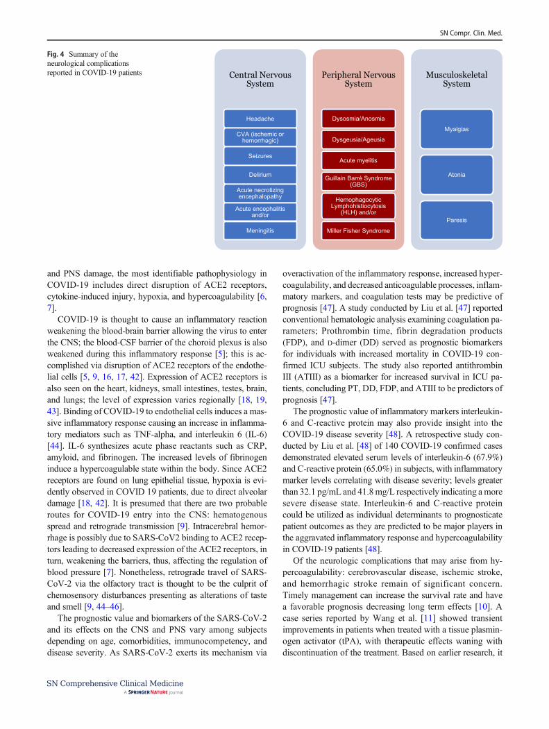
Figure 4 depicts all the neurologic complications reportedwith COVID-19 patients. It will be important to evaluate inthe recovered COVID-19 patients whether these pathophysi-ological manifestations will have any long term neurologicalcomplications.
Discussion
COVID-19 is the seventh and newest addition to the beta-coronaviridae (coronavirus) family; it is an enveloped, sin-gle-stranded, positive-sense, RNA virus. The mechanisms in-volved in causing neurological damage are aplenty [7].Despite the diverse mechanisms involved in causing CNS
SN Compr. Clin. Med.
and PNS damage, the most identifiable pathophysiology inCOVID-19 includes direct disruption of ACE2 receptors,cytokine-induced injury, hypoxia, and hypercoagulability [6,7].
COVID-19 is thought to cause an inflammatory reactionweakening the blood-brain barrier allowing the virus to enterthe CNS; the blood-CSF barrier of the choroid plexus is alsoweakened during this inflammatory response [5]; this is ac-complished via disruption of ACE2 receptors of the endothe-lial cells [5, 9, 16, 17, 42]. Expression of ACE2 receptors isalso seen on the heart, kidneys, small intestines, testes, brain,and lungs; the level of expression varies regionally [18, 19,43]. Binding of COVID-19 to endothelial cells induces a mas-sive inflammatory response causing an increase in inflamma-tory mediators such as TNF-alpha, and interleukin 6 (IL-6)[44]. IL-6 synthesizes acute phase reactants such as CRP,amyloid, and fibrinogen. The increased levels of fibrinogeninduce a hypercoagulable state within the body. Since ACE2receptors are found on lung epithelial tissue, hypoxia is evi-dently observed in COVID 19 patients, due to direct alveolardamage [18, 42]. It is presumed that there are two probableroutes for COVID-19 entry into the CNS: hematogenousspread and retrograde transmission [9]. Intracerebral hemor-rhage is possibly due to SARS-CoV2 binding to ACE2 recep-tors leading to decreased expression of the ACE2 receptors, inturn, weakening the barriers, thus, affecting the regulation ofblood pressure [7]. Nonetheless, retrograde travel of SARS-CoV-2 via the olfactory tract is thought to be the culprit ofchemosensory disturbances presenting as alterations of tasteand smell [9, 44–46].
The prognostic value and biomarkers of the SARS-CoV-2and its effects on the CNS and PNS vary among subjectsdepending on age, comorbidities, immunocompetency, anddisease severity. As SARS-CoV-2 exerts its mechanism via
overactivation of the inflammatory response, increased hyper-coagulability, and decreased anticoagulable processes, inflam-matory markers, and coagulation tests may be predictive ofprognosis [47]. A study conducted by Liu et al. [47] reportedconventional hematologic analysis examining coagulation pa-rameters; Prothrombin time, fibrin degradation products(FDP), and D-dimer (DD) served as prognostic biomarkersfor individuals with increased mortality in COVID-19 con-firmed ICU subjects. The study also reported antithrombinIII (ATIII) as a biomarker for increased survival in ICU pa-tients, concluding PT, DD, FDP, and ATIII to be predictors ofprognosis [47].
The prognostic value of inflammatory markers interleukin-6 and C-reactive protein may also provide insight into theCOVID-19 disease severity [48]. A retrospective study con-ducted by Liu et al. [48] of 140 COVID-19 confirmed casesdemonstrated elevated serum levels of interleukin-6 (67.9%)and C-reactive protein (65.0%) in subjects, with inflammatorymarker levels correlating with disease severity; levels greaterthan 32.1 pg/mL and 41.8mg/L respectively indicating a moresevere disease state. Interleukin-6 and C-reactive proteincould be utilized as individual determinants to prognosticatepatient outcomes as they are predicted to be major players inthe aggravated inflammatory response and hypercoagulabilityin COVID-19 patients [48].
Of the neurologic complications that may arise from hy-percoagulability: cerebrovascular disease, ischemic stroke,and hemorrhagic stroke remain of significant concern.Timely management can increase the survival rate and havea favorable prognosis decreasing long term effects [10]. Acase series reported by Wang et al. [11] showed transientimprovements in patients when treated with a tissue plasmin-ogen activator (tPA), with therapeutic effects waning withdiscontinuation of the treatment. Based on earlier research, it
Central Nervous System
Headache
CVA (ischemic or hemorrhagic)
Seizures
Delirium
Acute necrotizing encephalopathy
Acute encephalitis and/or
Meningitis
Peripheral Nervous System
Dysosmia/Anosmia
Dysgeusia/Ageusia
Acute myelitis
Guillain Barré Syndrome (GBS)
Hemophagocytic Lymphohistiocytosis
(HLH) and/or
Miller Fisher Syndrome
Musculoskeletal System
Myalgias
Atonia
Paresis
Fig. 4 Summary of theneurological complicationsreported in COVID-19 patients
SN Compr. Clin. Med.

is understood that COVID-19 activates platelets and theclotting cascade [9]. Thus, in the event of neurovascular dis-ease as a result of hypercoagulability, the prognosis is heavilyreliant on thrombolytic therapy administered promptly andshould be profoundly considered [9].
A study conducted by Carfì et al. [49] reported persistentneurological manifestations in a small number of subjects witha mean age of 56.5. Although fatigue (53.1%), dyspnea(43.4%), joint pain (27.3%), and chest pain (21.7%) have beencommonly reported, there is limited research suggesting long-term prognosis of the neurologic manifestations [49]. 44.1%of the elderly patients reported decreased quality of life anddid not confirm if this was due to CNS- or PNS-related pur-poses [49]. In terms of long-term prognosis, it can be hypoth-esized that complications may potentially arise as lingeringadverse effects of COVID-19 in this vulnerable population[49]. Prospective follow-up analytic investigations are crucialin determining the long-term outcomes of the SARS-CoV-2pandemic [8].
The number of COVID-19 cases compared with the num-ber of deaths worldwide reflects that the majority of patientsinfected with this virus predominately recover from their ill-ness [50]. However, the striking number of COVID-19 deathsis a cause for concern. Assessing major biomarkers seen inlaboratory testing can foresee the potential complications thatmay occur following the onset of disease, thus, predicting theprognosis and outcome [50]. Decreased number of lympho-cytes, markedly increased neutrophils to lymphocytes ratio(NLR), decreased platelet count, increased D-dimer, PT,LDH, ALT, and AST reflect poor prognosis possibly leadingto increased severity of disease and mortality [50].
Neurodegenerative disorders typically have late onset ofsymptoms commonly occurring after the age of 60. As of late,it is too early to predict if SARS-CoV-2 will be associatedwith neurodegenerative disease progression, severity, or ear-lier onset of illness. During this systematic review of the as-sociation among COVID-19 and the neuronal complexities ofthis age group, it can be hypothesized that viral particles en-tering the CNS may cause an additive effect on the inflamma-tory processes occurring in susceptible individuals. Furtherstudies are necessary to determine whether there is an associ-ation between neurodegenerative illnesses like Alzheimer’sand Parkinson’s disease with COVID-19.
Limitations
This literature review focuses on the neurologic manifesta-tions and complications of COVID-19 and poses several lim-itations such as subjects with multiple underlying comorbidconditions demonstrating a worse prognosis to COVID-19,low number of patient reports as information is largely basedon case reports, as well as a risk of bias is possible. It is not
very clear whether the neurological manifestations are occur-ring as a primary infection or secondary characteristics to sys-temic causes of disease. With rapidly emerging research re-garding COVID-19, more data is required to further supportexisting findings.
Conclusion
During this article, the authors conducted a literature review ofover 200 articles and used 52 articles of COVID-19-associated central nervous system– and peripheral nervoussystem–related manifestations and complications. In this liter-ature review, multiple studies confirm the presence of neuro-logical findings in COVID-19 patients; common manifesta-tions include headache, dizziness, alterations of taste andsmell, encephalopathy, encephalitis, GBS, cerebrovasculardisease, and skeletal muscle injuries in predisposed patients.This can affect the quality of life due to its debilitating natureand carries a significant risk for mortality.
Individuals with underlying health conditions have in-creased susceptibility to acquiring SARS-CoV-2 comparedwith subjects without comorbidities; hypertensive patientshave shown to be more prone to developing neurologicalmanifestations. Among the patient population affected withCOVID-19 that demonstrated neurologic manifestations,children were also susceptible to the development of symp-toms. The outcome in children was quite promising whencompared with the elderly population in terms of recovery.In fact, children presented with little to no underlying co-morbidities, and did not develop pulmonary manifestationsduring the presence of neurological exhibitions. It is possi-ble that neurological presence may have been secondary toPIMS. With the progression of the COVID-19 pandemicand increasing numbers of extrapulmonary and neurologi-cal cases, further studies and published articles are of greatimportance to better understand disease presentations, treat-ment, and outcomes. Throughout the systematic literaturereview of COVID-19 and the neurological manifestationsand complications, it is not entirely transparent if the signsand symptoms are primarily due to COVID-19 entering theCNS via the angiotensin-converting enzyme (ACE) II, orevents following hypoxia, hypercoagulability, or systemicinflammatory responses [32].
COVID-19 is affecting individuals worldwide and de-mands global public health measures to control the rapidspread of this debilitating virus. It is a collective duty to takeprecautionary action in order to prevent further spread ofCOVID-19. Disease prevention via isolation, quarantine,wearing a face mask, hand washing, and social distancingare essential public health measures to prevent the spread ofCOVID-19 within communities.
SN Compr. Clin. Med.

Authors’ Contribution I.P.: Conceptualization, drafting of the review ar-t icle , edi t ing, interpretat ion of data , and revis ion. M.P.:Conceptualization, review, editing, and supervision of review article writ-ing process. U.J.: Drafting the review article and interpretation of data.N.K.: Editing and drafting the review article.
Compliance with Ethical Standards
Conflict of Interest The authors declare that they have no conflict ofinterest.
References
1. Niazkar HR, Zibaee B, Nasimi A, Bahri N. The neurological man-ifestations of COVID-19: a review article. Neurol Sci. 2020;41(7):1667–71. https://doi.org/10.1007/s10072-020-04486-3 [AccessedJune 27, 2020 https://pubmed.ncbi.nlm.nih.gov/32483687/].
2. Ahmad I, Rathore FA. Neurological manifestations and complica-tions of COVID-19: a literature review. J Clin Neurosci. 2020;77:8–12. https://doi.org/10.1016/j.jocn.2020.05.017 [AccessedJune 27, 2020 https://www.jocn-journal.com/article/S0967-5868(20)31078-X/pdf].
3. World Health Organization. Coronavirus disease 2019 (COVID-19) situation report-160. [Accessed; August 27, 2020. https://www.who.int/docs/default-source/coronaviruse/situation-reports/20200824-weekly-epi-update.pdf?sfvrsn=806986d1_4 ].
4. Bridwell R, Long B, Gottlieb M. Neurologic complications ofCOVID-19. Am J Emerg Med. 2020;38(7):1549.e3–7. https://doi.org/10.1016/j.ajem.2020.05.024 [Accessed 30 June 2020, https://www.ajemjournal.com/article/S0735-6757(20)30364-8/pdf].
5. Wang L, Shen Y, Li M, et al. Clinical manifestations and evidenceof neurological involvement in 2019 novel coronavirus SARS-CoV-2: a systematic review and meta-analysis. J Neurol. 2020:1–13. https://doi.org/10.1007/s00415-020-09974-2 [published onlineahead of print, 2020 Jun 11] [Accessed July 8th, 2020 https://www.ncbi.nlm.nih.gov/pmc/articles/PMC7288253/].
6. Fan H, Tang X, SongY, Liu P, Chen Y. Influence of COVID-19 oncerebrovascular disease and its possible mechanism.Neuropsychiatr Dis Treat. 2020;16:1359–67. https://doi.org/10.2147/NDT.S251173 Published 2020 May 28. [AccessedJune 27,2020 https://www.dovepress.com/influence-of-covid-19-on-cerebrovascular-disease-and-its-possible-mech-peer-reviewed-article-NDT ].
7. Tsivgoulis G, Palaiodimou L, Katsanos AH, et al. Neurologicalmanifestations and implications of COVID-19 pandemic. TherAdv Neurol Disord. 2020;13:1756286420932036. https://doi.org/10.1177/1756286420932036 Published 2020 Jun 9. [AccessedJuly 8, 2020 https://www.ncbi.nlm.nih.gov/pmc/articles/PMC7284455/].
8. Paterson RW, Brown RL, Benjamin L, et al. The emerging spec-trum of COVID-19 neurology: clinical, radiological and laboratoryfindings. Brain. 2020:awaa240. https://doi.org/10.1093/brain/awaa240 [published online ahead of print, 2020 Jul 8] [AccessedJuly 12, 2020 https://pubmed.ncbi.nlm.nih.gov/32637987/].
9. Ghannam M, Alshaer Q, Al-Chalabi M, Zakarna L, Robertson J,Manousakis G. Neurological involvement of coronavirus disease2019: a systematic review. J Neurol. 2020:1–19. https://doi.org/10.1007/s00415-020-09990-2 [published online ahead of print,2020 Jun 19] [Accessed July 9th, 2020 https://www.ncbi.nlm.nih.gov/pmc/articles/PMC7304377/].
10. Wang D, Hu B, Hu C, et al. Clinical characteristics of 138 hospi-talized patients with 2019 novel coronavirus-infected pneumonia inWuhan, China. JAMA. 2020;323(11):1061–9. https://doi.org/10.
1001/jama.2020.1585 [published online ahead of print, 2020Feb 7]. [Acessed June 28,2020,https://jamanetwork.com/journals/jama/fullarticle/2761044].
11. Wang J, Hajizadeh N, Moore EE, et al. Tissue plasminogen activa-tor (tPA) treatment for COVID-19 associated acute respiratory dis-tress syndrome (ARDS): a case series. J Thromb Haemost.2020;18(7):1752–5. https://doi.org/10.1111/jth.14828 [AccessedJune 28, 2020].
12. Reddy ST, Garg T, Shah C, et al. Cerebrovascular disease in pa-tients with COVID-19: a review of the literature and case series.Case Rep Neurol. 2020;12(2):199–209. https://doi.org/10.1159/000508958 Published 2020 Jun 11. [Accessed July 10, 2020https://www.ncbi.nlm.nih.gov/pmc/articles/PMC7325208/].
13. Tang N, Bai H, Chen X, Gong J, Li D, Sun Z. Anticoagulanttreatment is associated with decreased mortality in severe corona-virus disease 2019 patients with coagulopathy. J Thromb Haemost.2020;18(5):1094–9. https://doi.org/10.1111/jth.14817 AccessedJune 28, 2020, https://onlinelibrary.wiley.com/doi/full/10.1111/jth.14817].
14. Tang N, Bai H, Chen X, Gong J, Li D, Sun Z. Anticoagulanttreatment is associated with decreased mortality in severe corona-virus disease 2019 patients with coagulopathy. J Thromb Haemost.2020;18(5):1094–9. https://doi.org/10.1111/jth.14817 AccessedJuly 10, 2020 https://onlinelibrary.wiley.com/doi/full/10.1111/jth.14817].
15. Sharifi-Razavi A, Karimi N, Rouhani N. COVID-19 and intracere-bral haemorrhage: causative or coincidental? New Microbes NewInfect. 2020;35:100669. https://doi.org/10.1016/j.nmni.2020.100669 Published 2020 Mar 27. [Accessed June 29, 2020, https://www.sciencedirect.com/science/article/pii/S2052297520300214].
16. Li YC, Bai WZ, Hashikawa T. The neuroinvasive potential ofSARS-CoV2 may play a role in the respiratory failure of COVID-19 patients. J Med Virol. 2020;92(6):552–5. https://doi.org/10.1002/jmv.25728 [Accessed July 8, 2020 https://www.ncbi.nlm.nih.gov/pmc/articles/PMC7228394/].
17. Jasti M, Nalleballe K, Dandu V, Onteddu S. A review of patho-physiology and neuropsychiatric manifestations of COVID-19. JNeurol. 2020:1–6. https://doi.org/10.1007/s00415-020-09950-w[published online ahead of print, 2020 Jun 3] [Accessed July 8,2020 https://www.ncbi.nlm.nih.gov/pmc/articles/PMC7268182/].
18. Cheng H, Wang Y, Wang GQ. Organ-protective effect ofangiotensin-converting enzyme 2 and its effect on the prognosisof COVID-19. J Med Virol. 2020;92(7):726–30. https://doi.org/10.1002/jmv.25785 [Accessed July 9th, 2020 https://www.ncbi.nlm.nih.gov/pmc/articles/PMC7317908/].
19. Yashavantha Rao HC, Jayabaskaran C. The emergence of a novelcoronavirus (SARS-CoV-2) disease and their neuroinvasive pro-pensity may affect in COVID-19 patients. J Med Virol.2020;92(7):786–90. https://doi.org/10.1002/jmv.25918 [AccessedJun 16th, 2020 https://www.ncbi.nlm.nih.gov/pmc/articles/PMC7264535/].
20. SheratonM, Deo N, Kashyap R, Surani S. A review of neurologicalcomplications of COVID-19. Cureus. 2020;12(5):e8192. https://doi.org/10.7759/cureus.8192 Published 2020 May 18. [AccessedJune 30, 2020, https://www.cureus.com/articles/32076-a-review-of-neurological-complications-of-covid-19].
21. Rodriguez-Morales AJ, Cardona-Ospina JA, Gutiérrez-Ocampo E,et al. Clinical, laboratory and imaging features of COVID-19: asystematic review and meta-analysis. Travel Med Infect Dis.2020;34:101623. https://doi.org/10.1016/j.tmaid.2020.101623[Accessed June 27, 2020 https://www.sciencedirect.com/science/article/pii/S1477893920300910?via%3Dihub].
22. Mao L, Jin H, Wang M, Hu Y, Chen S, He Q, et al. Neurologicmanifestations of hospitalized patients with coronavirus disease2019 in Wuhan, China. JAMA Neurol. 2020;77(6):1–9. https://doi.org/10.1001/jamaneurol.2020.1127 [published online ahead of
SN Compr. Clin. Med.

print, 2020 Apr 10] [Accessed June 27, 2020, https://jamanetwork.com/journals/jamaneurology/fullarticle/2764549].
23. Brüggemann R, Gietema H, Jallah B, Ten Cate H, Stehouwer C,Spaetgens B. Arterial and venous thromboembolic disease in apatient with COVID-19: a case report. Thromb Res. 2020;191:153–5. https://doi.org/10.1016/j.thromres.2020.04.046 [AccessedJuly 10th, 2020. https://www.ncbi.nlm.nih.gov/pmc/articles/PMC7252130/].
24. Chen T, Wu D, Chen H, et al. Clinical characteristics of 113 de-ceased patients with coronavirus disease 2019: retrospective study.BMJ. 2020;368:m1091. https://doi.org/10.1136/bmj.m1091Published 2020 Mar 26 [published correction appears in BMJ2020 Mar 31;368:m1295] [Accessed June 29, 2020, https://www.bmj.com/content/368/bmj.m1091].
25. Filatov A, Sharma P, Hindi F, Espinosa PS. Neurological compli-cations of coronavirus disease (COVID-19): encephalopathy.Cureus. 2020;12(3):e7352. https://doi.org/10.7759/cureus.7352Published 2020 Mar 21. [Accessed July 10, 2020 https://www.ncbi.nlm.nih.gov/pmc/articles/PMC7170017/].
26. Moriguchi T, Harii N, Goto J, et al. A first case of meningitis/encephalitis associated with SARS-Coronavirus-2. Int J InfectDis. 2020;94:55–8. https://doi.org/10.1016/j.ijid.2020.03.062[Accessed 30 June 2020, https://www.ijidonline.com/article/S1201-9712(20)30195-8/pdf].
27. Correia AO, Feitosa PWG,Moreira JLDS, Nogueira SÁR, FonsecaRB, Nobre MEP. Neurological manifestations of COVID-19 andother coronaviruses: a systematic review. Neurol Psychiatry BrainRes. 2020;37:27–32. https://doi.org/10.1016/j.npbr.2020.05.008Accessed July 3, 2020 https://www.ncbi.nlm.nih.gov/pmc/articles/PMC7261450/].
28. Poyiadji N, Shahin G, Noujaim D, Stone M, Patel S, Griffith B.COVID-19-associated acute hemorrhagic necrotizing encephalop-athy: CT and MRI features. Radiology. 2020:201187. https://doi.org/10.1148/radiol2020201187 [Accessed 30 June 2020, https://pubs.rsna.org/doi/10.1148/radiol.2020201187?url_ver=Z39.88-2003&rfr_id=ori%3Arid%3Acrossref.org&rfr_dat=cr_pub++0pubmed&.
29. Poyiadji N, Cormier P, Patel PY, et al. Acute pulmonary embolismand COVID-19. Radiology. 2020:201955. https://doi.org/10.1148/radiol.2020201955 [published online ahead of print, 2020 May 14][Accessed July 8th, 2020 https://pubs.rsna.org/doi/10.1148/radiol.2020201955].
30. Benger M, Williams O, Siddiqui J, Sztriha L. Intracerebral haem-orrhage and COVID-19: clinical characteristics from a case series.Brain Behav Immun. 2020;S0889–1591(20):31097. https://doi.org/10.1016/j.bbi.2020.06.005 [published online ahead of print, 2020Jun 7] [Accessed on July, 10, 2020 https://www.ncbi.nlm.nih.gov/pmc/articles/PMC7276127/].
31. Benger M, Williams O, Siddiqui J, Sztriha L. Intracerebral haem-orrhage and COVID-19: clinical characteristics from a case series.Brain Behav Immun. 2020;S0889–1591(20):31097. https://doi.org/10.1016/j.bbi.2020.06.005 [published online ahead of print, 2020Jun 7] [Accessed July 3, 2020 https://www.ncbi.nlm.nih.gov/pmc/articles/PMC7276127/].
32. Mei PA, Loeb L. COVID-19: a chronological review of the neuro-logical repercussions - what do we Know byMay, 2020? medRxiv.2020. https://doi.org/10.1101/2020.05.19.20107102 [Accessed 30June 2020. 10.1101/2020.05.19.20107102.].
33. Gane SB, Kelly C, Hopkins C. Isolated sudden onset anosmia inCOVID-19 infection. A novel syndrome? Rhinology. 2020;58(3):299–301. https://doi.org/10.4193/Rhin20.114 [Accessed July 12,2020https://pubmed.ncbi.nlm.nih.gov/32240279/].
34. Zhao H, Shen D, Zhou H, Liu J, Chen S. Guillain-Barré syndromeassociated with SARS-CoV-2 infection: causality or coincidence?Lancet Neurol. 2020;19(5):383–4. https://doi.org/10.1016/S1474-4422(20)30109-5 [Accessed on June 30, 2020 https://www.
thelancet.com/pdfs/journals/laneur/PIIS1474-4422(20)30109-5.pdf].
35. Sedaghat Z, Karimi N. Guillain Barre syndrome associated withCOVID-19 infection: a case report. J Clin Neurosci. 2020;76:233–5. https://doi.org/10.1016/j.jocn.2020.04.062 [AccessedJuly 3, 2020 https://www.ncbi.nlm.nih.gov/pmc/articles/PMC7158817/].
36. Sotoca J, Rodríguez-Álvarez Y. COVID-19-associated acute nec-rotizing myelitis. Neurol Neuroimmunol Neuroinflammation.2020;7(5):e803. https://doi.org/10.1212/NXI.0000000000000803Published 2020 Jun 10. [Accessed July 10, 2020 https://www.ncbi.nlm.nih.gov/pmc/articles/PMC7309521/].
37. MunzM,Wessendorf S, Koretsis G, et al. Acute transverse myelitisafter COVID-19 pneumonia. J Neurol. 2020;267(8):2196–7.https://doi.org/10.1007/s00415-020-09934-w [Accessed July 1,2020 https://link.springer.com/content/pdf/10.1007/s00415-020-09934-w.pdf].
38. AlKetbi R, AlNuaimi D, AlMulla M, et al. Acute myelitis as aneurological complication of Covid-19: a case report and MRIfindings. Radiol Case Rep. 2020;15(9):1591–5. https://doi.org/10.1016/j.radcr.2020.06.001 [Accessed July 3, 2020 https://www.ncbi.nlm.nih.gov/pmc/articles/PMC7275163/].
39. Kucuk A, Cumhur Cure M, Cure E. Can COVID-19 cause myalgiawith a completely different mechanism? A hypothesis. ClinRheumatol. 2020;39(7):2103–4. https://doi.org/10.1007/s10067-020-05178-1 Accessed July 3, 2020 https://www.ncbi.nlm.nih.gov/pmc/articles/PMC7249985/].
40. Han YN, Feng ZW, Sun LN, et al. A comparative-descriptive anal-ysis of clinical characteristics in 2019-coronavirus-infected childrenand adults. J Med Virol. 2020. https://doi.org/10.1002/jmv.25835[published online ahead of print, 2020 Apr 6] [Accessed July 8,2020 https://onlinelibrary.wiley.com/doi/full/10.1002/jmv.25835].
41. Abdel-Mannan O, Eyre M, Löbel U, et al. Neurologic and radio-graphic findings associated with COVID-19 infection in children.JAMA Neurol. 2020:e202687. https://doi.org/10.1001/jamaneurol.2020.2687 [published online ahead of print, 2020 Jul 1] [AccessedJuly 9th, 2020 https://jamanetwork.com/journals/jamaneurology/fullarticle/2767979].
42. Hamming I, TimensW, Bulthuis ML, Lely AT, Navis G, van GoorH. Tissue distribution of ACE2 protein, the functional receptor forSARS coronavirus. A first step in understanding SARS pathogen-esis. J Pathol. 2004;203(2):631–637. doi:https://doi.org/10.1002/path.1570 [Accessed July 3, 2020 https://www.ncbi.nlm.nih.gov/pmc/articles/PMC7167720/ ].
43. Rodríguez Y, Novelli L, Rojas M, et al. Autoinflammatory andautoimmune conditions at the crossroad of COVID-19. JAutoimmun. 2020:102506. https://doi.org/10.1016/j.jaut.2020.102506 [Accessed Jun 16th, 2020 https://europepmc.org/article/med/32563547].
44. Guo YR, Cao QD, Hong ZS, et al. The origin, transmission andclinical therapies on coronavirus disease 2019 (COVID-19) out-break - an update on the status. Mil Med Res. 2020;7(1):11.https://doi.org/10.1186/s40779-020-00240-0 Published 2020 Mar13. [Accessed July 3, 2020 https://pubmed.ncbi.nlm.nih.gov/32169119/].
45. Wu Y, Xu X, Chen Z, et al. Nervous system involvement afterinfection with COVID-19 and other coronaviruses. Brain BehavImmun. 2020;87:18–22. doi:https://doi.org/10.1016/j.bbi.2020.03.031 [Accessed July 3, 2020 https://www.ncbi.nlm.nih.gov/pmc/articles/PMC7146689/].
46. Desforges M, Le Coupanec A, Dubeau P, et al. Humancoronaviruses and other respiratory viruses: underestimated oppor-tunistic pathogens of the central nervous system? Viruses.2019;12(1):14. https://doi.org/10.3390/v12010014 Published2019 Dec 20. [Accessed July 3, 2020 https://www.ncbi.nlm.nih.gov/pmc/articles/PMC7020001/].
SN Compr. Clin. Med.

47. Liu Y, Gao W, Guo W, Guo Y, Shi M, Dong G, et al. Prominentcoagulation disorder is closely related to inflammatory responseand could be as a prognostic indicator for ICU patients withCOVID-19. J Thromb Thrombolysis. 2020:1–8 [Accessed August27, 2020 https://link.springer.com/article/10.1007/s11239-020-02174-9].
48. Liu F, Li L, XuM,Wu J, Luo D, Zhu YS, et al. Prognostic value ofinterleukin-6, C-reactive protein, and procalcitonin in patients withCOVID-19. J Clin Virol. 2020;127:104370. https://doi.org/10.1016/j.jcv.2020.104370 Accessed August 27, 2020 https://pubmed.ncbi.nlm.nih.gov/32344321/.
49. Carfì A, Bernabei R, Landi F, Gemelli. Against COVID-19 post-acute care study group. persistent symptoms in patients after acute
COVID-19. JAMA. 2020, 2020:e2012603. https://doi.org/10.1001/jama.2020.12603 [Accessed July 12, 2020 [published online aheadof print, 2020 Jul 9] https://pubmed.ncbi.nlm.nih.gov/32644129/].
50. Pourbagheri-Sigaroodi A, Bashash D, Fateh F, Abolghasemi H.Laboratory findings in COVID-19 diagnosis and prognosis.Clinica Chimica Acta. 2020. https://doi.org/10.1016/j.cca.2020.08.019 [Accessed August 27 2020 https://www.sciencedirect.com/science/article/pii/S0009898120304125].
Publisher’s Note Springer Nature remains neutral with regard to jurisdic-tional claims in published maps and institutional affiliations.
SN Compr. Clin. Med.