The many faces of What is Endometriosis? EndometriosisEndometriosis is a common cause of chronic !...
12
The many faces of Endometriosis Beryl Benacerraf M.D Harvard Medical School What is Endometriosis? • Endometriosis is defined as the presence of normal endometrial tissue occurring outside of the endometrial cavity. • This tissue responds to cyclic hormonal changes resulting in localized bleeding, inflammation and adhesion formation. Common sites of endometriosis • Ovaries • Uterine surface • Peritoneum • Uterosacral ligaments • Cul de sac • Bladder • Bowel wall • Scars in anterior abdominal wall • Rare – outside pelvis Common sites of endometriosis McKinney et al., 2000. How Common is Endometriosis? • Prevalence in the general population is estimated at 10% of women. • Present in up to 80% in patients with pelvic pain. • Present in up to 20% in infertility patients but in 3% of multiparous women. • Unknown • Reflux of menstrual tissue and blood through the fallopian tubes during menses. • Müllerian remnants in the rectovaginal region differentiate into endometrial tissue. • Lymphatics may be the conduate • shorter menstrual cycles, longer bleeding, and early menarche are risk factors – supports reflux theory Etiology
Transcript of The many faces of What is Endometriosis? EndometriosisEndometriosis is a common cause of chronic !...

The many faces of���Endometriosis
Beryl Benacerraf M.D���Harvard Medical School
What is Endometriosis?• Endometriosis is defined as the
presence of normal endometrial tissue occurring outside of the endometrial cavity.
• This tissue responds to cyclic hormonal changes resulting in localized bleeding, inflammation and adhesion formation.
Common sites of endometriosis• Ovaries• Uterine surface• Peritoneum• Uterosacral ligaments• Cul de sac• Bladder• Bowel wall• Scars in anterior abdominal wall• Rare – outside pelvis
Common sites of endometriosis
McKinney et al., 2000.
How Common is Endometriosis?
• Prevalence in the general population is estimated at 10% of women.
• Present in up to 80% in patients with pelvic pain.
• Present in up to 20% in infertility patients but in 3% of multiparous women.
• Unknown• Reflux of menstrual tissue and blood
through the fallopian tubes during menses. • Müllerian remnants in the rectovaginal
region differentiate into endometrial tissue.• Lymphatics may be the conduate• shorter menstrual cycles, longer bleeding,
and early menarche are risk factors – supports reflux theory
Etiology

Endometriosis is a common cause of chronic ���pelvic pain in premenopausal women
• The most recognized ultrasound appearance of endometriosis is the cyst known as an endometrioma.
• Traditionally, the sonographic diagnosis of endometriosis was reserved for patients with endometriomas, thus missing the cause of pelvic pain in many patients.
Endometrioma of the ovary
Deep implants• The pain associated with these implants
may be intense but these are small lesiona often not detected by a standard pelvic scan.
• Some patients with large endometriomas may have few symptoms whereas others with small deep implants have severe dysmenorrhea, dyspareunia and chronic pelvic pain.
Example: bowel endo
Adenomyosis:• Thought of as endometriosis of
the uterus.• Characterized by invasion of
endometrial glands into the neighboring myometrium.
• Symptoms: Dysmenorrhea ���abnormal bleeding, uterine enlargement and tenderness.

The curse of pelvic pain• Many patients never get a diagnosis and
live with chronic pain• 15% of women - as defined by pain for
> 6 months in women 18-50 years old. (Matthias et al OB GYN 1996;87:321)
• Pelvic pain accounts for 10% of referrals to a gynecologist and more than 40% of diagnostic laparoscopies
(Shwayder JM. Semin Reprod Med. 2008;26:25)
The examination ���of the patient���
suspected of endometriosis
Get a history during the exam• Acute or chronic• Diffuse or focal• Cyclical or constant• Sharp or dull or cramping• ? Prior surgery• Menopausal and hormonal status• Could she be pregnant?
During the scan• How tender is the patient?• Where is the tenderness? Focal?• Do organs slide past each other?• Push deliberately on each part of the
pelvis with the probe and other hand to determine where the pain comes from.
• Talk to the patient!
Adenomyosis: Invasion of ���endometrial glands into myometrium
Ultrasound appearance: – Mottled inhomogeneous myometrium– globular & asymmetrical uterus, – small subendometrial cysts – indistinct endometrial stripe.

Fuzzy borders of cavity due to adenomyosis
Adenomyoma versus fibroid
AshermansIs this adenomyosis?

Normal Asherman’s Is this adenomyosis?
Submucous fibroid
Typical endometriomas
Typical endometrioma with hydrosalpinx (common)
EndometriomaCystadenoma EndometriomaOv Cancer

Pitfall - struma ovariiIs this endometriosis? Endometrioma
mimicking dermoid
Is this a dermoid ?
Is this endometriosis?
Hint:Patient is pregnant
Decidualized endometrioma
Decidualized Endometrioma
• Cystic masses mimicking an ovarian malignancy during pregnancy, due to areas of nodularity containing blood flow by color Doppler.• A prospective diagnosis is possible when a pregnant women has a cyst with solid smoothly lobulated nodules and internal vascularity, stable over several weeks.

22 masses in 17 pregnant pts• 8 pts went to surgery, 9 (14 masses), had f/u
scans and surgery 3-34 weeks later• 8 of these masses showed no change and
one became smaller. • There were no characteristic sono features
identified to distinguish decidualized endometriomas from ovarian malignancy.
• Lesions showing no change over 4 weeks, or lacking solid components and vascularity are more likely to be benign.
Groszmann, Howitt, Bromley, Feltmate, Benacerraf, et al. In press JUM
Invasive ovarian cancer at 8 wks
Endo implant in CDS
Unusual appearances of endo implants anywhere
Endo in bowel wall
Endometriosis of the bladder wall
Tubal endometriosis

Extensive endometriosis 24 year old
Endo implant in utero-sacral ligament
Endo implant on appendix
Appendicitis!
Anterior abdominal wall scar
The comet shape
Endo in wall of sigmoid adherant to back of uterus

The comet-sign of endometriosis of the bowel wall

Bowel endo vs tumor
• Ultrasound sensitivity and specificity for detecting the disease was 78.5% and 95.2% respectively.
• The sensitivity was best for intestinal and bladder disease and slightly less accurate for utero-sacral and - rectovaginal lesions
Ultrasound for detecting deep pelvic endometriosis in 79 cases
Bazot et al. Obstet Gynecol 2004;24:180-5
Detection of Bowel Endometriosis���10 prospective studies – 1106 pts – ���prevalence of bowel endo 24-73%���
Sensitivity 71-98% (pooled data 91%)Specificity 92-100%(pooled data 98%)Accuracy 81-99%+LR 30.36-LR 0.09 Hudelist et al UOG 2011;37;257
Transvaginal scan in deep endometriosis���in 10 different pelvic sites
420 pts having ultrasound and laparoscopy (gold standard) for pelvic pain and infertility. Endo focus Sensitivity % Specificity %Bladder 61 99Recto-vaginal septum
52 96
Rectal 65 99Sigmoid 69 98
Fratelli et al. UOG 2013;41:69-75
Recto-vaginal septum
Ultrasound vs. MR���I98 pts with surgically ���
confirmed endometriosisAbrao et al. found that transvaginal ultrasound had a sensitivity, specificity and accuracy of 98%, 100% and 99% respectively compared to MRI’s sensitivity, specificity and accuracy of 83% 98% and 90% for recto-sigmoid endometriosis.
Abrao et al. Hum Reprod. 2007;22:3092-7
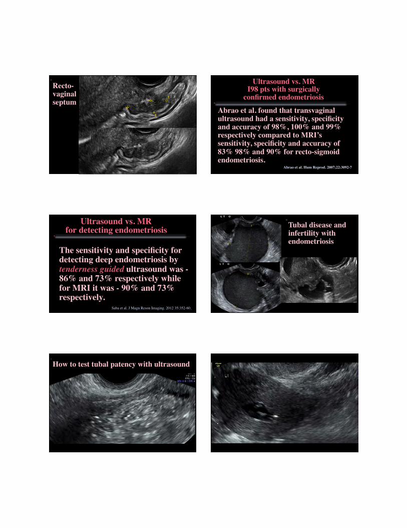
The sensitivity and specificity for detecting deep endometriosis by tenderness guided ultrasound was - 86% and 73% respectively while for MRI it was - 90% and 73% respectively.
Ultrasound vs. MR���for detecting endometriosis
Saba et al. J Magn Reson Imaging. 2012 35:352-60.
Tubal disease and infertility with endometriosis
How to test tubal patency with ultrasound

Conclusions• Pelvic pain in endometriosis is common
and impairs quality of life.• Accurate Dx requires an extended
ultrasound and history.• Patients with endometriosis deserve
more than just a series of standard pictures of the uterus and ovaries.
• Those that we help are among the most grateful of all our patients!
Long segment of bowel wall endo
Colon cancer
Crohn’s


![Pancreatic endometrial cyst mimics mucinous cystic neoplasm of … · 2017. 4. 29. · The most common sites of endometriosis are the pelvic organs[5]; however, endometriosis of the](https://static.fdocuments.in/doc/165x107/6117aa33d0c6a51c5b69412a/pancreatic-endometrial-cyst-mimics-mucinous-cystic-neoplasm-of-2017-4-29-the.jpg)