Laparoscopic Myomectomy Beware Adenomyosis - longdom.org · endometriosis, fibroids & adenomyosis,...
6
Laparoscopic Myomectomy Beware Adenomyosis John L Yovich 1,2* , Sunthra Lingam 1,2 , Philip K Rowlands 1 and Shanthi Srinivasan 1 PIVET Medical Centre, Cambridge Street, Perth, 6007 Western Australia, Australia Cairns Fertility Centre, McLeod Street, Cairns, Queensland, 4870 Australia * Corresponding author: John L Yovich, PIVET Medical Centre, Perth, Western Australia and Cairns Fertility Centre, Queensland, Australia, Tel: (08) 9422 5400 Fax: (08) 9382 4576; E-mail: [email protected] Received date: Accepted date: January 14, 2019; Published date: January 21, 2019 Copyright: © 2019 Yovich JL, et al. This is an open-access article distributed under the terms of the Creative Commons Attribution License, which permits unrestricted use, distribution and reproduction in any medium, provided the original author and source are credited. Abstract Objective: To assess the safety of laparoscopic myomectomy and consider the underlying reason for the most serious complication; that of uterine rupture. Design: 2 case studies with review of the recent literature. Setting: Two Australian private facilities under a single medical directorship providing comprehensive services in gynaecology, andrology and reproductive endocrinology for infertility management including in vitro fertilization (IVF). Experience with advanced laparoscopic surgery over 35 years including myomectomy over 27 years. Patients: Two women aged 37 years and 40 years respectively, who suffered uterine rupture post laparoscopic surgery to resect adenomyosis nodules, undertaken in order to improve their prognosis for IVF. Each woman now has a healthy child but one woman suffered an explosive uterine rupture which required emergency management at several levels. Both have been advised against further pregnancies. Intervention: Applying the same laparoscopic techniques that work so well for myomata, namely using intramural pitressin to minimise bleeding and a 2-layered suturing method to close the myometrial defect, nodules of focal adenomyosis were resected. Main outcome measures: Haemorrhage requiring blood transfusion and uterine rupture in ensuing pregnancy. Results: Whilst no surgical complications occurred in 1600 cases of laparoscopic myomectomy, 2 cases of excess bleeding and 2 cases of uterine rupture in the ensuing pregnancy occurred in 200 case of adenomyosis resection. Conclusion: Advanced diagnostic tests should be applied to differentiate those cases which have underlying adenomyosis and hormonal therapy may be preferred over surgical approaches for this enigmatic condition. Keywords: Laparoscopic myomectomy; Adenomyosis; Uterine rupture; Fibroids; Obstetric complications; Laparoscopic complications; IVF Introduction is article presents an extensive clinical experience which highlights, on the one hand, the comparative safety of laparoscopic myomectomy along with the potentially more hazardous procedure of wedge resection of nodular adenomyosis. We include two dramatic case reports which highlight that although the laparoscopic operations appear to mostly go very well, the subsequent pregnancy outcomes can be extremely hazardous. e implication is that the surrounding myometrium contains diffuse adenomyosis which causes suboptimal healing of the myometrium. Materials and Methods e first laparoscopic myomectomy procedure conducted from our facility was 27 years ago (July, 1991) and since then more than 1600 procedures have been conducted by our group. A further 200 cases of laparoscopic “myomectomy” proved to be cases of adenomyosis following recognition during surgery and subsequent histological evaluation. PIVET Medical Centre is a tertiary facility managing all aspects of Infertility along with associated pathologies including endometriosis, fibroids & adenomyosis, tubal & pelvic pathologies as well as advanced laparoscopic surgeries for benign conditions with authors being rated to the highest level 6 as categorised by AGES; the Australian Gynaecological Endoscopy Society [1]. Two members have attained AGES 6-B (benign gynaecological surgery) and 6-R (reproductive gynaecological surgery); the other 2 have attained AGES level 5. Cases of adenomyosis were sometimes obvious from the pre- operative evaluation due to associated symptomatology of dysmenorrhoea, generalised pelvic pain and bloating and tenderness J o u r n a l o f F e r t i l i z at i o n : I n V i t r o - I V F - W o r l d w i d e ISSN: 2375-4508 Journal of Fertilization: In Vitro - IVF Worldwide, Reproductive Medicine, Genetics & Stem Cell Biology Yovich et al., J Fertil In vitro IVF Worldw Reprod Med Genet Stem Cell Biol 2019, 6:1 DOI: 10.4172/2375-4508.1000212 Research Article Open Access J Fertil In vitro IVF Worldw Reprod Med Genet Stem Cell Biol, an open access journal ISSN:2375-4508 Volume 6 • Issue 1 • 212 1 2 December 01, 2018;
Transcript of Laparoscopic Myomectomy Beware Adenomyosis - longdom.org · endometriosis, fibroids & adenomyosis,...

Laparoscopic Myomectomy Beware AdenomyosisJohn L Yovich1,2*, Sunthra Lingam1,2, Philip K Rowlands1 and Shanthi Srinivasan1
PIVET Medical Centre, Cambridge Street, Perth, 6007 Western Australia, Australia
Cairns Fertility Centre, McLeod Street, Cairns, Queensland, 4870 Australia*Corresponding author: John L Yovich, PIVET Medical Centre, Perth, Western Australia and Cairns Fertility Centre, Queensland, Australia, Tel: (08) 9422 5400 Fax:(08) 9382 4576; E-mail: [email protected]
Received date: Accepted date: January 14, 2019; Published date: January 21, 2019
Copyright: © 2019 Yovich JL, et al. This is an open-access article distributed under the terms of the Creative Commons Attribution License, which permits unrestricteduse, distribution and reproduction in any medium, provided the original author and source are credited.
Abstract
Objective: To assess the safety of laparoscopic myomectomy and consider the underlying reason for the mostserious complication; that of uterine rupture.
Design: 2 case studies with review of the recent literature.
Setting: Two Australian private facilities under a single medical directorship providing comprehensive services ingynaecology, andrology and reproductive endocrinology for infertility management including in vitro fertilization (IVF).Experience with advanced laparoscopic surgery over 35 years including myomectomy over 27 years.
Patients: Two women aged 37 years and 40 years respectively, who suffered uterine rupture post laparoscopicsurgery to resect adenomyosis nodules, undertaken in order to improve their prognosis for IVF. Each woman nowhas a healthy child but one woman suffered an explosive uterine rupture which required emergency management atseveral levels. Both have been advised against further pregnancies.
Intervention: Applying the same laparoscopic techniques that work so well for myomata, namely using intramuralpitressin to minimise bleeding and a 2-layered suturing method to close the myometrial defect, nodules of focaladenomyosis were resected.
Main outcome measures: Haemorrhage requiring blood transfusion and uterine rupture in ensuing pregnancy.
Results: Whilst no surgical complications occurred in 1600 cases of laparoscopic myomectomy, 2 cases ofexcess bleeding and 2 cases of uterine rupture in the ensuing pregnancy occurred in 200 case of adenomyosisresection.
Conclusion: Advanced diagnostic tests should be applied to differentiate those cases which have underlyingadenomyosis and hormonal therapy may be preferred over surgical approaches for this enigmatic condition.
Keywords: Laparoscopic myomectomy; Adenomyosis; Uterinerupture; Fibroids; Obstetric complications; Laparoscopiccomplications; IVF
IntroductionThis article presents an extensive clinical experience which
highlights, on the one hand, the comparative safety of laparoscopicmyomectomy along with the potentially more hazardous procedure ofwedge resection of nodular adenomyosis. We include two dramaticcase reports which highlight that although the laparoscopic operationsappear to mostly go very well, the subsequent pregnancy outcomes canbe extremely hazardous. The implication is that the surroundingmyometrium contains diffuse adenomyosis which causes suboptimalhealing of the myometrium.
Materials and MethodsThe first laparoscopic myomectomy procedure conducted from our
facility was 27 years ago (July, 1991) and since then more than 1600procedures have been conducted by our group. A further 200 cases oflaparoscopic “myomectomy” proved to be cases of adenomyosisfollowing recognition during surgery and subsequent histologicalevaluation. PIVET Medical Centre is a tertiary facility managing allaspects of Infertility along with associated pathologies includingendometriosis, fibroids & adenomyosis, tubal & pelvic pathologies aswell as advanced laparoscopic surgeries for benign conditions withauthors being rated to the highest level 6 as categorised by AGES; theAustralian Gynaecological Endoscopy Society [1]. Two members haveattained AGES 6-B (benign gynaecological surgery) and 6-R(reproductive gynaecological surgery); the other 2 have attained AGESlevel 5.
Cases of adenomyosis were sometimes obvious from the pre-operative evaluation due to associated symptomatology ofdysmenorrhoea, generalised pelvic pain and bloating and tenderness
Jour
nal o
f Fer
tilization: In Vitro-IVF-W
orldwide
ISSN: 2375-4508
Journal of Fertilization: In Vitro - IVF Worldwide,Reproductive Medicine, Genetics & Stem Cell Biology
Yovich et al., J Fertil In vitro IVF Worldw Reprod Med Genet Stem Cell Biol 2019, 6:1 DOI: 10.4172/2375-4508.1000212
Research Article Open Access
J Fertil In vitro IVF Worldw Reprod Med Genet Stem Cell Biol, an open access journalISSN:2375-4508
Volume 6 • Issue 1 • 212
1
2
December 01, 2018;

on pelvic examination. Others were suspected from ultrasoundappearances, elevated CA125 levels and MRI studies although thelatter was rarely used. Most cases were diagnosed at laparoscopy withthe uterine irregularity proving to be non-discrete lumpiness withoutthe clear dissection planes typical of benign myomata. Both single-mass adenomyomata as well as diffuse adenomyosis patterns wereidentified and our operative procedures have included myolysis as wellas wedge resection of discrete and conglomerate adenomyosis masses.
Over the 27 year period we have conducted some “challenging”laparoscopic myomectomy procedures including the removal of 30separate fibroids in one case (who has subsequently achieved 2children spontaneously) and successful laparoscopic procedurescompleted where the uterus had sizes equating to 18-22 weeksgestation! It has been our long-standing practice to utilise intra-uterinepitressin (vasopressin; 20 units dissolved in 100 ml saline) at dosageslimited to 4 IU (20 ml of diluted solution) per hour, and we canindicate that no case required blood replacement. However, we havenow limited our cases to uterine size “16 weeks” in order to avoid longoperative procedures as cases of post-operative atelectasis andthrombo-embolic conditions occurred in occasional cases when theoperative time exceeded 3 hours; the patient positioned in steepTrendelenburg position for up to 5-hours in the most challengingcases.
At the beginning (1991-1995), myomectomy specimens weremorcellated by laparoscopic scissors, a tedious and time-consumingprocess where scissors became unusable after each case. When theStorz electronic morcellator became available in Australia in 1995, wepurchased the Rotocut device and continued to use it until currenttime. The device was a major advance reducing operating timesmarkedly and the vast majority of cases were conducted under 2.5 husually with the 12 mm but sometimes requiring the larger 15 mmrotor blade and we learnt to morcellate with minimised spinning andscatter of resected tissues.
First Case ReportA 36-year old woman G1P0 at 36+6 weeks gestation, experienced
acute abdominal pain and collapsed in the toilet of a friend’s house.Her husband extricated her from the toilet with some difficulty as shewas jammed against the door. Following a 7 km ambulance journey tothe Albany Regional Hospital in Western Australia, an urgentlaparotomy was performed revealing 3 litres of fresh blood in theperitoneal cavity. Her 2532 gram son was delivered by lower segmentcaesarean section with a low Apgar score of 2 and he required someresuscitation. Following his delivery the uterine fundus was deliveredthrough the abdominal wound and revealed an explosive 8 cm X 6 cmstellate full-thickness rupture in the right fundus (Figure 1a).
The infant was transferred by Royal Flying Doctor Serviceambulance to the Tertiary Obstetric Facility in Perth, some 490 kmdistant. He was returned to his mother 4 days later in good clinicalcondition. His mother was estimated to have lost 3 litres of blood intothe peritoneal cavity and was transfused 4 units of blood; she made anuneventful post-operative recovery.
Figure 1a: Uterine rupture at fundus noted at emergency caesareansection for acute pain and collapse at 36+6 weeks gestation. Rupturesite corresponds with site of wedge resection of uterineadenomyoma performed 12 months prior.
The scarred area was excised and the fundal rupture sutured in atwo-layer repair (Figure 1b).
Figure 1b: Fundal suturing after delivering the 2532 gram maleinfant by lower uterine segment caesarean section. The edges of therupture site were trimmed back to reveal vascularised myometriumprior to suturing.
Mother and child progressed well; the mother is using Implanon forcontraception and has been advised against further pregnancies. Herson has progressed well with full cognitive skills. However, he wassubsequently discovered to have a hemi-vertebra and is being managedby spinal brace. This congenital anomaly has no bearing on thematernal history of adenomyosis or the obstetric outcome.
It is undoubtedly relevant to note the woman’s previous historyincluded 5 laparoscopic procedures for pelvic endometriosis causinglong-standing dysmenorrhoea, dyspareunia and, subsequently,infertility. At PIVET, our main Fertility Centre in Perth, WesternAustralia, a further laparoscopic procedure was performed after 5embryo transfers failed (One fresh following IVF/ICSI and subsequent4 FETs). At this stage widespread adenomyosis was detected byultrasound and the laparoscopy revealed a deeply retroverted uteruswithin a frozen pelvis comprising grade IV endometriosis invading therectovaginal septum (Cullen’s disease). After block dissection of theendometriosis and ventro-suspension, a 4 cm uterine lump was alsoexcised from the uterine fundus (Figure 2).
2a) Injection of 4 IU Pitressin adjacent to fundal adenomyoma forvasoconstriction to minimise peri-operative bleeding. 2b) Unipolarhigh-frequency electro cautery incision to open overlying myometriumrevealing glandular intramural tissue rather than myoma which wasinitially expected. 2c) Highly vascular, glandular tissue excised bywedge resection as typical myoma plane absent. Although most of theadenomyomatous tissue was excised, lack of a clear plane and the
Citation: Yovich JL, Lingam S, Rowlands PK, Srinivasan S (2019) Laparoscopic Myomectomy Beware Adenomyosis. J Fertil In vitro IVF WorldwReprod Med Genet Stem Cell Biol 6: 212. doi:10.4172/2375-4508.1000212
Page 2 of 6
J Fertil In vitro IVF Worldw Reprod Med Genet Stem Cell Biol, an open access journalISSN:2375-4508
Volume 6 • Issue 1 • 212
associated diffuse adenomyosis meant some tissue inevitably remainsin-situ and is incorporated in the deeper layer of suturing, limiting thehealing process.
Figure 2: Excision of 4 cm adenomyoma 12 months prior to uterinerupture.
Zoladex implants 3.6 mg monthly were commenced and continuedfor the following 6 months. Further IVF procedures failed and anotherlaparoscopy undertaken for myolysis to treat expanding adenomyosis.The myolysis procedure constituted 12 sites of high frequency electro-cauterisation, avoiding the previous myomectomy site which however,was undetectable having a normal appearance, indicating it hadapparently healed well from the laparoscopic suturing.
The pregnancy ensued following the transfer of two cryopreservedblastocyst-stage embryos under hormone replacement treatment(FET/HRT). The ET was conducted under trans-vesical ultrasoundimaging and adjusted for the long cervical canal and bulky retroposeduterus. The clear fundal flash was detected at 9 cm from the externalcervical os. Pregnancy was subsequently diagnosed and monitoredthrough the first trimester as per PIVET protocol [2]. A normalsingleton intra-uterine fetus with heart activity was demonstrated at 7weeks and normal first trimester at 12 weeks. There after the patientwas managed in her regional town Albany, being 490 km from Perth byroad travel.
Second case reportA 40 year old woman with an IVF pregnancy achieved at our sister
clinic Cairns Fertility Centre, in Queensland, was referred to a localobstetrician after the 7 week scan determined a live intra-uterinepregnancy. He reported back that the pregnancy became complicatedby the development of gestational diabetes, requiring insulin therapy,and there was a major degree of placenta praevia. The patient had beenhospitalised on two occasions for vaginal bleeding during her antenatalcourse. She was managed by elective caesarean section at 37+5 weeksgestation and a healthy female infant weighing 2990 grams wasdelivered with excellent Apgar scores. However, after removal of theplacenta the obstetrician realized there was a defect in the posteriorwall of the uterus. This was quite a large defect and was covered byperitoneum only (Figure 3a).
Figure 3a: Fundal suturing after delivering the 2532 gram maleinfant by lower uterine segment caesarean section. The edges of therupture site were trimmed back to reveal vascularised myometriumprior to suturing.
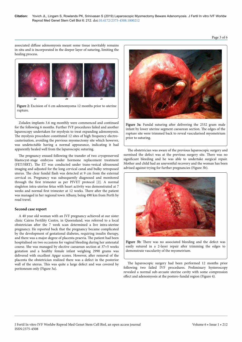
The obstetrician was aware of the previous laparoscopic surgery andsurmised the defect was at the previous surgery site. There was nosignificant bleeding and he was able to undertake surgical repair.Mother and child had an uneventful recovery and the woman has beenadvised against trying for further pregnancies (Figure 3b).
Figure 3b: There was no associated bleeding and the defect waseasily sutured in a 2-layer repair after trimming the edges todemonstrate vascularity of the myometrium.
The laparoscopic surgery had been performed 12 months priorfollowing two failed IVF procedures. Preliminary hysteroscopyrevealed a normal sub-arcuate uterine cavity with some compressioneffect and adenomyosis at the postero-fundal region (Figure 4).
Citation: Yovich JL, Lingam S, Rowlands PK, Srinivasan S (2019) Laparoscopic Myomectomy Beware Adenomyosis. J Fertil In vitro IVF WorldwReprod Med Genet Stem Cell Biol 6: 212. doi:10.4172/2375-4508.1000212
Page 3 of 6
J Fertil In vitro IVF Worldw Reprod Med Genet Stem Cell Biol, an open access journalISSN:2375-4508
Volume 6 • Issue 1 • 212

Figure 4: Preliminary hysteroscopy demonstrated cornual regionswith right and left tubal ostia. On the patient’s left side there was anapparent bulge into the uterine cavity matching the adenomyomaseen at the ensuing laparoscopy. The fundal area between the tubalostia demonstrates multiple glandular openings consistent withdiffuse adenomyosis.
Laparoscopy revealed a 5 cm intramural “fibroid” which was excisedunder intra-mural pitressin cover (Figure 5).
Figure 5: At laparoscopy the postero-fundal area of the uterus showsa 6 cm swelling. High frequency electrocautery incision through thecovering myometrium revealed glandular adenomyomatous tissue.There was no plane of delineation as seen with myomata hence awedge resection was undertaken to minimise the mass of tissue,removed in 4 segments prior to electronic morsellation.
During the dissection it became obvious that the nodule was not afibroid as there was no distinct plane of dissection, and the excisionbecame a wedge resection. None-the-less the cavity was closed quiteneatly in a 2 layered repair using a continuous Quill barbed suture(Figure 6). Areas of endometriosis at the uterosacral crus were ablatedand an incidental appendicectomy was also performed as the appendixwas noted to be nodular and surrounded by adhesions. A RediVacdrain was placed to the cul-de-sac. However, there was no bleedingduring this day-case operation and post-operative recovery wasentirely uneventful. Subsequent histology revealed normal proliferativeendometrium, the nodule was classical adenomyosis with nomalignant features and the appendix revealed extensive endometriosiswith no malignant features.
Figure 6: A neat result from 2-layered suturing with a continuousbarbed Quill suture. However, the inner cavity approximationinvolved myometrium diffusely invaded with adenomyosis. ARediVac drain is sited behind the uterus, removed 8 hours post-surgery as there was no bleeding. Although the final operativeappearance is neat, diffuse adenomyosis undoubtedly limits thehealing process.
Two months later IVF was repeated and 2 cleavage-stage embryos(Day 3) were transferred fresh under ultrasound control. The uteruswas noted to be ante-verted and was only mildly bulky. A clear fundalflash was seen at transfer indicating a well-sited procedure. Pregnancywas diagnosed 2 weeks later and ultrasound confirmed a singleton livefetus at 7 weeks (5 weeks post ET).
ResultsAs noted, our experience with more than 1600 laparoscopic
myomectomies indicated no operative complications, no casesrequiring blood transfusion and no cases of uterine rupture inpregnancy (a formal audit of cases is in preparation). In cases orprolonged steep Trendelenberg position over 3 h, there may beincreased risks for non-surgical complications such as atelectasis andthrombo-embolic phenomena which can be countered or avoidedcompletely. On the other hand, the experience with laparoscopic wedgeresection for adenomyosis and adenomyoma was vastly different inthat serious complications were encountered in 4 cases among the 200operations performed. These included 2 cases of uterine rupture in latepregnancy and one case of post-operative blood replacement (3-units)for post-operative bleeding via the routinely-placed Yates abdominaldrain. A fourth case with 14-week uterus had laparotomy conversionas the adenomyoma proved impossible to mobilise from the uterinedissection – that case had emergency hysterectomy 8 hours later asmassive secondary pelvic bleeding occurred from deep sacral vesselswhich had been excessively distracted (stretched) during the extractionprocess for the large, deeply embedded adenomyoma.
Our controlled morcellation technique, we have not experiencedany cases of parasitic implantation and, although we discovered that 3cases of our myomectomy specimens contained leiomyosarcoma athistology, none of these developed any metastases or displayedabdominal seeding and all 3 had successful management at theoncology service to which they were each referred.
The obstetric outcomes of post-myomectomy cases is the subject ofa separate report, and can be summarised as no untoward events;however 2 of the cases which had wedge resection of nodularadenomyosis masses suffered uterine rupture during the ensuingpregnancies.
Citation: Yovich JL, Lingam S, Rowlands PK, Srinivasan S (2019) Laparoscopic Myomectomy Beware Adenomyosis. J Fertil In vitro IVF WorldwReprod Med Genet Stem Cell Biol 6: 212. doi:10.4172/2375-4508.1000212
Page 4 of 6
J Fertil In vitro IVF Worldw Reprod Med Genet Stem Cell Biol, an open access journalISSN:2375-4508
Volume 6 • Issue 1 • 212

DiscussionThese 2 cases of uterine rupture ensued following excision of intra-
mural nodules. One case was explosive, occurring in somewhatunfavourable, remote circumstances and the woman was lucky to havesurvived the event. The second case occurred silently and the womanappeared in no imminent danger as she was already resting up inhospital awaiting elective caesarean delivery for a major degree ofplacenta praevia. These 2 cases occurred 4 and 5 years ago respectivelyin circumstances where our laparoscopic expertise was well establishedover more than 20 years, but where we were extending the surgeriesfrom “myomectomies” to wedge resection of adenomyosis nodules.Under pitressin cover, the surgeries appeared no more difficult thanmyomectomies, but it appears that the intramural tissue, beingdiffusely invaded by adenomyosis glandular tissue, may not have thesame healing capacity as the compressed normal myometrium whichsurrounds myomata (fibroids). We have learnt to become morecircumspect about diagnosing such cases by applying advances inultrasound technology, by increased awareness and by the increasinguse of magnetic resonance imaging (MRI) in uncertain cases. We havealso become more reticent about conservative surgery foradenomyosis. Like its sister condition endometriosis, adenomyosis isan enigmatic condition which still challenges the fertility worldregarding optimal management [3].
For cases desiring IVF, preliminary hormonal therapy includingGnRHa agents combined with progestogens (we favourmedroxyprogesterone acetate; Provera) should achieve reasonablecontrol prior to conducting IVF treatment. Such cases may be bestmanaged by cryopreservation of embryos whilst conducting severalmonths of hormonal suppression. The use of ulipristal acetate was alsoan advanced consideration but unfortunately its potential for livertoxicity will restrict its use [4-6]. None-the-less we can expect furtherresearch utilising selective estrogen receptor modulators (SERMS) andprogesterone modulators (SPRMS) will inevitably find saferalternatives. The idea of more complex surgeries requiring openlaparotomy, providing an answer, is diminished by reports of uterinerupture occurring even after the overlapping procedures described byOsada [7].
However, against this problem described with adenomyosis, thelaparoscopic myomectomy procedures appear quite safe and readilyamenable to day-surgery [4,8,9]. We have not encountered any sort ofsurgical complication from over 1600 procedures, although we havenow committed the very bulky cases to either sequential laparoscopiesor a single laparotomy to reduce the time at which a patient is held inthe steep Trendelenburg position required for Laparoscopicmyomectomy on the more challenging cases. Fibroids do causeimplantation problems as well as obstetric disorders hence an audit ofpregnancies arising after laparoscopic myomectomies is currentlyunderway from our series [10]. Our favourable experience has causedus to reduce the threshold for offering laparoscopic myomectomies inthe IVF setting expanding beyond the generally accepted indication ofTypes 0, 1, 2 and 3 fibroids according to FIGO categories [11,12]. Webelieve that all intramural fibroids can interrupt the inner uterinevasculature, even those categorised 4 and 5 by FIGO as was eminentlydescribed by Sampson a century ago and which can cause adverseeffects on implantation, even from locations in the outer myometrium,not just at the junctional zone, adjacent to the endometrium [13,14].
We conclude therefore that laparoscopic myomectomy should beoffered readily for those cases of infertility proving resistant tostandard treatments including IVF as well as those women suffering
recurrent miscarriage. However, we would express caution regardingany conservative surgical management for adenomyosis andfurthermore, diagnostic efforts should be undertaken to ensure thatfibroids are indeed myomas, and not masquerading with underlyingadenomyosis. The latter can be a cause of operative complications,especially haemorrhage, as well as serious obstetric complications,especially uterine rupture.
ConclusionWe conclude therefore that laparoscopic myomectomy should be
offered readily for those cases of infertility proving resistant tostandard treatments including IVF as well as those women sufferingrecurrent miscarriage. However, we would express caution regardingany conservative surgical management for adenomyosis andfurthermore, diagnostic efforts should be undertaken to ensure thatfibroids are indeed myomas, and not masquerading with underlyingadenomyosis. The latter can be a cause of operative complications,especially haemorrhage, as well as serious obstetric complications,especially uterine rupture.
AcknowledgementGrateful acknowledgement to the non-specialist obstetricians and
anaesthetists at Albany Regional hospital who handled the emergencyprocedures for the first case and provided the pictures for Figures 1aand 1b. Grateful thanks also to Dr. Thomas G Wright, obstetrician atCairns Private Hospital who managed the delivery of the second caseand provided the pictures for Figures 3a and 3b. Thanks also to theheroic mothers who each approved release of their medical details.
References1. https://obgyn.onlinelibrary.wiley.com/doi/pdf/10.1111/ajo.128742. https://books.google.co.in/books?id=MFR-
DwAAQBAJ&pg=PA120&lpg=PA120&dq=Yovich+JL.+Monitoring+the+stimulated+IVF+cycle&source=bl&ots=FccEzWJrEP&sig=o_CI8qlqrliARGfndr5atpWgKrw&hl=en&sa=X&ved=2ahUKEwiGm86TtOffAhWSP3AKHR1ZCZsQ6AEwAXoECAgQAQ#v=onepage&q=Yovich%20JL.%20Monitoring%20the%20stimulated%20IVF%20cycle&f=false
3. Dueholm M (2017) Uterine adenomyosis and infertility, review ofreproductive outcome after in vitro fertilization and surgery. Acta ObstetGynecol Scand 96: 715-726.
4. Donnez J, Dolmans MM (2016) Uterine fibroid management: from thepresent to the future. Hum Repro Update 22: 665-686.
5. Gracia M, Alcala M, Ferreri J, Rius M, Ros C, et al. (2018) Ulipristalacetate improves clinical symptoms in women with adenomyosis anduterine myomas. J Minim Invasive Gynec 25: 1274-1280.
6. Donnez J (2018) Liver injury and ulipristal acetate: an overstated tragedy?Fertil Steril 110: 593-595.
7. Osada H (2018) Uterine adenomyosis and adenomyoma: The surgicalapproach. Fertil Steril 109: 406-417.
8. Tentas I (2017) Uterine rupture following laparoscopic myomectomy: Anoverview. Obstet Gynecol Int J 6: 00212.
9. Mas A, Tarazona M, Carrasco JD, Estaca G, Cristobal I, et al. (2017)Updated approaches for management of uterine fibroids. Int J Women’sHealth 9: 607-617.
10. Ouyang DW, Economy KE, Norwitz ER (2006) Obstetric complications offibroids. Obstet Gynecol Clin N Am 33: 153-169.
11. Taylor HS (2018) Fibroids: when should they be removed to improve invitro fertilization success? Fertil Steril 109: 784-785.
Citation: Yovich JL, Lingam S, Rowlands PK, Srinivasan S (2019) Laparoscopic Myomectomy Beware Adenomyosis. J Fertil In vitro IVF WorldwReprod Med Genet Stem Cell Biol 6: 212. doi:10.4172/2375-4508.1000212
Page 5 of 6
J Fertil In vitro IVF Worldw Reprod Med Genet Stem Cell Biol, an open access journalISSN:2375-4508
Volume 6 • Issue 1 • 212

12. Yan L, Yuq ZY, Guo Z, Lee Z, Niu J, et al. (2018) Effect of type 3intramural fibroids on in-vitro fertilization-intracytoplasmic sperminjection outcomes: a retrospective cohort study. Fertil Steril 109:817-822.
13. Sampson JA (1918) The escape of foreign material from the uterine cavityinto the uterine veins. The Ameri J Obst Dis women Child 78: 161-175.
14. Yovich JL, Rowlands PK, Lingam S, Sillender M, Srinivasan S, et al. (2019)Advanced fibroid study; paying homage to John Sampson.
Citation: Yovich JL, Lingam S, Rowlands PK, Srinivasan S (2019) Laparoscopic Myomectomy Beware Adenomyosis . J Fertil In vitro IVF WorldwReprod Med Genet Stem Cell Biol 6: 212. doi:10.4172/2375-4508.1000212
Page 6 of 6
J Fertil In vitro IVF Worldw Reprod Med Genet Stem Cell Biol, an open access journalISSN:2375-4508
Volume 6 • Issue 1 • 212