The Lymphatic System. Functions Of The Lymphatic System Transport Excess Interstitial Fluid Back To...
22
The Lymphatic System
-
Upload
muriel-hall -
Category
Documents
-
view
225 -
download
0
Transcript of The Lymphatic System. Functions Of The Lymphatic System Transport Excess Interstitial Fluid Back To...

The Lymphatic System

The Lymphatic System
• Functions Of The Lymphatic System
• Transport Excess Interstitial Fluid Back To Bloodstream
• Transport Dietary Lipids
• House Lymphocytes
• Generate An Immune Response
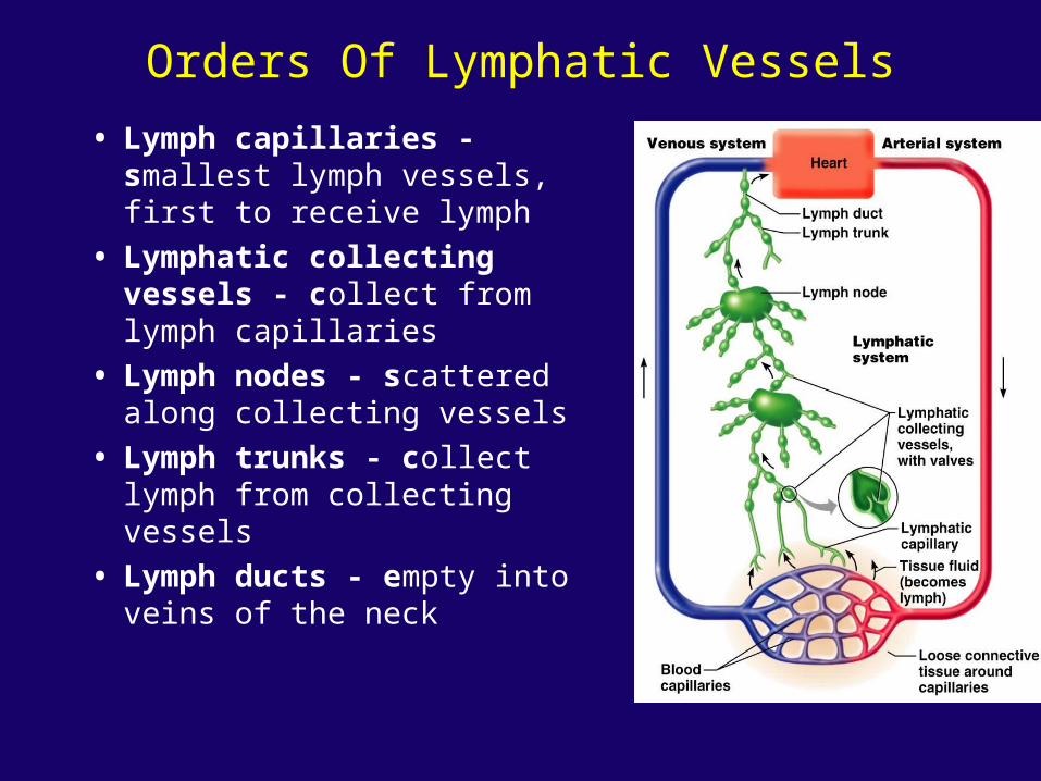
Orders Of Lymphatic Vessels
• Lymph capillaries - smallest lymph vessels, first to receive lymph
• Lymphatic collecting vessels - collect from lymph capillaries
• Lymph nodes - scattered along collecting vessels
• Lymph trunks - collect lymph from collecting vessels
• Lymph ducts - empty into veins of the neck

Lymphatic Capillaries• Located near blood capillaries
• Receive tissue fluid from CT
• Minivalve flaps open and allow fluid to enter
• Highly permeability allows entrance of tissue fluid, bacteria, viruses, and cancer cells
• Lacteals – specialized lymphatic capillaries
• Located in the villi of the small intestines
• Receive digested fats, Fatty lymph – chyle

Lymphatic Collecting Vessels
• Accompany blood vessels• Composed of the same three tunics as blood
vessels• Contain more valves than veins do
• helps direct the flow of blood
• Lymph propelled by:• contraction of skeletal muscles• pulse pressure of nearby arteries• Tunica media of the lymph vessels

Lymph Nodes• Cleanse the lymph of pathogens
• Human body contains around 500
• Lymph nodes are organized in clusters
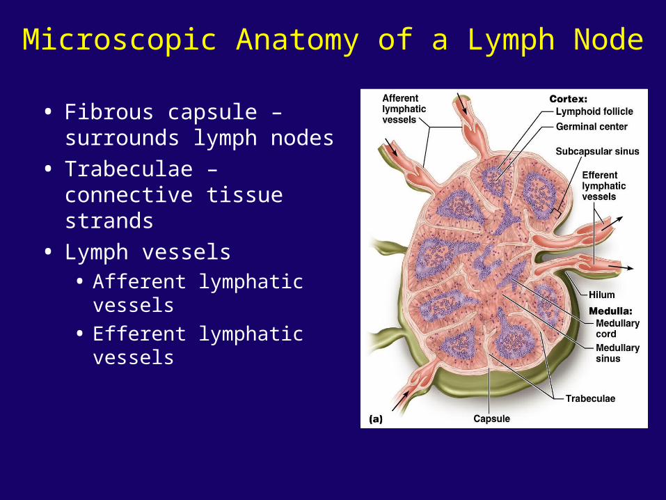
Microscopic Anatomy of a Lymph Node
• Fibrous capsule – surrounds lymph nodes
• Trabeculae – connective tissue strands
• Lymph vessels• Afferent lymphatic vessels
• Efferent lymphatic vessels

Lymph Trunks• Lymphatic collecting vessels
converge• Five major lymph trunks
• Lumbar trunks• Receives lymph from lower
limbs
• Intestinal trunk• Receives chyle from digestive
organs
• Bronchomediastinal trunks• Collects lymph from thoracic
viscera
• Subclavian trunks• Receive lymph from upper limbs
and thoracic wall
• Jugular trunks• Drain lymph from the head and
neck

Lymph Ducts
• Cisterna chyli - located at the union of lumbar and intestinal trunks
• Thoracic duct - ascends along vertebral bodies
• Empties into venous circulation
• Junction of left internal jugular and left subclavian veins
• Drains three quarters of the body
• Right lymphatic duct - empties into right internal jugular and subclavian veins

The Immune System
• Recognizes specific foreign molecules
• Destroys pathogens effectively
• Key cells – lymphocytes
• Also includes lymphoid tissue and lymphoid organs

Lymphocytes
• Infectious organisms attacked by inflammatory response, macrophages, then lymphocytes
• T Lymphocytes• Helper T-lymphocytes have receptors (CD4+) that can recognize an
antigen• Secrete cytokines (chemical signals that bind to receptors on other
lymphatic cells and activate them) and • Present the antigen to a B-lymphocyte.
• Cytotoxic T lymphocytes attack foreign cells directly• Receptors (CD8) bind to antigen-bearing cells• Perforates cell membrane• Signals cell to undergo apoptosis (self destruction)
• B lymphocytes• Become plasma cells• Secrete antibodies – bind and mark cells for destruction by macrophages

Lymphocyte Function
Figure 20.7

Lymphocyte Activation
• Lymphocytes originate in bone marrow
• Some travel to the thymus gland - T lymphocytes
• Some stay in bone marrow - B lymphocytes
• Able to recognize a unique antigen• Gain immunocompetence
• Travels through blood stream
• Meets and binds to a specific antigen

Lymphocyte Activation
• Activating T or B cells produce• Effector lymphocytes - short-lived, attack
immediately
• Memory lymphocytes - wait until body encounters their antigen again
• Basis of acquired immunity
• Guard against subsequent infections
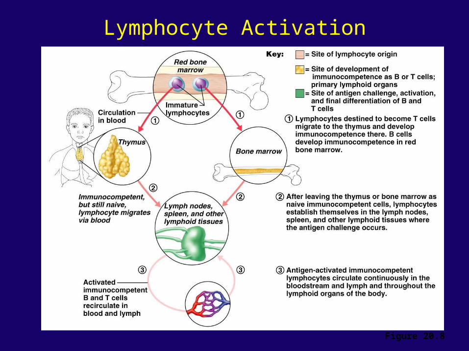
Lymphocyte Activation
Figure 20.8

Lymphoid Tissue
• Lymphoid tissue - areolar connective tissue and lymphocytes
• Most important tissue of the immune system• Mucous membranes of digestive, urinary,
respiratory, and reproductive tracts
• Mucosa-associated lymphoid tissue (MALT)• Makes up lymphoid organs (except thymus)

Lymphoid Organs
• Primary lymphoid organs
• Bone marrow
• Thymus
• Secondary lymphoid organs
• Designed to gather and destroy infectious microorganisms
• Lymph nodes, spleen, tonsils
• Aggregated lymphoid nodules - masses of lymphoid tissue NOT surrounded by a fibrous capsule.
• Appendix
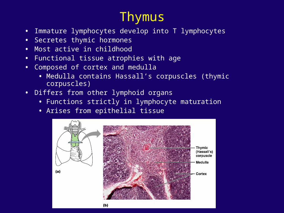
Thymus• Immature lymphocytes develop into T lymphocytes• Secretes thymic hormones • Most active in childhood• Functional tissue atrophies with age• Composed of cortex and medulla
• Medulla contains Hassall’s corpuscles (thymic corpuscles)• Differs from other lymphoid organs
• Functions strictly in lymphocyte maturation• Arises from epithelial tissue

Lymphoid Organs
• Lymph nodes• Lymph percolates through lymph sinuses
• Most antigenic challenges occur in lymph nodes
• Antigens destroyed – and activate B and T lymphocytes
• Spleen• Largest lymphoid organ
• Two main blood-cleansing functions• Removal of blood-borne antigens
• Removal and destruction of old or defective blood cells
• Site of hematopoiesis in the fetus
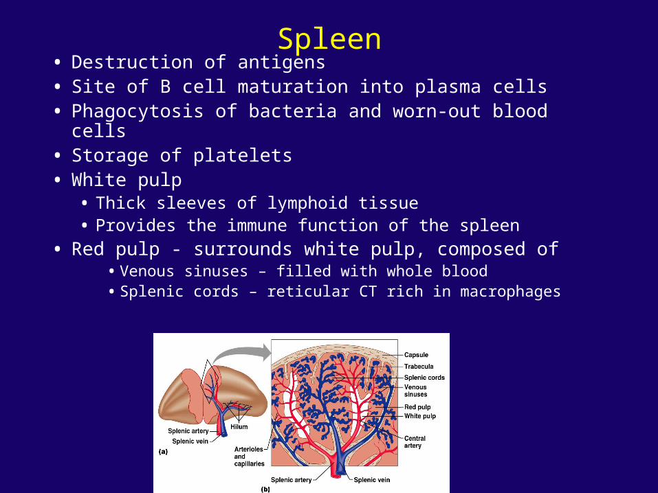
Spleen• Destruction of antigens• Site of B cell maturation into plasma cells• Phagocytosis of bacteria and worn-out blood cells• Storage of platelets• White pulp
• Thick sleeves of lymphoid tissue• Provides the immune function of the spleen
• Red pulp - surrounds white pulp, composed of• Venous sinuses – filled with whole blood • Splenic cords – reticular CT rich in macrophages

Tonsils• Simplest lymphoid organs
• Four groups of tonsils• Palatine, lingual, pharyngeal and tubal tonsils
• Arranged in a ring to gather and remove pathogens
• Underlying lamina propria consists of MALT
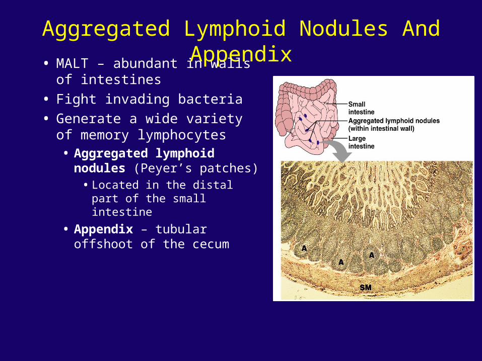
Aggregated Lymphoid Nodules And Appendix
• MALT – abundant in walls of intestines
• Fight invading bacteria
• Generate a wide variety of memory lymphocytes• Aggregated lymphoid
nodules (Peyer’s patches)• Located in the distal part of
the small intestine
• Appendix – tubular offshoot of the cecum