Fondamenti di anatomia e istologia - UniBG linfatico e...Structure of the lymphatic system In the...
46
Ingegneria delle tecnologie per la salute Fondamenti di anatomia e istologia Il sistema linfatico e immunitario
-
Upload
truongkien -
Category
Documents
-
view
220 -
download
2
Transcript of Fondamenti di anatomia e istologia - UniBG linfatico e...Structure of the lymphatic system In the...

Ingegneria delle tecnologie per la salute
Fondamenti di anatomia e istologia
Il sistema linfatico e immunitario

Definition
The immune system is the complex collection of
cells and organs that destroys or neutralizes
pathogens that would otherwise cause disease or
death.
The lymphatic system is the system of vessels,
cells, and organs that carries excess fluids to the
bloodstream and filters pathogens from the blood.
The lymphatic system is associated with the immune
system to such a degree that the two systems are
virtually indistinguishable.
Lymphatic
system

Functions of the lymphatic system
1. A major function of the lymphatic system is to drain body fluids and return them
to the bloodstream. Blood pressure causes leakage of fluid from the capillaries
→ accumulation of fluid in the interstitial space (spaces between individual cells
in the tissues) → about 20 L/die of interstitial fluid
17 liters is reabsorbed
directly by the blood vessels
3 liters enter in the lymphatic system
(becoming lymph) and are reintroduced
into the bloodstream via a series of
vessels, trunks, and ducts.

Functions of the lymphatic system
When the lymphatic system is damaged in some way a protein-rich interstitial
fluid accumulates in the tissue spaces → lymphedema that may lead to serious
medical consequences.

Functions of the lymphatic system
2.the network of lymphatic vessels transports
the cells of the immune system
3.lymphatic vessels transport dietary lipids
and fat-soluble vitamins absorbed in the gut

Cells of the immune system not only
use lymphatic vessels to make their
way from interstitial spaces back into
the
circulation, but they also use lymph
nodes as major staging areas for the
development of critical immune
responses. A
lymph node is one of the small,
bean-shaped organs located
throughout the lymphatic system.
The lymphatic vessels are open-ended
capillaries, which feed into larger and
larger lymphatic vessels, and
eventually empty into the bloodstream
by a series of ducts.
Along the way, the lymph travels
through the lymph nodes (major
staging areas for the development of
critical immune responses, about 500-
600), which are commonly found near
the groin, armpits, neck, chest, and
abdomen.
A major distinction between the
lymphatic and cardiovascular systems
in humans is that lymph is not actively
pumped by the heart, but is forced
through the vessels by the movements
of the body with the help of one-way
valves (semi-lunar valves) in lymphatic
vessels.
Structure of the lymphatic system

Structure of the lymphatic system
Lymphatic capillaries
are vessels where
interstitial fluid enters the
lymphatic system to
become lymph fluid.
Located in almost every
tissue in the body, these
vessels are interlaced
among the arterioles and
venules of the circulatory
system in the soft
connective tissues of the
body.
The central nervous system, bone marrow, bones, teeth, and the cornea of the eye, do
not contain lymph vessels. Lymphatic capillaries are formed by a one cell-thick layer of
endothelial cells.

Structure of the lymphatic system
In the small intestine, lymphatic capillaries called lacteals are critical for the transport of
dietary lipids and lipid-soluble vitamins to the bloodstream. In the small intestine, dietary
triglycerides combine with other lipids and proteins, and enter
the lacteals to form a milky fluid called chyle. The chyle then travels through the
lymphatic system, eventually entering the liver and then the bloodstream.
Structure of the lymphatic system
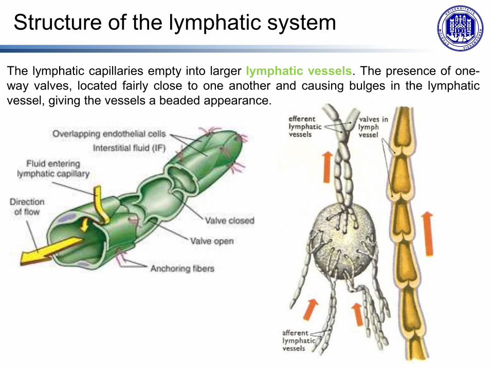
The lymphatic capillaries empty into larger lymphatic vessels. The presence of one-
way valves, located fairly close to one another and causing bulges in the lymphatic
vessel, giving the vessels a beaded appearance.

Structure of the lymphatic system
The superficial and deep lymphatics
eventually merge to form larger
lymphatic vessels known as
lymphatic trunks:
• On the right side of the body, the
right sides of the head, thorax, and
right upper limb drain lymph fluid
into the right subclavian vein via
the right lymphatic duct
• On the left side of the body, the
remaining portions of the body
drain into the larger thoracic duct,
which drains into the left
subclavian vein. The thoracic duct
itself begins just beneath the
diaphragmin the cisterna chyli, a
sac-like chamber that receives
lymph from the lower abdomen,
pelvis, and lower limbs by way of
the left and right lumbar trunks and
the intestinal trunk.

Structure of the lymphatic system

Structure of the lymphatic system
The overall drainage system of the body is asymmetrical. The right lymphatic duct
receives lymph from only the upper right side of the body. The lymph from the rest of
the body enters the bloodstream through the thoracic duct via all the remaining
lymphatic trunks.

Organization of the Immune system
The immune system is a collection of barriers, cells, and soluble proteins that interact
and communicate with each other. Immune function is organized into 3 phases based
on the timing of their effects.
Barrier defenses such as the skin and mucous membranes, which act
instantaneously to prevent pathogenic invasion
The rapid but nonspecific innate immune response, which is relatively rapid but
nonspecific and thus not always effective,
The slower but more specific and effective adaptive immune response, which
involves many cell types and soluble factors, but is primarily controlled by white
blood cells (leukocytes) known as lymphocytes. It is highly specific and effective
at attacking a wide variety of pathogens
The immune system’s first exposure to a pathogen is called a primary adaptive
response. Symptoms of a first infection, called primary disease, are always relatively
severe because it takes time for an initial adaptive immune response to a pathogen to
become effective. Upon re-exposure to the same pathogen, a secondary adaptive
immune response is generated, which is stronger and faster that the primary
response and eliminates a pathogen before it can cause significant tissue damage or
any symptoms. (immunological memory).
Organization of the Immune system
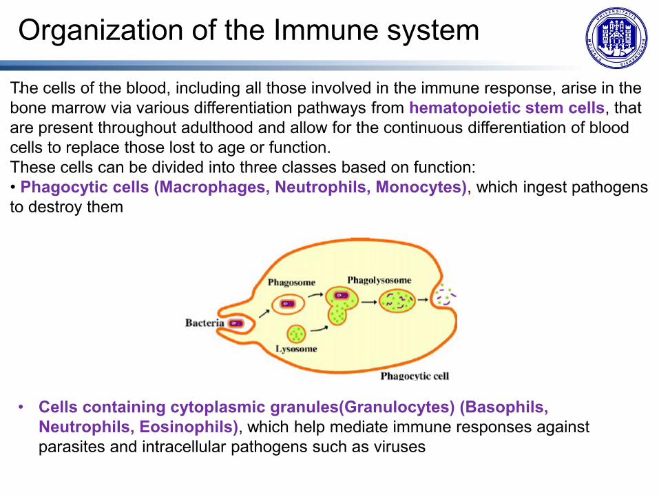
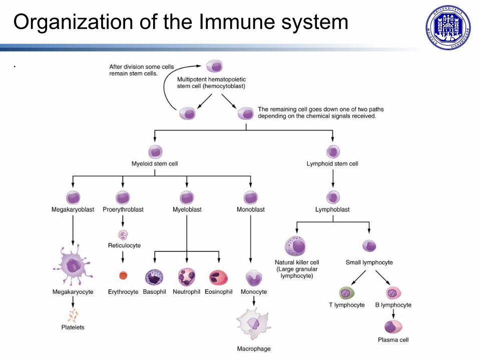
. The cells of the blood, including all those involved in the immune response, arise in the
bone marrow via various differentiation pathways from hematopoietic stem cells, that
are present throughout adulthood and allow for the continuous differentiation of blood
cells to replace those lost to age or function.
These cells can be divided into three classes based on function:
• Phagocytic cells (Macrophages, Neutrophils, Monocytes), which ingest pathogens
to destroy them
• Cells containing cytoplasmic granules(Granulocytes) (Basophils,
Neutrophils, Eosinophils), which help mediate immune responses against
parasites and intracellular pathogens such as viruses

Organization of the Immune system
. • Lymphocytes, which specifically coordinate the activities of adaptive immunity: T
lymphocytes (T cells), B lymphocytes (B cells), and natural killer (NK) cells.
o B cells are immune cells that function primarily by producing antibodies. An
antibody is a protein that binds specifically to pathogen-associated molecules
known as antigens. An antigen is a chemical structure on the surface of a
pathogen that binds to T or B lymphocyte antigen receptors. Once activated by
binding to antigen, B cells differentiate into cells that secrete a soluble form of
their surface antibodies. These activated B cells are known as plasma cells.
o T cells does not secrete antibody but performs a variety of functions in the
adaptive immune response. Different T cell types have the ability to either
secrete soluble factors (chemical messengers) that communicate with
other cells of the adaptive immune response or destroy cells infected with
intracellular pathogens.
o NK cells participy in the innate immune response. It contains cytotoxic (cell-
killing) granules in its extensive cytoplasm. They are especially genetically
programmed to recognize and kill certain altered cells: virally infected cells and
cancer cells.
o A plasma cell is a B cell that has differentiated in response to antigen binding,
and has thereby gained the ability to secrete soluble antibodies.
Organization of the Immune system
.

Organization of the Immune system
The two basic types of lymphocytes, B cells and T cells, are identical
morphologically with a large central nucleus surrounded by a thin layer of cytoplasm.
They are distinguished from each other by their surface protein markers as well as by
the molecules they secrete.
While B cells mature in red bone marrow and T cells mature in the thymus, they both
initially develop from bone marrow. T cells migrate from bone marrow to the thymus
gland where they further mature.
B cells are immune cells that function primarily by producing antibodies.
T cells does not secrete antibody but perform a variety of functions in the adaptive
immune response.

The primary lymphoid organs are where lymphocytes mature, proliferate, and are
selected, which enables them to attack pathogens without harming the cells of the
body.
The secondary lymphoid organs are where lymphocytes mount immune responses.
A naive lymphocyte is one that has left the primary organ and entered a secondary
lymphoid organ. Naive lymphocytes are fully functional immunologically, but have yet
to encounter an antigen to respond to. Lymphocytes circulete in blood and lymph and
concentrate in secondary lymphoid organs where remain as “sentinel” waiting to meet
an antigen.
Lymphoid Organs
The second lymphoid organs are: • Lymph nodes • Spleen • Lymphoid nodules
The primary lymphoid organs are: • Bone marrow • Thymus glands

Bone marrow is a highly cellular tissue
that is located in the medullary cavities
of the bone. Red bone marrow is the
principal site of blood cell formation, or
hemopoiesis.
In the embryo, blood cells are made in
the yolk sac. As development
proceeds, this function is taken over by
the spleen, lymph nodes, and liver.
Later, the bone marrow takes over most
hematopoietic functions
Bone Marrow

The yellow bone marrow is different from red bone marrow and it is a site of
energy storage, which consists largely of fat cells.
Bone Marrow

Bone Marrow
The B cell undergoes nearly all of its development in the red bone marrow, whereas
the immature T cell, called a thymocyte, leaves the bone marrow and matures
largely in the thymus gland.
Red bone marrow consists of densely
packed cords and islands of blood-
forming (hemopoietic) stem cells. They
are surrounded by numerous
macrophages and sinusoidal capillaries
opening into the thin venous sinuses.
A connective tissue stroma of reticular
cells and reticular fibers also form a
delicate meshwork that surrounds the
islands of hematopoietic cells and
provides support for the bone marrow.
In a section of the red bone marrow, all
types of developing blood cells are
difficult to distinguish.
The cells are densely packed, and
different cell types are intermixed.

Bone Marrow
Sinuses provide the main exit route through the openings in their endothelial lining for
the newly differentiated blood cells to enter the systemic circulation. The active red
bone marrow in selected bones provides a steady rate of blood cell renewal to replace
those that are worn out or lost.

Thymus gland
Thymus gland is a
capsulated bilobed organ.
The thymus gland is
surrounded by a
connective tissue capsule.
It is formed by different
lobules separated by
trabeculae. Under the
capsule there is the
cortex with an extensive
network of interconnecting
spaces.
These spaces become
colonized by immature
lymphocytes that migrate
here from hemopoietic
tissues in the developing
individual to undergo
maturation and
differentiation.

Thymus gland
The epithelial cells of the thymus gland provide structural support for the increased
lymphocyte population. In the lighter-staining medulla, the epithelial cells form a
coarser framework that contains fewer lymphocytes and whorls of epithelial cells
that combine to form thymic (Hassall) corpuscles.

Thymus
Located in the upper anterior mediastinum and lower part of the neck. The gland is most active during childhood, after which it undergoes slow involution, and, in adults, the lymphoid region is fi lled with adipose tissue. Endodermic and mesodermic orgin Max 25g

Thymus

Secodary lymphoid organs
Lymphocytes develop and mature in the primary lymphoid organs, but they mount
immune responses from the secondary lymphoid organs.
A naive lymphocyte is one that has left the primary organ and entered a secondary
lymphoid organ. Naive lymphocytes are fully functional immunologically, but have yet
to encounter an antigen to respond to.
In addition to circulating in the blood and lymph, lymphocytes concentrate in
secondary lymphoid organs: lymph nodes, spleen, and lymphoid nodules.
All of these tissues have many features in common, including the following:
• The presence of primary lymphoid follicles, the sites of accumulation of
lymphocytes, with specific B cell-rich and T cell-rich areas.
• When primary follicles are stimulated by specific antigens they activate and
became secondary follicles, that have a Germinal center, which is the site of
activated B-cells and memory B-cells
• Specialized post-capillary vessels known as high endothelial venules; the cells
lining these venules are thicker and more columnar than normal endothelial cells,
which allow cells from the blood to directly enter these tissues

Lymph nodes
Lymph nodes function to remove debris and pathogens from the lymph, (“filters of the
lymph”). Phagocytic cells within this organ internalize and kill many of the pathogens
that pass through, thereby removing them from the body. The lymph node is also the
site of adaptive immune responses mediated by T cells and B cells. Like the thymus,
the beanshaped lymph nodes are surrounded by a tough capsule of connective tissue
and are separated into compartments by trabeculae, the extensions of the capsule.
Each lymph node contains an outer cortex and an inner medulla
Cortex: nonencapsulated aggregations of
lymphocytes called lymphoid follicles.
Activated lymphoid follicles exhibit lighter-
staining central areas called germinal
centers
Medulla: medullary cords and medullary
sinuses. Medullary cords are networks of
reticular fibers filled with plasma cells,
macrophages, and lymphocytes separated
by capillary-like channels called medullary
sinuses.

Lymph nodes

Lymph nodes

Lymph nodes

Lymph nodes
The major routes into the lymph node are
via afferent lymphatic vessels.
Lymph enters the lymph node via the
subcapsular sinus, which is occupied
by phagocytic cells. Lymph flow throught
the lymphoid follicles and it is filtered.
As the lymph continues to flow through
the node, it enters the medulla, which
consists of medullary cords of B cells
and plasma cells, and the medullary
sinuses where the lymph collects before
leaving the node via the efferent
lymphatic vessels.

Ascellar station
Drains:
• Upper limb
• Thoracic wall
• breast
Lymph nodes stations

Drains:
• Heart
• Lungs
• Oesophagus
• Trachea
Thoracic stations
Lymph nodes stations

Inguinal station
Drains:
• Lower limb
• Perineum and genitalia
Lymph nodes stations

Cervical station
Drains:
• Head
• Mouth
• Neck
Lymph nodes stations

Sentinel lymphonode:
The first lymphonode that drain from a specific organ
Useful in surgical oncology:
Breast cancer
Melanoma
Lymph nodes and cancer
Cancer cell disseminate throught the lymphatic system and invade
lymphonode

Spleen
The spleen is a fragile organ with a thin capsule, and is dark red due to its extensive
vascularization.
“filter of the blood” for the presence of phagocytic cells that remove microbes and
dying red blood cells.
Surrounded by a capsule, the spleen is divided by
trabeculae into incomplete compartments called
splenic nodules.
Within each splenic nodule there are two areas:
• red pulp, consisting of mostly red blood cells
and formed by splenic cords and splenic
(blood) sinusoids. The splenic cords contain
networks of reticular fibers in which are found
numerous macrophages, lymphocytes, plasma
cells, and different blood cells.
• white pulp, consists of nodules of B cells
surrounded by T cells and accessory cells
(lymphatic nodules) that surround a blood
vessel called the central artery.

Spleen

Spleen
Upon entering the spleen, the splenic artery splits into several arterioles (surrounded
by white pulp) and eventually into sinusoids.
Blood from the capillaries subsequently collects in the venous sinuses and leaves via
the splenic vein.
Thus, the red pulp primarily
functions as a filtration
system of the blood, using
cells of the relatively
nonspecific immune
response, and white pulp is
where adaptive T and B
cell responses are
mounted.

Spleen
Parechimatous organ
Left hypochondrium, intraperitoneal
7x14 cm, 150-200 g
Mesodermic origin
Splenic artery (from celiac artery,
behind the pancreas)
Splenic vein portal system

Spleen

Spleen
Superiorly: diaphragm
Laterally: diaphragm and
thoracic wall
Medially: pancreas and
stomach
Posteriorly: dorsal wall,
left kidney
Anteriorly: stomach
Inferiorly: left colon

Lymphoid nodules
The other lymphoid tissues, the lymphoid nodules, have a simpler architecture than
the spleen and lymph nodes. They consist of a dense cluster of lymphocytes without a
surrounding fibrous capsule. These nodules are located in the respiratory and
digestive tracts, areas routinely exposed to environmental pathogens:
• Tonsils are lymphoid nodules located along the inner surface of the pharynx and
are important in developing immunity to oral pathogens

Lymphoid nodules
Palatine tonsil

Lymphoid nodules
• Mucosa-associated lymphoid tissue (MALT) consists of an aggregate of
lymphoid follicles directly associated with the mucous membrane epithelia of the
gastrointestinal tract, breast tissue, lungs, and eyes. Peyer’s patches, a type of
MALT in the small intestine, are especially important for immune responses against
ingested substances.
• Bronchus-associated
lymphoid tissue (BALT)
consists of lymphoid
follicular structures with
an overlying epithelial
layer found along the
bifurcations of the
bronchi, and between
bronchi and arteries.
These tissues, in addition
to the tonsils, are effective
against inhaled
pathogens.