The Influence of Rest Position of the Hyoid Bone upon ... · tiie influence of rest position of the...
66
Loyola University Chicago Loyola eCommons Master's eses eses and Dissertations 1976 e Influence of Rest Position of the Hyoid Bone upon Neuromusculature Adaptation to Forced Posterior Displacement of the Tongue Kent B. Augustson Loyola University Chicago is esis is brought to you for free and open access by the eses and Dissertations at Loyola eCommons. It has been accepted for inclusion in Master's eses by an authorized administrator of Loyola eCommons. For more information, please contact [email protected]. is work is licensed under a Creative Commons Aribution-Noncommercial-No Derivative Works 3.0 License. Copyright © 1976 Kent B. Augustson Recommended Citation Augustson, Kent B., "e Influence of Rest Position of the Hyoid Bone upon Neuromusculature Adaptation to Forced Posterior Displacement of the Tongue" (1976). Master's eses. Paper 2874. hp://ecommons.luc.edu/luc_theses/2874
Transcript of The Influence of Rest Position of the Hyoid Bone upon ... · tiie influence of rest position of the...

Loyola University ChicagoLoyola eCommons
Master's Theses Theses and Dissertations
1976
The Influence of Rest Position of the Hyoid Boneupon Neuromusculature Adaptation to ForcedPosterior Displacement of the TongueKent B. AugustsonLoyola University Chicago
This Thesis is brought to you for free and open access by the Theses and Dissertations at Loyola eCommons. It has been accepted for inclusion inMaster's Theses by an authorized administrator of Loyola eCommons. For more information, please contact [email protected].
This work is licensed under a Creative Commons Attribution-Noncommercial-No Derivative Works 3.0 License.Copyright © 1976 Kent B. Augustson
Recommended CitationAugustson, Kent B., "The Influence of Rest Position of the Hyoid Bone upon Neuromusculature Adaptation to Forced PosteriorDisplacement of the Tongue" (1976). Master's Theses. Paper 2874.http://ecommons.luc.edu/luc_theses/2874

TIIE INFLUENCE OF REST POSITION OF THE HYOID BONE UPON NEUROMUSCULATURE
ADAPTATION TO FORCED POSTERIOR DISPLACEMENT OF THE TONGUE
by
Kent B. Augustson D.D.S.
A Thesis Submitted to the Faculty of the Graduate School of
Loyola University in Partial Fulfillment of
the Requirements for the Degree
of Master of Science
June
1976
' - . .. ·-, ! ~ ~ .. _., L •. .)C, '· ' _·:

DEDICATION
to Carolyn, my wife and inspiration
ii

ACKNOWLEDGMENTS
I would like to extend my thanks to Dr. Douglas C. Bowman, my
thesis advisor, for his guidance and continued interest in this study.
My appreciation also goes to Dr. Bernard Pawlowski and Dr. Joseph
Gowgiel, members of my thesis advisory board.
I wish to acknowledge Drs. Cuozzo and Gobeille, whose work in this
area preceeded mine.
I am indebted to Loyola University and Drs. Braun, Madonia, and
Toto for making my graduate education possible.
I fondly acknowledge my parents, whose example in thought and
action provides a continuing inspiration.
Most importantly, I wish to acknowledge my wife Carolyn, to whom
this work is dedicated. I thank her for her love and understanding
during her marriage to a student.
iii

VITA
The author, Kent Binns Augustson, is the son of Elmer Moritz
Augustson and Geraldine Binns Augustson. He was born March 23, 1948,
in Galesburg, Illinois.
His elementary and secondary educations were obtained in the
public schools of Galesburg, Illinois. He graduated from Galesburg
Senior High School in 1966. In the fall of 1966 he entered the
University of Kansas in Lawrence, Kansas, where he majored in
Comparative Physiology and Biochemistry. He received the degree of
Bachelor of Arts in 1970. He attended Loyola University, School of
Dentistry from 1970 to 1974, when he received the degree Doctor of
Dental Surgery. Remaining at Loyola, in 1974 he began his studies
i~ the Orthodontic Certificate and Oral Biology Masters Programs.
iv

TABLE OF CONTENTS
ACKNOWLEDGMENTS
VITA
LIST OF TABLES •
LIST OF ILLUSTRATIONS
INTRODUCTION AND STATEMENT OF PROBLEM
REVIEI.J OF LITERATURE
Deglutition • • • • . • • • • • • • Hyoid Bone • • • • • . • • • Cinefluorography • • • • • • Myometrics • • • • •
METHODS AND MATERIALS
Selection of Subjects • • • • • • • ••• General Procedure • • • . • • • • • • • • Tongue Crib Appliance Fabrication and Placement Cinefluorographic Equipment and Technique Cinefluorographic Analysis • • • • Myometric Equipment and Technique • • Myometric Analysis • • • • •
RESULTS . . . ' . . . . . . . . DISCUSSION . . . " . . . . . . SUMMARY AND CONCLUSIONS . . . BIBLIOGRAPHY • • • • • • . . . . . . . . .
v
Page
iii
iv
vi
vii
1
2
2 8
15 16
21
21 22 22 25 26 27 29
30
46
51
53

,...
Table
I.
II.
.LIST OF TABLES
Quantified Results • • • • • . . . . . . . . . . . Subjects Exhibiting Extremes in Mandibular Plane-to-Hyoid Distance
vi
. . . . . . . . . .
Page
33
34

LIST OF ILLUSTRATIONS
Figure
1. Tongue Crib Appliance . . . . . . . . . . . . . . . . . 2. Appliance Viewed from Above Demonstrating
Anterior-Posterior Position of Crib Portion
3. Frontal View of Appliance Demonstrating Inferior Projection of Crib Portion •••.•••••••
4. Lingual View of Appliance Demonstrating Extension of Acrylic Portion • • • .
5. Plate Position for Control Myometric
. . . . . . . .
Recording . . . . . . . . . . Plate Position for Experimental Myometric Recording • • • • • • . . . . . . . . . . . .
7. Hyoid Position Before and Aftet' Placement of Tongue Crib Appliance
a. Subjects J.S. and K.A.
b. Subjects J.E. and H.Z.
c. Subjects C.B. and C.E.
d. Subjects C.H. and K.H.
e. Subjects D.P. and D.H. . f. Subjects A. H. and C.H. • • 41 • • .. •
g. Subjects A. C. and D.G.
h. Subjects M.S. and M.P. . . i. Subjects S.G. and M.E.
j. Subjects O.M. and D.T.
k. Subjects B.M. and S.N. . . . . vii
. . .
Page
23
23
24
24
28
28
35
35
36
37
38
39
40
41
42
43
44
45

INTRODUCTION AND STATEMENT OF PROBLEM
The purpose of this study was to determine whether the position
of the human hyoid bone, in its spatial relationship to the mandibular
plane, limits or influences the ability of the tongue neuromusculature
to adapt to forced posterior displacement.
In two previous studies at Loyola University, Cuozzo (1973) and
Gobeille (1974), the hyoid position did seem to influence tongue accom
modation. It was hypothesized that subjects with hyoids positioned
near the mandible have greater potential for tongue accomodation than
subjects presenting hyoids positioned further from the mandible.
This study was designed to test the hypothesis. Two groups of
subjects were chosen for the work. One group was comprised of girls
with the mandibular plane-to-hyoid distance relatively long. The other
group was comprised of girls with the mandibular plane-to-hyoid distance
relatively short. Tongue crib appliances were utilized to displace the
tongue posteriorly. With cinefluorographic and myometric techniques,
adaptive potential of the two groups was compared.
1

REVIEW OF THE LITERATURE
Deglutition
Magendie (1822) divided the act of swallowing into three stages.
Even today, Magendie's Theory of Constant Propulsion remains basically
correct, current studies with cinefluorography notwithstanding.
The three classic stages of swallowing are: (1) oral, (2) pharyn
geal and (3) esophageal. The oral stage is voluntary and conscious, the
pharyngeal involuntary, but still conscious, and the esophageal both in
voluntary and unconscious.
The oral or preparatory stage begins as the tongue collects the
substance to be swallowed and forms the bolus. The bolus is contained
in a cupped depression on the dorsal surface of the tongue. The bolus,
at this point, is circumscribed by a peripheral seal. Anteriorly, the
tip of the tongue is positioned against the palatal mucosa behind the
anterior teeth. The tongue seals laterally against the buccal teeth and
adjacent palatal mucosa. Posteriorly, the seal is formed by the tensor
depressed soft palate and the faucial pillars against the pharyngeal
portion of the tongue.
Passage of the bolus into the oral pharynx is accomplished with two
different actions. First, the lumen expands making room for the bolus
to pass. Then the lumen narrows and closes behind the bolus, propelling
it into the oral pharynx. Thus, the pharyngeal stage is begun by eleva
tion of the soft palate, relaxation of the faucial pillars, and depres-
2

sian and grooving of the posterior tongue. Closure of the lumen behind
the bolus is accomplished by pressure of the dorsum of the tongue on the
hard palate and then on the soft palate. The soft palate is carried
down by contraction of the faucial pillars. Then it is carried forward
to oppose the tongue by contraction of the superior and middle constric
tors of the pharynx. It has been noted in the literature that the ini
tial elevation of the soft palate to oppose the posterior pharyngeal
3
wall effectively seals against nasal leakage. Ardran (1954) likened
bolus propulsion to toothpaste being squeezed from a tube. Ramsey et al.,
(1955) calls the progressive narrowing and obliteration of the lumen
behind the bolus a "stripping wave." The further closing of the lumen
is affected by the constrictor muscles acting against the base of the
tongue. As the bolus passes through the laryngeal pharynx there is a
characteristic vigorous upward movement of the larynx and trachea, laryn
geal pharynx, hypopharynx, and upper esophagus. There is a concomitant
upward and forward movement of the hyoid. During the elevation of the
above mentioned structures, the true and false vocal folds are reflexly
approximated. Thus, the laryngeal additus is protected against bolus
penetration. The downward tilting of the epiglottis during this stage
tends to streamline the lumen and to some extent secondarily protect the
laryngeal additus. As the bolus reaches the hypopharyngeal constrictor
(cricopharyngeus) the sphincter is relaxed and the bolus enters the
esophagus.
Ramsey et al., (1955) stated that, with the exception of a few

reflex relationships, both the timing and order of the swallowing events
vary considerably in different individuals. They also vary in the same
individual under different conditions. He further stated that there are
persons who by training or habit vary markedly from the usual mechanism.
During the last twenty years the literature has been filled with studies
of deglutition. There has been a great deal written on these variations
to which Ramsey refers - tongue thrust swallow being the most controver
sial.
Straub (1960) professed these criteria for a normal swallow: (1)
teeth firmly together, (2) muscles of facial expression relaxed, and (3)
tongue remaining within the confines of the oral cavity. Conversely,
Straub felt that the abnormal swallow was hallmarked by a lack of com
plete tooth contact, protrusion of the tongue between the teeth at some
point, and tenseness in the perioral musculature. He felt that improper
bottle feeding was prime in the etiology of the tongue thrust swallow.
Straub stated that the abnormal swallowing habit usually produces an
open bite, and is capable of causing many "serious malocclusions." He
advocated function dictating form.
Rosenblum (1963) studied orofacial muscle activity during degluti
tion in twenty subjects with normal occlusions. A motion picture tech
nique was utilized. Analysis of the films showed that perioral activity
occurred in subjects with normal dentitions more than 50% of the time.
Wildman et al., (1964) studied fifty-two ten year old children, di
vided equally into normal and tongue thrust swallowers. Specifically,
4

static tongue posture and oral coordination of the two groups were in
vestigated. There was "no significant difference ••. found between the
two groups in either tongue carriage or repetitive ability."
The classical description of the clinically normal swallow came
under further question. Hedges et al., (1965) studied twelve to four
teen year old children with excellent occlusions and no speech problems.
These children demonstrated two distinct swallowing patterns: one with
teeth together and one with teeth apart. As a result, it was suggested
that "the term 'acceptable' be substituted for 'normal' as a more prac
tical and a more inclusive designation," when describing swallowing
patterns.
Cleall (1965) studied deglutition in adolescents divided into three
groups: normal - with excellent occlusion, Class II Division I malocclu
sions, and a tongue thrust group. He found variations from the classic
normal swallow in the group with excellent occlusions. Cleall stated
that 20% of the normal group exhibited no lip closure during swallowing,
11% protruded the tongue tip beyond the incisors, and 40% made no molar
contact. He concluded, "The concept of 'normal' swallowing in which the
teeth come into occlusion, the lips remain in repose, and the tongue re
mains within the confines of the oral cavity is no longer tenable."
Cleall placed palatal cribs in the tongue thrust patients, thereby chang
ing the local environment. From their ability to adapt, he surmised
that spontaneous correction of tongue thrust may result from orthodontic
correction - function adapting to form.
5

-Brauer and Holt (1965), after examining approximately two hundred
grade school and junior high school children, devised a tongue thrust
classification. The classification was based on deformities observed
rather than etiology. They intimated in their approach that function
was dictating form. Interestingly, two of the criteria in this study
for tongue thrust swallowing were perioral contraction and a lack of
tooth contact during the swallm-1.
A number of investigators concentrated on the tongue. Peat (1968)
stated that there are two postural positions of the tongue for each
individual. The first, an habitual position, exhibits tongue tip con
tact with the incisor teeth and/or lips. It also exhibits dorsum tongue
contact with the soft palate and sometimes with the soft palate and the
hard palate together. The second, a relaxed postural position, presents
increased convexity of the dorsum and contact with only the soft palate.
Fishman (1969) studied postural and dimensional aspects of the
tongue in rest position and occlusion. He included three groups in his
study: normals, a malocclusion group, and a group of lispers. His
observations on the normal group concurred with Peat's observations.
Fishman concluded, "Similarities and differences between normal, speech
and malocclusion groups have demonstrated that form and function have
some direct relationships to abnormal tongue posture and movement."
Harvold (1968) discussed animal experiments in which the dentition
exhibited sequellae when certain characteristics of the tongue (i.e.
size or position) were changed.
6

7
Cleall and Milne (1970) studied the posture and function of the
oropharyngeal structures during the period of mixed dentition. The
authors reported the oropharyngeal structures revealed a marked ability
to adapt to changes in the local dental environment. Specifically, the
forward position of the tongue was noted during the transition between
deciduous incisor loss and permanent incisor eruption.
Hanson et al., (1970) studied twenty-two factors in 193 subjects
and found only two "to be functionally associated with tongue-thrust
with any meaningful consistency. They are enlarged tonsils and lingual •
cross-bite." He contended that enlarged tonsils contribute to habitual
forward carriage of the tongue; and lingual cross-bite contributes by
forcing a narrowing and elongation of the tongue.
Subtelny (1970) observed swallowing behavior in ten normal subjects
and in thirty with malocclusions. He found appreciable differences in
tongue-tip function with differences in contiguous dentoskeletal form.
The interpretation was that the tongue was functionally adapting to the
specific anterior oral environment to achieve a seal during swallowing.
When the tongue-tip had adapted, the basic swallowing pattern was the
same in all the subjects. He concluded that the protrusive tongue activ-
ity could be a functional adaptation to its environment.
Subtelny (1973) reaffirmed his advocacy of form dictating function.
However, he mentions three factors which seem to contraindicate ortho-
dontic treatment and successful adaptation: abnormal skeletal relation-
ships, neurologic impairment in the control of orofacial muscle function,

- 8
and abnormal tongue size. Subtelny summarizes, "When form is modified
by orthodontic and/or surgical procedures within the anatomical and
physiologic limitations of the patient and within the reference of an
ticipated changes incident to growth and development, stable adjustments
in occlusion and favorable adaptations in orofacial muscle activity may
be anticipated."
Hanson et al., (1973) contended that more progress would be made
if researchers paid less attention to perpetuating the dichotomy between
form and function. Rather, he felt, researchers should consider more
seriously the possible reciprocation between form and function. He
pointed out that his research indicated crowding of the tongue, whether
it be a narrow maxillary arch, enlarged tonsils, or the presence of an
intruding thumb, might well promote a tongue-thrust habit. Hanson stated
that conversely, myometric research makes it difficult to avoid the con
clusion that the tongue does have the strength and persistence to cause
malocclusion.
Hyoid Bone
"The hyoid bone is shaped like a horseshoe, and is suspended from
the tips of the styloid processes of the temporal bones by the stylohyoid
ligaments.", Gray (1959). The bone is comprised of a body and paired
greater and lesser cornua.
Embryologically, according to Orban (1966), the hyoid has its ori
gins from the second and third branchial arches. The upper median part
of the body and the lesser cornua are derived from the second arch. While

The greater cornua and the majority of the body come from the third
arch.
The body of the hyoid is quadrilateral in form. It gives origin
to the Hyoglossus muscle. Insertions for the Geniohyoid, Mylohyoid,
Sternohyoid, and Omohyoid muscles are found on the body.
The greater cornua project back from the lateral borders of the
body. To the end of each is fixed the lateral hyothyroid ligament. The
greater cornua give origin to the Hyoglossus and Constrictor pharyngis
medius muscles. While the Thyrohyoid and Stylohyoid muscles insert in
the same.
The lesser cornua are small conical eminences found at the junction
between the greater cornua and the body. The attachment to the stylo
hyoid ligament is at the apex of each cornua. The Chondroglossus muscle
originates from the medial base.
By these attachments the hyoid bone is connected to and influenced
by: (1) the tongue, (2) the mandible, (3) the base of the skull, (4) the
sternum, (5) the scapula, (6) the thyroid cartilage, and (7) the pharnyx.
Sicher (1970) refers to the hyoid as the "skeleton of the tongue."
With this in mind, a brief discussion of tongue musculature is apropos.
The tongue musculature is divided into extrinsic and intrinsic groups.
The extrinsic muscles are the Genioglossus, the Hyoglossus, the Chondro
glossus (sometimes described as part of the Hyoglossus), and the Stylo
glossus. The intrinsic group is comprised of the Longitudinalis superi
or, the Longitudinalis inferior, the Transversus, and the Verticalis.
9

10
The Genioglossi can act to move the tongue forward, backward, or down
ward. The Hyoglossi, of particular interest in this study, depress the
tongue and draw its sides down. The Styloglossi draw the tongue upward
and backward. The intrinsic muscles are mainly concerned with changing
the shape of the tongue, i.e. shortening, narrowing, and curving actions.
In a cephalometric study of mandibular movements, Thompson (1941)
reported that movements of the mandible influenced hyoid position. He
noted that the hyoid moved only slightly posteriorly during the opening
rotation of the mandible.
Mainland (1945) stated that the hyoid acts as a platform. By fixing
one set of muscles, the platform is stabilized such that another set of
muscles can work from it.
Correlating the movements of the head and the hyoid, Wood (1956),
found that when the head is in dorsiflexion the hyoid is elevated. When
the head is in ventriflexion, however, the hyoid is directed downward.
Smith (1956) showed that the hyoid moves forward and slightly up
ward as the mandible moves into protrusive position. On maximum opening
of the mandible, downward and backward hyoid movement was pointed out.
Hyoid position in Class I, II, and III malocclusions was observed
by Grant (1959). He found, from a representative cephalometric film of
each subject, the hyoid position to be constant. Grant stated that mus
culature, not the occlusion of the teeth, determines hyoid position.
Shelton et al., (1960) studied cinefluorographically, tongue, hyoid
and laryngeal displacement during swallowing. Three phases of displace-

- 11
ment were described. Phase 1 "included simultaneous cephalad displace
ment of the hyoid, elevation of the larynx and usually a dorsad movement
of the pharyngeal portion of the tongue." Hyoid movement here was some
times slightly dorsad or ventrad, but always secondary to the cephalad
movement. Phase 2 "included simultaneous ventrad or ventrad and cepha
lad displacement of the hyoid and elevation and closure of the larynx."
Here hyoid displacement ranged from directly ventrad to obliquely ceph
aloventrad. Phase 3 "included simultaneous descent of the hyoid either
obliquely dorsad and caudad or dorsad and caudad and then more directly
caudad." There was also ventrad movement of the pharyngeal portion of
the tongue and descent and opening of the larynx.
Brodie (1961) called attention to the fact that the hyoid's role
as a functional part of the skeletal system has been a recent evolution
ary development. He related the actions of the hyoid to maintenance of
an ainmy during mandibular movements.
Durzo and Brodie (1962) conducted a longitudinal cephalometric study
of five normal occlusions. They found the hyoid to be positioned supero
inferiorly opposite the lower portion of the third and upper portion of
the fourth vertebrae. It was stated that position anteroposteriorly de
pends on the relative length of the muscles running from the hyoid to
the base of the cranium and the mandibular symphysis. They stated that
during development the hyoid descends as cervical vertebrae grow and as
the posterior cranial base and mandible descend. But the relative posi
tion of the hyoid is invariable.

In a study of 165 subjects, Bench (1963), found that the hyoid
gradually descends from a position opposite the lower half of the third
and the upper half of the fourth cervical vertebra (at age 3) to a posi
tion opposite the fourth cervical vertabra (at adulthood).
Wildman (1964), in his cinefluorographic study of deglutition,
stated that since the hyoid bone serves as the "posterior pedestal" on
which the tongue is mounted, it is a particularly helpful structure to
study in relation to the swallowing act.
A cephalometric positional study of the hyoid bone was attempted
by Stepovich (1965). He stated that hyoid position could not be dupli
cated from one cephalometric film to the next. He attributed the prob
lem in duplicating the hyoid position from one film to the next to two
factors: (1) the hyoid is totally suspended by muscles and ligaments,
and (2) the hyoid cannot be easily related to a fixed point in the head,
which is itself unstable.
12
Sloan et al., (1967), in a study of comparative hyoid movement dur
ing swallowing in Class I and Class II malocclusions, showed the antero
posterior location of the hyoid was consistently found to be near the
anterior root of the pterygoid plates. He found that the Class I malo
cclusions, although exhibiting no skeletal differences from the other
classes of malocclusion in the study, exhibited significantly lower and
more posterior hyoid locations (relative to the mandible). The Class II
malocclusions, conversely, showed higher and more forward hyoid positions.
The position of the hyoid bone and that of the mandible in retruded

---13
contact position, intercuspal position, and postural position in 144
persons were studied by means of cephalometric films by Ingervall et al.,
(1970). The following observations were made: (1) The position of the
hyoid bone varied less in superoinferior direction on repeated determin
ations in the postural position of the mandible than the contact posi
tions. (2) The hyoid moved downward and backward from intercuspal to
retruded contact position. (3) The postural position showed the hyoid
occupying a more superior position than in the intercuspal position.
Ingervall (1970), in a follow-up study, sought to determine whether
the size of hyoid movement concomitant with movement of the mandible be
tween postural, intercuspal and retruded contact positions, varies with
facial and dental arch morphology. He found that if the height of the
face is small, the hyoid bone tends to move inferiorly on movement of
the lower jaw from intercuspal position to postural position. But if
the facial height is great, the hyoid bone will move superiorly.
A cinefluorographic technique was used by Milne and Cleall (1970),
to measure changes occurring in the posture and function of the oro
pharyngeal structures during the transitional dentition stage. They
stated that the oropharyngeal structures demonstrated a marked ability
to adapt to changes in the local dental environment. It was pointed out
as being probable that the adaptive movements of the hyoid bone are limi
ted to those which would not interfere with the maintenance of an ade
quate airway.
Yip and Cleall (1971) conducted a cinefluorographic study of the

resting posture and the pattern of movement of the oronasopharyngeal
structures before and after surgical removal of both tonsils and ade
noids on twenty-eight children. "The positions of the hyoid bone ap
peared to be in a more upward and forward position both at rest and dur
ing all the stages of swallowing after the operation."
In a later study, Ingervall et al., (1971) recorded the act of
deglutition cineradiographically to study the movements of the hyoid
bone and to clarify whether contact between the teeth occurs in inter
cuspal or retruded contact position. It was confirmed that the hyoid
undergoes marked supero-anterior movement during the later part of the
act of swallowing and reaches its most supero-anterior position when
the bolus is in the lower part of the oropharynx. The pattern of hyoid
movement seemed independent of the position of the mandible during con
tact. It even seemed to be independent of whether or not there was in
fact contact during swallowing.
14
Wickwire (1972) found that when the mandible is set back in Class
III correction, the tongue is carried lower in the mouth, as demonstrated
by a change in hyoid position.
Cuozzo and Bowman (1975) performed a study to determine the amount
of change in positioning of the hyoid bone during deglutition following
forced distal positioning of the tongue by a tongue crib. Ten female
subjects, ranging in age from nineteen to thirty years, were included.
They were Class I normal occlusions. Cuozzo and Bowman judged accommo
dation in terms of hyoid repositioning (determined from cinefluorographic

-films) and myometric results. They found a strong correlation between
initial hyoid position and the ability to adapt. From these results,
they hypothesized that individuals with hyoids held relatively close to
the mandibular plane can reposition the tongue posteriorly or inferior
ly. Conversely, those with hyoid bones held relatively distant from
the mandibular plane would be expected to find such accommodation dif
ficult due to encroachment on the airway space.
Gobeille (1974), in a follow-up of Cuozzo's work, studied ten open
bite, tongue-thrust patients. His method of study as well as his re
sults were similar to Cuozzo's. Gobeille stated, "The hypothesis of
hyoid-mandibular plane distance was again born out."
Cinefluorography
Cinefluorography is the roentgen method for investigating function.
There have been numerous studies reporting various aspects of swallowing
behavior as revealed by cinefluorography.
15
Perhaps the first such investigators were Rushmer and Hendron (1951).
Referring to their study of deglutition, Saunders et al., (1951) stated,
" ••• the development of relatively high speed cineradiography offers the
most favorable opportunity of analyzing details of the mechanism and the
precise phase and sequence of the motions."
Sloan et al., (1964) stated that the cinefluorographic record pro
vides these basic data: (1) motion of the structures, (2) variation in
their radiographic density, and (3) chronology of the changes occurring
during the functional cycle. In this study, Sloan establishes eight

basic steps necessary to the correlation of cephalometric analysis and
cinefluorographic craniopharyngeal films.
Cleall et al., (1966) in a cinefluorographic study of head posture
and its relationship to deglutition stated several characteristics of
cinefluorography. The greater degree of magnification in cinefluoro
graphic equipment compared with the conventional cephalometric set-up
16
is due to a shorter x-ray source to patient distance and a larger patient
to image distance. Further, cinefluorographic film is such that while
the structures are plainly visible in motion, single frames are much
less clear than cephalometric radiographs.
Cinefluorography enables the researcher to view the previously in
accessible oral cavity and its related pharyngeal complements. The
quantity of literature in the area is growing. The following studies,
mentioned within this literature review, utilized cinefluorography:
Ardran and Kemp (1954); Ramsey et al., (1955); Shelton et al., (1960);
Wildman et al.' (1964); Brauer and Holt (1965); Cleall (1965); Hedges
et al,, (1965); Sloan et al., (1967); Milne and Cleall (1970); Hanson
et al., (1970); Subtelny (1970); Ingervall (1971); Yip and Cleall (1971);
Cuozzo and Bowman (1975); and Gobeille (1974).
Myometrics
Tomes (1873) stated, "The agency of the lips and tongue is that
which determines the position of the teeth themselves." Since, there
has been much research in the area of determining quantitatively muscle
influence on the dentition.

17
Kydd (1956) studied maximum tongue force in a thirty year old eden-
tulous patient. Strain gauges were mounted on a mandibular denture base.
He found the maximum anterior force to be about 5 lb. and the maximum
lateral force in the second bicuspid region to be approximately 2.5 lb.
Muscle pressures in seven subjects with normal occlusion were in-
vestigated by Winders (1956). The results indicated an apparent im-
balance of forces exerted by the tongue and perioral musculature. The
tongue was found to exert greater force. In a later study, 'vinders
(1958) recorded buccal-lingual pressures in both Angle Class I and Class
II Division I subjects to determine muscle effect on tooth position.
He found that there was no statistically significant correlation between
the swallowing pressures and the anteroposterior position of the teeth.
He further stated, "During function, there is an imbalance of myometric
forces acting on the dentition - the tongue exerting a much greater force
than the perioral musculature." In still a later study, Winders (1962)
once again reaffirmed his theory that the tongue exerts a greater force
than the perioral musculature. He also noted greater lingual pressure
in tongue-thrust subjects. However, it was found that this swallowing
pattern can be superimposed on any occlusal pattern.
Comparing a group of tongue-thrusters to a group of normals, Kydd
and Neff (1964) found the former to swallow at a lower frequency. The
tongue-thrust subjects, however, exhibited twice the force for a longer
duration in swallowing. They concluded that the effective pressures were
similar.

18
Lear and Moorrees (1964), considering force per swallow, frequency
of swallow, and resting force, concluded that the force contribution
during deglutition, if converted to terms relating to continuous rather
than spasmodic function, is only 1/40 - 1/20 as great as was made by
resting forces.
The adaptability of labiolingual musculature was studied by McNulty
et al., (1967). Partial dentures were fabricated for three subjects re-
placing maxillary incisors. One partial denture was set up with the
incisors identical to the subject's original denture, the other with
the incisors positioned in a three millimeter protrusion. Muscle force
increased labially and decreased lingually initially. Within twenty-four
hours, however, the musculature had largely adapted.
Lear and Moorrees (1969) studied seven young men with normal occlu-
sions in an attempt to determine dentition stability and symmetry re-
lated to buccolingual muscle function. Only one of the seven subjects
exhibited a close counterbalance between buccal and lingual pressures in
both arches. They stated that their findings should not be construed
as either vindicating or confounding the concept of balance between
buccal and lingual forces.
Using pressure transducers embedded in plastic palatal appliances,
Proffit et al., (1969) observed linguopalatal pressure in a group of
five to eight year old children. No positive correlation between lateral
pressure and arch width was found. It was suggested, however, that lat-
eral pressure tends to decrease with increased intermolar width.
~ r '
I ,, i

19
Proffit and McGlone (1972) performed a study on nine children with
oral cavities showing a wide range of sizes. Functional tongue pres
sures against the maxillary arch were measured. Correlations between
these oral activities and cavity size were small. It was suggested that
functional activities contribute only to a limited extent to oral cavity
growth compared to the lesser resting forces. The authors used the term
"semi-functional" matrix referring to this limited contribution of the
functional activities.
Brader (1972) studied Winders data and reaffirmed the concept of
equilibrium between the tongue and the perioral musculature. He hypoth
esized that the radius of curvature of the dental arch influences the
stresses on it. This, he proposed, added to the resting buccolingual
pressures confirms the equilibrium statement.
Maximum perioral and tongue forces in both normal and malocclusions
were studied by Posen (1972). A significant relationship was shown to
exist between maximum strength and force of the lips and the final posi
tion and angulation of the incisors. Conversely, there was strong evi
dence that the role of the tongue on the same is minimal, except where
there is a perverted position of the tongue during function or rest.
Muscle pressures and tooth positions were compared in North American
whites and Australian aborigines by Proffit (1975). It was found that,
despite their expanded arches, there was no indication that expanding
tongue forces for the aborigines are even as great as in American sub
jects. The restraining resting lip pressures, on the other hand, were

20
almost precisely the same in both groups. Proffit warned against "physi
ologic reactance" (defined as alteration of the physiologic activity
being studied by the presence of instrumentation). He contended that
tongue activity can be altered as the tongue avoids the pressure trans
ducers. Proffit concludes by stating that it appears the form of the
dental arch dictates the functional pattern of the tongue and lips to
a much greater extent than function alters form. He further states,
that to the extent to which function does alter form, resting pressures
seem more important than functional pressures.

METHODS AND MATERIALS
Selection of Subjects
Forty-seven adult females, judged to have good Class I occlusion,
were screened for this study. The screening process consisted of a
lateral cephalometric x-ray. From this cephalogram, the distance on
a perpendicular from the mandibular plane to the inferior-most point
on the body of the hyoid bone was measured. On this basis, two groups
of twelve were chosen for the study. One group was comprised of sub-
jects with mandibular plane-to-hyoid distances ranging from 16-19 mm.
The second group of subjects' mandibular plane-to-hyoid distances ranged
from 26-39 mm. A cinefluorographic sequence of each subject's normal
swallowing pattern was taken as part of the control data. As a result,
this original grouping of subjects, based on measurements taken from
~ateral cephalometric x-rays, was re-evaluated. Some of the mandibular
plane-to-hyoid measurements varied significantly from the cephalogram to
the cinefluorograph. It was determined that the hyoid rest position as
portrayed by the cinefluorograph was more accurate. Consequently, the
subjects were redistributed into three groups based upon their mandibular
plane-to-hyoid distances determined from cinefluorographic sequences:
(1) subjects with mandibular plane-to-hyoid distances of 24-32 mm., (2)
subjects with mandibular planes-to-hyoid distances of 19-24 mm., and (3)
subjects with mandibular plane-to-hyoid distances of 14-17 mm.
21
I
'I

22
General Procedure
For each subject the following control data was collected: (1) a
myometric recording, and (2) cinefluorographic sequences of normal
deglutition.
A tongue crib appliance was fabricated for each subject. The pur-
pose of the crib was to restrain the tongue posterior to its normal
position,
Experimental data was collected after the appliance had been in
place for 24 hours. The myometric recording and cinefluorographic se-
quence were repeated prior to crib removal.
Tongue Crib Appliance Fabrication and Placement
The appliance used in this study was modelled after that of Gobeille
(1974) ~ee Fig. 1). The crib superstructure was borne by a straight
length of .045 inch diameter orthodontic wire soldered at each end to
cuspid orthodontic bands. This wire spanned the palate approximately
15 mm. distal to the central incisors (See Fig. 2). The superstructure
consisted of: (1) a "U" shaped crib, and (2) an acrylic palate. Both
the "U" shaped crib portion and the acrylic portion were attached to a
length of .045 inch inside diameter orthodontic tubing which fitted over
the transoral .045 inch wire. This allowed the entire superstructure to
rotate around the transoral wire soldered to the cuspids.
The "U" shaped crib projected inferiorly from the tubing approxi-
mately 10 mm (See Fig, 3). When the subject was in occlusion, the crib
projected into the lingual aspect of the mandibular arch. It served to

Figure 1. Tongue Crib Appliance
Figure 2. Appliance Viewed from Above Demonstrating Anterior-Posterior Position of Crib Portion
23

Figure 3. Frontal View of Appliance Demonstrating Inferior Projection of Crib Portion
Figure 4. Lingual View of Appliance Demonstrating Extension of Acrylic Portion
24

prevent the tongue from slipping inferiorly and anteriorly during deglu
tition.
The acrylic portion extended anteriorly to the cingula of the max
illary incisors. Laterally it was cut away from the canines and pre
molars. The portion at the posterior of the crib was trimmed to mimic
the lingual aspect of the incisor teeth and the anterior hard palate
(See Fig. 4) •
The crib design enabling the superstructure to rotate around the
transoral wire was an attempt to allow for near physiologic feedback
during deglutition. Anterior pressure exerted by the tongue was trans
mitted to the incisors and anterior palate.
25
The appliance was cemented in each subject for a period of 24 hours.
Cinefluorographic Equipment and Technique
A Picker Cinefluorograph with a high image intensifying screen was
used for the deglutition film sequences. Mounted at one end of a "C"
arm was the x-ray head. The other end held the image amplifier with
camera and optical system. The "C" arm was adjustable in the vertical
dimension, capable of being locked in any position. A cephalostat was
attached to the "C" arm. The ear rod nearest the image amplifier was
stationary. This provided constant subject-to-film distance.
The subject \vas seated in a chair of fixed height. The head was
stabilized with the ear rods after asking the subject to "sit up as
straight as possible." Head position was further adjusted until the
Frankfort plane was parallel to the floor. An adjustable stand with a

26
horizontal arm was used to stabilize the chin in this position. The
height of the chin support was recorded for duplication on subsequent
sittings.
The subject was given 3-4 cc. of barium and instructed to swallow
on command. Two swallows were recorded per sitting, one with barium
and one with residual oral fluids, The sequence was shot at 60 frames
per second on 16 mm. Kodak Shellburst Film. The unit was set for 90
kvp, and 13m, Each subject received approximately .75 r. total ra-
diation.
Cinefluorographic Analysis
A Vanguard Motion Analyzer was utilized in viewing the film sequen-
ces, Viewing speed was variable from 5 to 30 frames per second. It
was also possible to view individual frames.
The following structures were traced viewing the control film:
(J} palate, (2) maxillary central incisor, (3) mandibular central in-
cisor, (4) lower border of mandible and mandibular symphysis, and (5)
the hyoid bone, A template was used for the incisors and the hyoid.
The hyoid template was made from a lateral cephalometric film taken of
each subject during screening. The hyoid was traced in three positions:
(1) rest position, (2) most posterior superior position, and (3) most
anterior superior position. The control tracing was then superimposed
on the experimental film sequence and the hyoid was again traced in three
positions.
Analysis was further accomplished by superimposing a grid on each

subject's tracing. The mandibular plane was used as the X axis. The
Y axis was constructed by bisecting the mandibular plane and dropping
a perpendicular at this point. This allowed quantitative analysis of
hyoid changes.
Myometric Equipment and Technique
The anterior component of tongue force during deglutition was meas
ured using a Myograph C pressure transducer, manufactured by Narco-Bio
Systems. This transducer was capable of measuring in the range of
0-500 gm. The recordings of the transducer were registered by pen de
flections on the graph paper of a polygraph. This instrument was a
Narco Physiograph.
27
A plate soldered to the end of a length of .014 inch diameter or
thodontic wire was placed intraorally. This had soldered to it a length
of .045 inch diameter orthodontic wire which enabled the entire unit to
"couple" into a .045 inch inside diameter sheath at the end of an exten
sion arm on the pressure transducer. The plate bearing unit was slipped
beneath the contact of the maxillary central incisors for the control
recording. The plate extended approximately 2 mm. distal to the incisive
papilla (See Fig. 5). For the experimental recording, the plate bearing
unit was threaded through a section of .045 inch inside diameter tubing
mounted on the crib. The plate extended approximately 2 mm. distally
from the appliance (See Fig. 6).
During myometric recording sessions, the subject's head was stabili
zed with the head rest of a dental chair. Water was introduced with an

Figure 5. Plate Position for Control Myornetric Recording
Figure 6. Plate Position for Experimental Myometric Recording
28
I I
,

29
eye dropper, facilitating normal repetitive swallowing.
Myometric Analysis
Ten representative swallows were chosen from each recording. Cal-
ibration was accomplished by hanging standard weights from the myograph
at the end of each session. The myograph used was linear in its record-
~ng. The heights of pen deflection for the ten representative swallows
were averaged. This average height was then extrapolated to the height
pen deflection of a known weight to determine the force value in grams.
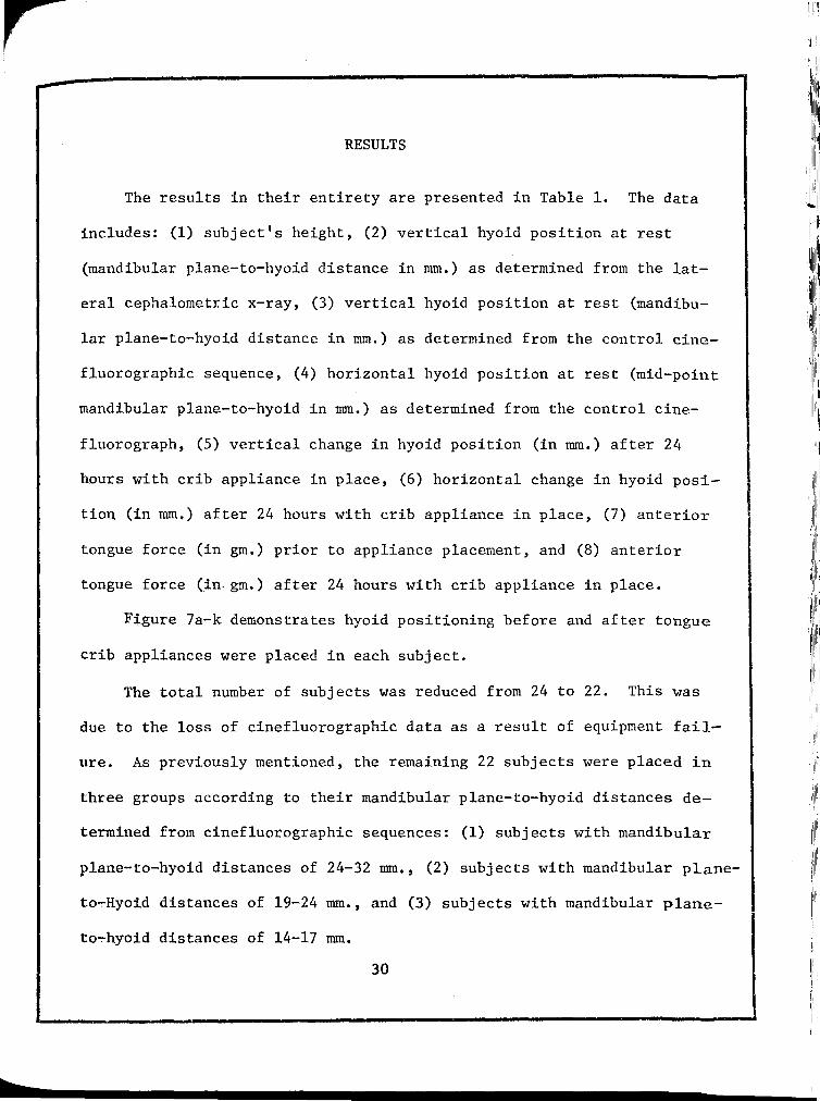
RESULTS
The results in their entirety are presented in Table 1. The data
includes: (1) subject's height, (2) vertical hyoid position at rest
(mandibular plane-to-hyoid distance in rom.) as determined from the lat-
eral cephalometric x-ray, (3) vertical hyoid position at rest (mandibu-
lar plane-to--hyoid distance in rom.) as determined from the control cine-
fluorographic sequence, (4) horizontal hyoid position at rest (mid-point
mandibular plane-to-hyoid in rom.) as determined from the control cine-
fluorograph, (5) vertical change in hyoid position (in rom.) after 24
hours with crib appliance in place, (6) horizontal change in hyoid posi-
tion (in rom.) after 24 hours with crib appliance in place, (7) anterior
tongue force (in gm.) prior to appliance placement, and (8) anterior
tongue force (in. gm.) after 24 hours with crib appliance in place.
Figure 7a-k demonstrates hyoid positioning before and after tongue
crib appliances were placed in each subject.
The total number of subjects was reduced from 24 to 22. This was
due to the loss of cinefluorographic data as a result of equipment fail-
ure. As previously mentioned, the remaining 22 subjects were placed in
three groups according to their mandibular plane-to-hyoid distances de-
termined from cinefluorographic sequences: (1) subjects with mandibular
plane-to-hyoid distances of 24-32 rom., (2) subjects with mandibular plane-
to~Hyoid distances of 19-24 rom., and (3) subjects with mandibular plane-
to~hyoid distances of 14-17 mm.
30

I I I
31
Group 1, with mandibular plane-to-hyoid distances ranging from
24-32 mm., generally did not exhibit hyoid repositioning in the infer-
!or-posterior direction. There was one exception. K.A. showed dramatic
inferior-posterior repositioning.
Group 2, with mandibular plane-to-hyoid distances ranging from
19-24 mm., exhibited diversity in hyoid response. Four subjects (K.H.,
D.P~, D.H., and C.M.) showed hyoid repositioning in the inferior-poster-
ior repositioning.
Group 3, with mandibular plane-to-hyoid distances ranging from
14-17 mm., generally exhibited inferior-posterior repositioning of the
hyoid. Two subjects (S.G. and D.T.) did not follow this pattern.
Table 2 depicts the subjects in Groups 1 and 3. These subjects
exhibited the sample's extremes in terms of hyoid rest position. T
tests were run to determine whether hyoid repositioning in either the
vertical or horizontal dimension was statistically significant. The
difference between the two groups' vertical repositioning was calculated
to be significant at the .05 level, (T value of 2.491). The horizontal
repositioning difference was not found to be significant at the .05 level.
It fell in the probability range of .20>P>.l0, (T value of 1.531).
Myometric results were varied throughout the three groups. Hyoid
repositioning in the inferior-posterior dimension was deemed adaptation
to the posterior tongue displacement. Myometric values in some cases
correlated with such adaptive hyoid repositioning or lack thereof, and
in other cases seemed independent of the hyoid reaction.
I
II I , I,
! I

32
Seven subjects (K.H., D.H., M.S., M.P., M.E., O.M., and S.N.) ex-
hibited what were appraised as being adaptive anterior tongue force
levels coinciding with their adaptive hyoid repositioning. Three more
subjects (J.E., C.B., and D.T.) exhibited increased force values com-
mensurate with their lack of hyoid adaptation.
Conversely, three subjects (D.P., C.M., and D.G.) had adaptive
hyoid changes but adaptation was not shown myometrically. And, six sub-
jects (J.S., H.Z., C.E., A.H., A.C., and S.G.) did not exhibit adaptive
hyoid repositioning, yet myometrically anterior tongue force levels in-
dicated adaptation.
Finally, three subjects (K.A., C.H., and B.M.) showed anterior
tongue force levels that made adaptation judgment in terms of myometrics
difficult.


SUBJECT
Group 1
J.S. K.A. J.E. H. Z. C.B. C,E. C,H.
Group 3
D.G. M.S. M.P. S.G. M.E. O.M. D.T. B.M. S.N.
TABLE II
SUBJECTS EXHIBITING EXTREMES IN MANDIBULAR PLANE-TO-HYOID DISTANCE
Hyoid to GoGn in nun.
32 29 28 26 25 25 24
17 17 17 17 16 15 15 14 14
Hyoid to Mid-Mand Corpus in nnn.
19 4 10 10 34 2 11
10 14 19 25. 13 8 23 12 11
Vert. Change in nun.
+5 -11 +4 -4 +2 +1 +3
-7 -5 -8 +1 -11 -5 -1 +2 -6
Horiz. Change in nun.
+2 -2 +2 +10 +3 -1
0
+1 -2 -3 -1 +1 +4 +1 -6 -4
34

35 Figure 7a
KA
Control Q Experimental ~

j
I
I I I j
I j
36 Figure 7b
HZ
Control 0 Experimental ~

I I
I I I
Figure 7c
Control 0 Experimental ~
37
CB
CE

~~~~~~11! 11
1':"11 ~ ' II
r
Figure 7d
1,11111111
38 1
111''11 1,,11,
I 1
1
111111
llill11.1!
'II~ !1~1 r:
1
11rli
CH
11
1
11111
:1111il
II ~ II
11!111
,rrll11
1
l
/liiiJii
!IIIII
i 1
1:111
!li~ 111)1:111,
1 11~ '11.!1111
1"1111[1
ill~ 1
1!111ilrl1
111
111]11
· Ill I 111 !:
'I :1
KH i!i ! 1',11111
·1111
11'1
·1:1)1.
ll,ill
~ :::
1
11
l,il,'"' 1','1'
:11!1
''I Ill I!
Control 0 '/'I I
I lj
, Ill
Experimental ~ 1:1

' 1 39 I Figure 7e
DP
DH
Control 0 Experimental ~

40 Figure 7f
AH
CM
Control 0 Experimental ~

Figure 7g
AC
DG
Control Q
Experimental ~
41
II, I I
I, II'
II I'
'.1
!

r Figure 7h
Control 0 Experimental ~
42
MS
MP

r j
43 Figure 7i
SG
ME
Control 0 Experimental ~

r Figure 7j
Control 0 Experimental ~
44
OM
DT

r Figure 7k
Control Q
Experimental ~
45
BM
SN

r DISCUSSION
As mentioned, the relative vertical position of the hyoid bone, re-
lated to the mandibular plane, varied considerably from the lateral
cephalometric x-ray to the control cinefluorographic sequence. The
lateral cephalometric x-ray provides a static reading on hyoid position.
This poses a problem. The hyoid is a "floating bone", completely sup-
ported by muscle and ligamentous attachments. Cinefluorographic data
however, is dynamic. It allows the viewer to visualize physiologic
functional patterns. As a result, hyoid rest position can, in the au-
thor's opinion, be more accurately determined from cinefluorographic
sequences. Ingervall (1970) pointed out significant variations in hyoid
position between intercuspal and postural positions of the mandible. He
further found lateral cephalometric x-rays taken with the mandible in
postural position allowed reproducable hyoid positioning. The cephalo-
grams taken in this study for original screening purposes were shot in
the traditional intercuspal position. This could explain the variance
between the cephalometric and cinefluorographic hyoid rest position
noted. For the clinician to utilize lateral cephalometric x-rays to
monitor hyoid rest position, they should be taken with the mandible in
postural position.
For purposes of this study, repositioning of the hyoid bone in an
inferior-posterior direction was deemed adaptation. Two of the three
main sets of extrinsic tongue muscles, the hyoglossi and the genioglossi,
46
I ,li

r have attachments on the hyoid bone. Any repositioning of the hyoid bone
in an inferior-posterior direction accommodates posterior displacement
of the tongue.
47
The lack of adaptive repositioning of the hyoid bone in group 1 was
judged to be a result of the initial hyoid position. These subjects pre
sented with hyoid positions relatively distant from the mandibular plane.
Hyoid repositioning in an inferior-posterior direction could have en
croached on air-way space. Cuozzo (1975) mentioned this possibility in
explaining similar results. Brodie (1961) noted specifically the an
terior suprahyoids (mylohyoids and geniohyoids) as holding the larynx
forward to insure patency of the air-\vay.
One subject in group 1 (K.A.) did show adaptive hyoid repositioning
despite the initial mandibular plane-to-hyoid distance of 29 mm. The
initial horizontal position of the hyoid bone provided a possible ex
planation. The hyoid was positioned near the middle of the corpus. Only
one other subject in this group (C.E.) exhibited similar anterior hyoid
rest position. This forward carriage of the hyoid bone could account for
K.A.'s ability to show hyoid accommodation without embarrassing the air
way. C.E. exhibited myometric adaptation. Possibly the adaptive poten
tial of her intrinsic tongue musculature made hyoid repositioning un
necessary.
Group 2 subjects had mandibular plane-to-hyoid distances ranging
from 19-24 mm. K.H., with a distance of 24 mm., was placed in this group
even though C.H. was placed in group 1. It was determined that while a

r mandibular plane-to-hyoid distance of 24 mm. is relatively long, it was
judged to be in the average or medium range for someone of K.H. 's stat
ure. It should be noted that K.H. showed adaptive hyoid repositioning,
which supports this judgement.
48
The remaining subjects in group 2 showed varied hyoid response. As
alluded to above, to accurately predict hyoid adaptive potential overall
anatomic considerations, i.e. body size and structure, must be evaluated.
These factors become increasingly important when dealing with the middle
range of mandibular plane-to-hyoid distances. In such cases secondary
factors (if in fact they are only secondary) such as size of subject,
hyoid position in the horizontal dimension, and intrinsic tongue muscula
ture adaptation become increasingly important determinants in adaption
patterns.
Group 3, with hyoids positioned relatively near the mandibular
plane, generally showed adaptive hyoid repositioning in the inferior
posterior direction. It can be theorized that this hyoid stature allows
adaptation because the air-way space is not encroached upon by such ac
commodation. It should be noted that the two exceptions in this group
(S.G. and D.T.) exhibited hyoid rest positions quite posterior relative
to the mandibular corpus. It is hypothesized that despite the relatively
high carriage of the hyoid, adaptation in an inferior-posterior direction
was limited due to the relative posterior position of the bone. From
this posterior position it is theorized that adaptive positional changes
might embarrass the air-way. The hyoid behavior of K.A. in group 1 and

r S.G. and D.T. mentioned here suggests the horizontal hyoid position may
be a significant determinant of hyoid adaptive potential.
49
In interpreting the statistical results, it should be stressed that
groups 1 and 3 were comprised of the sample extremes in terms of mandib
ular plane-to-hyoid distance. The groups were compared to determine if
the vertical hyoid position had any statistical effect on the hyoid bone's
adaptive potential in the inferior-posterior direction. The T test run
on the changes in vertical dimension revealed that the two groups indeed
differed in their ability to move the hyoid inferiorly. This offers
statistical support for the theory first proposed by Cuozzo (1975). It
appears hyoid adaptive potential is influenced by its spatial relation
ship to· the mandibular plane.
The horizontal adaptive changes were not significantly different
(.20>P>.lO), when the two groups were compared. When interpreting this
it must be kept in mind, however, that the subjects studied were grouped
on the basis of vertical hyoid position. Thus, only initial vertical
position's effect on horizontal repositioning was tested. This is not
to say that horizontal position is not a factor in hyoid adaptation.
It has been mentioned that myometric results were varied when re
lated to adaptive changes in hyoid positioning. It must, however, be
remembered that hyoid repositioning is only one way in which the tongue
might adapt to forced posterior displacement. The intrinsic tongue mus
culature plays a role. It was not monitered in this study. Nevertheless,
it is reasonable to assume that the intrinsic tongue musculature's own

adaptive potential accounts for some of the apparent discrepancy between
myometric and cinefluorographic results.
Also, there are inherrent difficulties in any myometric recording.
50
These must be realized. First, there is always the possibility of alter
ing physiologic activity with the instrumentation used. Proffit (1975)
refers to this as "physiologic reactance". Secondly, it has been well
documented that there is a great deal of variability in the "normal"
swallowing pattern. The accuracy of myometric recordings as performed
in this study depended on the tongue tip placement against the recording
plate. There is the possibility that because of normal variation in
tongue position some subjects' anterior tongue force was only partially
captured at one or both recording sessions.

SUMMARY AND CONCLUSIONS
The purpose of this study was to determine whether the position of
the human hyoid bone, in its spatial relationship to the mandibular
plane, limits of influences the ability of the tongue musculature to
adapt to forced posterior displacement.
Twenty-two adult female subjects were included in the study. My
ometric recordings and cinefluorographic sequences of deglutition were
taken. Tongue crib appliances were placed in each subject. After 24
hours wear, and prior to removal of the appliances, the myometric re
cordings and cinefluorographic sequences were repeated.
Conclusions:
(1) The mandibular plane-to-hyoid distance does seem to
influence the ability of the tongue neuromusculature
to adapt to forced posterior displacement.
(2) Hyoid bones positioned relatively distant from the
mandibular plane seemed unable to reposition in an
inferior··posterior direction in response to forced
posterior tongue displacement.
(3) Hyoid bones positioned relatively near the mandibular
plane seemed to possess the potential for inferior
posterior adaptive repositioning in response to forced
posterior tongue displacement.
(4) The horizontal position of the hyoid bone seemed also
to influence its physiologic adaptive capacity.
51

(5) Lateral cephalometric x-rays taken in the traditional
intercuspal position were found to be unreliable
monitors of hyoid rest position.
(6) From the lack of correlation between myometric and
cinefluorographic data, it was hypothesized that the
intrinsic tongue musculature plays a significant role
in adaptive behavior of the tongue.
52

BIBLIOGRAPHY
Ardran, G. M., and Kemp, F. H., "Radiographic Study of Movements of the Tongue in Swallowing", Brit. Soc. Study of Orth., Trans., pp. 117-126, disc. 127-128, 1954.
Bench, R. W., "Growth of the Cervical Vertebrae as Related to the Tongue, Face, and Denture Behavior", Am. J. Orth., 49: 183-214, 1963.
Brader, A. C., "Dental Arch Form Related with Intraoral Forces: PR=C", Am. J. Orth., 61:541-561, June, 1972.
Brauer, James S., and Holt, Townsend V., "Tongue Thrust Classification", Angle Orth., 35: 106-112, Apr., 1965.
Brodie, A. G. Congenital Anomalies of the Face and Associated Structures, (ed., S. Pruzansky), Springfield, Illinois, Chas. C. Thomas, 1961.
Cleall, J. F. , J. Ortho.,
"Deglutition: A Study of Form and Function", 51: 566-594, Aug., 1965.
Am.
C1eall, J. F., Alexander, W. J., and Mcintyre, M. D., "Head Posture and Its Relationship to Deglutition", Angle Orth., 36: 335-350, Oct., 1966.
Cuozzo, G. S., and Bowman, D. C., "Hyoid Positioning During Deglutition Following Forced Positioning of the Tongue", Am. J. Orth., 68: 564-570, Nov., 1975.
Durzo, C. A., and Brodie, A. G., "Growth and Behavior of the Hyoid Bone", Angle Orth., 32: 193-204, July, 1962.
Fishman, Leonard S., "Postural and Dimensional Changes in the Tongue from Rest Position to Occlusion'', Angle Orth., 39: 109-113, Apri., 1969.
Grant, L.A., "A Radiographic Study of the Hyoid Bone Position in Angles Class I, II, And III Malocclusions", Unpublished Masters Thesis, University of Kansas City, 1959.
Gray, H., Anatomy of the Human Body, ed. 26, Philadelphia, Lea and Febiger, 1956.
53

Gobeille, D. M., "Adaptability of Muscle and Hence Hyoid Position Following Forced Distal Repositioning of the Tongue in Open Bite Patients", Unpublished Masters Thesis, Loyola University, 1974.
Hanson, Marv:f.n L. , Barnard, Logan W. , and Case, James L. , "Tonguethrust in Preschool Children Part II: Dental Occlusal Patterns", Am. J. Orth., 57: 15-22, Jan. 1970.
Hanson, Marvin L., and Cohen, Melvin S., "Effects of Form and Function on Swallowing and the Developing Dentition", Am. J. Orth., 64: 63-82, July, 1973.
Harvold, Egil P., "The Role of Function in the Etiology and Treatment of Malocclusion", Am. J. Orth., 54: 883-898, Dec., 1968.
Hedges, Robert B., McLean, C. Donald, and Thompson, Frederic A., "A Cinefluorographic Study of Tongue Patterns in Function", Angle Orth., 35: 253-268, Oct., 1965.
Ingervall, B., Carlsson, G. E., and Helkima, M., "Change in Location of Hyoid Bone with Mandibular Positions", Acta Odont. Scand., 28: 337-361, 1970.
Ingervall, B., "Positional Changes of the Mandible and Hyoid Bone Relative to Facial and Dental Arch Morphology", Acta Odont. Scand., 28: 867-894, 1970.
Ingervall, B., Bratt, C. M., Carlsson, G. E., Helkima, M., and Lantz, B., "Positions and Movements of Mandible and Hyoid Bone During Swallmving", Acta Odont. Scand. , 29: 549-562, 1971.
Kydd, H. L., "Quantitative Analysis of Forces of the Tongue", J. Dent. Res., 35: 171-174, Apr., 1956.
Kydd, lv. L. , and Neff, C. Wayne, "Frequency of Deglutition of Tongue-thrusters Compared to a Sample Populatio" of Normal Swallowers", J. Dent. Res., 43: 363-369, 1964.
Lear, C. S., and Moorrees, C. F., "Measurement of Orofacial Muscle Forces", J. Dent. Res., 43: 906, 1964.
Lear, S. C., and Moorrees, C. F., "Buccolingual Muscle Force and Dental Arch Form", Am. J. Orth., 56: 379-393, Oct., 1969.
54

Magendie, F., A Summary of Physiology, Baltimore, Straub Publ., 1822.
Mainland, D., Anatomy as a Basis for Medical and Dental Practice, New York, Paul Hoeber and Co., 1945.
McNulty, E. C., Lear, C. S., and Moorrees, C. F., "Measurements of Labiolongual Forces on Central Incisors in Normal and Protrusive Positions", Am. J. Orth., 53: 137, Feb., 1967.
Milne, I. M., and Cleall, J. F., "Cinefluorographic Study of Functional Adaptation of the Oropharyngeal Structures", Angle Orth., 40: 267-283, Oct., 1970.
Orban, B. J., Orban's Oral Histology and Embryology, ed. 5, St. Louis, The C.V. Mosby, Co., 1962.
Peat, John H., "A Cephalometric Study of Tongue Position", Am. J. Orth., 54: 339-351, May, 1968.
Posen, A. L., "The Influence of Maximum Perioral and Tongue Force on the Incisor Teeth", Angle Orth., 42: 285-310, Oct., 1972.
Proffit, W. R., Chastain, B. B., and Norton, L.A., "Linguopalatal Pressure in Children", Am. J. Orth., 55: 154-166, Feb., 1969.
Proffit, W. R., and McGlone, R. E., "Correlation Between Functional Lingual Pressure and Oral Cavity Size", Cleft Palate Journal, 9: 229-235, 1972.
Proffit, W. R., "Muscle Pressures and Tooth Position: North American vlhites and Australian Aborigines", Angle Orth., 45: 1-12, Jan., 1975.
Ramsey, G. H., Watson, J. S., Gramiak, R., and Weinberg, S. A., "A Cinefluorographic Analysis of the Mechanism of Swallowing", Radiology, 64: 498-518, 1955.
Rushmer, R. F., and Hendron, J. A., "The Act of Deglutition: A Cinefluorographic Study", J. Appl. Physiol., 3: 622-630, Apr., 1951.
Saunders, J. B., Davis, C., and Miller, E. R., "The Mechanism of Deglutition as Revealed by Cineradiography", Annals of Otology, Rhinology, and Laryngology, 60: 897-916, Dec., 1951.
55

Shelton, R. L., Bosma, J. F., and Sheets, B. V., "Tongue, Hyoid and Larynx Displacement in Swallow and Phonation", J. Applied Physio., 15: 283-288, 1960.
Sieber, H., and DuBrul, E. L., Oral Anatomy, ed. 5, St. Louis, The C.V. Mosby Co., 1970.
Sloan, R. F., Ricketts, R. M., Bench, R. W., Hahn, E., Westover, J., and Brummett, S., "The Application of Cephalometries to Cinefluorography", Angle Orth., 34: 132-141, Apr., 1964.
Sloan, R. F., Bench, R. W., Hulick, J. F., Ricketts, R. M., Brummett, S. W., and Hestover, J. L., "The Application of Cephalometries to Cinefluorography: Comparative Analysis of Hyoid Movement Patterns During Deglutition in Class I and Class II Orthodontic Patients", Angle Orth., 37: 26-33, Jan., 1967.
Smith, J. A., "A Cephalometric Radiographic Study of the Hyoid Bone in Relation to the Mandible in Certain Functional Positions", Unpublished Masters Thesis, Northwestern University, 1956.
Stepovich, M. L., "A Cephalometric Positional Study of the Hyoid Bone", Am. J. Orth., 51: 882-900, Dec., 1965.
Straub, R., "Malfunction of Tongue: Part I. The Abnormal Swallowing Habit; Its Cause, Effects and Results in Relation to Orthodontic Treatment and Speech Therapy", Am. J. Orth., 46: 404-424, June, 1960.
Subtelny, J.D., "Malocclusions, Orofacial Huscle Adaptation", July, 1970.
Orthodontic Corrections and Angle Orth., 40: 170-201,
Subtelny, J. D., and Subtelny, J.D., "Oral Habits- Studies in Form, Function, and Therapy", Angle Orth., 43: 347-383, Oct., 1973.
Thompson, J. R., Mandible",
"A Cephalometric Study of the Movements of the J.A.D.A., 28: 750-761, 1941.
Tomes, C. S., "The Bearing of the Development of the Jaws on Irregularities", Dental Cosmos., 15: 292-296, 1873.
,/Wickwire, N. A., White, R. P., and Proffit, W. R., "The Effect of Mandibular Osteotomy on Tongue Position", J. Oral Surg., 30: 184-190, 1970.
56

Wildman, A. J., Fletcher, S. G., and Cox, Barbara, "Patterns of Deglutition", Angle Orth., 34: 271-291, Oct., 1964.
Winders, R. V., "A Study in the Development of an Electronic Technique to Measure Force Exerted on the Dentition by the Perioral and Lingual Musculature", Am. J. Orth., 26: 645-657, Sept., 1956.
Winders, R. V., "Forces Exerted on the Dentition by the Perioral and Lingual Musculature During Swallowing", Angle Orth., 28: 226-235, Apr., 1958.
Winders, R. V., "Recent Findings in Myometric Research", Angle Orth., 32: 38-43, Jan., 1962.
Wood, B., "An Electromyographic and Cephalometric Radiographic Investigation of the Positional Changes of the Hyoid Bone in Relation to Head Posture", Unpublished Master's Thesis, Northwestern University, 1956.
Yip, A. S., and Cleall, J. F., "Cinefluorographic Study of Velarpharyngeal Function Before and After Removal of Tonsils and Adenoids", Angle Orth., 41: 251-263, Oct., 1971.
57

APPROVAL SHEET
The thesis submitted by Kent B. Augustson has been read and approved by the following committee:
Dr. Douglas C. Bowman, Director Associate Professor, Physiology and Pharmacology, Loyola
Dr. Bernard M. Pawlowski Clinical Associate Professor, Orthodontics, Loyola
Dr. Joseph M. Gowgiel Associate Professor and Chairman of Anatomy Loyola
The final copies have been examined by the director of the thesis and the signature which appears below verifies the fact that any necessary changes have been incorporated and that the thesis is now given final approval by the Committee with reference to content and form.
The thesis is therefore accepted in partial fulfillment of the requirements for the degree of Master of Science.
L~kc.o~ Direc~'s Signature Date


![Figure 7 - Sinoe Medical Associationsinoemedicalassociation.org/AP/theskullpresentation.pdf · the skull • = 22 bones [actually 29] • all fused except one.:hyoid bone • joints](https://static.fdocuments.in/doc/165x107/5afd0b577f8b9a994d8ceec6/figure-7-sinoe-medical-associationsin-skull-22-bones-actually-29-all.jpg)