The importance of the postmortem interval for the ...
8
Autopsy and Case Reports. ISSN 2236-1960. Copyright © 2019. This is an Open Access article distributed under the terms of the Creative Commons Attribution Non-Commercial License, which permits unrestricted non-commercial use, distribution, and reproduction in any medium provided the article is properly cited. a Università degli Studi di Milano, Facoltà di Medicina e Chirurgia, Sezione di Medicina Legale e delle Assicurazioni, Dipartimento di Scienze Biomediche per la Salute. Milano, Italy. b Università degli Studi di Bologna, Facoltà di Medicina e Chirurgia, Sezione di Medicina Legale, Dipartimento di Scienze mediche e Chirurgiche (DIMEC). Bologna, Italy. c Ospedale S. Carlo Borromeo, Servizio di Immunoematologia e Medicina Trasfusionale (SIMT). Milano, Italy. d Ospedale di Bergamo, Azienda Socio Sanitaria Papa Giovanni XXIII, Responsabile USS Medicina Legale. Bergamo, Italy. The importance of the postmortem interval for the diagnosis of Waterhouse–Friderichsen syndrome by Neisseria meningitidis in a series of forensic cases Guendalina Gentile a , Alberto Amadasi b , Paolo Bailo a , Michele Boracchi a , Francesca Maciocco c , Matteo Marchesi d , Riccardo Zoja a How to cite: Gentile G, Amadasi A, Bailo P, et al. The importance of the postmortem interval for the diagnosis of Waterhouse–Friderichsen syndrome by Neisseria meningitidis in a series of forensic cases. Autops Case Rep [Internet]. 2019 Jul-Sep;9(3):e2019103. https://doi.org/10.4322/acr.2019.103 Article / Autopsy Case Report ABSTRACT The effective value of microbiological post-mortem examinations stands as fundamental in forensic cases involving microbiology. We ran these analyses on five victims, who suddenly died after showing persistent fever. The examinations were conducted between 48 hours and 10 days after death, and adrenal gland apoplexy was detected in all the cases. Microbiological examinations identified Neisseria meningitidis, which was accountable for Waterhouse–Friderichsen syndrome. Diplococci were isolated from three cadavers that underwent forensic dissection between 2 and 3 days after death. The remaining two cadavers showed polymicrobial contamination, and a polymerase chain reaction technique was necessary to identify the pathogen. We assumed that the microbial overlap could lead to diagnostic mistakes and conceal the identification of the lethal pathogen. Therefore, we suggest using molecular techniques for a postmortem interval (PMI) longer than 72 hours. Classical microbiological examination should be performed for PMI within 72 hours. Keywords Autopsy; Forensic Microbiology; Waterhouse-Friderichsen Syndrome; Post-Mortem Interval. INTRODUCTION Waterhouse–Friderichsen syndrome (WFS) is a catastrophic sepsis 1 with a higher incidence in children, 2-4 and is usually associated with adrenal gland hemorrhage, which is considered-along with the disseminated intravascular coagulation (DIC)-as the most fearsome complication. Due to the fact that symptoms may be non-specific, the in-vivo diagnosis of adrenal gland hemorrhage is difficult to define 5 and is usually identified at autopsy. 1 Despite their protected position in the retroperitoneal space (an area rich in adipose-connective tissue), 6 at autopsy examination hemorrhage of the adrenal glands can be found in several conditions as traumatism, 7 or rare cases of infection-related 8 sudden death, 9 such as WFS. People suffering from WFS may initially show unspecific symptoms, such as fever—with a temperature always
Transcript of The importance of the postmortem interval for the ...
Autopsy and Case Reports. ISSN 2236-1960. Copyright © 2019. This is an Open Access article distributed under the terms of the Creative Commons Attribution Non-Commercial License, which permits unrestricted non-commercial use, distribution, and reproduction in any medium provided the article is properly cited.
a Università degli Studi di Milano, Facoltà di Medicina e Chirurgia, Sezione di Medicina Legale e delle Assicurazioni, Dipartimento di Scienze Biomediche per la Salute. Milano, Italy.
b Università degli Studi di Bologna, Facoltà di Medicina e Chirurgia, Sezione di Medicina Legale, Dipartimento di Scienze mediche e Chirurgiche (DIMEC). Bologna, Italy.
c Ospedale S. Carlo Borromeo, Servizio di Immunoematologia e Medicina Trasfusionale (SIMT). Milano, Italy.d Ospedale di Bergamo, Azienda Socio Sanitaria Papa Giovanni XXIII, Responsabile USS Medicina Legale. Bergamo, Italy.
The importance of the postmortem interval for the diagnosis of Waterhouse–Friderichsen syndrome by Neisseria meningitidis in a series of forensic cases
Guendalina Gentilea , Alberto Amadasib , Paolo Bailoa , Michele Boracchia , Francesca Macioccoc, Matteo Marchesid, Riccardo Zojaa
How to cite: Gentile G, Amadasi A, Bailo P, et al. The importance of the postmortem interval for the diagnosis of Waterhouse–Friderichsen syndrome by Neisseria meningitidis in a series of forensic cases. Autops Case Rep [Internet]. 2019 Jul-Sep;9(3):e2019103. https://doi.org/10.4322/acr.2019.103
Article / Autopsy Case Report
ABSTRACT
The effective value of microbiological post-mortem examinations stands as fundamental in forensic cases involving microbiology. We ran these analyses on five victims, who suddenly died after showing persistent fever. The examinations were conducted between 48 hours and 10 days after death, and adrenal gland apoplexy was detected in all the cases. Microbiological examinations identified Neisseria meningitidis, which was accountable for Waterhouse–Friderichsen syndrome. Diplococci were isolated from three cadavers that underwent forensic dissection between 2 and 3 days after death. The remaining two cadavers showed polymicrobial contamination, and a polymerase chain reaction technique was necessary to identify the pathogen. We assumed that the microbial overlap could lead to diagnostic mistakes and conceal the identification of the lethal pathogen. Therefore, we suggest using molecular techniques for a postmortem interval (PMI) longer than 72 hours. Classical microbiological examination should be performed for PMI within 72 hours.
Keywords Autopsy; Forensic Microbiology; Waterhouse-Friderichsen Syndrome; Post-Mortem Interval.
INTRODUCTION
Waterhouse–Friderichsen syndrome (WFS) is a catastrophic sepsis1 with a higher incidence in children,2-4 and is usually associated with adrenal gland hemorrhage, which is considered-along with the disseminated intravascular coagulation (DIC)-as the most fearsome complication. Due to the fact that symptoms may be non-specific, the in-vivo diagnosis of adrenal gland hemorrhage is difficult to define5 and is
usually identified at autopsy.1 Despite their protected position in the retroperitoneal space (an area rich in adipose-connective tissue),6 at autopsy examination hemorrhage of the adrenal glands can be found in several conditions as traumatism,7 or rare cases of infection-related8 sudden death,9 such as WFS. People suffering from WFS may initially show unspecific symptoms, such as fever—with a temperature always
2-8 Autops Case Rep (São Paulo). 2019 Jul-Sep;9(3):e2019103
The importance of the postmortem interval for the diagnosis of Waterhouse–Friderichsen syndrome by Neisseria meningitidis in a series of forensic cases
higher than 38°C—then, suddenly, the general conditions worsen and progressive hypotension occurs along with shock and disseminated intravascular coagulation. In 50%-60% of cases2 cutaneous purpura may be observed. The clinical development of the disease is variable; it may be: (i) mild and undergo spontaneous resolution; (ii) sub-acute with an insidious and slow development; or (iii) fulminant and lead to death in a short period of time. The latter event usually occurs after the setting of the first symptoms, and these episodes—with or without involvement of the meninges—are complicated by the septic process, the disseminated intravascular disease, and massive hemorrhage of the adrenal glands (distinct WFS). There are three fundamental elements necessary for the identification of this lethal form:10
1. Etiological factors (meningococcal hematogenous dissemination or fulminant pneumococcal hematogenous dissemination)
2. Clinical presentation (severe and rapid progression of the infectious setting)
3. Peculiar anatomical lesions (cerebral-spinal meningitis signs with compromised neural-axons and bilateral adrenal gland hemorrhage (cases of unilateral adrenal gland hemorrhage have been described in literature).11
The mortality rates of the meningococcal/pneumococcal disease, not associated with WFS, are in the range of 10%-30%,3 sensibly increasing to 95% in cases where WFS is associated. The aim of this study is to (i) verify the sensitivity of post-mortem microbial examination on victims deceased due to certified infective pathologies (WFS) who undergo forensic dissection at different periods of time; and (ii) try to delineate the time interval in which it is still possible to microbiologically detect Neisseria meningitidis.
CASE REPORTS
Case 1
A 2-month-old male baby was carried to Accident & Emergency (A&E) with persistent fever. After clinical examination, the patient was prescribed with antipyretics (paracetamol), but the same day he developed spreading petechiae. The medical
staff requested cerebrospinal fluid bacterial exams. Unfortunately, the little patient died before the results of the exams became available. An autopsy was performed 10 days after death. The cadaver had several cutaneous petechiae and adrenal glands hemorrhage. The microbiological post-mortem exams showed an autolytic multi-bacterial proliferation on leptomeninges and cardiac blood. A molecular investigation (polymerase chain reaction [PCR]) on the same tissue and blood resulted positive for N. meningitidis. Histological examinations were not allowed by the Judicial Authority.
Case 2
A 5-month-old male baby was carried to A&E with fever. The medical staff prescribed an antipyretic (paracetamol). The following day, the baby became cyanotic and developed a cutaneous exanthema. The baby died while clinical exams were still being carried out. The autopsy was performed 3 days after death. The cadaver had several cutaneous petechiae, congestion of the leptomeninges with opaque coloration, deposits of fibrin, purulent exudate of yellow-greenish color on the convexities of the hemispheres, and bilateral apoplexy of the adrenal glands. Post-mortem microbial exams revealed the presence of N. meningitidis on cerebral swabbing. Histological examinations of brain, cerebellum, and pons showed a pronounced edema of the leptomeninges, inflammatory granulocyte infiltration, few lymphocytes, and marked dilatation and hematic stasis of the leptomeningeal vascular system. Histological examinations of the adrenal glands showed acute hemorrhagic infiltration of both glands.
Case 3
A 3-year-old girl was carried to A&E presenting petechiae, vomiting, and fever. The physicians suspected meningitis and administered an antibiotic (ceftriaxone), but she suddenly died. The autopsy examination was performed 4 days later. The cadaver had widespread cutaneous petechiae, necrotic hemorrhage of adrenal glands, and purulent exudate in the frontal-parietal lobes and cerebellum. Postmortem microbiological exams revealed Gram-negative bacterial cells on sampled purulent areas. The molecular investigations (PCR) were positive for N. meningitidis. Histological examinations showed dilated and marked leukostasis

Gentile G, Amadasi A, Bailo P, et al.
3-8Autops Case Rep (São Paulo). 2019 Jul-Sep;9(3):e2019103
(formed by neutrophils) in the leptomeninges, widening of perivascular and pericellular spaces (due to severe cellular edema), adrenal glands apoplexy, septal microbial micro-abscesses, and marked edematous dissociation of cardiac fibers and pericardial connective tissue.
Case 4
A 17-year-old boy was taken to A&E with fever (39.5 °C) and sore throat. The physicians suspected meningitis and administered an antibiotic (ceftriaxone). The boy died after 1 day. The forensic examination was performed 3 days after death. The corpse had opaque leptomeninges at cerebral convexities. The adrenal glands showed areas with necrotic-hemorrhagic aspect. Postmortem microbial investigations revealed the presence of N. meningitidis on cardiac blood and brain samples. Histological examinations revealed early signs of meningitis on the leptomeninges, and hemorrhagic infiltration of both adrenal glands.
Case 5
A 23-year-old female presented with fever, tremor, pain, and developed paresthesia to the limbs 24 hours after the onset of the first symptoms. She was taken to one of the main hospitals of the city of Milan where she died minutes after admission. The autopsy examination was performed 5 days after death.
The corpse showed diffuse cutaneous petechiae, which was also detected at dissection on the temporal muscles, brain, esophagus, pleurae, pericardium, stomach, intestines, and liver. The adrenal glands showed bilateral hemorrhagic infiltration. Postmortem microbiological cultures of viscera and fluids were negative. The PCR was positive for N. meningitidis. The histological examinations showed a multi-organ hemorrhagic petechial syndrome, a perivascular sleeve of neutrophils in an intra-parenchymal brain vessel with an initial infiltration of the nearby parenchyma (mild stage meningitis), bilateral adrenal gland hemorrhage, and acute multi-focal bacterial myocarditis.
In all cases, the cause of death was identified in WFS caused by N. meningitidis. (Table 1).
Autopsy presentation
Diagnosis of WFS can be arduous: cadavers coming to the attention of the forensic practitioner with early symptoms and with a lack of cutaneous macroscopic signs, can pose difficulties in terms of differential diagnosis between WFS and a milder viral infection. On the other hand, in cadavers showing an intra-vitam history of high fever, spreading cutaneous exanthema, and progressive shock, WFS must always be suspected.2 In these cases, autopsy examinations request the adoption of adequate measures of protection. In a paper edited by the Centers for Disease
Table 1. Demographic data, autopsy and microbiological results of the 5 cases of Waterhouse-Friderichsen Syndrome
Case Sex AgeAutopsy sampling
Skin BrainAdrenal apoplexy
Other organs’ signs
AgentCause of death
1 M 2 m Meninges, cardiac blood WP yes N. meningitidis
(with PCR) WFS
2 M 5 m cerebral swabs WP
opaque meninges, congestion,
purulent depositsyes N. meningitidis WFS
3 F 3 y purulent material WP
opaque meninges,
purulent depositsyes
endocardial petechiae, bacterial involvement of the
myocardium
N. meningitidis (with PCR) WFS
4 M 17 y brain and cardiac blood opaque meninges yes N. meningitidis WFS
5 F 23 y fluids and organs WP
petechiae in the brain, cerebellum
and temporal muscles
yes
Visceral petechiae, bacterial
involvement of the myocardium, renal
micro - thrombi
N. meningitidis (with PCR) WFS
F = feminine; M = masculine; m = month; PCR = polymerase chain reaction; y = year; WFS = Waterhouse Friderichsen Syndrome; WP = Widespread Petechiae.

4-8 Autops Case Rep (São Paulo). 2019 Jul-Sep;9(3):e2019103
The importance of the postmortem interval for the diagnosis of Waterhouse–Friderichsen syndrome by Neisseria meningitidis in a series of forensic cases
Control and Prevention (CDC),12 since the pathology is contagious and airborne, the preventive measures recommended for the microbiological safety of the laboratory personnel potentially exposed to aerosolized N. meningitidis should be vaccines; however, nothing is stated about preventive measures for mortuary staff.
When WFS is full-blown, it can be detected macroscopically at the external examination thanks to characteristic signs, such as cutaneous petechiae (Figure 1A), and at internal dissection due to purulent meningeal and cerebral matter and adrenal gland hemorrhage —small bleedings usually located at the cortical-medullary junction that can be detected with a careful histological examination.6 The sampling of these cadavers requests a befitting selection of the biological fluids that must be collected with sterile syringes and stored in sealed vials: cardiac blood9,13 and cerebrospinal fluid3,14 are the most fitting samples; moreover, the examiner can proceed with the sampling of cadaveric parenchyma with sterile lancet and clamps, and storage in sealed vials. It is also advisable to sample the spleen,9,14 heart, and liver.15 In addition, an extensive cerebral sampling is recommended, including: leptomeninges,3 cerebral hemispheres’ base and convexity (frontal, temporal, parietal, and occipital lobes), cerebellum (hemispheres and vermis), brainstem (midbrain, pons, and medulla oblongata), and spinal cord (cervical, thoracic, abdominal, and sacrum-coccygeal). The CDC guidelines12 recommend to start a post-exposure antimicrobial chemoprophylaxis within 24 hours.
This study was conducted at the Legal Medicine Section of the University of Milan. We selected five
patients (three males and two females aged between 2 months and 23 years) who suddenly died after showing symptoms of persistent fever. The autopsy was performed on all five patients between 48 hours and 10 days after death. Three cases (2, 3, 4) had adrenal apoplexy (Figure 1B) and meningitis signs (Figure 1C). Two cases (1, 5) did not show signs of meningitis.
Laboratory Procedures
During autopsy, a certain amount of biological fluids was destined for microbiological examination with a thin smear on at least two microscope slides. One of these slides was dyed with Gram cytological staining in order to highlight the presence of possible bacterial cells while the other slide was dyed with cytological hematoxylin and eosin (H&E) in order to evaluate the possible presence of inflammatory infiltrate. The remaining samples were addressed to standard cultural exams. If any cultures were negative or characterized by postmortem autolytic growth, it was considered necessary to continue with higher level procedures, such as molecular tests.
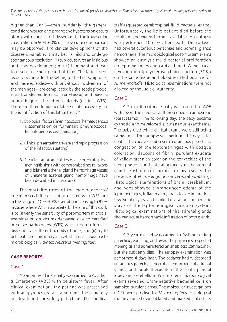
In order to proceed with standard histopathological analysis, slides had to be stained with H&E, histologic Gram staining, and phosphotungstic acid hematoxylin. After the staining process, fibrin clots were detected (Figure 2A) along with neutrophil infiltration in cerebral parenchyma (Figure 2B), cortical hemorrhage of the adrenal glands (Figure 2C), and multifocal acute bacterial myocarditis with several neutrophil infiltrations (Figure 2D).
Figure 1. (Case 3) – A – Gross examination of the corpse’s body showing diffuse cutaneous petechiae; B and C – opacification of the meninges with deposition of purulent material over the frontal and parietal lobes, vermis and cerebellar lobes.
Gentile G, Amadasi A, Bailo P, et al.
5-8Autops Case Rep (São Paulo). 2019 Jul-Sep;9(3):e2019103
A recent acquisition in Forensic Sciences is the assessing of molecular and genetic amplification. This procedure, using a specific gene primer, enables the detection of CrgA—an adhesion regulatory protein responsible for the adhesion of N. meningitidis to its target cells,6—in cerebrospinal fluid, cerebral parenchyma with leptomeninges (especially the arachnoid mater), spleen tissue,14,15 and cutaneous purpura lesions.16 International studies1 use the serum concentration of procalcitonin as a postmortem biochemical marker of sepsis. Normal blood levels of 0.5 ng/mL, or lower, can rise to 10 ng/mL in patients deceased due to sepsis; recently, vitreous humor has also been considered as a useful substrate to isolate
N. meningitidis, since this humor is resistant to blood biochemical postmortem changes.17
Etiology
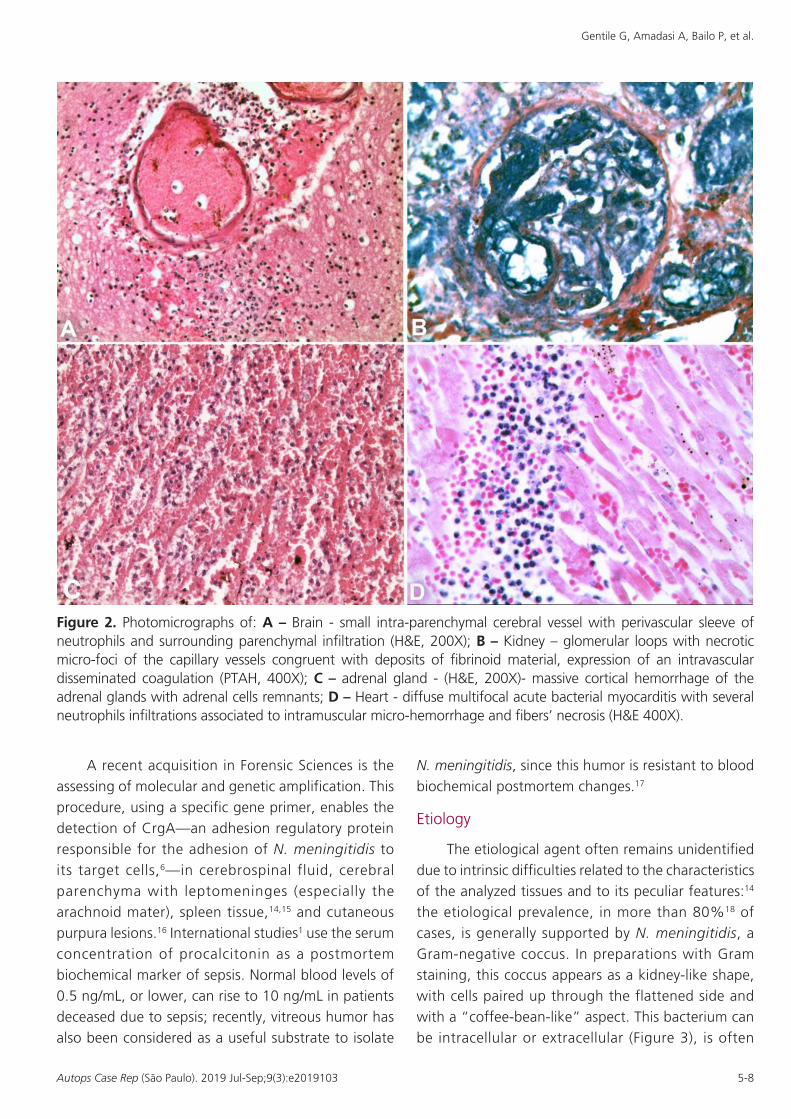
The etiological agent often remains unidentified due to intrinsic difficulties related to the characteristics of the analyzed tissues and to its peculiar features:14 the etiological prevalence, in more than 80%18 of cases, is generally supported by N. meningitidis, a Gram-negative coccus. In preparations with Gram staining, this coccus appears as a kidney-like shape, with cells paired up through the flattened side and with a “coffee-bean-like” aspect. This bacterium can be intracellular or extracellular (Figure 3), is often
Figure 2. Photomicrographs of: A – Brain - small intra-parenchymal cerebral vessel with perivascular sleeve of neutrophils and surrounding parenchymal infiltration (H&E, 200X); B – Kidney – glomerular loops with necrotic micro-foci of the capillary vessels congruent with deposits of fibrinoid material, expression of an intravascular disseminated coagulation (PTAH, 400X); C – adrenal gland - (H&E, 200X)- massive cortical hemorrhage of the adrenal glands with adrenal cells remnants; D – Heart - diffuse multifocal acute bacterial myocarditis with several neutrophils infiltrations associated to intramuscular micro-hemorrhage and fibers’ necrosis (H&E 400X).
6-8 Autops Case Rep (São Paulo). 2019 Jul-Sep;9(3):e2019103
The importance of the postmortem interval for the diagnosis of Waterhouse–Friderichsen syndrome by Neisseria meningitidis in a series of forensic cases
arranged in couples (diplococcus), and is found in the nasopharynx of 3%-30% of healthy individuals,15 who can be considered as asymptomatic carriers but can develop meningitis during or after flu infections.19 Other virulent organisms like Klebsiella,20 Ewingella,9 Haemophilus influenzae,21 Streptococcus pyogenes,22 Streptococcus pneumonia,7,22 and Pasteurella23 occasionally can be detected as etiological agents of WFS and isolated in cadaveric samples. The etiological agent was not immediately identified with standard microbiological techniques in three cases (1, 3, 5) with a postmortem interval (PMI) longer than 72 hours; instead, the bacterium was identified with real-time PCR. In the remaining cases (2, 4) with a PMI within 72 hours, N. meningitidis was identified with routine microbiological cultures.
DISCUSSION
We wanted to verify the sensitivity of the postmortem microbial testing on a group of five patients who died due to WFS and underwent forensic dissections at different times—PMI between 2 and 10 days—to try to evaluate the interval time in which it was still possible to microbiologically detect
the bacterial agent. We can draw the following conclusions:
1. The PMI should be less than 72 hours in order to perform a reliable microbiological examination on cadaveric samples: in the cases with PMI greater than 72 hours (1, 3, 5) it was not possible to isolate and detect the lethal microbial agent; therefore, real-time PCR was necessary. The other two cadavers (2, 4) were stored for less than 72 hours in refrigerating rooms. We were able to investigate both with cultures of cadaveric matter and with cerebral sampling stained with cytological Gram. These two methods were simpler than the molecular ones. We concluded that there is a multi-microbial overlap due to a PMI longer than 72 hours. This can lead to diagnostic mistakes and conceal the identity of the pathogen involved in the mechanisms of death.24 Therefore, it is essential to use a molecular bacterial method in all cases of suspected WFS with a wide postmortem interval.
2. Leptomeninges are targeted by meningococci in WFS cases even if they are not macroscopically involved. In fact, the leptomeninges were clean at the macroscopic examination of cases 4 and 5, but the histological examination revealed a variable stage—mild to moderate,3—of meningitis.
3. The septic invasion from meningococci, as in cases 3 and 5, must be validated by histological exam of samples taken from different organs, especially myocardium. Bacterial myocarditis is commonly observed because of the septic processes. Therefore, the actual cause of death may be due to shock, DIC, or myocarditis3 in such cases.
CONCLUSION
WFS is characterized by high mortality, sudden onset of symptoms, rapid clinical development, and unexpected death. These factors open various medical-legal issues related to the cause of death, the search for sources of infection, and the assessment of any medical malpractice. What must be stated is that WFS has to be distinguished from sudden infant death syndrome (SIDS), since autopsy results always provide proof of the cause of death, while macroscopic and microscopic investigations in cases of SIDS are negative to the assessment of a precise cause of death.3 Moreover, in terms of medical malpractice, the answers
Figure 3. Cerebral smear with Gram staining highlighting the diagnostic difficulties on a substrate of granulocytes neutrophils undergoing the autolytic process, but still recognizable. Evidence of Gram-negative diplococci with “coffee bean-like” aspect, isolated or paired in couples or tetrads, organized both in the cellular cytoplasm of the neutrophils (black circles) and outside the cytoplasm (blue circles).
Gentile G, Amadasi A, Bailo P, et al.
7-8Autops Case Rep (São Paulo). 2019 Jul-Sep;9(3):e2019103
requested by forensic investigations are whether or not there was a missed or delayed diagnosis/treatment, which may have prevented lethal outcomes.4 Victims’ relatives, especially when children are involved, often blame the general practitioner or pediatrician who took the little patient for the first clinical observations; however, in these patients, it is not easy to discern such a lethal infection from common flu or enteritis,3 since symptoms such as fever or inflammation of the upper airways may be commonly expressed.4 When the diagnosis of WFS becomes clear, its development cannot be stopped, and there is no effective therapy for it.
REFERENCES
1. Tsokos M. Postmortem measurement of serum procalcitonin concentration in Waterhouse-Friderichsen syndrome. Virchows Arch. 2002;441(6):629-31. http://dx.doi.org/10.1007/s00428-002-0675-0. PMid:12587602.
2. Varon J, Chen K, Sternbach GL. Rupert Waterhouse and Carl Friderichsen: adrenal apoplexy. J Emerg Med. 1998;16(4):643-7. http://dx.doi.org/10.1016/S0736-4679(98)00061-4. PMid:9696186.
3. Sperhake JP, Tsokos M. Pathological features of Waterhouse-Friderichsen syndrome in infancy and childhood. In: Tsokos M, editor. Forensic pathology reviews. Totowa: Humana Press; 2004. http://dx.doi.org/10.1007/978-1-59259-786-4_9.
4. Morini O, Zaccaria G. La sindrome di Waterhouse-Friderichsen: aspetti anatomo-clinici e medico legali. Arch Med Leg Ass. 1992;14:1-9.
5. Vella A, Nippoldt TB, Morris JC 3rd. Adrenal hemorrhage: a 25-year experience at the Mayo Clinic. Mayo Clin Proc. 2001;76(2):161-8. http://dx.doi.org/10.1016/S0025-6196(11)63123-6. PMid:11213304.
6. Tormos LM, Schandl CA. The significance of adrenal hemorrhage: undiagnosed Waterhouse-Friderichsen syndrome, a case series. J Forensic Sci. 2013;58(4):1071-4. http://dx.doi.org/10.1111/1556-4029.12099. PMid:23458363.
7. Verzeletti A, Bonfanti C, Leide A, et al. Streptococcus pneumoniae detection a long time after death in a fatal case of Waterhouse-Friderichsen syndrome. Am J Forensic Med Pathol. 2017;38(1):18-20. PMid:28009598.
8. Saukko P, Knight B. Complication of injury. In: Saukko P, Knight B, editors. Knight’s forensic pathology. 3rd ed. London: Hodder Arnold; 2004. http://dx.doi.org/10.1201/b13642.
9. Tsokos M. Fatal Waterhouse-Friderichsen syndrome due to Ewingella americana infection. Am J Forensic Med Pathol. 2003;24(1):41-4. http://dx.doi.org/10.1097/01.PAF.0000051704.91568.A6. PMid:12604997.
10. Harris P, Bennett A. Waterhouse-Friderichsen syndrome. N Engl J Med. 2001;345(11):841. http://dx.doi.org/10.1056/NEJM200109133451117. PMid:11556316.
11. Miihlig K, Theile J, Dalitz E. Einseitige nebennierenblutung-e i n W a t e r h o u s e - F r i d e r i c h s e n S y n d r o m e ? Ultraschall Med. 1995;16(6):293-6. http://dx.doi.org/10.1055/s-2007-1003222. PMid:8584912.
12. Cohn AC, MacNeil JR, Clark TA, et al. Prevention and control of meningococcal disease: recommendations of the Advisory Committee on Immunization Practices (ACIP). MMWR Recomm Rep [Internet]. 2005 [cited 2019 Mar 10];62:1-28. Available from: https://www.cdc.gov/mmwr/preview/mmwrhtml/rr6202a1.htm.
13. Saegeman V, Cohen MC, Alberola J, et al. How is post-mortem microbiology appraised by pathologists? Results from a practice survey conducted by ESGFOR. Eur J Clin Microbiol Infect Dis. 2017;36(8):1381-5. http://dx.doi.org/10.1007/s10096-017-2943-6. PMid:28236029.
14. Waloszczyk P, Szydłowski L. Waterhouse-Friderichsen cases in the archives of ZMS PAM-epidemiological conditions and diagnostic difficulties. Arch Med Sadowej Kryminol. 2005;55(1):7-10. PMid:15984111.
15. Taha MK. Molecular detection and characterization of Neisseria meningitidis. Expert Rev Mol Diagn. 2002;2(2):143-50. http://dx.doi.org/10.1586/14737159.2.2.143. PMid:11962334.
16. Böhm N. Adrenal, cutaneous and myocardial lesions in fulminating endotoxinemia (Waterhouse-Friderichsen syndrome). Pathol Res Pract. 1982;174(1-2):92-105. http://dx.doi.org/10.1016/S0344-0338(82)80032-0. PMid:7134067.
17. Garland J, Tse R, Cala AD. Neisseria meningitidis isolated in postmortem vitreous humor in a death due to meningococcal sepsis. Am J Forensic Med Pathol. 2016;37(4):233-5. http://dx.doi.org/10.1097/PAF.0000000000000269. PMid:27584014.
18. Koneman EW. Neisseria. In: Koneman EW, editor. Testo atlante di Microbiologia Diagnostica. Roma: Antonio Delfino editore; 1987.
19. Morse SA. Gram-negative cocci and coccobacilli. In: Braude AI, editor. Medical microbiology and infectious disease. Philadelphia: WB Saunders; 1981.
20. Hori K, Yasoshima H, Yamada A, et al. Adrenal haemorrhage associated with Klebsiella oxytoca bacteraemia. Intern Med. 1998;37(11):990-4. http://dx.doi.org/10.2169/internalmedicine.37.990. PMid:9868968.
8-8 Autops Case Rep (São Paulo). 2019 Jul-Sep;9(3):e2019103
The importance of the postmortem interval for the diagnosis of Waterhouse–Friderichsen syndrome by Neisseria meningitidis in a series of forensic cases
21. Morrison U, Taylor M, Sheahan DG, Keane CT. Waterhouse-Friderichsen syndrome without purpura due to Haemophilus influenzae group B. Postgrad Med J. 1985;61(711):67-8. http://dx.doi.org/10.1136/pgmj.61.711.67. PMid:3873065.
22. Hamilton D, Harris MD, Foweraker J, Gresham GA. Waterhouse-Friderichsen syndrome as a result of non-meningococcal infection. J Clin Pathol. 2004;57(2):208-9. ht tp : / /dx .do i .org/10.1136/ jcp .2003.9936. PMid:14747454.
23. Ip M, Teo JG, Cheng AF. Waterhouse-Friderichsen syndrome complicating primary biliary sepsis due to Pasteurella multocida in a patient with cirrhosis. J Clin Pathol. 1995;48(8):775-7. http://dx.doi.org/10.1136/jcp.48.8.775. PMid:7560209.
24. Maujean G, Guinet T, Fanton L, Malicier D. The interest of post-mortem bacteriology in putrified bodies. J Forensic Sci. 2013;58(4):1069-70. http://dx.doi.org/10.1111/1556-4029.12155. PMid:23551205.
Author contributions: All authors have equally contributed to the manuscript conception, proofread the final version and approved to publication.
This manuscript is in accordance with the Institutional Ethics Committee and thereby with the Italian Laws ruling on the matter of use of Sensible Data and Scientific Divulgation.
Conflict of interest: None
Financial support: None
Submitted on: April 3rd, 2019 Accepted on: June 6th, 2019
Correspondence Dr. Guendalina Gentile Università degli Studi – Sezione di Medicina Legale Via Luigi Mangiagalli, 37 – Milano – Italy 20133 Phone: +39 0250315720; Fax: +39 0250315724 [email protected]