THE IMPORTANCE OF SINGLET DELTA OXYGEN IN … · THE IMPORTANCE OF SINGLET DELTA OXYGEN ......
3
R&DUPDATES ______________________________________________________ _ JOHN G. PARKER THE IMPORTANCE OF SINGLET DELTA OXYGEN IN CANCER PHOTORADIATION THERAPY Cancer treatment in the form of photoradiation therapy and the involvement of electronically ex- cited oxygen in the presence of visible light and a suitable injectable dye are discussed. A recently developed method of cancer treatment known as photo radiation therapy] or, synonymous- ly, as photochemotherapy involves the combined ef- fects of an intravenously injected hematoporphyrin derivative dye and subsequent irradiation of the diseased area with visible light in the presence of dis- solved oxygen. This therapy has been particularly ef- fective in the treatment of early-stage lung cancer. 2 Hematoporphyrin derivative is a mixture of com- pounds obtained by reacting hematoporphyrin with acetic acid in the presence of sulfuric acid. Hemato- porphyrin, in turn, is obtained from the hemoglobin of natural blood. Light interacting with the hemato- porphyrin derivative causes it to be excited from its ground singlet state to a metastable triplet state with a lifetime of a few millionths of a second during which it is capable of rapidly energizing dissolved ox- ygen to its first excited electronic state. This energetic and reactive state of oxygen, lying one electron volt above its ground triplet state (corresponding to a temperature equivalent of 10,000 K), is also short- lived but very reactive. Oxygen in this state is referred to as singlet delta molecular oxygen; it is designated symbolically as ]O 2 to differentiate it from the con- ventional, stable, ground triplet state species 3 O 2 • The effectiveness of ] O 2 in biological systems stems from its reactive oxidative behavior. It has been shown to produce physical rupture of cell mem- branes, to impair membrane transport functions, to inactivate internal organelles such as mitochondria, and to a lesser degree to induce DNA strand scission. Since the lifetime of ]O 2 in cellular media is short (a few microseconds in the ·surrounding and internal watery phase of a cell and tens of microseconds in such fatty (lipid) environments as the cell mem- brane), it is clear that, in order to be effective in cell destruction, ] O 2 must be generated close to the vul- nerable target it is intended to destroy. In water, for example, the distance through which] O 2 is capable of diffusing during its lifetime is of the order of 10 - 5 centimeters, which is small compared with the diame- 48 ter of a typical cell (10 - 3 centimeters) but large com- pared with the thickness of a typical membrane (10 - 6 centimeters). Because lifetimes in a lipid environment are significantly longer, the effective "reaction radius" is somewhat increased, being limited primar- ily by the intrinsic high viscosity (slow diffusion rates). Also of importance is the small membrane thickness and high associated escape probability. If ] O 2 is to be generated close to a vulnerable target, it is obviously imperative that the sensitizer (hemato- porphyrin derivative) also be located close by. One particular characteristic of hematoporphyrin derivative that distinguishes it from other sensitizers is that it is both water soluble and lipid soluble. Water solubility is essential if the cardiovascular sys- tem is to provide the means by which the sensitizer is to be transported through the body to the diseased area. Lipid solubility, on the other hand, is required for biological activity, i.e., cell inactivation. In addi- tion, it satisfies a third very important requirement: it is tumor specific, being selectively retained in cancer- ous cells while normal cells are able to clear them- selves of it. Thus, if radiation treatment is started several days after injection, the hematoporphyrin de- rivative will have left the noncancerous regions but will still be retained by the cancer cells that are to be destroyed. This specificity toward tumors, as con- trasted with hematoporphyrin, was first established by Lipson et al. in 1961 in an important series of investigations 3 in which the relative fluorescence in- tensities of both substances as a means of tumor lo- calization were compared. Dougherty and colleagues, ] using high-pressure li- quid chromatography techniques to separate the hematoporphyrin derivative into its various consti- tuents, have found that the effectiveness for tumor destruction resides primarily in a single substance. Recently they have identified it as a hematoporphyrin dimer existing in the form of an ether (oxygen link- age) that is referred to as dihematoporphyrin ether. 4 Other components are much less active. Johns H opkins A PL Techni cal Digest
Transcript of THE IMPORTANCE OF SINGLET DELTA OXYGEN IN … · THE IMPORTANCE OF SINGLET DELTA OXYGEN ......
R&DUPDATES ______________________________________________________ _
JOHN G. PARKER
THE IMPORTANCE OF SINGLET DELTA OXYGEN IN CANCER PHOTORADIATION THERAPY
Cancer treatment in the form of photoradiation therapy and the involvement of electronically excited oxygen in the presence of visible light and a suitable injectable dye are discussed.
A recently developed method of cancer treatment known as photo radiation therapy] or, synonymously, as photochemotherapy involves the combined effects of an intravenously injected hematoporphyrin derivative dye and subsequent irradiation of the diseased area with visible light in the presence of dissolved oxygen. This therapy has been particularly effective in the treatment of early-stage lung cancer. 2
Hematoporphyrin derivative is a mixture of compounds obtained by reacting hematoporphyrin with acetic acid in the presence of sulfuric acid. Hematoporphyrin, in turn, is obtained from the hemoglobin of natural blood. Light interacting with the hematoporphyrin derivative causes it to be excited from its ground singlet state to a metastable triplet state with a lifetime of a few millionths of a second during which it is capable of rapidly energizing dissolved oxygen to its first excited electronic state. This energetic and reactive state of oxygen, lying one electron volt above its ground triplet state (corresponding to a temperature equivalent of 10,000 K), is also shortlived but very reactive. Oxygen in this state is referred to as singlet delta molecular oxygen; it is designated symbolically as ] O 2 to differentiate it from the conventional, stable, ground triplet state species 3 O 2 •
The effectiveness of ] O2 in biological systems stems from its reactive oxidative behavior. It has been shown to produce physical rupture of cell membranes, to impair membrane transport functions, to inactivate internal organelles such as mitochondria, and to a lesser degree to induce DNA strand scission. Since the lifetime of ] O2 in cellular media is short (a few microseconds in the ·surrounding and internal watery phase of a cell and tens of microseconds in such fatty (lipid) environments as the cell membrane), it is clear that, in order to be effective in cell destruction, ] O 2 must be generated close to the vulnerable target it is intended to destroy. In water, for example, the distance through which] O2 is capable of diffusing during its lifetime is of the order of 10 - 5
centimeters, which is small compared with the diame-
48
ter of a typical cell (10 - 3 centimeters) but large compared with the thickness of a typical membrane (10 - 6
centimeters). Because lifetimes in a lipid environment are significantly longer, the effective "reaction radius" is somewhat increased, being limited primarily by the intrinsic high viscosity (slow diffusion rates). Also of importance is the small membrane thickness and high associated escape probability. If ] O 2 is to be generated close to a vulnerable target, it is obviously imperative that the sensitizer (hematoporphyrin derivative) also be located close by.
One particular characteristic of hematoporphyrin derivative that distinguishes it from other sensitizers is that it is both water soluble and lipid soluble. Water solubility is essential if the cardiovascular system is to provide the means by which the sensitizer is to be transported through the body to the diseased area. Lipid solubility, on the other hand, is required for biological activity, i.e., cell inactivation. In addition, it satisfies a third very important requirement: it is tumor specific, being selectively retained in cancerous cells while normal cells are able to clear themselves of it. Thus, if radiation treatment is started several days after injection, the hematoporphyrin derivative will have left the noncancerous regions but will still be retained by the cancer cells that are to be destroyed. This specificity toward tumors, as contrasted with hematoporphyrin, was first established by Lipson et al. in 1961 in an important series of investigations3 in which the relative fluorescence intensities of both substances as a means of tumor localization were compared.
Dougherty and colleagues, ] using high-pressure liquid chromatography techniques to separate the hematoporphyrin derivative into its various constituents, have found that the effectiveness for tumor destruction resides primarily in a single substance. Recently they have identified it as a hematoporphyrin dimer existing in the form of an ether (oxygen linkage) that is referred to as dihematoporphyrin ether. 4
Other components are much less active.
J ohns H opkins A PL Technical Digest
An important question immediately arose as to whether the singular biological behavior of the ether results primarily from its ability to be selectively retained by tumors or from the fact that it is an extraordinarily good generator of 102 , In assessing the effectiveness of any photosensitizer in the production of 102 , it is essential that this capability be measured both in aqueous and lipid environments. This excludes the use of chemical acceptors of 102 (so-called "102 traps") because by nature they are either water soluble or lipid soluble, but not both.
However, use of the remote optical means of monitoring 102 production, developed at APL and described in earlier publications,5-7 is not limited in this regard. Thus, using that optical technique, a determination of the relative abilities of dihematoporphyrin ether and of hematoporphyrin to generate 102 in the lipid or the aqueous phase was carried out. A similar comparison had indicated that hematoporphyrin and hematoporphyrin derivative were essentially equivalent. 7 Following established laboratory procedure, octanol was used to simulate the lipid environment characterizing the cell membrane. 8
The ether used in this comparison was supplied by K. R. Weishaupt of Photofrin Medical, Inc. under the name Photofrin II and was specified to contain 90% dihematoporphyrin ether. The sample, as received, was diluted to yield the same optical absorbance as a standard solution containing 1 x 10 - 4
mole hematoporphyrin buffered to a pH of 7.2. The two corresponding aqueous solutions were placed in separate cuvettes, each being filled with 1 cubic centimeter of liquid. An equal amount of l-octanol was then carefully added and the solutions were put in the dark for four days to allow ample time for diffusion of the sensitizer from the lower aqueous phase to the octanol phase. Next, the cuvettes were placed in a holder and initially oriented so that the exciting pulsed laser beam irradiated only the aqueous phase of each; the corresponding 102 emissions were recorded as a function of time. After that, the cuvette holder was lowered so that the laser beam was located entirely in the octanol and the temporal 102
emissions were again recorded. The results indicated that neither the hematopor
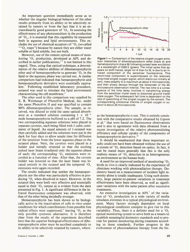
phyrin nor the ether was particularly effective in producing 10 2 when dissolved in water. However, in the octanol they were both very effective and essentially equal in their 102 output as is evident from the data presented in Fig. 1. A significant difference in the infrared fluorescence component was observed, with that of the ether being decidedly stronger.
Hematoporphyrin has been shown to be biologically active in the inactivation of cells in vitro under conditions for which contributions from possible free radical effects were suppressed,9 leaving 102 as the only possible cytotoxic alternative. It is therefore clear from the results of the experiment described here that the superior biological effectiveness of dihematoporphyrin ether must be ascribed completely to its ability to be selectively retained by tumors, where-
Volume 5, Number 1, 1984
1.0 ,-----,-----r--,----,,-----,-----r--,----,- -,---,
~ 'Vi c: OJ
OJ+-, > c:
.~.~ 0.5 Qj 0
ex: :~ E OJ
2 4 6 8 1 0 12 14 16 18 20
t (,us)
Figure 1 - Comparison of the relative singlet oxygen emission intensities of dihematoporphyrin ether (trace A) and hematoporphyrin (trace B) following pulsed laser excitation at a wavelength of 5320A (green). The initial spike that appears on both traces, larger for A than for B, is due to an infrared component of the sensitizer fluorescence. This short-lived component is superimposed on the relatively long-lived singlet oxygen signal, which starts out initially at zero, rises steadily to a maximum at about 4 microseconds, and then slowly decays through the remainder of the 20-microsecond observation interval. The rise time is a consequence of the time delay involved in transferring energy from the sensitizer triplet state to dissolved ground triplet state oxygen. The subsequent fall time is dependent on the rate of quenching of the singlet oxygen by the solvent. The corresponding collisional lifetime of singlet oxygen in octanol is about 20 microseconds.
as the hematoporphyrin is not. This is entirely consistent with the comparative results obtained by Lipson et al. 3 that were based on fluorescence observations and is also in agreement with data obtained from a recent investigation of the relative photosensitizing efficiency and cellular uptake of the components of hematoporphyrin derivative. 10
It should be emphasized that these important results could not have been obtained without the use of a system of 102 detection based on optics. In fact, it can be stated more generally that this is the only realistic means of 1 O2 detection in as heterogeneous an environment as the human body.
A need for an improved method of monitoring 102
levels in vivo is clearly indicated by the experience of clinicians working with photoradiation therapy. Dosimetry based on a measurement of incident light intensity alone is totally inadequate. Using such dosimetry, large patient-to-patient variations in treatment effectiveness have been observed, as well as significant variations with the same patient after successive treatments.
An extensive investigation at APL 7 of the variation of 102 production in a wide variety of media simulates extremes in a typical physiological environment. Many factors strongly dependent on local physiological conditions conspire to give rise to this variability. Thus, there is a definite need for a 102
optical monitoring system to serve both as a means to establish meaningful dosimetry standards and to provide for patient monitoring during treatment according to those standards. Further progress in the evolvement of photoradiation therapy from the hit-
49
J. G. Parker - Singlet Delta Oxygen in Cancer Photoradiation Therapy
and-miss conditions that now prevail to the level of an approved modality will be greatly impeded unless an effective means for in vivo monitoring along these lines is developed. Efforts are now under way to bring about modifications necessary to convert the existing optical laboratory system into one having application in the clinic.
REFERENCES IT. J . Dougherty, D. G . Boyle, K. R. Weishaupt, B. A. Henderson, W. R. Potter, D. A. Bellnier, and K. E. Wityk, "Photoradiation Therapy Clinical and Drug Advances, " in Porphyrin Photosensitization, D. Kessel and T. J . Dougherty, eds. , Plenum Press, New York (1983) .
2y . Hayata, H . Kato , C. Konaka , J . Ono, and N. Takizawa, "Hematoporphyrin Derivative and Laser Photoradiation in the Treatment of Lung Cancer, " Chest 81,269-277 (1982) .
3R. L. Lipson, E. J . Baldes, and A . M. Olsen, " The Use of a Derivative of Hematoporphyrin in Tumor Detection, " J. Nat. Cancer Inst. 26, 1-8 (1961).
50
4T. J . Dougherty, W. R. Potter , and K. R. Weishaupt, "The Structure of the Active Component of Hematoporphyrin Derivative," in Proc. Clayton Foundation Symp. on Porphyrin Localization and Treatment of Tumors, D. R. Doiron , ed., A. R. Liss, Inc. (1983) .
5J . G . Parker and W. D. Stanbro, " Energy Transfer Processes Accompanying Laser Excitation of Hematoporphyrin in Various Solvents," Johns HopkinsAPL Tech. Dig. 2, 196-199(1981).
6J . G . Parker and W. D. Stanbro, " Optical Determination of the Collisional Lifetime of Singlet Molecular Oxygen [0 2(1 ~g )l in Acetone and Deuterated Acetone, " J. A m. Chem. Soc. 104, 2067-2U69 (1982).
7J. G. Parker and W. D. Stanbro, " Dependence of Photosensitized Singlet Oxygen Production on Porphyrin Structure and Solvent," in Proc. Clay ton Foundation Symp. on Porphyrin Localization and Treatment of Tumors, D. R. Doiron, ed ., A. R. Liss, Inc. (1983) .
80 . Kessel, " Transport and Binding of Hematoporphyrin Derivative and Related Porphyrins by Murine Leukemia Ll210 Cells," Cancer Res. 41, 1318-1323 (1981).
9S. Sandberg and I. Romslo, "Porphryi n-Induced Photodamage at the Cellular and the Subcellular Level as Related to the Solubility of the Porphyrin, " Clin. Chim. A cta 109, 193-201 (1981) .
10J . Moan, J. B. McGhie, and T . Christensen, " Hematoporphyrin Derivative: Photosensitizing Efficiency and Cellular Uptake of its Components," Photochem. Photobiophys. 4, 337-345 (1982) .
Johns Hopkins A PL Technical Digest