The impact of RASopathy-associated mutations on CNS ...
17
REVIEW Open Access The impact of RASopathy-associated mutations on CNS development in mice and humans Minkyung Kang 1,2 and Yong-Seok Lee 1,2,3* Abstract The RAS signaling pathway is involved in the regulation of developmental processes, including cell growth, proliferation, and differentiation, in the central nervous system (CNS). Germline mutations in the RAS signaling pathway genes are associated with a group of neurodevelopmental disorders, collectively called RASopathy, which includes neurofibromatosis type 1, Noonan syndrome, cardio-facio-cutaneous syndrome, and Costello syndrome. Most mutations associated with RASopathies increase the activity of the RAS-ERK signaling pathway, and therefore, most individuals with RASopathies share common phenotypes, such as a short stature, heart defects, facial abnormalities, and cognitive impairments, which are often accompanied by abnormal CNS development. Recent studies using mouse models of RASopathies demonstrated that particular mutations associated with each disorder disrupt CNS development in a mutation-specific manner. Here, we reviewed the recent literatures that investigated the developmental role of RASopathy-associated mutations using mutant mice, which provided insights into the specific contribution of RAS-ERK signaling molecules to CNS development and the subsequent impact on cognitive function in adult mice. Keywords: RAS, MAPK, neurodevelopmental disorders, cognition, mutant strains mouse Introduction The RAS-extracellular signal-regulated kinase (ERK) pathway is a highly conserved signaling cascade that transduces signals from membrane receptors to the cytoplasm and nucleus by protein–protein interactions and phosphorylation [1–3]. It plays a critical role in con- trolling various cellular processes, including cell growth, survival, proliferation, and differentiation, in developing and adult tissues, such as the brain [2, 4]. RAS, which is composed of a multigene family that includes HRAS, KRAS, and NRAS, encodes a small guanosine nucleotide- bound GTPase protein, and the activation of the RAS- ERK signal transduction is initiated by the binding of growth factors to G-protein-coupled receptors, such as receptor tyrosine kinases (RTKs) and cytokine receptors. RAS is activated by guanine nucleotide exchange factors (GEFs), such as SOS1, whose activity is regulated by multiple adaptor proteins, including GAB1 and GRB2 (Fig. 1)[5]. On the contrary, GTPase activating proteins (GAPs), such as NF1, switch RAS activity off by hydro- lyzing GTP to GDP. The GTP-bound form of active RAS leads to the activation of its direct downstream ef- fector, RAF. RAF encodes a serine/threonine kinase and represents the RAF family, which also includes ARAF, BRAF, and RAF1. RAF phosphorylates and activates the MAPK kinase, MAPK/ERK kinase 1/2 (MEK1/2), which in turn activates ERK1 and ERK2 by phosphorylating the tyrosine and threonine residues on ERK1/2 [6]. ERK1 and ERK2 are homologous subtypes of the ERK family and are final effectors of the RAS-ERK pathway. ERK1/2 affect a large number of downstream molecules, such as nuclear components, transcription factors, and mem- brane proteins [7]. Since the RAS-ERK pathway is critically involved in mul- tiple biological processes, germline mutations in RAS-ERK signaling components can cause a class of developmental disorders that are collectively called RASopathy [3, 8, 9]. © The Author(s). 2019 Open Access This article is distributed under the terms of the Creative Commons Attribution 4.0 International License (http://creativecommons.org/licenses/by/4.0/), which permits unrestricted use, distribution, and reproduction in any medium, provided you give appropriate credit to the original author(s) and the source, provide a link to the Creative Commons license, and indicate if changes were made. The Creative Commons Public Domain Dedication waiver (http://creativecommons.org/publicdomain/zero/1.0/) applies to the data made available in this article, unless otherwise stated. * Correspondence: [email protected] 1 Department of Physiology, Seoul National University College of Medicine, 103 Daehak-ro, Jongro-gu, Seoul 03080, South Korea 2 Department of Biomedical Sciences, Seoul National University College of Medicine, Seoul 03080, Korea Full list of author information is available at the end of the article Kang and Lee Molecular Brain (2019) 12:96 https://doi.org/10.1186/s13041-019-0517-5
Transcript of The impact of RASopathy-associated mutations on CNS ...

REVIEW Open Access
The impact of RASopathy-associatedmutations on CNS development in miceand humansMinkyung Kang1,2 and Yong-Seok Lee1,2,3*
Abstract
The RAS signaling pathway is involved in the regulation of developmental processes, including cell growth,proliferation, and differentiation, in the central nervous system (CNS). Germline mutations in the RAS signalingpathway genes are associated with a group of neurodevelopmental disorders, collectively called RASopathy, whichincludes neurofibromatosis type 1, Noonan syndrome, cardio-facio-cutaneous syndrome, and Costello syndrome.Most mutations associated with RASopathies increase the activity of the RAS-ERK signaling pathway, and therefore,most individuals with RASopathies share common phenotypes, such as a short stature, heart defects, facialabnormalities, and cognitive impairments, which are often accompanied by abnormal CNS development. Recentstudies using mouse models of RASopathies demonstrated that particular mutations associated with each disorderdisrupt CNS development in a mutation-specific manner. Here, we reviewed the recent literatures that investigatedthe developmental role of RASopathy-associated mutations using mutant mice, which provided insights into thespecific contribution of RAS-ERK signaling molecules to CNS development and the subsequent impact on cognitivefunction in adult mice.
Keywords: RAS, MAPK, neurodevelopmental disorders, cognition, mutant strains mouse
IntroductionThe RAS-extracellular signal-regulated kinase (ERK)pathway is a highly conserved signaling cascade thattransduces signals from membrane receptors to thecytoplasm and nucleus by protein–protein interactionsand phosphorylation [1–3]. It plays a critical role in con-trolling various cellular processes, including cell growth,survival, proliferation, and differentiation, in developingand adult tissues, such as the brain [2, 4]. RAS, which iscomposed of a multigene family that includes HRAS,KRAS, and NRAS, encodes a small guanosine nucleotide-bound GTPase protein, and the activation of the RAS-ERK signal transduction is initiated by the binding ofgrowth factors to G-protein-coupled receptors, such asreceptor tyrosine kinases (RTKs) and cytokine receptors.RAS is activated by guanine nucleotide exchange factors
(GEFs), such as SOS1, whose activity is regulated bymultiple adaptor proteins, including GAB1 and GRB2(Fig. 1) [5]. On the contrary, GTPase activating proteins(GAPs), such as NF1, switch RAS activity off by hydro-lyzing GTP to GDP. The GTP-bound form of activeRAS leads to the activation of its direct downstream ef-fector, RAF. RAF encodes a serine/threonine kinase andrepresents the RAF family, which also includes ARAF,BRAF, and RAF1. RAF phosphorylates and activates theMAPK kinase, MAPK/ERK kinase 1/2 (MEK1/2), whichin turn activates ERK1 and ERK2 by phosphorylating thetyrosine and threonine residues on ERK1/2 [6]. ERK1and ERK2 are homologous subtypes of the ERK familyand are final effectors of the RAS-ERK pathway. ERK1/2affect a large number of downstream molecules, such asnuclear components, transcription factors, and mem-brane proteins [7].Since the RAS-ERK pathway is critically involved in mul-
tiple biological processes, germline mutations in RAS-ERKsignaling components can cause a class of developmentaldisorders that are collectively called RASopathy [3, 8, 9].
© The Author(s). 2019 Open Access This article is distributed under the terms of the Creative Commons Attribution 4.0International License (http://creativecommons.org/licenses/by/4.0/), which permits unrestricted use, distribution, andreproduction in any medium, provided you give appropriate credit to the original author(s) and the source, provide a link tothe Creative Commons license, and indicate if changes were made. The Creative Commons Public Domain Dedication waiver(http://creativecommons.org/publicdomain/zero/1.0/) applies to the data made available in this article, unless otherwise stated.
* Correspondence: [email protected] of Physiology, Seoul National University College of Medicine,103 Daehak-ro, Jongro-gu, Seoul 03080, South Korea2Department of Biomedical Sciences, Seoul National University College ofMedicine, Seoul 03080, KoreaFull list of author information is available at the end of the article
Kang and Lee Molecular Brain (2019) 12:96 https://doi.org/10.1186/s13041-019-0517-5

RASopathy affects approximately 1 in 1,000 live birthsworldwide and shares a common molecular mechanism,such as mutations in RAS-ERK signaling components [4].Representatively, RASopathy includes 1) neurofibromatosistype 1, which is caused by loss of function mutations inNF1; 2) Noonan syndrome, caused by gain of function mu-tations in PTPN11, SOS1, SHOC2, CBL, KRAS, NRAS,BRAF, RAF1, and MEK1; 3) Noonan syndrome with mul-tiple lentigines that is caused by mutations in PTPN11 andRAF1; 4) cardio-facio-cutaneous syndrome, which is causedby either gain of function or loss of function mutations inBRAF, KRAS, MEK1, and MEK2; 5) Costello syndrome,caused by gain of function mutations in HRAS; and 6)neurofibromatosis type 1-like syndrome (NFLS or Legiussyndrome) that is also caused by loss of function mutationsin NF1. RASopathies share typical characteristics, such as ashort stature, craniofacial dysmorphism, cardiac defects,and neurocognitive impairments that are accompanied byabnormal brain development [10]. However, each RASopa-thy also displays distinct and unique symptoms, dependingon the mutated genes [3, 11]. Consistently, recent studiesusing mouse models of RASopathies have demonstrated
that each disorder also shows disease-specific abnor-malities in central nervous system (CNS) develop-ment. Here, we review the distinctive roles of RAS-ERK signaling molecules in CNS development thatwere revealed by investigating the deficits in CNS develop-ment of RASopathies (Tables 1 and 2). Furthermore, wealso review how RASopathy-associated mutations affectcognitive function in mice and human.
RAS-ERK signaling and nervous system developmentThe RAS-ERK signaling pathway is tightly regulated dur-ing CNS development and many studies have demon-strated that the dysregulation of this signaling pathwayresults in aberrant brain development. There are a num-ber of studies demonstrating that ERK1/2, the final ef-fectors of RAS-ERK signaling, are involved in cellproliferation and differentiation in the nervous system[110]. Activation of ERK signaling is required for neuralstem cells (NSCs) to maintain their ability to self-renewand form neurospheres, indicating that ERK may act asa critical regulator in the maintenance of NSCs [111]. Inaddition, it has also been shown that ERK signaling
Fig. 1 The RAS-ERK signaling pathway and associated disorders. A simplified RAS-ERK signaling pathway. Genes frequently mutated in RASopathyare colored based on the RASopathy and are displayed as a polygon depending on their functional categories. NS/NSML, Noonan syndrome/Noonan syndrome with multiple lentigines; NF1, Neurofibromatosis type 1; CS, Costello syndrome; CFCS, Cardio-facio-cutaneous syndrome; GEF,guanine exchange factor; GAP, GTPase activating protein.
Kang and Lee Molecular Brain (2019) 12:96 Page 2 of 17

promotes neuronal survival by multiple mechanisms[112, 113]. For example, an ERK-activated kinase, ribo-somal S6 Kinase (RSK), phosphorylates the pro-apoptotic protein BAD and suppresses BAD-mediatedapoptosis in neurons [112]. ERK was also shown to regu-late the activation of anti-apoptotic regulators, such asBcl-2, CREB, and STAT3/5, and subsequently promotecell survival [112, 114, 115]. However, in spite of thecrucial role of ERK in neuronal survival, aberrant andlong-lasting ERK activation has also been implicated inneurodegenerative diseases [116, 117].Several studies have implied that the MEK/ERK signal-
ing cascade has a crucial role in neurogenesis. ERK2 is ne-cessary for regulating the proliferation of neurogenicprecursors and the positive regulation of neurotrophin-induced neurogenesis by the MEK-C/EBP pathway duringcortical development [118, 119]. Despite the evidence thatMEK is required for neurogenesis, in vivo and in vitrostudies have demonstrated that ERK also regulates andmaintains the pool of glial populations in the developingbrain [109]. NSC-specific ablation of Mek1/2 induces acomplete blockade of glial specificity and gliogenesis fail-ure, while Mek1 gain of function promotes precociousglial progenitor specification in mice [109]. Several studieshave demonstrated that in vitro, Erk1 and Erk2 are criticalcomponents of proliferation in cultured rat astrocytes, andthat MEK/ERK signaling induces gliogenic signals, such asSDF-1a and FGF2 [120–122]. Consistently, treatment withthe MEK inhibitor PD98059 induced a reduction in astro-cytic growth, suggesting that MEK/ERK signaling is in-volved in astrocyte proliferation [122]. In addition, thechemical inhibition of MEK also impairs the ability ofoligodendrocyte precursors to differentiate into mature
oligodendrocyte in vitro, suggesting that both oligoden-drocytes and astrocytes are regulated by ERK signaling[103]. Several studies demonstrated that the pharmaco-logical inhibition of ERK1/2 signaling in oligodendrocyteprogenitors negatively regulates differentiation and thetransition of early progenitors to late oligodendrocyte pro-genitors [123–125]. Furthermore, ERK signaling promotesoligodendrocyte myelination [126]. However, there areconflicting results about the role of ERK signaling in thedifferentiation of oligodendrocyte progenitors into matureoligodendrocytes. Recently, Suo and colleagues demon-strated that MEK inhibitors significantly enhance the dif-ferentiation of oligodendrocyte precursor cells intooligodendrocytes in vitro and in vivo [127]. Consistently,many studies have suggested that increased ERK activitynegatively regulates oligodendrocyte differentiation. Forexample, ERK1/2 activation, which is induced by highdose stimulation of neuregulin-1 or fibroblast growthfactor-2 in mature oligodendrocytes, results in downregu-lated myelin proteins and aberrant cell cycle re-entry[128–130].The RAS-ERK signaling pathway also regulates the ex-
pression of transcription factors, such as cell fate determi-nants. Numerous studies demonstrated that the enhancedactivity of RAS-ERK signaling induces the expression ofthe transcription factor OLIG2, which promotes the fateof NSCs to the glial lineage [85, 90, 108]. Furthermore, theactivation of RAS-ERK signaling promotes the expressionof the pro-neural gene Achaete scute-like 1 (Ascl1) butblocks pro-neural gene Neurogenin 2 (Neurog2) expres-sion. Neurog2 specifies glutamatergic neuronal cell fate indorsal progenitors, while Ascl1 specifies neocorticalgamma-aminobutyric acidergic (GABAergic) neurons and
Table 1 Human patients with RASopathies and their phenotypes
Disease Associated genes CNS structural phenotypes Other phenotypes
Neurofibromatosis type 1 NF1 (95%) [12] Neurofibromas , abnormal corticaldevelopment [13], abnormal glialdevelopment [14], macrocephaly
Below-average IQ, ADHD, impairedexecutive functioning, deficits invisual-spatial skills [15, 16],hyperpigmentation of melanocytes,hamartomas of the iris [17, 18], bonemalformation, cardiac defects [19, 20]
Noonan syndrome,Noonan syndrome withmultiple lentigines
PTPN11 (>50%) [21], RAF1(3-17%) [22, 23], SOS1(9-13%) [24] KRAS (<2%)[25, 26], BRAF (<2%) [22],MEK1/2 (<2%) [27]
Cerebellar ectopia [28, 29], temporallobe anomaly, hydrocephalus,cerebral abscess [30–32], epilepsy,cortical dysplasia [33]
Neurocognitive delay [33–35], typicalfacial abnormalities, short stature,motor delay, increased risk ofcancer, cardiac defects [34–40]
Cardio-facio-cutaneoussyndrome
BRAF (43-78%) [41–43],MEK1/2 (7-11%) [42, 43],KRAS (5-8%) [25, 43]
Ventriculomegaly, hydrocephalus[44–50], atrophy [44, 46, 51–54],migration and myelinationabnormalities, agenesis ofcorpus callosum [50, 52, 55–57]
Neurological abnormalities, seizures,tactile defensiveness, learningdisabilities [4, 50, 55], craniofacialdefects, cardiac defects [4, 58, 59],motor delay, hypotonia [4, 50, 55]
Costello syndrome HRAS (85-90%) [60–62],KRAS (7%) [63], BRAF(4-6%) [27], MEK1/2(2-3%) [27]
Ventricular abnormalities [64–67],cerebral malformations [64, 65, 67–71],cerebellar abnormalities [66, 69, 71–74],macrocephaly [59, 60]
Mental retardation [59, 60], facialfeatures, loose skin, severe failureto thrive, predisposition totumors [59, 60]
IQ Intelligence quotient, ADHD Attention deficit hyperactivity disorder;
Kang and Lee Molecular Brain (2019) 12:96 Page 3 of 17
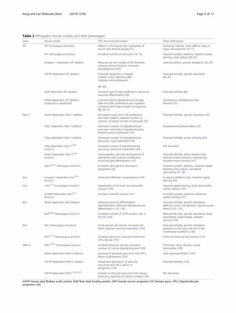
Table 2 RASopathy mouse models and their phenotypesGene Mouse model CNS structural phenotypes Other phenotypes
Nf1 Nf1 homozygous knockout Defects in the neural tube, hyperplasia ofneural crest-derived ganglia [75]
Embryonic lethality, heart defects, delay inorgan development [76, 77]
Nf1 heterozygous knockout Increased number of astrocytes [78, 79] Impaired synaptic plasticity, impaired spatiallearning, heart defects [80–82]
Synapsin 1-dependent Nf1 ablation Reduced size and weight of the forebrain,reduced cortical thickness, increasedastrogliogenesis [83]
Learning deficits, growth retardation [83, 84]
hGFAP-dependent Nf1 ablation Increased gliogenesis, enlargedcerebral cortex, defective GNPmigration and proliferation
[85–88]
Postnatal lethality, growth retardation[86, 87]
BLBP-dependent Nf1 ablation Increased glial lineage proliferation, abnormalneuronal differentiation [89]
Postnatal lethality [89]
Nestin-dependent Nf1 ablation(induction in adulthood)
Unlocked latent oligodendrocyte lineage,defective GNP proliferation and migration,increased adult hippocampal neurogenesis[88, 90, 91]
Spontaneous antidepressive-likebehavior [91]
Ptpn11 Nestin-dependent Ptpn11 ablation Decreased neural stem cell proliferation,lamination defects, reduced number ofneurons, increased number of astrocytes [92]
Postnatal lethality, growth retardation [92]
Olig1-dependent Ptpn11 ablation Decreased number of oligodendrocyteprecursors and mature oligodendrocytes,reduced axonal myelination [93]
Developmental abnormalities [93]
Olig2-dependent Ptpn11 ablation Decreased number of oligodendrocyteprecursors, hypomyelination [94]
Postnatal lethality, severe shivering [94]
Olig2-dependent Ptpn11Q79R
knock-inIncreased number of oligodendrocyteprecursors, abnormal myelination [94]
Not described
Nestin-dependent Ptpn11E76K
knock-inHydrocephalus, aberrant development ofependymal cells, reduced proliferation,enhanced glial differentiation [95]
Postnatal lethality, dome-shaped head,reduced anxiety behavior, hyperactivity,impaired motor function [95]
Ptpn11D61G herozygous knock-in Increased neurogenesis, decreasedgliogenesis [96]
Impaired synaptic plasticity, impaired spatiallearning, short stature, craniofacialdysmorphia [97, 98]
Kras Synapsin1-dependent KrasG12V
knock-inEnhanced GABAergic synaptogenesis [99] Increased inhibitory tone, impaired spatial
learning [99]
Hras HrasG12V homozygous knock-in Hypertrophy of the brain and pyramidalneurons [100]
Impaired spatial learning, facial dysmorphia,cardiac defects [100]
aCaMKII-dependent HrasG12V
knock-inIncrease in docked vesicles [101] Increased synaptic plasticity, enhanced
spatial learning [101]
Braf Nestin-dependent Braf ablation Impaired neuronal differentiation,dysmyelination, defective oligodendrocytedifferentiation [102, 103]
Postnatal lethality, growth retardation,defective motor coordination, neuromusculardefects [102, 103]
BrafV600E heterozygous knock-in Increased number of GFAP positive cells inthe DG [104]
Reduced life span, growth retardation, facialdysmorphia, cardiomegaly, epilepticseizures [104]
Raf1 Raf1 heterozygous knockout Small granule cell volume, increased celldeath, reduced neuronal maturation [105]
Postnatal lethality, growth retardation,apoptosis in the lung and liver, limbscoordination problems [106]
Raf1L613V heterozygous knock-in Increased density of astrocytes, enhancedOPCs density [107]
Enhanced learning and memory [107]
Mek1/2 Mek1Y130C homozygous knock-in Increased astrocyte density, increasednumber of cortical oligodendrocytes [108]
Pulmonary artery stenosis, cranialdysmorphia [108]
Nestin-dependent Mek1/2 ablation Decrease of astrocyte precursors and OPCs,failure of gliogenesis [109]
Early postnatal lethality [109]
hGFAP-dependent Mek1/2 ablation Suppressed generation of astrocyteprecursors and OPCs, failure ofgliogenesis [109]
Postnatal lethality [109]
hGFAP-dependent Mek1S218E,S222E Increase in astrocyte precursors and matureastrocytes, reduction of neuron number [109]
Not described
hGFAP Human glial fibrillary acidic protein, BLBP Brain lipid binding protein, GNP Granule neuron progenitor, DG Dentate gyrus, OPCs Oligodendrocyteprogenitor cells
Kang and Lee Molecular Brain (2019) 12:96 Page 4 of 17
oligodendrocyte precursor cells [131–133]. Therefore,during normal early developmental stages, RAS-ERK sig-naling activity is kept low so that Neurog2 is able to pro-mote glutamatergic neuronal differentiation of embryoniccortical progenitors. However, in an abnormal contextwhere the RAS-ERK signaling is elevated, Neurog2 expres-sion is switched to Ascl1 expression [134]. During moder-ate activation of RAS-ERK signaling, Ascl1 expressionpromotes GABAergic neuronal differentiation, while Ascl1promotes proliferative glioblast phenotypes when RAS-ERK signaling is highly active [134].RAS interacts with and regulates other signaling path-
ways in addition to the MEK/ERK cascade. As one of themain effector pathways of RAS, the phosphatidylinositol3-kinase (PI3K)-AKT pathway regulates protein synthesisand variety of cellular processes such as cell growth, cycleentry, and cellular survival [135–137]. The Ras and PI3K-AKT pathway were shown to activate and inhibit eachother via multiple cross-talks [138]. Studies using rodentmodels have reported distinct phenotypes and revealed apivotal role of PI3K signaling in nervous systems. For in-stance, deleting a PI3K isoform PI3Kγ in mice impairedsynaptic plasticity and behavioral flexibility, while its over-expression through viral vector resulted in impaired syn-aptic plasticity and spatial learning [139, 140]. The Januskinase (JAK)-signal transducer and activator of transcrip-tion (STAT) pathway is also a well characterized cascadeknown to interact with RAS-ERK [141]. JAK activationstimulates cell proliferation, differentiation, cell migrationand apoptosis, and there are compelling evidences thatJAK-STAT pathway plays essential roles in synaptic plasti-city [142].
RASopathies and central nervous system developmentNeurofibromatosis type 1Neurofibromatosis type 1 (NF1) is a relatively commondevelopmental disease that affects 1 in 3,000 individualsand is diagnosed by both somatic and behavioral symptoms[20, 143]. NF1 is caused by loss of function mutations inNF1 alleles [10, 143, 144]. The NF1 gene encodes a GAPfor RAS, neurofibromin, which promotes the conversion ofactive RAS-GTP to inactive RAS-GDP, thus, negativelyregulating the RAS-ERK signaling pathway [145, 146].Therefore, loss of function mutations in NF1 result in thehyperactivation of RAS-ERK signaling. As mutations in theNF1 gene lead to abnormal cell growth, proliferation, anddifferentiation, individuals with NF1 frequently displayneurofibromas, hyperpigmentation of melanocytes, andhamartomas of the iris [17, 18]. Additionally, common fea-tures of NF1 include bone malformations, cardiac defects,and neurocognitive impairments [19, 20]. More than 75%of NF1 patients suffer from cognitive deficits, such asbelow-average IQ and specific deficits in attention, execu-tive functioning, and visual-spatial skills [15, 16].
Although tumor development in the peripheral ner-vous system is a hallmark of NF1, a variety of CNS ab-normalities, including neurofibroma, have been reportedin NF1 patients [147]. For example, abnormal corticallamination and a compressed cerebral cortex were ob-served in the brains of NF1 patients, indicating a criticalrole for NF1 in cortical development [13]. Interestingly,several studies have also suggested that NF1 is associatedwith deficits in glial development. For example, childrenwith NF1 display abnormalities in astrocyte growth regu-lation and tend to develop astrocytoma [14, 148]. Simi-larly, a postmortem study reported that three NF1brains exhibited extensively increased astrogliogenesis[149]. Specifically, an association between an enlargedcorpus callosum and severe learning disabilities in a sub-population of NF1 patients has been reported [150, 151].Moore and colleagues also reported that the total brainvolume, especially the gray matter, was significantly lar-ger in NF1 subjects than in children and adolescentswithout NF1. The gray matter volume in NF1 subjectswas inversely correlated with their degree of learningdisability [150]. Taken together, individuals with NF1display CNS developmental abnormalities, includingpromoted astrogliogenesis and structural malformation,which might be associated with learning disabilities.Nf1 homozygous knockout mice (Nf1-/-) die in utero
because of severe heart malformations, a delay in renal,hepatic, and skeletal muscle development, and hyperpla-sia of neural crest-derived sympathetic ganglia [76, 77].In addition, Nf1-deficient mouse embryos exhibit defectsin the neural tube, including exencephaly or the thinningof the dorsal telencephalic wall, although the targeted al-lele in this study was slightly different from previous in-vestigations [75]. Therefore, a heterozygous knockoutmouse line (Nf1+/-) has been extensively used to investi-gate the cellular mechanisms underlying NF1 etiology[80, 81, 83, 84, 152, 153]. Silva and colleagues showedthat Nf1+/- mice display impaired spatial learning andimpaired hippocampal synaptic plasticity [80, 81]. Mech-anisms underlying the deficits in learning and synapticplasticity in NF1 mouse models have been extensivelyreviewed in previous publications [8, 154]. In line withhuman patients, Nf1 heterozygous mutant mice showeddevelopmental abnormalities in the heart and neuralcrest-derived tissues, and an increased number of astro-cytes with high levels of glial fibrillary acidic protein(GFAP) in the periaqueductal grey, nucleus accumbens,and hippocampus [76, 79].Ablation of Nf1 only in neurons by using the Synapsin
I promoter (Nf1Syn1) led to growth retardation, includingreduced body weight and size, that was sustained intoadulthood [83]. Nf1Syn1 conditional knockout (CKO)mice exhibited reduced size and weight of the forebrain,but not other brain regions [83]. Histological analyses of
Kang and Lee Molecular Brain (2019) 12:96 Page 5 of 17
CKO mice also revealed remarkable defects in the cere-bral cortex, such as a reduction in cortical thickness[83]. Neuronal loss in mutant cortices was not detected;however, interestingly, CKO mice displayed extensiveGFAP immunoreactivity throughout the cerebral cortex,hippocampus, and brainstem, which indicates increasedastrogliogenesis [83]. These results indicate that Nf1 hasan indispensable role in CNS development, and thatNf1-deficient neurons induce astroglial hypertrophy andGFAP induction through a paracrine effect [83, 155].Several studies suggested that neurofibromin might be
required for NSCs or neuroglial progenitor function, andthat Nf1 mutations affect both astroglial and neuronal lin-eages. Studies using a well-characterized human GFAP(hGFAP)-Cre transgenic mouse line have demonstratedthat Nf1 plays a critical role in CNS development. Typic-ally, hGFAP-Cre expression is first detected in radial glia,which give rise to both neuronal and glial lineage cells,around embryonic day 13 [156]. Mutant Nf1hGFAP CKOmice, which lack neurofibromin in the majority of theircortical neurons and astrocytes, were born in normalnumbers, but became noticeably smaller than their litter-mates over time, and typically died by four months of age[86, 87]. Nf1hGFAP CKO mice displayed enlarged cerebralcortices and an increased brain to body weight ratiocaused by the enlarged cortex [85, 88]. The mutant micealso exhibited a notably smaller cerebellum, comparedwith littermates, and defective migration and proliferationof granule neuron progenitors [88]. In addition, Nf1hGFAP
CKO mice failed to form cortical barrels in the somato-sensory cortex, although segregation of thalamic axonswithin the somatosensory cortex was unaffected [87].Consistent with NF1 patients, the mutant mice displayedincreased GFAP-positive astrocytes throughout both thegray and the white matter, including the corpus callosumand anterior commissure [86]. Wang and colleagues alsoshowed that the Nf1hGFAP CKO mice display increasedgliogenesis at the expense of neurogenesis in the neonatalperiod and during adulthood [85]. Due to the altered ratioof glia to neurons, Nf1hGFAP CKO mice displayed a smallerolfactory bulb and an enlarged corpus callosum, providinga link between brain structural abnormalities and cogni-tive impairments in animal models and those seen in NF1patients [85]. Similarly, Nf1 inactivation in neuroglial pro-genitors using a brain lipid binding protein (BLBP)-Cremouse strain also led to increased glial proliferation andabnormal neuronal differentiation in vivo [89]. However,it is also noteworthy to mention that deleting Nf1 usingGFAP-Cre did not impair either learning or synaptic plas-ticity in adult mice [84].Recent studies reported that Nf1 regulates cell fate
specificity and cellular processes in both the develop-mental stage and in adulthood. Inactivation of Nf1 inadult NSCs unlocked a latent oligodendrocyte lineage
and allowed NSCs to produce all three lineages in vivo[90]. Similarly, postnatal Nf1 ablation using Nestin-CreERT2 was sufficient to cause cerebellar abnormal-ities, including defective cerebellar foliation, granuleneuron progenitors (GNPs) proliferation, and migration[88]. Also, deletion of Nf1 in adult hippocampal neuralprogenitor cells led to enhanced proliferation and an in-crease in new neurons in the dentate gyrus [91].Since Nf1 also functions as a tumor suppressor gene,
in vitro studies in various cell types have suggested thatNf1 mutations are associated with growth abnormalities,such as increased proliferation of oligodendrocyte pre-cursors in the embryonic spinal cord [157] and Schwanncells [158]. Particularly, Nf1-/- and Nf1+/- NSCs generateincreased numbers of morphologically abnormal, imma-ture astroglial cells in vitro [159]. The increase in astro-glial progenitors and proliferating cells seen in vitro wasalso observed in Nf1-/- and Nf1+/- embryonic brains andNf1+/- adult brains in vivo [159]. In addition, Lee andcolleagues showed that Nf1-/- NSCs from the brainstemexhibit increased proliferation and glial cell differenti-ation in vitro and in vivo; however, the lack of effect onneocortex NSCs proliferation or gliogenesis suggests thatthe effects of Nf1 gene inactivation are brain region-specific [160].What would be an underlying mechanism for the en-
hanced glial population in NF1? It has been demonstratedthat Nf1 inactivation in neural stem/progenitor cells canalter glia/neuron fate specification by promoting the ex-pression of Olig2, a basic-helix-loop-helix transcriptionfactor that is required for oligodendrocyte progenitor cellspecification [161]. Nf1hGFAP CKO and Nf1BLBP CKO mu-tant mice showed increased Olig2 expression, suggestingthat Nf1 suppresses Olig2 expression and the oligodendro-cyte progenitor lineage in neonatal subventricular zoneprogenitor cells [85, 160]. In concordance with the neo-natal study, inactivation of Nf1 in adult NSCs also resultedin increased Olig2 expression [90]. In conclusion, thesestudies with Nf1 mutant mice revealed the essential roleof NF1 in CNS development, including the gross morph-ology and proper formation of several brain region struc-tures, and the regulation of cell fate.Along with structural abnormalities in CNS, several
lines of evidence suggest that the distribution of NF1 insingle neuronal cell type may also contribute to cognitivedeficits in NF1. Transcriptome analyses of mouse brainhave unveiled the enriched NF1 expression in inhibitoryneurons rather than the in excitatory neurons, and pro-vided a clue as to how NF1 mainly carries out its role ininhibitory synaptic function [162]. Furthermore, based onthe conserved expression pattern of NF1 in human brain,it is suggested that the enriched expression of NF1 in in-hibitory neurons may underlie cell type-specific patho-physiology and cognitive deficits in NF1 [163].
Kang and Lee Molecular Brain (2019) 12:96 Page 6 of 17

Nf1 mutant mice mimic most of the CNS featuresfound in NF1 human patients, including increased brainvolume, enlarged corpus callosum and cortical area, andespecially, enhanced gliogenesis, which may be closelyassociated with structural abnormalities. Despite com-pelling evidences of the expression of glial lineage tran-scription factors such as Olig2 increasing as RAS-ERKhighly activates [85, 90, 108], yet it is unclear how RAS-ERK pathway regulates cell fate determinants. Thus, forunderstanding CNS abnormalities in NF1 patients, it isworth investigating the expression regulations of cell fatedeterminants with regard to RAS-ERK activity.
Noonan syndrome and Noonan syndrome with multiplelentiginesNoonan syndrome (NS) is an autosomal dominant gen-etic disorder with an incidence of 1 in 2,500 live births[31, 164, 165]. This complex disorder occurs both in fa-milial and sporadic forms [166]. Germline mutations ingenes involved in RAS-ERK signaling pathway have beenreported to be associated with NS, such as the gain offunction mutations in protein tyrosine phosphatase non-receptor type 11 (PTPN11), son of sevenless homolog 1(SOS1), Kirsten rat sarcoma viral oncogene homolog(KRAS), neuroblastoma RAS viral oncogene homolog(NRAS), Raf-1 proto-oncogene (RAF1), BRAF, soc-2 sup-pressor of clear homolog (SHOC2), and MEK1, and theloss of function mutations in Cbl proto-oncogene (CBL)[25, 63, 167]. Above all, mutations in PTPN11, which en-codes the non-receptor protein phosphatase SHP2, ac-count for approximately 50% of NS cases [167]. Patientswith NS are characterized by typical facial abnormalities,such as a broad forehead, sparse eyebrows, a low-set andposteriorly rotated ear, and a webbed neck, while otherimportant features include a short stature, motor delay,increased risk of cancer, and cardiac defects [34–40].Noonan syndrome with multiple lentigines (NSML) pa-tients have most of the clinical symptoms observed inindividuals with NS, but they also display increasedpenetrance of hypertrophic cardiomyopathy and lentigi-nes [168]. Distinct from NS, PTPN11 loss of functionmutations result in NSML [168].Between 30%-50% of NS patients show a variable degree
of neurocognitive delay, but there are relatively few reportsof CNS malformations in NS individuals [34, 35]. Twocases of NS were reported to be associated with cerebellarectopia [28, 29]. In addition, there are several reports of NSbeing associated with a temporal lobe anomaly, hydroceph-alus, cerebral abscess, and malignant Schwannoma [30–32].In particular, Saito and colleagues reported one case of anNS patient with severe mental retardation and intractableepilepsy [33]. The patient also displayed cortical dysplasia,including dilated perivascular spaces and a dysplastic lesionin the left temporal lobe [33].
Mutant mice harboring NS-associated Sos1E846K,KrasV14I, and Raf1L613V displayed a short stature, facial dys-morphia, growth retardation, and cardiac defects, which arecharacteristic features of NS patients [169–172]. SincePTPN11 mutations are the majority among NS cases, Shp2mutant mice are one of the most studied models of NS[96–98, 173, 174]. A subpopulation of NS patients have aconstitutively active mutation Shp2D61G, which has a highlyincreased phosphatase activity [175, 176]. The homozygousShp2D61G mutation was eventually embryonically lethal, asthe embryos were grossly hemorrhagic and edematous,showed a decreased liver size, and had cardiac defects [98].However, half of heterozygous Shp2D61G mice that carriedonly one copy of the mutant allele (Shp2D61G/+) survived,and displayed a short stature and craniofacial dysmorphia,such as wide-set eyes, a broad forehead, and a triangularface, which were similar to NS patients [98]. HeterozygousShp2D61G mice also showed deficits in spatial learning andmemory and had impaired synaptic plasticity [97]. Micecarrying a milder mutation, Shp2N308D, displayed some car-diac defects and mild impairment to spatial learning andmemory that was consistent with human cases [97, 98].Neural crest cell-specific Shp2Q79R resulted in craniofacialdefects and growth retardation [170]. Neural stem cell-specific expression of Shp2E76K by using Nestin-Cre re-sulted in hydrocephalus due to aberrant development ofependymal cells [95]. In addition, Shp2E76K-expressing miceshowed hyperactivity accompanied by reduced anxiety be-havior, and impaired motor function [95]. Global Shp2D61Y
expression resulted in embryonic lethality, while epiblast-specific Shp2D61Y expression induced embryonic cardiacdefects [173].SHP2 is a growth factor-regulated phosphatase that
modulates both the RAS-ERK and the gp130-JAK-STATpathways [177, 178]. Since both pathways are known toplay critical roles in cell proliferation and differentiation,several studies demonstrated that SHP2 affects cell pro-liferation and differentiation in large range of cell types[179–183]. For example, SHP2 is required for the initi-ation of retinal neurogenesis and it regulates the pattern-ing of optic vesicles by mediating retinal progenitorfactors and cell proliferation [184]. Huang and colleagueshave shown that the suppression of SHP2 activity reducescell migration and neurite outgrowth, and that it decreasesthe differentiation-induced activation of FAK, Src, paxillin,and ERK1/2 [185]. Also, the authors demonstrated thatSHP2 is recruited to focal adhesions in NSCs and that itregulates focal adhesion formation [185].Recent studies have suggested that Shp2 is involved in
oligodendrocyte development in the telencephalon. Invitro studies using rat cortical cultures demonstrated dif-ferent roles for Shp2 in either oligodendrocyte precursorcell proliferation or maturation [186, 187]. The in vivofunction of Shp2 in oligodendrocyte differentiation was
Kang and Lee Molecular Brain (2019) 12:96 Page 7 of 17

also investigated by Zhu and colleagues using condi-tional mutant mice with a selective Shp2 deletion inOlig1-expressing cells in the ventral spinal cord [93].The mutant mice displayed a dramatic reduction in thenumber of both oligodendrocyte precursor cells and ma-ture oligodendrocytes and decreased axonal myelinationin the developing CNS, suggesting that Shp2 is a criticalregulator of oligodendrocyte proliferation and differenti-ation [93]. Similarly, Ehrman and colleagues investigatedthe role of Shp2 in ventricular zone progenitor cells ofthe ventral telencephalon and in cells of the oligo-dendrocyte lineage by deleting Shp2 in Olig2-positivecells [94]. Olig2-specific Shp2 null mutant mice showeda significant decrease in the number of oligodendrocyteprogenitor cells, at embryonic and postnatal stages, andsevere hypomyelination [94]. Moreover, expressing anNS-associated mutation Shp2Q79R using Olig2-Cre in-creased the number of oligodendrocyte precursor cellsin the embryonic and postnatal brain, but also inducedabnormal myelination and fewer myelinated axons inthe white matter [94].SHP2 has been shown to play a role in cell fate deci-
sions as it promotes neurogenesis and suppresses astro-gliogenesis through the repression of the JAK-STATpathway, which is required for astrocyte formation inthe developing brain. Gauthier and colleagues reportedthat germline Shp2D61G heterozygous mice showed moreneurons and fewer astrocytes in the hippocampus anddorsal cortex at postnatal day 2, and suggested that NS-associated mutations cause brain abnormalities by dis-rupting the balance of CNS populations [96]. Ke andcolleagues also demonstrated that SHP2 is an importantplayer in mammalian brain development by generating anovel mutant mouse in which Shp2 is selectively elimi-nated in neural precursor cells [92]. The mutant mouseshowed early postnatal lethality, decreased proliferationof NSCs, and lamination defects in the developing cere-bral cortex [92]. Mutant mice showed a reduced numberof neurons and an increased number of astrocytes,which imply defective neuronal differentiation and mod-estly enhanced astrogliogenesis, supporting the idea thatShp2 promotes neurogenesis and suppresses astrocyto-genesis [92]. The peripheral nervous system of Wnt1-Cre or Krox20-Cre conditional Shp2 floxed mice dis-played severe deficits in Schwann cell development andthe hypomyelination of peripheral nerves [188].There are other NS mouse models in addition to Shp2
mutant mice. Heterozygous Raf1-deficient mice displaysmaller granule cell layer volumes at postnatal day 30 anda substantial number of abnormal, chromophilic, fast div-iding cells in the subgranular zone and dentate gyrus[105]. In addition, Raf1-deficient neural progenitor cellsshowed an increased rate of cell death and reduced neur-onal maturation [105]. Recently, Holter and colleagues
reported that mice expressing the NS-associated gain offunction mutation Raf1L613V have a significantly greaterdensity of GFAP-positive astrocytes in the cortex andhippocampus. In addition, the number of Olig-positiveoligodendrocyte progenitor cells were also increased incortical area of Raf1L613V mutant mice [107]. Interestingly,Raf1L613V mice showed enhanced performance in severallearning tasks [107]. NS-associated KrasG12V mutant miceshowed enhanced GABAergic synaptogenesis and im-paired spatial learning when the mutation was selectivelyexpressed in synapses [99].Although it is known that transcription factors for glial
lineage become highly expressed in accordance with in-creasing RAS-ERK activity [85, 90, 108], RAS-activatingmutation SHP2D61G promotes neuronal lineage ratherthan glial lineage, by direct interaction with JAK-STATpathway [96]; however, the expression of glial transcrip-tion factors that may have been affected by the increase inRAS-ERK activity is yet to be examined. On the contrary,other NS-linked mutations such as Raf1L613V rather en-hanced glial lineage [107]. Although the underlying mech-anism for the discrepancy in cellular phenotypes is notclear, these results suggest that there are distinct patho-physiology according to each NS-associated mutation. Itwould be interesting to examine the neuron-glia ratio ineither NS patient-derived iPSCs or postmortem brain tis-sues harboring specific PTPN11 or RAF1 mutations.
Cardio-facio-cutaneous syndromeCardio-facio-cutaneous syndrome (CFCS) is a rare RASo-pathy that is caused by mutations in the genes that encodedownstream effectors of RAS [41, 42, 44], including BRAF[41, 42], KRAS [41], and MEK1/2 [42]. Importantly, het-erozygous BRAF mutations are found in over 70% ofCFCS patients [58]. BRAF encodes a serine/threonine kin-ase, and, interestingly, both the kinase-active and kinase-impaired mutations of BRAF are associated with CFCS[41, 42]. Heterozygous missense mutations in MEK1 andMEK2 are found in approximately 25% of CFCS individ-uals [58]. MEK1 and MEK2 are threonine/tyrosine ki-nases, and all the MEK mutants associated with CFCS areactivating mutations [42, 189]. CFCS patients display mul-tiple congenital abnormalities which overlap with thoseseen in NS and Costello syndrome, including craniofacialdefects, hypertrophic cardiomyopathy, pulmonary arterystenosis, and neurocognitive delay [58]. CFCS individualsexhibit NS-like faces, with macrocephaly, low-set ears, ashort nose, a broad forehead, and down-slanting palpebralfissures with ptosis [4, 59]. Cardiac abnormalities are alsosimilar to those of NS and Costello syndrome, with pul-monic stenosis, septal defects, and hypertrophic cardiomy-opathy (HCM) having the highest prevalence [59].Neurological abnormalities, including hypotonia, motordelay, seizures, tactile defensiveness, speech delay, and
Kang and Lee Molecular Brain (2019) 12:96 Page 8 of 17

learning disabilities, are present at varying degrees[4, 50, 55]. Failure to thrive caused by gastrointes-tinal dysfunction, including vomiting, oral aversion,reflux, and constipation, is also typical in CFCS individualsin infancy [50]. However, CNS abnormalities are signifi-cant diagnostic features of CFCS. Previous studies re-ported the abnormalities in brain structures, includingventriculomegaly and hydrocephalus, in CFCS patients[44–50]. Volume loss in the brain due to cortical atrophy,cerebral atrophy, brain stem atrophy, and white matter at-rophy have also emerged in a subpopulation of patients[44, 46, 51–54]. Additionally, migration abnormalities,myelination abnormalities, and corpus callosum ab-normalities, such as hypoplasia and lipoma were alsorevealed by brain imaging [50, 52, 55–57]. In linewith brain abnormalities, most CFCS patients are di-agnosed with varying degrees of cognitive deficits andintellectual disabilities [50].Recently, patient-derived induced pluripotent stem
cells (iPSCs) have contributed to advancements in theunderstanding of disease-associated mutations. Yeh andcolleagues generated iPSC from a patient harboringBRAFQ257R, the most frequent CFCS mutation [190].This mutation resulted in a depletion of neural progeni-tor pool, induced by decreased phosphorylation of AKT,and early neuronal maturation [190]. Due to the deple-tion of progenitors, the number of late-born cells, suchas the upper-layer cortical neurons and glia, was de-creased [191]. The number of GABAergic interneuronswas increased, indicating that the high prevalence of sei-zures in CFCS individuals may be caused by an imbal-ance between excitation and inhibition [191].Fewer animal models of CFCS have been reported
likely due to its lower prevalence (1 in 810,000) com-pared to other RASopathies. Transgenic mouse modelscarrying gain of function mutations that are associatedwith CFCS recapitulate multiple aspects of human CFCSpatients [108, 192]. Since BRAF is the most prevalentgene that is mutated in CFCS, a majority of animal stud-ies in CFCS have focused on Braf. Prior to the review ofgain of function mouse models associated with CFCS,we first reviewed loss of function studies that investi-gated the role of Braf in various biological processes.Wiese and colleagues identified that cultured embryonicsensory and motor neurons lacking Braf could not sur-vive in the presence of neurotrophic factors while Raf1-deficient neurons could survive, suggesting that Braf isessential for survival [193]. A Braf null mutant mousewas embryonically lethal due to the vascular defects atmidgestation [194]. In addition, the ablation of Braf inNSCs using Nestin-Cre resulted in abnormal morpho-genesis of the CNS, such as a decreased cerebellum withfuzzy granule cell layer borders and a diminished hippo-campus granule cell layer, due to reduced differentiation
of dentate gyrus progenitor cells into mature granule cellneurons [102]. Nestin-Cre specific Braf-deficient mice alsodisplayed severe dysmyelination and defective oligo-dendrocyte differentiation, implicating Braf in postnatalCNS development [103]. Forebrain excitatory neuron-specific Braf knockout mice showed deficits in hippocam-pal long-term potentiation and impaired hippocampal-dependent learning and memory, while the impact of Brafdeletion on CNS development in this knockout remainsto be investigated [195].The first mutant mouse model of CFCS was a knock-
in of the constitutively active form of Braf, BrafV600E,which recapitulates several CFCS characteristics, includ-ing a reduced life span, growth retardation, facial dys-morphia, cardiomegaly, and epileptic seizures [104].Mice expressing the conditional knock-in BrafL597V mu-tation also recapitulated CFCS symptoms of a short stat-ure, facial dysmorphia, and cardiac enlargement [196].The most prevalent CFCS mutation, BrafQ241R, inducedembryonic/neonatal lethality with multiple congenitaldefects that included embryonic skeletal abnormalities,lymphatic defects, cardiac defects, and liver necrosis inthe C57BL/6J background, and lethality between birthand 24 weeks, growth retardation, sparse and ruffled fur,liver necrosis, and atrial septal defects on the mixedbackground (BALB/c and C57BL/6J) [192, 197]. Inaddition, BrafQ241R/- mice showed growth retardation, ahunched appearance, craniofacial dysmorphism, andlearning deficits on ICR background [192].Mouse models carrying Mek1Y130C, the most common
MEK1 mutation in CFCS patients, showed increased ERKactivation in response to growth factors, pulmonary arterystenosis, cranial dysmorphia, and neurological anomalies[108]. Moreover, Mek1Y130C/Y130C mice showed a higherdensity of GFAP-positive astrocytes in the sensory cortexand hippocampal CA1 regions [108]. In addition, the totalcortical oligodendrocyte population, as analyzed by Olig2immunolabeling, was increased in the sensory cortex ofMek1Y130C/Y130C mice [108]. As addressed earlier,patients-derived iPSC containing BRAFQ257R exhibitedearly neuronal maturation and decreased late-born glialpopulations, whereas either CFCS-associated Mek1Y130C
or BrafV600E expressing adult mice exhibited an increasednumber of GFAP-positive cells in hippocampal and cor-tical areas [104, 108, 190]. Although, BRAFQ257R is a gainof function mutation, the activation of ERK was decreasedin neural progenitor cells, which might have been due tocell context-dependent role of BRAF, and these results in-dicate that the decreased ERK activation may be respon-sible for the decreased glia in BRAFQ257R iPSCs [190]. Inaddition to mouse models, zebrafish models expressingCFCS Braf or Mek variants were also generated, and theseCFCS mutant alleles interfered with convergence-extension cell movements during gastrulation to cause
Kang and Lee Molecular Brain (2019) 12:96 Page 9 of 17
similar developmental phenotypes [189]. Taken together,each of the CFCS-associated genes play essential roles inCNS development, including oligodendrocyte precursormaturation and proliferation, myelination, and neuronaldifferentiation. However, our knowledge regarding thecausal relationship between CNS abnormalities and cogni-tion in CFCS is still limited. Further studies using mutantanimals with more specific temporal and spatial manipula-tion of CFCS genes would provide understanding of thepathophysiology of cognitive deficits in CFCS.
Costello syndromeCostello syndrome (CS) is a rare multiple congenital ab-normality syndrome that affects 1 in 1,250,000 people andshares many features with other RASopathies [198–200].CS is mostly caused by gain of function mutations in theHRAS gene, most of which have been previously reportedas somatic or oncogenic mutations in various tumors[60, 62, 201]. HRAS activating mutations are highlyprevalent in CS individuals; they disrupt guanine nucleo-tide binding and induce a decrease in intrinsic and GAP-induced GTPase activity, allowing mutant HRAS proteinsto remain in the active state [202]. In addition, BRAF,KRAS, and MEK1 mutations are also associated with asmall population of CS individuals [27, 63, 203]. CS pa-tients are typical characterized by coarse facial features, re-dundant and loose skin, severe failure to thrive, mentalretardation, cardiomyopathy, and a predisposition to tu-mors [59, 60]. There is no single feature that is unique toCS, and this syndrome phenotypically overlaps with NF1,NS, NSML, and CFCS [60, 204]. Typical and coarse facialfeatures associated with CS involve macrocephaly with aprominent forehead, a short nose with a depressed nasalbridge and a broad base, and low-set, posteriorly rotatedears with thickened helices and lobes. Most CS patientshave cardiac abnormalities, including hypertrophic cardio-myopathy, valve abnormalities, septal defects, andarrhythmia [205]. Failure to thrive due to gastrointestinaldysfunction often involves reflux, oral aversion, and con-stipation during early infancy [67, 200]. Structural andelectrophysiological neurological malformations are alsocommon in CS. For example, ventricular abnormalities,such as mild ventricular dilatation, are observed in morethan 40% of CS individuals [64–67]. Cerebral malforma-tions in CS include cerebral atrophy, leukomalacia, poorgray-white matter differentiation, a small corpus callosum,and MRI signal abnormalities [64, 65, 67–71]. Cerebellarabnormalities include malformation, cerebellar atrophy,deviation of the cerebellar tonsils, and demyelinization ofthe basal tonsil [66, 69, 71–74].Krencik and colleagues have shown that human iPSCs
carrying HRASG12S that were derived from CS patientsexhibited hyperplasia and differentiated into astrogliamore rapidly in vitro than iPSCs derived from control
cell lines with normal HRAS. CS-derived iPSCs also gen-erated an abundance of extracellular matrix remodelingfactors and proteoglycans [206]. Moreover, HRASG12S
iPSC-derived neurons had a longer progenitor phase,unlike the phenotype reported in BRAFQ257R iPSC-derived neurons that originated from CFCS patients[190, 207]. Thus, postnatal progressive cerebellar over-growth of the brain in CS individuals could be caused bythe extended progenitor phase [208].As with CFCS, only few animal models were generated
for CS. Both homozygous and heterozygous HrasG12V
knock-in mice closely phenocopied some of the featuresobserved in individuals with CS, including facial dys-morphia, cardiomyopathies, and alterations to the homeo-stasis of the cardiovascular system [209]. In addition, laterstudies with homozygous HrasG12V knock-in mice demon-strated that they have neurocognitive deficits, such ashyperactivity, increased anxiety-like behavior and milddeficit in spatial memory [210]. However, Viosca and col-leagues did not observe significant changes in either theactivity or the expression of downstream of Hras such asphospho-CREB and c-fos [210]. Transgenic mice withforebrain excitatory neuron-specific expression ofHrasG12V under the control of the αCaMKII promoter dis-played several synaptic phenotypes, including a high dens-ity of docked neurotransmitter vesicles in glutamatergicterminals and increased synaptic plasticity which may beassociated with the dramatically enhanced hippocampal-dependent learning [101]. Schreiber and colleagues havealso shown that the homozygous HrasG12V knock-in miceexhibit spatial learning deficits, which are accompanied byrobust upregulation of Erk signaling in hippocampal ly-sates, neuronal hypertrophy, increased brain volume, andimpaired mGluR-dependent long-term depression (LTD)[100]. Notably, mice expressing CS-associated HrasG12V
or HrasG12S mutations in cortical precursors displayedpromoted precursor cell proliferation and premature glio-genesis, but inhibited neurogenesis [211]. Consistently, ei-ther form of Hras mutations also promoted precursor cellproliferation and astrogenesis, but inhibited neurogenesisin cultured cortical precursors [211]. These findings frommultiple experimental systems such as iPSCs, mice modelsand cultured cells commonly suggest the essential role ofHRAS in neural precursor cell proliferation and gliogen-esis, which might strongly affect the structure and func-tion of CNS including increased brain volume in CSpatients.
Concluding remarksHere, we reviewed that mutations in different compo-nents of the RAS-ERK signaling pathway associated withdifferent RASopathies have distinct impacts on CNS de-velopment in a cell type-specific manner (Fig. 2). How-ever, it is still unclear how some mutations affect
Kang and Lee Molecular Brain (2019) 12:96 Page 10 of 17

neurons and others affect glia. One hypothesis is thatdifferent signaling molecules are expressed at distinctphases during development. So far, we do not have theexpression profiles of RAS signaling molecules duringbrain development in high spatial and temporal reso-lution. However, most of the key RAS-ERK components,such as NF1, SHP2, BRAF, and MEK1/2, begin to beexpressed before either embryonic day 10 or 15, whichare initiation time points of neurogenesis or astroglio-genesis, respectively [212]. Recently, it has been shownthat the expression of RAS-ERK signaling molecules wasremarkably different between excitatory and inhibitoryneurons in mouse hippocampus [162]. Thus, it would beinteresting to examine whether the expression levels ofvarious signaling molecules are differentially regulated inNSCs over different developmental stages. Advancedtools, such as single cell RNA-seq, might be useful to
answer this question. It should also be considered thatthere are multiple cross-talks between RAS and othersignaling pathways. Different components in RAS signal-ing interact with distinct signaling networks, which mayaccount for the cell type-specific developmental deficitsin each RASopathy.Treatments for the cognitive deficits found in RASopa-
thies are not available yet. Since most RASopathy-associated mutations increase RAS-ERK activation, down-regulating the activity of RAS or its downstream effectorsis an obvious strategy to develop treatments for RASopa-thies. Although statins, which can reduce RAS activity byinhibiting the farnesylation of RAS, have been proposedfor the improvement of learning disabilities in NF1 chil-dren [213], the results from various clinical trials havebeen inconsistent [214–216]. The reason for these dis-crepant results remains unclear. However, considering the
Fig. 2 Effect of RAS signaling components on neural stem cell differentiation. Neural stem cells are able to generate progeny cells that terminallydifferentiate into neurons, oligodendrocytes, and astrocytes. a NF1 inactivation led to decreased neurogenesis in neonatal and adult mousebrains [85]. PTPN11 positively regulates neurogenesis at the expense of gliogenesis [96]. b NF1 negatively regulates gliogenesis, thus NF1inactivation increases the number of glial progenitor cells and gliogenesis [78, 79, 85, 89, 159, 160]. PTPN11 suppresses gliogenesis by directlyinteracting with the JAK-STAT pathway, which promotes gliogenesis [92, 96]. c Hyperactivation of RAF1 induces the increase of glial lineagepopulations, including oligodendrocyte progenitor cells and astrocytes [107]. MEK is required for gliogenesis, and the hyperfunction of MEK1leads to increase in glial populations [108, 109]. d BRAF and RAF1 positively regulate neuronal differentiation, and the disruption of BRAF or RAF1impairs the ability of progenitor cells to differentiate into mature neurons in mouse brain [102, 105]. In consistent, iPSC containing hyperactivatedBRAF mutant showed early maturation of neurons [190]. e Oligodendroglial lineage potential is restricted by NF1 in the adult hippocampus, andinactivation of NF1 allows the adult hippocampus to generate oligodendrocytes [85]. f BRAF is required for oligodendrocyte maturation andmyelination during postnatal development [103]. g Hyperactivated HRAS leads to an acceleration of astroglial maturation [206, 211]. Blue and redarrows indicate positive and negative regulation, respectively.
Kang and Lee Molecular Brain (2019) 12:96 Page 11 of 17
ubiquitous expression of RAS in many cell types and mul-tiple organs, directly regulating RAS activity may have un-known confounding effects. Thus, it would be better totarget specific molecules other than RAS in a disease-specific manner. For example, Omrani and colleaguesshowed that inhibitory neuron-specific attenuation ofhyperpolarization-activated cyclic nucleotide-gated (HCN)currents can be an underlying mechanism for the cogni-tive deficits in Nf1+/- mice when they used an HCN agon-ist to rescue cognitive deficits in Nf1+/- mice [217].Recently, Ryu and colleagues showed that selectively redu-cing the interaction between mutant SHP2 and Gab1 inexcitatory neurons reversed the physiological and behav-ioral deficits in a mouse model of NS [162]. Conditionalmutant mice with higher spatial and temporal resolutionwill provide clues when, where, and which cell types aremost suited for interventions.Lastly, it should be noted that most of the RASopathy
mechanism studies have used mice as a model system.Mouse models have many advantages and can be usedto study neuropsychiatric disorders because the majorityof neuropsychiatric drugs used in humans were shownto be, at least partially, effective in mouse models [218].However, caution is still warranted. Nowadays, it has be-come relatively easy to model diseases in vitro usingiPSC and several iPSC lines are available to study RASo-pathies, allowing for parallel and comparative analysesin vitro and in vivo.
AbbreviationsAscl1: Achaete scute-like 1; BLBP: Brain lipid binding protein; CFCS: Cardio-facio-cutaneous syndrome; CKO: Conditional knockout; CNS: Central nervoussystem; CS: Costello syndrome; ERK: Extracellular signal-regulated kinase;GABAergic: Gamma-aminobutyric acidergic; GAPs: GTPase activating proteins;GEFs: Guanine nucleotide exchange factors; GFAP: Glial fibrillary acidicprotein; GNPs: Granule neuron progenitors; HCM: Hypertrophiccardiomyopathy; iPSCs: Induced pluripotent stem cells; JAK: Janus kinase;KRAS: Kirsten rat sarcoma viral oncogene homolog; MEK1/2: MAPK/ERKkinase 1/2; Neurog2: Neurogenin 2; NF1: Neurofibromatosis type 1;NRAS: Neuroblastoma RAS viral oncogene homolog; NS: Noonan syndrome;NSCs: Neural stem cells; NSML: Noonan syndrome with multiple lentigines;PI3K: Phosphatidylinositol 3-kinase; PTPN11: Protein tyrosine phosphatasenon-receptor type 11; RTKs: Receptor tyrosine kinases; SHOC2: Soc-2suppressor of clear homolog; SOS1: Son of sevenless homolog 1;STAT: Signal transducer and activator of transcription
AcknowledgementsAuthors thank Jae Yoon Hwang for proofreading the manuscript and all themembers of laboratory of neurophysiology for their comments on themanuscript.
Authors' contributionsY-SL and MK contributed to conception. MK and Y-SL reviewed the literaturesand wrote the manuscript. MK generated figures and tables. Both authors readand approved the final manuscript.
FundingThis work was supported by the NRF-2016H1A2A1907206 grant to M.K. andthe NRF-2019R1A2C1084232, NRF-2019R1A4A2001609 and NRF-2017M3C7A1026959 grants to Y.-S.L.
Availability of data and materialsNot applicable
Ethics approval and consent to participateNot applicable
Consent for publicationNot applicable
Competing interestsThe authors declare that they have no competing interests.
Author details1Department of Physiology, Seoul National University College of Medicine,103 Daehak-ro, Jongro-gu, Seoul 03080, South Korea. 2Department ofBiomedical Sciences, Seoul National University College of Medicine, Seoul03080, Korea. 3Neuroscience Research Institute, Seoul National UniversityCollege of Medicine, 103 Daehak-ro, Jongro-gu, Seoul 03080, South Korea.
Received: 23 July 2019 Accepted: 28 October 2019
References1. Kolch W. Meaningful relationships: the regulation of the Ras/Raf/MEK/ERK
pathway by protein interactions. Biochem J. 2000;351(Pt 2):289–305.2. Boguski MS, McCormick F. Proteins regulating Ras and its relatives. Nature.
1993;366:643–54.3. Ryu HH, Lee YS. Cell type-specific roles of RAS-MAPK signaling in learning
and memory: Implications in neurodevelopmental disorders. NeurobiolLearn Mem. 2016;135:13–21.
4. Rauen KA. The RASopathies. Annu Rev Genomics Hum Genet. 2013;14:355–69.
5. Innocenti M, Tenca P, Frittoli E, Faretta M, Tocchetti A, Di Fiore PP, Scita G.Mechanisms through which Sos-1 coordinates the activation of Ras andRac. J Cell Biol. 2002;156:125–36.
6. Zhao L, Brinton RD. Vasopressin-induced cytoplasmic and nuclear calciumsignaling in embryonic cortical astrocytes: dynamics of calcium andcalcium-dependent kinase translocation. J Neurosci. 2003;23:4228–39.
7. Deak M, Clifton AD, Lucocq LM, Alessi DR. Mitogen- and stress-activatedprotein kinase-1 (MSK1) is directly activated by MAPK and SAPK2/p38, andmay mediate activation of CREB. EMBO J. 1998;17:4426–41.
8. Shilyansky C, Lee YS, Silva AJ. Molecular and cellular mechanisms of learningdisabilities: a focus on NF1. Annu Rev Neurosci. 2010;33:221–43.
9. Kim YE, Baek ST. Neurodevelopmental Aspects of RASopathies. Mol Cells.2019;42:441–7.
10. Tidyman WE, Rauen KA. The RASopathies: developmental syndromes of Ras/MAPK pathway dysregulation. Curr Opin Genet Dev. 2009;19:230–6.
11. Jindal GA, Goyal Y, Burdine RD, Rauen KA, Shvartsman SY. RASopathies:unraveling mechanisms with animal models. Dis Model Mech. 2015;8:769–82.
12. Ferner RE, Huson SM, Thomas N, Moss C, Willshaw H, Evans DG, UpadhyayaM, Towers R, Gleeson M, Steiger C, et al. Guidelines for the diagnosis andmanagement of individuals with neurofibromatosis 1. J Med Genet. 2007;44:81–8.
13. Rosman NP, Pearce J. The brain in multiple neurofibromatosis (vonRecklinghausen's disease): a suggested neuropathological basis for theassociated mental defect. Brain. 1967;90:829–38.
14. Listernick R, Charrow J, Greenwald MJ, Esterly NB. Optic gliomas in childrenwith neurofibromatosis type 1. J Pediatr. 1989;114:788–92.
15. Krab LC, Aarsen FK, de Goede-Bolder A, Catsman-Berrevoets CE, Arts WF,Moll HA, Elgersma Y. Impact of neurofibromatosis type 1 on schoolperformance. J Child Neurol. 2008;23:1002–10.
16. Shilyansky C, Karlsgodt KH, Cummings DM, Sidiropoulou K, Hardt M, JamesAS, Ehninger D, Bearden CE, Poirazi P, Jentsch JD, et al. Neurofibrominregulates corticostriatal inhibitory networks during working memoryperformance. Proc Natl Acad Sci U S A. 2010;107:13141–6.
17. Cichowski K, Jacks T. NF1 tumor suppressor gene function: narrowing theGAP. Cell. 2001;104:593–604.
18. Zhu Y, Parada LF. Neurofibromin, a tumor suppressor in the nervous system.Exp Cell Res. 2001;264:19–28.
Kang and Lee Molecular Brain (2019) 12:96 Page 12 of 17
19. Brems H, Legius E. Legius syndrome, an Update. Molecular pathology ofmutations in SPRED1. Keio J Med. 2013;62:107–12.
20. Williams VC, Lucas J, Babcock MA, Gutmann DH, Korf B, Maria BL.Neurofibromatosis type 1 revisited. Pediatrics. 2009;123:124–33.
21. Tartaglia M, Kalidas K, Shaw A, Song X, Musat DL, van der Burgt I, BrunnerHG, Bertola DR, Crosby A, Ion A, et al. PTPN11 mutations in Noonansyndrome: molecular spectrum, genotype-phenotype correlation, andphenotypic heterogeneity. Am J Hum Genet. 2002;70:1555–63.
22. Razzaque MA, Nishizawa T, Komoike Y, Yagi H, Furutani M, Amo R, KamisagoM, Momma K, Katayama H, Nakagawa M, et al. Germline gain-of-functionmutations in RAF1 cause Noonan syndrome. Nat Genet. 2007;39:1013–7.
23. Pandit B, Sarkozy A, Pennacchio LA, Carta C, Oishi K, Martinelli S, Pogna EA,Schackwitz W, Ustaszewska A, Landstrom A, et al. Gain-of-function RAF1mutations cause Noonan and LEOPARD syndromes with hypertrophiccardiomyopathy. Nat Genet. 2007;39:1007–12.
24. Tartaglia M, Pennacchio LA, Zhao C, Yadav KK, Fodale V, Sarkozy A, Pandit B,Oishi K, Martinelli S, Schackwitz W, et al. Gain-of-function SOS1 mutationscause a distinctive form of Noonan syndrome. Nat Genet. 2007;39:75–9.
25. Schubbert S, Zenker M, Rowe SL, Boll S, Klein C, Bollag G, van der Burgt I,Musante L, Kalscheuer V, Wehner LE, et al. Germline KRAS mutations causeNoonan syndrome. Nat Genet. 2006;38:331–6.
26. Carta C, Pantaleoni F, Bocchinfuso G, Stella L, Vasta I, Sarkozy A, Digilio C,Palleschi A, Pizzuti A, Grammatico P, et al. Germline missense mutationsaffecting KRAS Isoform B are associated with a severe Noonan syndromephenotype. Am J Hum Genet. 2006;79:129–35.
27. Nava C, Hanna N, Michot C, Pereira S, Pouvreau N, Niihori T, Aoki Y,Matsubara Y, Arveiler B, Lacombe D, et al. Cardio-facio-cutaneous andNoonan syndromes due to mutations in the RAS/MAPK signalling pathway:genotype-phenotype relationships and overlap with Costello syndrome. JMed Genet. 2007;44:763–71.
28. Peiris A, Ball MJ. Chiari (type 1) malformation and syringomyelia in a patientwith Noonan's syndrome. J Neurol Neurosurg Psychiatry. 1982;45:753–4.
29. Kobayashi I, Aikawa T, Takemiya T, Maruyama S, Takano K. Noonan’ssyndrome with syringomyelia. Jpn J Psychiatry Neurol. 1986;40:101–4.
30. Gorke W. Cerebral defects in Noonan's syndrome (author's transl). KlinPadiatr. 1980;192:577–81.
31. Noonan JA. Hypertelorism with Turner phenotype. A new syndrome withassociated congenital heart disease. Am J Dis Child. 1968;116:373–80.
32. Raman Unnithan R, Bahuleyan CG, Matnew Roy VC. Noonan syndrome. JAssoc Physicians India. 1985;33:177–9.
33. Saito Y, Sasaki M, Hanaoka S, Sugai K, Hashimoto T. A case of Noonansyndrome with cortical dysplasia. Pediatr Neurol. 1997;17:266–9.
34. Cesarini L, Alfieri P, Pantaleoni F, Vasta I, Cerutti M, Petrangeli V, Mariotti P,Leoni C, Ricci D, Vicari S, et al. Cognitive profile of disorders associated withdysregulation of the RAS/MAPK signaling cascade. Am J Med Genet A. 2009;149A(2):140–6.
35. Pierpont EI, Pierpont ME, Mendelsohn NJ, Roberts AE, Tworog-Dube E,Seidenberg MS. Genotype differences in cognitive functioning in Noonansyndrome. Genes Brain Behav. 2009;8:275–82.
36. Aoki Y, Niihori T, Banjo T, Okamoto N, Mizuno S, Kurosawa K, Ogata T,Takada F, Yano M, Ando T, et al. Gain-of-function mutations in RIT1 causeNoonan syndrome, a RAS/MAPK pathway syndrome. Am J Hum Genet.2013;93:173–80.
37. Lee DA, Portnoy S, Hill P, Gillberg C, Patton MA. Psychological profile ofchildren with Noonan syndrome. Dev Med Child Neurol. 2005;47:35–8.
38. Razzaque MA, Komoike Y, Nishizawa T, Inai K, Furutani M, HigashinakagawaT, Matsuoka R. Characterization of a novel KRAS mutation identified inNoonan syndrome. Am J Med Genet A. 2012;158A(3):524–32.
39. Roberts AE, Allanson JE, Tartaglia M, Gelb BD. Noonan syndrome. Lancet.2013;381:333–42.
40. van der Burgt I, Thoonen G, Roosenboom N, Assman-Hulsmans C, GabreelsF, Otten B, Brunner HG. Patterns of cognitive functioning in school-agedchildren with Noonan syndrome associated with variability in phenotypicexpression. J Pediatr. 1999;135:707–13.
41. Niihori T, Aoki Y, Narumi Y, Neri G, Cave H, Verloes A, Okamoto N, HennekamRC, Gillessen-Kaesbach G, Wieczorek D, et al. Germline KRAS and BRAFmutations in cardio-facio-cutaneous syndrome. Nat Genet. 2006;38:294–6.
42. Rodriguez-Viciana P, Tetsu O, Tidyman WE, Estep AL, Conger BA, Cruz MS,McCormick F, Rauen KA. Germline mutations in genes within the MAPKpathway cause cardio-facio-cutaneous syndrome. Science. 2006;311:1287–90.
43. Narumi Y, Aoki Y, Niihori T, Neri G, Cave H, Verloes A, Nava C, Kavamura MI,Okamoto N, Kurosawa K, et al. Molecular and clinical characterization ofcardio-facio-cutaneous (CFC) syndrome: overlapping clinical manifestationswith Costello syndrome. Am J Med Genet A. 2007;143a:799–807.
44. Reynolds JF, Neri G, Herrmann JP, Blumberg B, Coldwell JG, Miles PV, OpitzJM. New multiple congenital anomalies/mental retardation syndrome withcardio-facio-cutaneous involvement--the CFC syndrome. Am J Med Genet.1986;25:413–27.
45. Neri G, Sabatino G, Bertini E, Genuardi M. The CFC syndrome--report of thefirst two cases outside the United States. Am J Med Genet. 1987;27:767–71.
46. Chrzanowska K, Fryns JP, Van den Berghe H. Cardio-facio-cutaneous (CFC)syndrome: report of a new patient. Am J Med Genet. 1989;33:471–3.
47. Ades LC, Sillence DO, Rogers M. Cardiofaciocutaneous syndrome. ClinDysmorphol. 1992;1:145–50.
48. Young TL, Ziylan S, Schaffer DB. The ophthalmologic manifestations of thecardio-facio-cutaneous syndrome. J Pediatr Ophthalmol Strabismus. 1993;30:48–52.
49. Krajewska-Walasek M, Chrzanowska K, Jastrzbska M. The cardio-facio-cutaneous (CFC) syndrome--two possible new cases and review of theliterature. Clin Dysmorphol. 1996;5:65–72.
50. Yoon G, Rosenberg J, Blaser S, Rauen KA. Neurological complications ofcardio-facio-cutaneous syndrome. Dev Med Child Neurol. 2007;49:894–9.
51. Somer M, Peippo M, Aalto-Korte K, Ritvanen A, Niemi KM. Cardio-facio-cutaneous syndrome: three additional cases and review of the literature.Am J Med Genet. 1992;44:691–5.
52. Grebe TA, Clericuzio C. Neurologic and gastrointestinal dysfunction incardio-facio-cutaneous syndrome: identification of a severe phenotype. AmJ Med Genet. 2000;95:135–43.
53. Manci EA, Martinez JE, Horenstein MG, Gardner TM, Ahmed A, Mancao MC,Gremse DA, Gardner DM, Nimityongskul P, Maertens P, et al.Cardiofaciocutaneous syndrome (CFC) with congenital peripheralneuropathy and nonorganic malnutrition: an autopsy study. Am J MedGenet A. 2005;137:1–8.
54. Baraitser M, Patton MA. A Noonan-like short stature syndrome with sparsehair. J Med Genet. 1986;23:161–4.
55. Armour CM, Allanson JE. Further delineation of cardio-facio-cutaneoussyndrome: clinical features of 38 individuals with proven mutations. J MedGenet. 2008;45:249–54.
56. Papadopoulou E, Sifakis S, Sol-Church K, Klein-Zighelboim E, Stabley DL,Raissaki M, Gripp KW, Kalmanti M. CNS imaging is a key diagnostic tool inthe evaluation of patients with CFC syndrome: two cases and literaturereview. Am J Med Genet A. 2011;155a:605–11.
57. Wieczorek D, Majewski F, Gillessen-Kaesbach G. Cardio-facio-cutaneous(CFC) syndrome--a distinct entity? Report of three patients demonstratingthe diagnostic difficulties in delineation of CFC syndrome. Clin Genet. 1997;52:37–46.
58. Roberts A, Allanson J, Jadico SK, Kavamura MI, Noonan J, Opitz JM, Young T,Neri G. The cardiofaciocutaneous syndrome. J Med Genet. 2006;43:833–42.
59. Schulz AL, Albrecht B, Arici C, van der Burgt I, Buske A, Gillessen-Kaesbach G,Heller R, Horn D, Hubner CA, Korenke GC, et al. Mutation and phenotypicspectrum in patients with cardio-facio-cutaneous and Costello syndrome.Clin Genet. 2008;73:62–70.
60. Aoki Y, Niihori T, Kawame H, Kurosawa K, Ohashi H, Tanaka Y, Filocamo M,Kato K, Suzuki Y, Kure S, et al. Germline mutations in HRAS proto-oncogenecause Costello syndrome. Nat Genet. 2005;37:1038–40.
61. Kerr B, Delrue MA, Sigaudy S, Perveen R, Marche M, Burgelin I, Stef M, TangB, Eden OB, O'Sullivan J, et al. Genotype-phenotype correlation in Costellosyndrome: HRAS mutation analysis in 43 cases. J Med Genet. 2006;43:401–5.
62. Estep AL, Tidyman WE, Teitell MA, Cotter PD, Rauen KA. HRAS mutations inCostello syndrome: detection of constitutional activating mutations incodon 12 and 13 and loss of wild-type allele in malignancy. Am J MedGenet A. 2006;140:8–16.
63. Zenker M, Lehmann K, Schulz AL, Barth H, Hansmann D, Koenig R,Korinthenberg R, Kreiss-Nachtsheim M, Meinecke P, Morlot S, et al.Expansion of the genotypic and phenotypic spectrum in patients with KRASgermline mutations. J Med Genet. 2007;44:131–5.
64. Der Kaloustian VM, Moroz B, McIntosh N, Watters AK, Blaichman S. Costellosyndrome. Am J Med Genet. 1991;41:69–73.
65. Zampino G, Mastroiacovo P, Ricci R, Zollino M, Segni G, Martini-Neri ME,Neri G. Costello syndrome: further clinical delineation, natural history,genetic definition, and nosology. Am J Med Genet. 1993;47:176–83.
Kang and Lee Molecular Brain (2019) 12:96 Page 13 of 17

66. Gripp KW, Scott CI Jr, Nicholson L, Figueroa TE. Second case of bladdercarcinoma in a patient with Costello syndrome. Am J Med Genet. 2000;90:256–9.
67. Gripp KW, Scott CI Jr, Nicholson L, McDonald-McGinn DM, Ozeran JD, JonesMC, Lin AE, Zackai EH. Five additional Costello syndrome patients withrhabdomyosarcoma: proposal for a tumor screening protocol. Am J MedGenet. 2002;108:80–7.
68. Fryns JP, Vogels A, Haegeman J, Eggermont E, van den Berghe H. Costellosyndrome: a postnatal growth retardation syndrome with distinctphenotype. Genet Couns. 1994;5:337–43.
69. Johnson JP, Golabi M, Norton ME, Rosenblatt RM, Feldman GM, Yang SP,Hall BD, Fries MH, Carey JC. Costello syndrome: phenotype, natural history,differential diagnosis, and possible cause. J Pediatr. 1998;133:441–8.
70. Kerr B, Eden OB, Dandamudi R, Shannon N, Quarrell O, Emmerson A,Ladusans E, Gerrard M, Donnai D. Costello syndrome: two cases withembryonal rhabdomyosarcoma. J Med Genet. 1998;35:1036–9.
71. van Eeghen AM, van Gelderen I, Hennekam RC. Costello syndrome: reportand review. Am J Med Genet. 1999;82:187–93.
72. Say B, Gucsavas M, Morgan H, York C. The Costello syndrome. Am J MedGenet. 1993;47:163–5.
73. Delrue MA, Chateil JF, Arveiler B, Lacombe D. Costello syndrome andneurological abnormalities. Am J Med Genet. 2003;123a:301–5.
74. Okamoto N, Chiyo H, Imai K, Otani K, Futagi Y. A Japanese patient with theCostello syndrome. Hum Genet. 1994;93:605–6.
75. Lakkis MM, Golden JA, O'Shea KS, Epstein JA. Neurofibromin deficiency inmice causes exencephaly and is a modifier for Splotch neural tube defects.Dev Biol. 1999;212:80–92.
76. Brannan CI, Perkins AS, Vogel KS, Ratner N, Nordlund ML, Reid SW,Buchberg AM, Jenkins NA, Parada LF, Copeland NG. Targeted disruption ofthe neurofibromatosis type-1 gene leads to developmental abnormalities inheart and various neural crest-derived tissues. Genes Dev. 1994;8:1019–29.
77. Jacks T, Shih TS, Schmitt EM, Bronson RT, Bernards A, Weinberg RA. Tumourpredisposition in mice heterozygous for a targeted mutation in Nf1. NatGenet. 1994;7:353–61.
78. Gutmann DH, Loehr A, Zhang Y, Kim J, Henkemeyer M, Cashen A.Haploinsufficiency for the neurofibromatosis 1 (NF1) tumor suppressorresults in increased astrocyte proliferation. Oncogene. 1999;18:4450–9.
79. Rizvi TA, Akunuru S, de Courten-Myers G, Switzer RC 3rd, Nordlund ML,Ratner N. Region-specific astrogliosis in brains of mice heterozygous formutations in the neurofibromatosis type 1 (Nf1) tumor suppressor. BrainRes. 1999;816:111–23.
80. Costa RM, Federov NB, Kogan JH, Murphy GG, Stern J, Ohno M, KucherlapatiR, Jacks T, Silva AJ. Mechanism for the learning deficits in a mouse model ofneurofibromatosis type 1. Nature. 2002;415:526–30.
81. Silva AJ, Frankland PW, Marowitz Z, Friedman E, Laszlo GS, Cioffi D, Jacks T,Bourtchuladze R. A mouse model for the learning and memory deficitsassociated with neurofibromatosis type I. Nat Genet. 1997;15:281–4.
82. Costa RM, Yang T, Huynh DP, Pulst SM, Viskochil DH, Silva AJ, Brannan CI.Learning deficits, but normal development and tumor predisposition, inmice lacking exon 23a of Nf1. Nat Genet. 2001;27:399–405.
83. Zhu Y, Romero MI, Ghosh P, Ye Z, Charnay P, Rushing EJ, Marth JD, ParadaLF. Ablation of NF1 function in neurons induces abnormal development ofcerebral cortex and reactive gliosis in the brain. Genes Dev. 2001;15:859–76.
84. Cui Y, Costa RM, Murphy GG, Elgersma Y, Zhu Y, Gutmann DH, Parada LF,Mody I, Silva AJ. Neurofibromin regulation of ERK signaling modulatesGABA release and learning. Cell. 2008;135:549–60.
85. Wang Y, Kim E, Wang X, Novitch BG, Yoshikawa K, Chang LS, Zhu Y. ERKinhibition rescues defects in fate specification of Nf1-deficient neuralprogenitors and brain abnormalities. Cell. 2012;150:816–30.
86. Zhu Y, Harada T, Liu L, Lush ME, Guignard F, Harada C, Burns DK, BajenaruML, Gutmann DH, Parada LF. Inactivation of NF1 in CNS causes increasedglial progenitor proliferation and optic glioma formation. Development.2005;132:5577–88.
87. Lush ME, Li Y, Kwon CH, Chen J, Parada LF. Neurofibromin is required for barrelformation in the mouse somatosensory cortex. J Neurosci. 2008;28:1580–7.
88. Sanchez-Ortiz E, Cho W, Nazarenko I, Mo W, Chen J, Parada LF. NF1regulation of RAS/ERK signaling is required for appropriate granule neuronprogenitor expansion and migration in cerebellar development. Genes Dev.2014;28:2407–20.
89. Hegedus B, Dasgupta B, Shin JE, Emnett RJ, Hart-Mahon EK, Elghazi L,Bernal-Mizrachi E, Gutmann DH. Neurofibromatosis-1 regulates neuronal
and glial cell differentiation from neuroglial progenitors in vivo by bothcAMP- and Ras-dependent mechanisms. Cell Stem Cell. 2007;1:443–57.
90. Sun GJ, Zhou Y, Ito S, Bonaguidi MA, Stein-O'Brien G, Kawasaki NK, Modak N,Zhu Y, Ming GL, Song H. Latent tri-lineage potential of adult hippocampalneural stem cells revealed by Nf1 inactivation. Nat Neurosci. 2015;18:1722–4.
91. Li Y, Li Y, McKay RM, Riethmacher D, Parada LF. Neurofibromin modulatesadult hippocampal neurogenesis and behavioral effects of antidepressants.J Neurosci. 2012;32:3529–39.
92. Ke Y, Zhang EE, Hagihara K, Wu D, Pang Y, Klein R, Curran T, Ranscht B,Feng GS. Deletion of Shp2 in the brain leads to defective proliferation anddifferentiation in neural stem cells and early postnatal lethality. Mol Cell Biol.2007;27:6706–17.
93. Zhu Y, Park J, Hu X, Zheng K, Li H, Cao Q, Feng GS, Qiu M. Control ofoligodendrocyte generation and proliferation by Shp2 protein tyrosinephosphatase. Glia. 2010;58:1407–14.
94. Ehrman LA, Nardini D, Ehrman S, Rizvi TA, Gulick J, Krenz M, Dasgupta B,Robbins J, Ratner N, Nakafuku M, et al. The protein tyrosine phosphataseShp2 is required for the generation of oligodendrocyte progenitor cells andmyelination in the mouse telencephalon. J Neurosci. 2014;34:3767–78.
95. Zheng H, Yu WM, Waclaw RR, Kontaridis MI, Neel BG, Qu CK. Gain-of-functionmutations in the gene encoding the tyrosine phosphatase SHP2 inducehydrocephalus in a catalytically dependent manner. Sci Signal. 2018;11:eaao1591.
96. Gauthier AS, Furstoss O, Araki T, Chan R, Neel BG, Kaplan DR, Miller FD.Control of CNS cell-fate decisions by SHP-2 and its dysregulation in Noonansyndrome. Neuron. 2007;54:245–62.
97. Lee YS, Ehninger D, Zhou M, Oh JY, Kang M, Kwak C, Ryu HH, Butz D, ArakiT, Cai Y, et al. Mechanism and treatment for learning and memory deficitsin mouse models of Noonan syndrome. Nat Neurosci. 2014;17:1736–43.
98. Araki T, Mohi MG, Ismat FA, Bronson RT, Williams IR, Kutok JL, Yang W, PaoLI, Gilliland DG, Epstein JA, et al. Mouse model of Noonan syndrome revealscell type- and gene dosage-dependent effects of Ptpn11 mutation. NatMed. 2004;10:849–57.
99. Papale A, d'Isa R, Menna E, Cerovic M, Solari N, Hardingham N, CambiaghiM, Cursi M, Barbacid M, Leocani L, et al. Severe Intellectual Disability andEnhanced Gamma-Aminobutyric Acidergic Synaptogenesis in a NovelModel of Rare RASopathies. Biol Psychiatry. 2017;81:179–92.
100. Schreiber J, Grimbergen LA, Overwater I, Vaart TV, Stedehouder J,Schuhmacher AJ, Guerra C, Kushner SA, Jaarsma D, Elgersma Y. Mechanismsunderlying cognitive deficits in a mouse model for Costello Syndrome aredistinct from other RASopathy mouse models. Sci Rep. 2017;7:1256.
101. Kushner SA, Elgersma Y, Murphy GG, Jaarsma D, van Woerden GM, HojjatiMR, Cui Y, LeBoutillier JC, Marrone DF, Choi ES, et al. Modulation ofpresynaptic plasticity and learning by the H-ras/extracellular signal-regulatedkinase/synapsin I signaling pathway. J Neurosci. 2005;25:9721–34.
102. Pfeiffer V, Gotz R, Xiang C, Camarero G, Braun A, Zhang Y, Blum R, HeinsenH, Nieswandt B, Rapp UR. Ablation of BRaf impairs neuronal differentiationin the postnatal hippocampus and cerebellum. PLoS One. 2013;8:e58259.
103. Galabova-Kovacs G, Catalanotti F, Matzen D, Reyes GX, Zezula J, Herbst R,Silva A, Walter I, Baccarini M. Essential role of B-Raf in oligodendrocytematuration and myelination during postnatal central nervous systemdevelopment. J Cell Biol. 2008;180:947–55.
104. Urosevic J, Sauzeau V, Soto-Montenegro ML, Reig S, Desco M, Wright EM,Canamero M, Mulero F, Ortega S, Bustelo XR, et al. Constitutive activation ofB-Raf in the mouse germ line provides a model for human cardio-facio-cutaneous syndrome. Proc Natl Acad Sci U S A. 2011;108:5015–20.
105. Pfeiffer V, Gotz R, Camarero G, Heinsen H, Blum R, Rapp UR. Impairedneuronal maturation of hippocampal neural progenitor cells in mice lackingCRAF. PLoS One. 2018;13:e0192067.
106. Preiss T, Hentze MW. From factors to mechanisms: translation andtranslational control in eukaryotes. Curr Opin Genet Dev. 1999;9:515–21.
107. Holter MC, Hewitt LT, Koebele SV, Judd JM, Xing L, Bimonte-Nelson HA,Conrad CD, Araki T, Neel BG, Snider WD, et al. The Noonan Syndrome-linkedRaf1L613V mutation drives increased glial number in the mouse cortex andenhanced learning. PLoS Genet. 2019;15:e1008108.
108. Aoidi R, Houde N, Landry-Truchon K, Holter M, Jacquet K, Charron L,Krishnaswami SR, Yu BD, Rauen KA, Bisson N, et al. Mek1(Y130C) micerecapitulate aspects of human cardio-facio-cutaneous syndrome. Dis ModelMech. 2018;11:dmm031278.
109. Li X, Newbern JM, Wu Y, Morgan-Smith M, Zhong J, Charron J, Snider WD.MEK Is a Key Regulator of Gliogenesis in the Developing Brain. Neuron.2012;75:1035–50.
Kang and Lee Molecular Brain (2019) 12:96 Page 14 of 17

110. Ivanova T, Karolczak M, Beyer C. Estrogen stimulates the mitogen-activatedprotein kinase pathway in midbrain astroglia. Brain Res. 2001;889:264–9.
111. Campos LS, Leone DP, Relvas JB, Brakebusch C, Fassler R, Suter U. ffrench-Constant C. Beta1 integrins activate a MAPK signalling pathway in neuralstem cells that contributes to their maintenance. Development. 2004;131:3433–44.
112. Bonni A, Brunet A, West AE, Datta SR, Takasu MA, Greenberg ME. Cellsurvival promoted by the Ras-MAPK signaling pathway by transcription-dependent and -independent mechanisms. Science. 1999;286:1358–62.
113. Xia Z, Dickens M, Raingeaud J, Davis RJ, Greenberg ME. Opposing effects ofERK and JNK-p38 MAP kinases on apoptosis. Science. 1995;270:1326–31.
114. Michaelidis TM, Sendtner M, Cooper JD, Airaksinen MS, Holtmann B, MeyerM, Thoenen H. Inactivation of bcl-2 results in progressive degeneration ofmotoneurons, sympathetic and sensory neurons during early postnataldevelopment. Neuron. 1996;17:75–89.
115. Krasilnikov M, Ivanov VN, Dong J, Ronai Z. ERK and PI3K negatively regulateSTAT-transcriptional activities in human melanoma cells: implicationstowards sensitization to apoptosis. Oncogene. 2003;22:4092–101.
116. Kulich SM, Chu CT. Sustained extracellular signal-regulated kinase activationby 6-hydroxydopamine: implications for Parkinson's disease. J Neurochem.2001;77:1058–66.
117. Russo C, Dolcini V, Salis S, Venezia V, Violani E, Carlo P, Zambrano N, RussoT, Schettini G. Signal transduction through tyrosine-phosphorylatedcarboxy-terminal fragments of APP via an enhanced interaction with Shc/Grb2 adaptor proteins in reactive astrocytes of Alzheimer's disease brain.Ann N Y Acad Sci. 2002;973:323–33.
118. Pucilowska J, Puzerey PA, Karlo JC, Galan RF, Landreth GE. Disrupted ERKsignaling during cortical development leads to abnormal progenitorproliferation, neuronal and network excitability and behavior, modelinghuman neuro-cardio-facial-cutaneous and related syndromes. J Neurosci.2012;32:8663–77.
119. Menard C, Hein P, Paquin A, Savelson A, Yang XM, Lederfein D, Barnabe-Heider F, Mir AA, Sterneck E, Peterson AC, et al. An essential role for a MEK-C/EBP pathway during growth factor-regulated cortical neurogenesis.Neuron. 2002;36:597–610.
120. Song MR, Ghosh A. FGF2-induced chromatin remodeling regulates CNTF-mediated gene expression and astrocyte differentiation. Nat Neurosci. 2004;7:229–35.
121. Morrow T, Song MR, Ghosh A. Sequential specification of neurons and gliaby developmentally regulated extracellular factors. Development. 2001;128:3585–94.
122. Bajetto A, Barbero S, Bonavia R, Piccioli P, Pirani P, Florio T, Schettini G.Stromal cell-derived factor-1alpha induces astrocyte proliferation throughthe activation of extracellular signal-regulated kinases 1/2 pathway. JNeurochem. 2001;77:1226–36.
123. Baron W, Metz B, Bansal R, Hoekstra D, de Vries H. PDGF and FGF-2signaling in oligodendrocyte progenitor cells: regulation of proliferation anddifferentiation by multiple intracellular signaling pathways. Mol CellNeurosci. 2000;15:314–29.
124. Guardiola-Diaz HM, Ishii A, Bansal R. Erk1/2 MAPK and mTOR signalingsequentially regulates progression through distinct stages ofoligodendrocyte differentiation. Glia. 2012;60:476–86.
125. Fyffe-Maricich SL, Karlo JC, Landreth GE, Miller RH. The ERK2 mitogen-activated protein kinase regulates the timing of oligodendrocytedifferentiation. J Neurosci. 2011;31:843–50.
126. Xiao J, Ferner AH, Wong AW, Denham M, Kilpatrick TJ, Murray SS.Extracellular signal-regulated kinase 1/2 signaling promotes oligodendrocytemyelination in vitro. J Neurochem. 2012;122:1167–80.
127. Suo N, Guo YE, He B. Gu H, Xie X. Glia: Inhibition of MAPK/ERK pathwaypromotes oligodendrocytes generation and recovery of demyelinatingdiseases; 2019.
128. Fressinaud C, Vallat JM, Labourdette G. Basic fibroblast growth factor down-regulates myelin basic protein gene expression and alters myelin compactionof mature oligodendrocytes in vitro. J Neurosci Res. 1995;40:285–93.
129. Bansal R, Pfeiffer SE. FGF-2 converts mature oligodendrocytes to a novelphenotype. J Neurosci Res. 1997;50:215–28.
130. Canoll PD, Kraemer R, Teng KK, Marchionni MA, Salzer JL. GGF/neuregulininduces a phenotypic reversion of oligodendrocytes. Mol Cell Neurosci.1999;13:79–94.
131. Casarosa S, Fode C, Guillemot F. Mash1 regulates neurogenesis in theventral telencephalon. Development. 1999;126:525–34.
132. Britz O, Mattar P, Nguyen L, Langevin LM, Zimmer C, Alam S, Guillemot F,Schuurmans C. A role for proneural genes in the maturation of corticalprogenitor cells. Cereb Cortex. 2006;16:i138–51.
133. Parras CM, Hunt C, Sugimori M, Nakafuku M, Rowitch D, Guillemot F. Theproneural gene Mash1 specifies an early population of telencephalicoligodendrocytes. J Neurosci. 2007;27:4233–42.
134. Li S, Mattar P, Dixit R, Lawn SO, Wilkinson G, Kinch C, Eisenstat D, KurraschDM, Chan JA, Schuurmans C. RAS/ERK signaling controls proneural geneticprograms in cortical development and gliomagenesis. J Neurosci. 2014;34:2169–90.
135. Vivanco I, Sawyers CL. The phosphatidylinositol 3-Kinase AKT pathway inhuman cancer. Nat Rev Cancer. 2002;2:489–501.
136. Castellano E, Downward J. Role of RAS in the regulation of PI 3-kinase. CurrTop Microbiol Immunol. 2010;346:143–69.
137. Downward J. Targeting RAS signalling pathways in cancer therapy. Nat RevCancer. 2003;3:11–22.
138. Mendoza MC, Er EE, Blenis J. The Ras-ERK and PI3K-mTOR pathways: cross-talk and compensation. Trends Biochem Sci. 2011;36:320–8.
139. Kim JI, Lee HR, Sim SE, Baek J, Yu NK, Choi JH, Ko HG, Lee YS, Park SW, KwakC, et al. PI3Kgamma is required for NMDA receptor-dependent long-termdepression and behavioral flexibility. Nat Neurosci. 2011;14:1447–54.
140. Choi JH, Park P, Baek GC, Sim SE, Kang SJ, Lee Y, Ahn SH, Lim CS, Lee YS,Collingridge GL, et al. Effects of PI3Kgamma overexpression in thehippocampus on synaptic plasticity and spatial learning. Mol Brain. 2014;7:78.
141. Rawlings JS, Rosler KM, Harrison DA. The JAK/STAT signaling pathway. J CellSci. 2004;117:1281–3.
142. Nicolas CS, Peineau S, Amici M, Csaba Z, Fafouri A, Javalet C, Collett VJ,Hildebrandt L, Seaton G, Choi SL, et al. The Jak/STAT pathway is involved insynaptic plasticity. Neuron. 2012;73:374–90.
143. Krab LC, Goorden SM, Elgersma Y. Oncogenes on my mind: ERK and MTORsignaling in cognitive diseases. Trends Genet. 2008;24:498–510.
144. Bentires-Alj M, Kontaridis MI, Neel BG. Stops along the RAS pathway inhuman genetic disease. Nat Med. 2006;12:283–5.
145. Cawthon RM, Weiss R, Xu GF, Viskochil D, Culver M, Stevens J, Robertson M,Dunn D, Gesteland R, O'Connell P, et al. A major segment of theneurofibromatosis type 1 gene: cDNA sequence, genomic structure, andpoint mutations. Cell. 1990;62:193–201.
146. Martin GA, Viskochil D, Bollag G, McCabe PC, Crosier WJ, Haubruck H,Conroy L, Clark R, O'Connell P, Cawthon RM, et al. The GAP-related domainof the neurofibromatosis type 1 gene product interacts with ras p21. Cell.1990;63:843–9.
147. Gutmann DH, Aylsworth A, Carey JC, Korf B, Marks J, Pyeritz RE, RubensteinA, Viskochil D. The diagnostic evaluation and multidisciplinary managementof neurofibromatosis 1 and neurofibromatosis 2. Jama. 1997;278:51–7.
148. Listernick R, Charrow J, Gutmann DH. Intracranial gliomas inneurofibromatosis type 1. Am J Med Genet. 1999;89:38–44.
149. Nordlund ML, Rizvi TA, Brannan CI, Ratner N. Neurofibromin expression andastrogliosis in neurofibromatosis (type 1) brains. J Neuropathol Exp Neurol.1995;54:588–600.
150. Moore BD 3rd, Slopis JM, Jackson EF, De Winter AE, Leeds NE. Brain volumein children with neurofibromatosis type 1: relation to neuropsychologicalstatus. Neurology. 2000;54:914–20.
151. Pride N, Payne JM, Webster R, Shores EA, Rae C, North KN. Corpus callosummorphology and its relationship to cognitive function in neurofibromatosistype 1. J Child Neurol. 2010;25:834–41.
152. Costa RM, Silva AJ. Molecular and cellular mechanisms underlying thecognitive deficits associated with neurofibromatosis 1. J Child Neurol. 2002;17:622–6 discussion 7-9, 46-51.
153. Li W, Cui Y, Kushner SA, Brown RA, Jentsch JD, Frankland PW, Cannon TD,Silva AJ. The HMG-CoA reductase inhibitor lovastatin reverses the learningand attention deficits in a mouse model of neurofibromatosis type 1. CurrBiol. 2005;15:1961–7.
154. Acosta MT. Challenges of cognitive research in neurofibromatosis type 1.Lancet Neurol. 2013;12:1040–1.
155. Rabchevsky AG, Weinitz JM, Coulpier M, Fages C, Tinel M, Junier MP. A rolefor transforming growth factor alpha as an inducer of astrogliosis. JNeurosci. 1998;18:10541–52.
156. Zhuo L, Theis M, Alvarez-Maya I, Brenner M, Willecke K, Messing A. hGFAP-cre transgenic mice for manipulation of glial and neuronal function in vivo.Genesis. 2001;31:85–94.
Kang and Lee Molecular Brain (2019) 12:96 Page 15 of 17

157. Bennett MR, Rizvi TA, Karyala S, McKinnon RD, Ratner N. Aberrant growthand differentiation of oligodendrocyte progenitors in neurofibromatosistype 1 mutants. J Neurosci. 2003;23:7207–17.
158. Kim HA, Ling B, Ratner N. Nf1-deficient mouse Schwann cells areangiogenic and invasive and can be induced to hyperproliferate: reversionof some phenotypes by an inhibitor of farnesyl protein transferase. Mol CellBiol. 1997;17:862–72.
159. Dasgupta B, Gutmann DH. Neurofibromin regulates neural stem cellproliferation, survival, and astroglial differentiation in vitro and in vivo. JNeurosci. 2005;25:5584–94.
160. Lee DY, Yeh TH, Emnett RJ, White CR, Gutmann DH. Neurofibromatosis-1regulates neuroglial progenitor proliferation and glial differentiation in abrain region-specific manner. Genes Dev. 2010;24:2317–29.
161. Zhou Q, Anderson DJ. The bHLH transcription factors OLIG2 and OLIG1couple neuronal and glial subtype specification. Cell. 2002;109:61–73.
162. Ryu HH, Kim T, Kim JW, Kang M, Park P, Kim YG, Kim H, Ha J, Choi JE, Lee J,et al. Excitatory neuron-specific SHP2-ERK signaling network regulatessynaptic plasticity and memory. Sci Signal. 2019;12:eaau5755.
163. Ryu HH, Kang M, Park J, Park SH, Lee YS. Enriched expression of NF1 ininhibitory neurons in both mouse and human brain. Mol Brain. 2019;12:60.
164. Tartaglia M, Gelb BD. Noonan syndrome and related disorders: genetics andpathogenesis. Annu Rev Genomics Hum Genet. 2005;6:45–68.
165. Romano AA, Allanson JE, Dahlgren J, Gelb BD, Hall B, Pierpont ME, RobertsAE, Robinson W, Takemoto CM, Noonan JA. Noonan syndrome: clinicalfeatures, diagnosis, and management guidelines. Pediatrics. 2010;126:746–59.
166. Chen PC, Yin J, Yu HW, Yuan T, Fernandez M, Yung CK, Trinh QM, PeltekovaVD, Reid JG, Tworog-Dube E, et al. Next-generation sequencing identifiesrare variants associated with Noonan syndrome. Proc Natl Acad Sci U S A.2014;111:11473–8.
167. Tartaglia M, Mehler EL, Goldberg R, Zampino G, Brunner HG, Kremer H, vander Burgt I, Crosby AH, Ion A, Jeffery S, et al. Mutations in PTPN11, encodingthe protein tyrosine phosphatase SHP-2, cause Noonan syndrome. NatGenet. 2001;29:465–8.
168. Sarkozy A, Digilio MC, Dallapiccola B. Leopard syndrome. Orphanet J RareDis. 2008;3:13.
169. Chen PC, Wakimoto H, Conner D, Araki T, Yuan T, Roberts A, Seidman C,Bronson R, Neel B, Seidman JG, et al. Activation of multiple signalingpathways causes developmental defects in mice with a Noonan syndrome-associated Sos1 mutation. J Clin Invest. 2010;120:4353–65.
170. Nakamura T, Gulick J, Pratt R, Robbins J. Noonan syndrome is associatedwith enhanced pERK activity, the repression of which can preventcraniofacial malformations. Proc Natl Acad Sci U S A. 2009;106:15436–41.
171. Hernandez-Porras I, Fabbiano S, Schuhmacher AJ, Aicher A, Canamero M,Camara JA, Cusso L, Desco M, Heeschen C, Mulero F, et al. K-RasV14Irecapitulates Noonan syndrome in mice. Proc Natl Acad Sci U S A. 2014;111:16395–400.
172. Wu X, Simpson J, Hong JH, Kim KH, Thavarajah NK, Backx PH, Neel BG, ArakiT. MEK-ERK pathway modulation ameliorates disease phenotypes in amouse model of Noonan syndrome associated with the Raf1(L613V)mutation. J Clin Invest. 2011;121:1009–25.
173. Araki T, Chan G, Newbigging S, Morikawa L, Bronson RT, Neel BG. Noonansyndrome cardiac defects are caused by PTPN11 acting in endocardium toenhance endocardial-mesenchymal transformation. Proc Natl Acad Sci U SA. 2009;106:4736–41.
174. Liu X, Zheng H, Li X, Wang S, Meyerson HJ, Yang W, Neel BG, Qu CK. Gain-of-function mutations of Ptpn11 (Shp2) cause aberrant mitosis and increasesusceptibility to DNA damage-induced malignancies. Proc Natl Acad Sci U SA. 2016;113:984–9.
175. Yoshida R, Hasegawa T, Hasegawa Y, Nagai T, Kinoshita E, Tanaka Y, KaneganeH, Ohyama K, Onishi T, Hanew K, et al. Protein-tyrosine phosphatase,nonreceptor type 11 mutation analysis and clinical assessment in 45 patientswith Noonan syndrome. J Clin Endocrinol Metab. 2004;89:3359–64.
176. Keilhack H, David FS, McGregor M, Cantley LC, Neel BG. Diverse biochemicalproperties of Shp2 mutants. Implications for disease phenotypes. J BiolChem. 2005;280:30984–93.
177. Neel BG, Gu H, Pao L. The 'Shp'ing news: SH2 domain-containing tyrosinephosphatases in cell signaling. Trends Biochem Sci. 2003;28:284–93.
178. Ernst M, Jenkins BJ. Acquiring signalling specificity from the cytokinereceptor gp130. Trends Genet. 2004;20:23–32.
179. Feng GS. Shp-2 tyrosine phosphatase: signaling one cell or many. Exp CellRes. 1999;253:47–54.
180. Chong ZZ, Maiese K. The Src homology 2 domain tyrosine phosphatasesSHP-1 and SHP-2: diversified control of cell growth, inflammation, andinjury. Histol Histopathol. 2007;22:1251–67.
181. Marin TM, Clemente CF, Santos AM, Picardi PK, Pascoal VD, Lopes-Cendes I,Saad MJ, Franchini KG. Shp2 negatively regulates growth in cardiomyocytesby controlling focal adhesion kinase/Src and mTOR pathways. Circ Res.2008;103:813–24.
182. Neel BG. Structure and function of SH2-domain containing tyrosinephosphatases. Semin Cell Biol. 1993;4:419–32.
183. Qu CK. The SHP-2 tyrosine phosphatase: signaling mechanisms andbiological functions. Cell Res. 2000;10:279–88.
184. Cai Z, Feng GS, Zhang X. Temporal requirement of the protein tyrosinephosphatase Shp2 in establishing the neuronal fate in early retinaldevelopment. J Neurosci. 2010;30:4110–9.
185. Huang YS, Cheng CY, Chueh SH, Hueng DY, Huang YF, Chu CM, Wu ST, TaiMC, Liang CM, Liao MH, et al. Involvement of SHP2 in focal adhesion,migration and differentiation of neural stem cells. Brain and Development.2012;34:674–84.
186. Kuo E, Park DK, Tzvetanova ID, Leiton CV, Cho BS, Colognato H. Tyrosinephosphatases Shp1 and Shp2 have unique and opposing roles inoligodendrocyte development. J Neurochem. 2010;113:200–12.
187. Liu X, Li Y, Zhang Y, Lu Y, Guo W, Liu P, Zhou J, Xiang Z, He C. SHP-2promotes the maturation of oligodendrocyte precursor cells through Aktand ERK1/2 signaling in vitro. PLoS One. 2011;6:e21058.
188. Grossmann KS, Wende H, Paul FE, Cheret C, Garratt AN, Zurborg S, FeinbergK, Besser D, Schulz H, Peles E, et al. The tyrosine phosphatase Shp2(PTPN11) directs Neuregulin-1/ErbB signaling throughout Schwann celldevelopment. Proc Natl Acad Sci U S A. 2009;106:16704–9.
189. Anastasaki C, Estep AL, Marais R, Rauen KA, Patton EE. Kinase-activating andkinase-impaired cardio-facio-cutaneous syndrome alleles have activityduring zebrafish development and are sensitive to small moleculeinhibitors. Hum Mol Genet. 2009;18:2543–54.
190. Yeh E, Dao DQ, Wu ZY, Kandalam SM, Camacho FM, Tom C, Zhang W,Krencik R, Rauen KA, Ullian EM, et al. Patient-derived iPSCs show prematureneural differentiation and neuron type-specific phenotypes relevant toneurodevelopment. Mol Psychiatry. 2017;23(8):1687–98.
191. Yeh TH, Lee DY, Gianino SM, Gutmann DH. Microarray analyses revealregional astrocyte heterogeneity with implications for neurofibromatosistype 1 (NF1)-regulated glial proliferation. Glia. 2009;57:1239–49.
192. Moriya M, Inoue S, Miyagawa-Tomita S, Nakashima Y, Oba D, Niihori T, HashiM, Ohnishi H, Kure S, Matsubara Y, et al. Adult mice expressing a Braf Q241Rmutation on an ICR/CD-1 background exhibit a cardio-facio-cutaneoussyndrome phenotype. Hum Mol Genet. 2015;24:7349–60.
193. Wiese S, Pei G, Karch C, Troppmair J, Holtmann B, Rapp UR, Sendtner M.Specific function of B-Raf in mediating survival of embryonic motoneuronsand sensory neurons. Nat Neurosci. 2001;4:137–42.
194. Wojnowski L, Zimmer AM, Beck TW, Hahn H, Bernal R, Rapp UR, Zimmer A.Endothelial apoptosis in Braf-deficient mice. Nat Genet. 1997;16:293–7.
195. Chen AP, Ohno M, Giese KP, Kuhn R, Chen RL, Silva AJ. Forebrain-specificknockout of B-raf kinase leads to deficits in hippocampal long-termpotentiation, learning, and memory. J Neurosci Res. 2006;83:28–38.
196. Andreadi C, Cheung LK, Giblett S, Patel B, Jin H, Mercer K, Kamata T, Lee P,Williams A, McMahon M, et al. The intermediate-activity (L597V)BRAFmutant acts as an epistatic modifier of oncogenic RAS by enhancingsignaling through the RAF/MEK/ERK pathway. Genes Dev. 2012;26:1945–58.
197. Inoue S, Moriya M, Watanabe Y, Miyagawa-Tomita S, Niihori T, Oba D, Ono M,Kure S, Ogura T, Matsubara Y, et al. New BRAF knockin mice provide apathogenetic mechanism of developmental defects and a therapeutic approachin cardio-facio-cutaneous syndrome. Hum Mol Genet. 2014;23:6553–66.
198. Gripp KW, Hopkins E, Sol-Church K, Stabley DL, Axelrad ME, Doyle D,Dobyns WB, Hudson C, Johnson J, Tenconi R, et al. Phenotypic analysis ofindividuals with Costello syndrome due to HRAS p.G13C. Am J Med GenetA. 2011;155a:706–16.
199. Abe Y, Aoki Y, Kuriyama S, Kawame H, Okamoto N, Kurosawa K, Ohashi H,Mizuno S, Ogata T, Kure S, et al. Prevalence and clinical features of Costellosyndrome and cardio-facio-cutaneous syndrome in Japan: findings from anationwide epidemiological survey. Am J Med Genet A. 2012;158a:1083–94.
200. Costello JM. A new syndrome: mental subnormality and nasal papillomata.Aust Paediatr J. 1977;13:114–8.
201. Gripp KW, Lin AE, Stabley DL, Nicholson L, Scott CI Jr, Doyle D, Aoki Y,Matsubara Y, Zackai EH, Lapunzina P, et al. HRAS mutation analysis in
Kang and Lee Molecular Brain (2019) 12:96 Page 16 of 17

Costello syndrome: genotype and phenotype correlation. Am J Med GenetA. 2006;140:1–7.
202. Gibbs JB, Sigal IS, Poe M, Scolnick EM. Intrinsic GTPase activity distinguishesnormal and oncogenic ras p21 molecules. Proc Natl Acad Sci U S A. 1984;81:5704–8.
203. Bertola DR, Pereira AC, Brasil AS, Albano LM, Kim CA, Krieger JE. Furtherevidence of genetic heterogeneity in Costello syndrome: involvement ofthe KRAS gene. J Hum Genet. 2007;52:521–6.
204. Gripp KW, Lin AE. Costello syndrome: a Ras/mitogen activated proteinkinase pathway syndrome (rasopathy) resulting from HRAS germlinemutations. Genet Med. 2012;14:285–92.
205. Lin AE, Alexander ME, Colan SD, Kerr B, Rauen KA, Noonan J, Baffa J,Hopkins E, Sol-Church K, Limongelli G, et al. Clinical, pathological, andmolecular analyses of cardiovascular abnormalities in Costello syndrome: aRas/MAPK pathway syndrome. Am J Med Genet A. 2011;155a:486–507.
206. Krencik R, Hokanson KC, Narayan AR, Dvornik J, Rooney GE, Rauen KA, WeissLA, Rowitch DH, Ullian EM. Dysregulation of astrocyte extracellular signalingin Costello syndrome. Sci Transl Med. 2015;7:286ra66.
207. Rooney GE, Goodwin AF, Depeille P, Sharir A, Schofield CM, Yeh E, Roose JP,Klein OD, Rauen KA, Weiss LA, et al. Human iPS Cell-Derived NeuronsUncover the Impact of Increased Ras Signaling in Costello Syndrome. JNeurosci. 2016;36:142–52.
208. Gripp KW, Hopkins E, Doyle D, Dobyns WB. High incidence of progressivepostnatal cerebellar enlargement in Costello syndrome: brain overgrowthassociated with HRAS mutations as the likely cause of structural brain andspinal cord abnormalities. Am J Med Genet A. 2010;152a:1161–8.
209. Schuhmacher AJ, Guerra C, Sauzeau V, Canamero M, Bustelo XR, Barbacid M.A mouse model for Costello syndrome reveals an Ang II-mediatedhypertensive condition. J Clin Invest. 2008;118:2169–79.
210. Viosca J, Schuhmacher AJ, Guerra C, Barco A. Germline expression of H-Ras(G12V) causes neurological deficits associated to Costello syndrome.Genes Brain Behav. 2009;8:60–71.
211. Paquin A, Hordo C, Kaplan DR, Miller FD. Costello syndrome H-Ras allelesregulate cortical development. Dev Biol. 2009;330:440–51.
212. Daston MM, Ratner N. Neurofibromin, a predominantly neuronal GTPaseactivating protein in the adult, is ubiquitously expressed duringdevelopment. Dev Dyn. 1992;195:216–26.
213. Krab LC, de Goede-Bolder A, Aarsen FK, Pluijm SM, Bouman MJ, van derGeest JN, Lequin M, Catsman CE, Arts WF, Kushner SA, et al. Effect ofsimvastatin on cognitive functioning in children with neurofibromatosistype 1: a randomized controlled trial. Jama. 2008;300:287–94.
214. van der Vaart T, Plasschaert E, Rietman AB, Renard M, Oostenbrink R, VogelsA, de Wit MC, Descheemaeker MJ, Vergouwe Y, Catsman-Berrevoets CE,et al. Simvastatin for cognitive deficits and behavioural problems in patientswith neurofibromatosis type 1 (NF1-SIMCODA): a randomised, placebo-controlled trial. Lancet Neurol. 2013;12:1076–83.
215. Payne JM, Barton B, Ullrich NJ, Cantor A, Hearps SJ, Cutter G, Rosser T, WalshKS, Gioia GA, Wolters PL, et al. Randomized placebo-controlled study oflovastatin in children with neurofibromatosis type 1. Neurology. 2016;87:2575–84.
216. Stivaros S, Garg S, Tziraki M, Cai Y, Thomas O, Mellor J, Morris AA, Jim C,Szumanska-Ryt K, Parkes LM, et al. Randomised controlled trial ofsimvastatin treatment for autism in young children with neurofibromatosistype 1 (SANTA). Molecular autism. 2018;9:12.
217. Omrani A, van der Vaart T, Mientjes E, van Woerden GM, Hojjati MR, Li KW,Gutmann DH, Levelt CN, Smit AB, Silva AJ, et al. HCN channels are a noveltherapeutic target for cognitive dysfunction in Neurofibromatosis type 1.Mol Psychiatry. 2015;20:1311–21.
218. Howe JR, Bear MF, Golshani P, Klann E, Lipton SA, Mucke L, Sahin M, SilvaAJ. The mouse as a model for neuropsychiatric drug development. CurrBiol. 2018;28:R909–R14.
Publisher’s NoteSpringer Nature remains neutral with regard to jurisdictional claims inpublished maps and institutional affiliations.
Kang and Lee Molecular Brain (2019) 12:96 Page 17 of 17